Journal of Chemical and Pharmaceutical Research, 2018, 10(4): 114-121
Research Article
CODEN(USA) : JCPRC5
ISSN : 0975-7384
114
Niosomes: Applications and Future Prospectives
Mohammad Atif*, Satya Prakash Singh and Arun Kumar
Faculty of Pharmacy, Integral University, 226026, Lucknow India
_____________________________________________________________________________
ABSTRACT
Niosomes are globular, microscopic, more stable, inexpensive, nontoxic formulation plays a versatile role in the development of novel drug delivery system is potentially applicable in various pharmacological uses for their action against various diseases. These are bilayer nonionic surfactant and used as promising drug carrier made by association of nonionic surfactant in an aqueous phase. It entrapped hydrophilic as well as lipophilic drug. The preparation method of niosome is merely same as liposome, these are equiactive in drug delivery potential and both enhances drug efficacy as compared with that of free drug.
Keywords: Noisomes; Nonionic surfactants; Vesicles; Controlled drug release; Multi lamellar
_____________________________________________________________________________
INTRODUCTION
Noisomes are a novel drug delivery system, in which the drugs are encapsulated in a vesicle. The vesicle is composed of a bilayer of non-ionic surface active agents thus the name noisome. The niosomes are small, microscopic in size. Their size deceits in the nano metric scale. Niosomes are structurally analogous to liposomes, but they offer few advantages over them. Niosomes have recently been shown to prominently increase transdermal drug delivery and also used in targeted drug delivery, and therefore increased study in these structures can provide new methods for drug delivery [1]. Niosomes are non-ionic surfactant based vesicles obtained on hydration of synthetic nonionic surfactants, with or without incorporation of cholesterol or other lipids. They are vesicular systems like liposomes and can be used as carriers of amphiphilic and lipophilic drugs. Niosomes are promising vehicle for drug delivery and being non-ionic. It is less toxic and improves the therapeutic index of drug by restricting its action to target cells.
Niosomes were first described in late seventies as a feature of the cosmetic industry by the scientists- Vanlerberghe et al, Handjani-vila et al. After that Van Abbe explained that the non-ionic surfactants are chosen because the irritation power of surfactants decreases in the following order: anionic > ampholytic> non-ionic.
Component of Niosomes [2]
Two main constituents used for the preparation of niosomes that is, 1. Cholesterol
2. Non-ionic surfactants
1. Cholesterol: Cholesterol used to provide rigidity, appropriate shape and inflexibility to the niosomal preparations. 2. Non-ionic surfactants: The ionic surfactant plays key role in the development of niosomes. Following non-ionic surfactants are generally used for the preparation of niosomes.
E.g. Spans (span 60, 40, 20, 85,80). Tweens (tween 20, 40, 60, 80). Brijs (brij 30, 35, 52, 58, 72, 76).
115
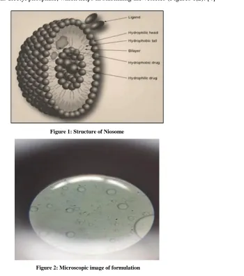
Structure of Niosomes [image:2.612.181.531.115.511.2]Niosome vesicles would consist of a vesicle making amphiphile i.e. a non-ionic surfactants such as Span-60,80,Tween-20,60,80 which is commonly stabilized by the adding together with cholesterol and a reduced amount of anionic surfactants such as dicetyl phosphate, which helps in stabilizing the vesicles (Figures 1,2). [4]
Figure 1: Structure of Niosome
Figure 2: Microscopic image of formulation
Formulation of Niosomes
The methods of development should be selected according to the make use of the niosomes, because the development methods influence the number of bilayers, size, size distribution, and entrapment efficiency of the aqueous phase and membrane permeability of the vesicles.
Ether injection method
The ether injection method provides a means of building niosomes by gradually introducing a solution of surfactant dissolved in diethyl ether into hot water at 60°C. The surfactants mixture is injected in ether through 16-gauge needle into an aqueous solution of material. Vaporization of ether takes place which leads to formation of single layered vesicles. Depending upon the conditions used, the diameter of the vesicle range from 50 to 1000 nm. [5]
Hand shaking method (Thin film hydration technique)
116
SonicationThe sonication method is a typical method for the preparation of vesicles by sonication of solution as described by Cable. In this method an aliquot of drug solution in buffer is added to the surfactant/cholesterol mixture in a 10 ml glass vial.
The mixture is sonicated at 60°C for 3 minutes using a probe sonicator with a titanium probe to produce small and uniform size of niosomes [6].
Micro fluidization
This is recent technique used for the development of uni-lamellar vesicles of defined size. Method is based on submerged jet principle in which two fluidized streams interact at ultra-high velocities, in precisely defined micro channels within the interaction chamber. The impingement of thin liquid sheet along a common front is arranged such that the energy supplied to the system remains within the area of niosomes formation. The result is a greater uniformity, smaller size and better reproducibility of niosomes formed [6].
Multiple membrane extrusion method
In this method the mixture of surfactants, cholesterol and dicetyl phosphate in chloroform is prepared into thin film by evaporation. The film is hydrated with aqueous drug polycarbonate membranes, solution and the resulting suspension is extruded through which are placed in series for up to 8 passages. It is a good method for controlling niosomes size [7].
Reverse Phase Evaporation Technique (REV)
Cholesterol and surfactant (1:1) both are dissolved in a combined mixture of ether and chloroform. An aqueous phase phosphate buffer solution containing drug is added and the resulting two phases are sonicated at 50°C for 5 minutes. The clear gel formed which is further sonicated after the addition of a small amount of phosphate buffer saline (PBS). The organic phase is removed at 40°C under low pressure using a rotary vaccume evaporator until the thin film was formed inside the flask. The resulting film was hydrated with PBS and heated on a water bath at 60°C for 10 min to yield niosomes [6,7].Raja Naresh et al. prepare the niosomes of Diclofenac Sodium by using Tween 85 with this method.
G.Trans membrane pH gradient (inside acidic) Drug Uptake Process (remote Loading technique)
Surfactant and cholesterol both are dissolved in chloroform in 1:1 ratio. The solvent is evaporated under reduced pressure to get a thin film on the wall of the round bottom flask. The film is hydrated by using 30 ml citric acid (pH 4.0) with the help of vortex mixing. The multi lamellar vesicles are frozen and thawed 3 times and later sonicated. To this niosomal suspension, aqueous solution containing 10 mg/ml of drug is added and vortexed. The pH of the sample is raised to 7.0-7.2 with 1M disodium phosphate. This mixture is later heated at 60°C for 10 minutes to give niosomes [7]
The “Bubble” Method
It is fresh technique for the one step preparation of liposomes and niosomes without use of organic solvents. The bubbling unit made up of RBF with three necks positioned in water bath to control the temperature. Water-cooled reflux and thermometer is placed in the first and second neck and nitrogen supply through the third neck. Cholesterol and surfactant both are dispersed together in this buffer (pH 7.4) at 70°C, the dispersion mixed for 15 seconds with high shear homogenizer and instantly afterwards “bubbled” at 70°C using nitrogen gas [6].
Types of Niosomes [7]
The niosomes are classified upon the function of the number of bilayer (e.g. MLV, SUV) or as a function of size. (e.g. LUV, SUV) or as a function of the method of preparation (e.g. REV, DRV). The various types of niosomes are described below:
117
Multi lamellar vesicles (MLV):It consists of a number of bilayer surrounding the aqueous lipid compartment individually. The approximate size of these vesicles is 0.5-10 μm diameter. Multi lamellar vesicles are the most widely used niosomes. These vesicles are highly suited as drug carrier for lipophilic compounds.
Large uni lamellar vesicles (LUV):
Niosomes of this type have a high aqueous/lipid compartment ratio, so that larger volumes of bio-active materials can be entrapped with a very inexpensive use of membrane lipids.
Small unilamellar vesicles (SUV):
These small unilamellar vesicles are generally prepared from multilamellar vesicles by sonication technique, French press extrusion electrostatic stabilization is the inclusion of dicetyl phosphate in 5(6)-carboxy fluorescein (CF) loaded Span 60 based niosomes.
Niosomes in Comparison with Liposomes
Niosomes are now commonly studied as an unconventional to liposomes, which show certain disadvantages such as they are luxurious, their ingredients like phospholipids are chemically unstable because of their predisposition, oxidative degradation, they have need of unique storage, handling and purity of natural phospholipids is variable. Niosomes are prepared from uncharged single-chain surfactants and cholesterol whereas liposomes are prepared from double chain phospholipids (neutral or charged). Niosomes act in-vivo similar to liposomes, prolonging the circulation of entrapped drug and altering its organ distribution and metabolic stability. Encapsulation of various antineoplastic agents in these carrier vesicles has been exposed to decrease drug induced toxic effects, at the same time as maintaining, or in some instances, greater than ever the anti-tumor efficiency. Such vesicular drug carrier systems alter the plasma clearance kinetics, tissue distribution, metabolism and cellular interaction of the drug. They can be estimated to target the drug to its preferred site of action and/or to control its release.
Advantage of Niosomes
Niosomes used first by L’Oreal in cosmetics, they have following advantages [8,9]
The vesicle suspension being water based so it offers a greater patient compliance over oil based systems.
Since the structure of the niosome offers place to accommodate hydrophilic, lipophilic as well as amphiphilic drug moieties, they can be used for a variety of drugs.
The characteristics such as size, lamellarity etc. of the vesicle can be varied depending on the requirement. The vesicle act as depot to release the drug slowly and offers a controlled release.
Other advantages of Niosomes
Osmotically active and stable. Increase the stability of the entrapped drug. Do not require any special conditions for the handling and storage of surfactants. It can increase the oral bioavailability of drugs.
It can enhance the skin penetration of drugs.
They have used for oral, parenteral as well as topical.
The surfactants are biodegradable, biocompatible, and non-immunogenic.
It can be improve the therapeutic performance of the drug by protecting it from the biological environment and restricting effects to target cells, thereby reducing the clearance of the drug.
The niosomal dispersions in an aqueous phase can be emulsified in a non-aqueous phase to control the release rate of the drug and administer normal vesicles in external non-aqueous phase.
Characterization of Niosomes
The characterization of niosome is necessary for the clinical applications. Characterization parameters have a direct impression on the stability of niosomes and a significant outcome on their in vivo performance. Therefore these parameters such as morphology, size, polydispersity index (PI), number of lamellae, zeta potential, encapsulation efficiency, and stability must be evaluated.
Size and Morphology
118
simultaneously cumulative information of particle size and valuable information on the homogeneity of the solution. A single sharp peak in the DLS profile implies existence of a single population of scatterers. The PI is helpful in this respect. It less than 0.3 corresponds to a homogenous population for colloidal systems [11]. The microscopic approaches are generally used to characterize the morphology of the niosomes.
Zeta Potential
Surface zeta potential of niosomes can be determined using zetasizer and DLS instruments. The surface charge of niosome plays an important role in the behavior of niosomes. In general, charged niosomes are more stable against aggregation than uncharged vesicles. Bayindir and Yuksel prepared paclitaxel loaded niosomes and investigated the physicochemical properties such as zeta potential of niosomes. They found that negative zeta potential values ranging between −41.7 and −58.4 mV are sufficiently high for electrostatic stabilization of niosomes [13].
Bilayer Characterization
Bilayer characteristics of niosomes have an importance on drug entrapment efficiency. The number of lamellae can be determined by AFM, NMR, and small angle X-ray scattering (SAXS) for multi lamellar vesicles [14]. Membrane rigidity of niosomal formulations can be measured by means of the mobility of fluorescence probe as a function of temperature. DPH (1,6 diphenyl-1,3,5-hexatriene) is most used fluorescent probe and added to niosomal dispersion. DPH normally exists in hydrophobic region in the bilayer membrane. The micro viscosity of niosomal membrane is determined by fluorescence polarization. High fluorescence polarization means high microviscosity of the membrane [15]. Moreover, the bilayer thickness can be characterized using the latter method, together with the insitu energy-dispersive X-ray diffraction (EDXD) [16].
Entrapment Efficiency
Entrapment efficiency (%EE) is defined as the portion of the applied drug which is entrapped by the niosomes. Un encapsulated free drug can be removed from the niosomal solution using centrifugation, dialysis, or gel chromatography [17]. After this step the loaded drug can be released from niosomes by destruction of vesicles. Niosomes can be destroyed with the addition of 0.1% Triton X-100 or methanol to niosomal suspension. The loaded and free drug concentration can be determined by a spectrophotometer [18] or high-performance liquid chromatography (HPLC) [19].
Stability
The stability of niosomes can be evaluated by determining mean vesicle size, size distribution, and entrapment efficiency over several month storage periods at different temperatures. During storage the niosomes are sampled at regular intervals of time and the percentage of drug which is retained into the niosomes is analyzed by UV spectroscopy or HPLC methods [17,20].
In vitro Drug Release
One often applied method to study in vitro release is based on using of dialysis tubing. A dialysis bag is washed and soaked in distilled water. After 30 mins, the drug loaded niosomal suspension is transferred, into this bag. The bag containing the vesicles is immersed in buffer solution with constant shaking at 25°C or 37°C. At specific time intervals, samples were removed from the outer buffer (release medium) and replaced with the same volume of fresh buffer. The samples are analyzed for the drug content by an appropriate assay method [21].
Applications of Niosomes [22-31]
The application of niosomal technology is commonly diverse and used to treat a number of diseases.
Niosomes as Drug Carriers
Niosomes have also been used as carriers for iobitridol, a diagnostic agent used for X-ray imaging. Topical niosomes may serve as solubilization matrix, as a local depot for sustained release of dermally active compounds, as penetration enhancers, or as rate-limiting membrane barrier for the modulation of systemic absorption of drugs (Table 1).
Drug Targeting
119
the niosome for clearance. Such localization of drugs is utilized to treat tumors in animals known to metastasize to the liver and spleen. This localization of drugs can also be used for treating parasitic infections of the liver. Niosomes can also be utilized for targeting drugs to organs other than the RES. A carrier system (such as antibodies) can be attached to niosomes (as immunoglobulin’s bind readily to the lipid surface of the niosome) to target them to specific organs.
Anti-neoplastic Treatment
Most antineoplastic drugs cause severe side effects. Niosomes can alter the metabolism; prolong circulation and half-life of the drug, thus decreasing the side effects of the drugs. Niosomes are decreased rate of proliferation of tumor and higher plasma levels accompanied by slower elimination.
Leishmaniasis
Leishmaniasis is a disease in which a parasite of the genus Leishmania invades the cells of the liver and spleen. Use of niosomes in tests conducted showed that it was possible to administer higher levels of the drug without the triggering of the side effects, and thus allowed greater efficacy in treatment.
Delivery of Peptide Drugs
Oral peptide drug delivery has long been faced with a challenge of bypassing the enzymes which would breakdown the peptide. Use of niosomes to successfully protect the peptides from gastrointestinal peptide breakdown is being investigated. In an in vitro study conducted by oral delivery of a vasopressin derivative entrapped in niosomes showed that entrapment of the drug significantly increased the stability of the peptide.
Use in Studying Immune Response
Due to their immunological selectivity, low toxicity and greater stability, niosomes are being used to study the nature of the immune response provoked by antigens. Nonionic surfactant vesicles have clearly demonstrated their ability to function as adjuvant following parenteral administration with a number of different antigens and peptides.
Niosomes as Carriers for Haemoglobin
Niosomes can be used as carriers for haemoglobin within the blood. The niosomal vesicle is permeable to oxygen and hence can act as a carrier for haemoglobin in anaemic patients.
Other Applications [23,24]
Sustained release:
Sustained release action of niosomes can be applied to drugs with low therapeutic index and low water solubility since those could be maintained in the circulation via niosomal encapsulation.
Localized drug action:
Drug delivery through niosomes is one of the best approaches to achieve localized drug action, since their size and low penetrability through epithelium and connective tissue keeps the drug localized at the site of administration.
Future prospectives: [25-30]
There are many technological challenges to be met in developing the following techniques:
Niosomal drug delivery systems deliver large quantities of drugs to specific areas and released in controlled ways. Controllable release profiles, especially for sensitive and toxic drugs.
Materials for nanoparticles that is biocompatible and biodegradable. Architectures / structures, such as biomimetic polymers, nanotubes. Technologies for self-assembly.
Functions (active drug targeting, on-command delivery, intelligent drug release devices, bioresponsive triggered systems, self-regulated delivery systems, systems interacting with the body, smart delivery).
Virus-like systems for intracellular delivery.
Nanoparticles to improve devices such as implantable devices/nanochips for nanoparticle release, or multi reservoir drug delivery-chips.
And also in the development of Combined therapy and medical imaging, for example, nanoparticles for diagnosis and manipulation during surgery (e.g. thermotherapy with magnetic particles).
120
User-friendly lab on a chip devices for point-of care, disease prevention and control at home.
Devices for detecting changes in magnetic or physical properties after specific binding of ligands on paramagnetic nanoparticles that can correlate with the amount of ligand.
Better disease markers in terms of sensitivity and specificity.
Table 1: Drug used in Niosomal delivery system [31-37] Route Of Administration Examples of Drugs
Intravenous Route Doxorubicin, Methotrexate, Vincristine, Diclofenac Sodium, Zidovudine, Insulin, AmphotericinB, 5Flurouracil Peroral Route DNA Vaccines, Peptides, Proteins, Ciprofloxacin, Norflaxocine, Insulin
Transdermal Route Nimesulide, Flurbiprofen, Piroxicam, Estradiol, Levonorgesterol, Ketoconazole, Ketorolac
Ocular Route Timolol maleate
Nasal Route Influenza Vaccines
Marketed Preparation [24]
[image:7.612.218.394.259.413.2]Lancôme has come out in the market with a variety of anti-ageing property which is based on niosomes formulations. L’Oreal is also conducting research on many cosmetic products. Niosomes Preparation present in the Market is –Lancôme (Figure 3).
Figure 3: Niosomes Preparation present in the market
CONCLUSION
The concept of drug targeting at specific tissue site by incorporating drug into niosomes is widely accepted by researchers and academicians. Niosomes is somewhat similar to liposomes in structure which shows certain advantages like cost, stability etc over liposomes. Also they have the potential to encapsulate various types of drugs like anti-infective, anticancer drugs within them. Niosomes can also be used as diagnostic imaging, vaccine adjuvants. Further these areas needs to be explored more. Various types of drug delivery approaches can be possible using niosomes like ophthalmic, parenteral, targeting, etc. It has wide applications in cosmaceuticals also.
ACKNOWLEDGEMENT
The authors express their sincere thanks to Integral University, Lucknow to inspire them by its research atmosphere and it’s all valuable supports. The authors also thankful to Dean R&D, Integral University for providing manuscript communication number (MCN IU/R&D/2018-MCN000254).
CONFLICT OF INTEREST
121
REFERENCES[1] M Nvs; A Saini. Int J Res Pharm Chem. 2011, 1, 498-511. [2] C Hu; DG Rhodes. Int J Pharm.1999, 185(1), 23-35.
[3] G Parthasarathi; N Udupa; GK Pillai. Indian J Pharm Sci. 1994, 56(3). [4] http//en.wikipedia.org/wiki/Niosomes, Structure of Niosomes.
[5] RR Naresh; GK Pillai; N Udupa; G Chandrashekar. Indian J Pharm. 1994, 26(1), 46-48. [6] AIB Welsh; DG Rhodes. Pharm Res. 1994, 18(5), 656-661.
[7] P Gadhiya; S Shukla; D Modi. P Bharadia. Int J Pharm Res Scholars. 2012, 2, 61. [8] SS Biju; S Talegaonkar; PR Misra; RK Khar. Indian J Pharm. 2006, 68, 141-153. [9] A Alsarra; A Bosela; SM Ahmed; GM Mahrous. Eur J Pharm Biopharm. 2004, 2(1), 1-6.
[10] L Tavano; R Aiello; G Ioele; N Picci; R Muzzalupo. Colloids Surfaces B: Biointerfaces. 2014, 118, 7-13. [11] A Priprem; K Janpim; S Nualkaew; P Mahakunakorn. AAPS Pharm Sci Tech. 2016, 17(3), 631-639. [12] W Hua; T Liu. Colloids Surfaces A: Physicochem Eng Aspects. 2007, 302(1), 377-382.
[13] ZS Bayindir; N Yuksel. J Pharm Sci. 2010, 99(4), 2049-2060.
[14] T Liu; R Guo; W Hua; J Qiu. Colloids Surfaces A: Physicochem Eng Aspects. 2007, 293(1-3), 255-261. [15] A Manosroi; P Wongtrakul; J Manosroi. Colloids Surfaces B: Biointerfaces.2003, 30, 129-138.
[16] D Pozzi; R Caminiti; C Marianecci. Langmuir.2010, 26(4), 2268-2273.
[17] M Tabbakhian; S Daneshamouz; N Tavakoli; MR Jaafari. Iranian J Pharm Sci. 2005, 1(3), 119-130. [18] SK Mehta; N Jindal. Colloids Surfaces B: Biointerfaces. 2013, 101, 434-441.
[19] AY Waddad; S Abbad; F Yu. Int J Pharm. 2013, 456, 446-458. [20] Y Hao; F Zhao; N Li; Y Yang; K Li. Int J Pharm. 2002, 244, 73-80.
[21] D Akhilesh; KB Bini; JV Kamath. Int J Res Pharm Biomed Sci. 2012, 3, 6-12.
[22] K Ruckmani; B Jayakar; SK Ghosal. Drug Development Industrial Pharm. 2000, 26, 217.
[23] M Conacher; J Alexanderand; JM Brewer; M Conacher; J Alexander. Int Publishers Distributors Ltd. 2000, 185-205.
[24] https://www.google.co.in/search?q=lancome+niosome+image.
[25] WN Charman; HK Chan; BC Finnin; SA Charman. Drug Development Res.1999, 46, 316-327. [26] JT SantiniJr; AC Richards; R Scheidt; MJ Cima; R Langer. Chem Int Ed. 2000, 39, 2396-2407. [27] J Kopecek. European J Pharm Sci. 2003, 20, 1-16.
[28] VP Torchilin. J Control Release. 2001, 73, 137-172.
[29] PD Reddy; D Swarnalatha. Int J Pharm Tech Res. 2010, 2(3), 2025-2027.
[30] KM Kazi; AS Mandal; N Biswas; A Guha; S Chatterjee; M Behera; K Kuotsu. J Adv Pharm Technol Res. 2010, 1(4), 374.
[31] NK Verma; A Roshan. Int J Res Pharm Life Sci. 2014, 2(1), 182-184.
[32] MA Theresa. Drugs published by Adis international Ltd. 1998, 56(5), 747-756.
[33] G Buckton; Harwood. Interfacial Phenomena in Drug Delivery and Targeting Academic Publishers, Switzerland.1995, 154-155.
[34] CA Hunter. J Pharm Pharmacol.1988, 161. [35] A Shahiwala; A Misra. J Pharma Sci. 2002, 220.