Journal of Chemical and Pharmaceutical Research, 2014, 6(3):1279-1285
Research Article
CODEN(USA) : JCPRC5
ISSN : 0975-7384
Formulation development, physico-chemical characterization and evaluation
of anti-microbial activity of herbal tooth gel
Mridul Haque
1, Anil Kumar Singh
2, Santosh K Maurya
1and Ankit Seth
1*1Ayurvedic Pharmacy Research Laboratory, Rajiv Gandhi South Campus, Banaras Hindu University, Barkachha,
Mirzapur
2Department of Dravyaguna, Faculty of Ayurveda, Institute of Medical Science, Banaras Hindu University,
Varanasi
_____________________________________________________________________________________________
ABSTRACT
Several synthetic drugs have been evaluated over the years for their antimicrobial effect in oral cavity; however, all are associated with numerous side effects that prohibit their regular long term use. Natural remedies are more acceptable in the belief that they are safer with fewer side effects than the synthetic ones. Therefore the objective of the current research was to formulate, characterize and evaluate a gel based dental which is more stable in terms of rheological behaviour, long term protection and stability in diverse conditions with minimum adverse effects. Anti- microbial activity of the formulated dental gel was carried out by disc diffusion method and was compared for antimicrobial activity with Ciprofloxacin (5µg/disc) as standard. The formulated batches F2 (5%) and F4 (10%) showed significant zone of inhibition. Formulated batch F4 (10%) gel showed maximum zone of inhibition.
Keywords: gel, dental, antimicrobial, herbal, disc diffusion method.
_____________________________________________________________________________________________
INTRODUCTION
Dental disease is considered to be a major health problem throughout the world. Dental disease may be chronic or acute and long term treatment is often required. The efficient use of anti bacterial agents for the treatment of various dental problems requires a sufficient drug concentration at the site of action without any adverse effects [1]. Dentifrice is a preparation for cleansing and polishing the teeth. There are two types of dentifrices, one is simple cleansing dentifrice and another is therapeutic dentifrice [2]. Dental products are available in paste, gel or powder form. Dental products are usually applied with a toothbrush for cleaning and polishing the teeth and to maintain the oral hygiene. It typically consists of a mild abrasive, detergent, flavoring agent, fluoride and binder. Other common ingredients include deodorants, humectants, desensitizers, and several medications to prevent dental caries, plaque and many other gum diseases such as halitosis [3].
Toothpaste is not intended to be swallowed due to the presence of fluoride and the other pharmaceutical ingredients, but is usually not very harmful if swallowed accidentally in small quantity. Medicated toothpaste must have effective cleansing power, pleasant odour and taste and it must be non-decalcifying and non-poisonous in nature. Commercially available toothpaste have the properties like anti plaque, antimicrobial activity, tooth whitening property but there are some demerits, such as most of the companies are using fluoride as an ingredient of tooth paste which may produce many associated adverse effect such as dental fluorosis [4, 5].
catesbiana [7], while another linked triclosan exposure with reduced sperm production in male rats [8].The
hypothesis proposed is that triclosan blocks the metabolism of thyroid hormone, because it chemically mimics thyroid hormone, and binds to the hormone receptor sites, blocking them, so that endogenous hormones cannot be used. Although the chemical structure of triclosan closely resembles certain estrogens, a study on a Japanese species of fish did not demonstrate estrogenic effects. However, it did find that triclosan is weakly androgenic, causing changes in fin length and sex ratios [9]. A more recent paper shows that triclosan can hinder estrogen sulfotransferase in sheep placenta, an enzyme which helps metabolize the hormone and transport it to the developing fetus. A doubt is that whether or not triclosan would be dangerous in pregnancy if it is likely to reach the placenta at a concentration that will affect the enzyme [10].
Addition of above mention properties of good quality dentifrice, efforts are made to bring a new gel based polyherbal formulation which is more stable in terms of rheological behavior, long term protection and stability in diverse conditions with minimum adverse effects. To prepare the tooth gel carbopol (940) and sodium carboxymethyl cellulose was used. Glycerin is used as a humectants; triethanolamine is used as a ph neutralizer and also produce transparent gel.
The plants which are used in this formulation are well known or have reported anti-microbial activity and to cure tooth related problem such as tooth ache, halitosis, and gum disease, namely clove or Lang (Syzygium aromaticum Linn.)[11], turmeric or haldi (Curcuma longa Linn.) [12], acacia or babul (Acacia nilotica Linn.) [13], liquorice or yasthimadhu (Glycyrizha glabra Linn.) [14] , tomarbeej or tejova in Sanskrit (Zanthoxylum armatum Dc.), miswak or tooth brush tree or pilu (Salvadora persica Linn.)[15], Indian madder or manjistha (Rubia cordifolia Linn.) etc. Clove oil has anti-bacterial activity and it also heal tooth ache related problem. Turmeric, acacia, tomarbeej is used for anti-microbial activity. Manjistha is used as coloring agent. Liquorice is used as sweetening purpose and it also work as an anti-microbial agent.
EXPERIMENTAL SECTION
Plant collection and authentication:
The useful parts of all seven plants viz; seeds of tomarbeej, roots of yasthimadhu and manjistha, rhizome of haldi, stem bark of babula and miswak and buds of lang were collected from the market and identified by Prof. Anil Kumar Singh, Department of Dravyaguna, Faculty of Ayurveda, IMS, BHU, Varanasi.
Chemicals:
Carbopol-940 (Loba chemicals), Sodium Carboxy methyl cellulose (S.D. Fine- Chem. Ltd.), Poly ethylene glycol- 4000 (Central Drug House), Sodium lauryl sulphate (Central Drug House), Tri-ethanolamine (Loba chemicals), Sodium saccharine (Loba chemicals), Sodium benzoate (Loba chemicals) were purchased from the market.
Extraction process of the plant drug:
The fresh and shade dried coarsely powdered drugs were accurately weighed in equal proportion i.e. 100 gram each of seven drugs viz; Tomarbeej, yasthimadhu, Lang, haldi, babula, miswak and manjistha. Each drug was transferred in conical flask separately and then water and ethanol were mixed in equal proportion (1:1). The plant drugs mixed with hydro alcoholic solvents were kept for 2 weeks with occasional shaking. After the extraction of two weeks, each extract was filtered with filter paper followed by cotton cloth. The filtered extracts were heated on water bath to evaporate the ethanol and concentrate the extract. The concentrated extracts were used for the development of gel based dental formulation, which was further evaluated for anti- microbial activity.
Preparation of Gel:
Different proportions of Carbopol 940 and Sodium CMC were dispersed in 50 ml of distilled water or deionized water with continuous stirring with the help of mechanical stirrer. 5 ml of distilled water was taken and required quantity of sodium benzoate was dissolved by heating on water bath. Solution was cooled and polyethylene glycol-4000 was added and mixed with first solution. Sodium saccharine and required quantity of sodium lauryl sulphate was taken to dissolve it properly with water and after that it was mixed to the first one. Further required quantity of seven plant extracts were mixed to the above mixture and volume was make up by adding remaining distilled water. Finally full mixed ingredients were mixed properly to the Carbopol 940 gel with continuous stirring and triethanolamine was added drop wise to the formulation for adjustment of required pH (6.8-7) and to obtain the gel in required consistency [16].
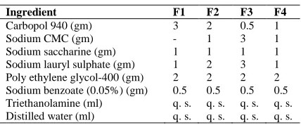
Table 1: Chemical Composition
[image:3.595.200.414.87.176.2]Ingredient F1 F2 F3 F4 Carbopol 940 (gm) 3 2 0.5 1 Sodium CMC (gm) - 1 3 1 Sodium saccharine (gm) 1 1 1 1 Sodium lauryl sulphate (gm) 1 2 3 1 Poly ethylene glycol-400 (gm) 2 2 2 2 Sodium benzoate (0.05%) (gm) 0.5 0.5 0.5 0.5 Triethanolamine (ml) q. s. q. s. q. s. q. s. Distilled water (ml) q. s. q. s. q. s. q. s.
Table 2: Hydroalcoholic Plant Extract
Ingredients: F1 (ml)
F2 (ml)
F3 (ml)
F4 (ml) Lavanga (Syzygium aromaticum) 10 5 5 10 Haldi (Curcuma longa) 10 5 5 10 Yasthimadhu (Glycyrizha glabra) 10 5 5 10 Pilu (Salvadora persica) 10 5 5 10 Babul (Acacia Arabica) 10 5 5 10 Tejova (Zanthoxylum alatum) 10 5 5 10 Manjistha (Rubia cordifolia) 10 5 5 10
EVALUATION OF DENTAL GEL FORMULATION: Transparency, smoothness and relative density:
5ml of gel formulation was taken in the 10ml test tube and its transparency was checked visually. The smoothness of the formulation was tested by rubbing the gel between the fingers and was observed that whether the gel is smooth, clumped, homogenous or rough. The relative density of the formulation or weight/ml of the was determined by taking the weight in gm of 10ml formulation & 10ml distilled water using RD bottle.
Viscosity, pH and microbial growth:
Viscosity is an important feature to determine the resistance of flow of gel formulation so that it can spread on the skin properly. It was determined with the help of viscometer (Brookfield) using 2 number spindles.
pH of the formulation was determined by using pH meter. In this method, electrode was washed with double distilled water, dried with the help of tissue paper and then dipped in 20ml gel formulation. The pH of the gel formulations were recorded at ambient condition.
Nutrient agar media was used in microbial growth study. In this method the blank and sample petriplates were used and the gel samples were aseptically transferred on to the sample plates in a cross pattern. The microbial growth was observed daily for 14 days
Extrudability:
The gel formulations were filled in standard capped collapsible aluminum tubes and sealed by crimping to the end. The weights of the tubes were recorded. The tubes were placed between two glass slides and were clamped. 500 gm was placed over the slides and then the cap was removed. The amount of the extruded gel was collected and weighed. The percent of the extruded gel was calculated (>90% extrudability: excellent, >80%extrudability: good, >70% extrudability: fair) [17].
Spreadability:
In this method spreadability was measured on the basis of slip and drag characteristics of gels. An excess of gel (about 2 g) under study was placed on the ground slide. The gel was then sandwiched between this slide and another glass slide having the dimension of fixed ground slide and provided with a hook. 1 kg weight was placed at the top of the two slides for 5 minutes to expel air and to provide a uniform film of the gel between the slides. Excess of the gel was scrapped off from the edges. The top plate was then subjected to pull of 80 g with the help of string attached to the hook and the time (in seconds) required by the top slide to cover a distance of 7.5 cm was noted. A shorter interval indicated better spreadability [16].
Spreadability was calculated using the following formula:
Where,
S = Spreadibility
M = Weight in the pan (tied to the upper slide) L = Length moved by the glass slide
T = Time (in sec.) taken to separate the upper slide from the ground slide.
Stability study:
The stability study was performed as per ICH guidelines. The formulated gel was filled in collapsible tubes and stored at different temperatures and humidity conditions, viz. 25ºC± 2ºC / 60% ± 5% RH, 30º C ± 2ºC / 65% ± 5% RH, 40ºC ± 2ºC / 75% ± 5% RH for a period of three months and studied for appearance, pH and spreadability.
In vitro antibacterial activity [18- 21]
Method of antibacterial susceptibility testing:
Antimicrobial susceptibility testing methods was adopted by disc diffusion method. The pathogenic organism was grown on Mueller-Hinton agar in the presence of various antimicrobial impregnated filter paper disks. The presence or absence of growth around the disks was an indirect measure of the ability of that compound to inhibit that organism, known as zone of inhibition.
Test microorganisms:
The four bacterial strains were used in the present study which obtained from Department of Microbiology, Institute of Medical Sciences, Banaras Hindu University Varanasi. The bacterial strains used were Escherichia coli,
Salmonella typhi, Staphylococcus aureus and Pseudomonas aeroginosa. The effects of the formulation on the
bacterial strains were assayed by disc diffusion method.
Preparation of Mueller-Hinton Agar Medium:
22.8 gm of MH agar media (purchased from Himedia laboratories Pvt. Ltd.) was suspended into 600 ml distill water and mixed properly. After that it was heated to dissolve the media completely. Sterilized by autoclaving at 15 Lbs pressure and 1210C temperature for 15 minutes, mixed well before use.
Preparation of MH plates:
Approximately 25 ml of liquid MH media was poured (to a depth of 4 mm) into Petri plates and allowed to solidify
at room temperature, stored at 4 to 80C temperature, pH of the MH agar should fall between 7.2 and 7.4 at room
temperature after solidification.
Procedure for disc diffusion test Inoculum Preparation:
At least three to five well isolated colonies of the same morphological type were selected from an agar plate culture. The top of each colony was touched with a loop and the growth was transferred into a tube containing 4 to 5 ml of a suitable broth medium, such as soy broth. The broth culture was incubated at 35°C until it achieves or exceeds the turbidity of the 0.5 McFarland standards (usually 2 to 6 hours). The turbidity of the actively growing broth culture was adjusted with sterile saline to obtained turbidity optically comparable to that of the 0.5 McFarland standards.
Inoculation of test plates:
Optimally, within 15 minutes after adjusting the turbidity of the inoculums suspension, a sterile cotton swab was dipped into the adjusted suspension. The swab was rotated several times and pressed firmly on the inside wall of the tube above the fluid level to remove excess inoculum from the swab. The dried surface of a Mueller-Hinton agar plate was inoculated by streaking the swab over the entire sterile agar surface. This procedure was repeated by streaking two more times, rotating the plate approximately 60° each time to ensure an even distribution of inoculums. As a final step the rim of the agar was swabbed.
The lid may be left partly opened for 3 to 5 minutes, but not more than 15 minutes, to allow for any excess surface moisture to be absorbed before applying the drug impregnated disks.
Application of discs to inoculated Agar plates:
instantaneously, a disc should not be relocated once it has come into contact with the agar surface. The plates were inverted and placed in an incubator set to 35°C within 15 minutes after the discs applied [22].
Reading plates and interpreting results:
After 14 to 16 hours of incubation, each plate was examined. If the plate was satisfactorily streaked, and the inoculum was correct, the resulting zones of inhibition should be uniformly circular and a confluent lawn of growth. The diameters of the zones of complete inhibition (as judged by the unaided eye) were measured, including the diameter of the disc. Zones were measured to the nearest whole millimeter with the help of a ruler, which was held on the back of the inverted Petri plates. The Petri plate was held a few inches above a black, nonreflecting background and illuminated with reflected light. The zone margin should be taken as the area showing no apparent, visible growth that can be detected with the unaided eye. Faded growth of tiny colonies, which can be detected only with a magnifying lens at the edge of the zone of inhibited growth, should be ignored. After measured the diameter of zone of inhibition the data was noted and interpreting the result [23].
RESULTS AND DISCUSSION
Herbal dental gel formulation was prepared form Hydroalcoholic extract of various plant part or natural ingredient and small amount synthetic ingredient. At the time of formulation development four batches (F1, F2, F3and F4) were prepared among them two batches (F1 and F3) were discarded due to the problem viz, homogeneity, spreadability & viscosity. Rest of the batches (F2 and F4) was selected for further study. The developed herbal dental gel was reddish brown in colour, translucent in appearance (Table no.-5.2) and showed good homogeneity with absence of lumps. The formulated batch F2 was much clear and transparent as compared to F4 formulation.
The values of spreadability indicate that the gel is easily spreadable by small amount of shear.
Spreadability of formulated dental gels (F2 and F4) was 15.75; 19.78 g cm/sec. respectively. (Table no.-5.4) Hence spreadability of F4 formulation was good as compared to F2 formulation. During the accelerated stability studies the appearance was clear and no significant variation in pH was observed and spreadability is 17.82 in F4 formulation after 3 months where as spreadability in F2 was 15.12 (Table no.-5.8). pH was also maintained throughout the study which was found to be 6.91 to 7.0 (Table no.-5.8). The initial viscosities of developed gels were measured using Brookfield viscometer with spindle. The extrudability of the dental gel formulation was excellent at the time of preparation (initial month), because the value of extrudability amount was 91.73 percent. (Table no.-5.5)
Anti-bacterial activity of the dental gel was carried out by disc diffusion method against gram negative microorganisms such as Pseudomonas aeroginosa, E.coli and gram positive microorganisms such as Staphylococus
aureus. The plates were incubated for 24 hours at 37ºC. Dental gel formulation containing hydro-alcoholic extracts
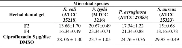
of various plants, showed significant zone of inhibition for F2 (5%) and F4 (10%) gel. F4 (10%) gel showed maximum inhibition when comparing with F2 (5%) gel. Ciprofloxacin (5µg/disc) was used as a standard anti bacterial agent. By comparing the inhibition of F4 (10%) gel with the standard anti bacterial agent (Ciprofloxacin) we could be interprets that the zone of inhibition was near about similar to the standard. In case S.typhi the F4 (10%) gel and the standard both possessed similar results. The results of zone of inhibition were mentioned in (Table no5.9).
Physical evaluation (color, appearance, transparency, smoothness, relative density, viscosity, pH etc.) of herbal dental gel formulation:
Table 3
Batch Color Appearance Transparency Smoothness F2 Reddish brown Homogeneous Translucent Smooth F4 Reddish brown Homogeneous Translucent Smooth
Table 4
Batch Relative density Viscosity
(Dyn.s/cm2) Microbial growth pH F2 10.52 1.6*10-3 No growth 7.2
Table 5: Spreadability of dental gel
Batch Spreadability (g.cm/sec) F2 15.75 F4 19.78
Extrudability of the herbal dental gel (F4) at the time of preparation (Mean ± SEM):
Table 6
Extrudability Mean of three tubes (Initial month) Net wt of formulation in tube (g) 12.34±0.011
Wt. of gel extruded (g) 11.32±0.014 Excrudability amount percentage 91.73±0.005
Stability testing at 25oC ± 2oC/ 60% ± 5% RH (3rd months) of herbal dental gel:
Table 7
Formulation Color Appearance Spreadability pH F2 Reddish brown Homogenous 15.65 7 F4 Reddish brown Homogenous 18.40 6.98
Stability testing at 30oC ± 2oC/ 65% ± 5% RH (3rd months) of herbal dental gel:
Table 8
Formulation Color Appearance Spreadability pH F2 Reddish brown Homogenous 15.32 6.94 F4 Reddish brown Homogenous 18.34 6.97
Stability testing at 40oC ± 2oC/ 75% ± 5% RH (3rd months) of herbal dental gel:
Table 9
Formulation Color Appearance Spreadability pH F2 Reddish brown Homogenous 15.12 6.91 F4 Reddish brown Homogenous 17.82 6.96
[image:6.595.154.458.494.575.2]Antimicrobial activity expressed as Zone of Inhibition (in mm) mean ± sem:
Table 10
Microbial species
Herbal dental gel
E. coli
(ATCC 35218)
S. typhi
(MTCC 3216)
P. aeruginosa (ATCC 27853)
S. aureus
(ATCC 25323) F2 13.66±1.70 20.67±0.49 17.34±1.22 15±0.68 F4 16.34±0.49 23.34±0.71 21.34±0.88 18.16±0.78 Ciprofloxacin 5 µg/disc
DMSO 28. 06 ± 1.30 23.7 ± 1.05 24.76 ± 0.76 29.93 ± 0.49
CONCLUSION
Natural remedies are more acceptable in the belief that they are safer with fewer side effects than the synthetic ones. Herbal formulations have growing demand in the world market. It was a very good attempt to establish the herbal dental gel containing plant extract. The studies revealed that the developed poly herbal dental gel formulation F2 and F4 consisting plant extract was comparatively better than the other batches F1 and F3 on every aspect.
In this study disc diffusion method was followed. It is the standard and convenient method to screen the herbal extracts with antibacterial property. Standard drug Ciprofloxacin showed greater activity (zone of inhibition) when compared to the formulated dental gel (F2 or 5%) and near about similar activity to the (F4 or 10%) gel. Both F2 and F4 formulation showed considerable anti microbial activity.
further pharmacological evaluation and total clinical justification. We are totally confident that this would be a successful step ahead towards dental research, and for the total fulfillment of intended activity.
REFERENCES
[1] A Katiyar; SK Prajapati; A Akhtar; SK Vishwakarma. Int. Res. J Pharm., 2012, 3(10), 143-148.
[2] JB Wilkinson; RJ Moore. Harry’s Cosmeticology, 7th Edition, Chemical Publishing, New York, 1982; 608-622.
[3] Mithal BM; Saha RN. A handbook of cosmetics, 2nd edition, Vallabh Prakashan, New Delhi, 2006; 52-60.
[4] D Browne; H Whelton; DO Mullane. J Dent., 2005, 33(3), 177-186. [5] DG Pendrys; RV Katz. Am. J Epidemiol., 1989, 130(6), 1199-1208.
[6] LM Zorrilla; EK Gibson; SC Jeffay; KM Crofton; WR Setzer; RL Cooper; TE Stoker. Toxicol. Sci., 2009, 107(1), 56–64.
[7] N Veldhoen; RC Skirrow; H Osachoff; H Wigmore; DJ Clapson; MP Gunderson; GV Aggelen; CC Helbing.
Aquat. Toxicol., 2006, 80(3), 217-27.
[8] V Kumar; A Chakraborty; MR Kural; P Roy. Reprod. Toxicol., 2009, 27(2), 177- 185. [9] CM Foran; ER Bennett; WH Benson. Mar. Environ. Res., 2000, 50(1), 153-156.
[10] MO James; W Li; DP Summerlot; L Rowland-Faux; CE Wood. Environ. Int., 2010, 36(8), 942-949
[11] J Briozzo;L Nuncez; J Chirife; L Herszage; M Daquino. J Appl. Bacteriol., 1989, 66(1), 69-75.
[12] KJ Kim; HH Yu; JD Cha, SJ Seo; NY Choi; YO You. Phytother. Res., 2005, 19(7), 599-604.
[13] OM Abd el Nabi; EC Reisinger; FF Reinthaler; F Still; U Eibel; GJ Krejs. J Ethnopharmacol.,1992, 37(1), 77-9.
[14] W Li, Y Asada; T Yoshikawa. Planta. Med., 1998, 64(8), 746-7. [15] FA Al-Bayati; KD Sulaiman. Turk. J Biol., 2008, 32, 57-62. [16] S Dwivedi; S Gupta. Acta. Chim. Pharm. Indica., 2012, 2(1), 54-59.
[17] A Negi; N Sharma; MF Singh. J Pharmacognosy Phytochem., 2012, 1(4), 112-116.
[18] S Suganya; R Bharathidasan; G Senthilkumar; P Madhanraj. A Panneerselvam. J Chem. Pharm. Res., 2012, 4(3), 1846-1850.