cell ALL patients
Cameron J. Turtle, … , Stanley R. Riddell, David G. Maloney
J Clin Invest. 2016;126(6):2123-2138. https://doi.org/10.1172/JCI85309.
BACKGROUND. T cells that have been modified to express a CD19-specific chimeric antigen receptor (CAR) have antitumor activity in B cell malignancies; however, identification of the factors that determine toxicity and efficacy of these T cells has been challenging in prior studies in which phenotypically heterogeneous CAR–T cell products were prepared from unselected T cells.
METHODS. We conducted a clinical trial to evaluate CD19 CAR–T cells that were manufactured from defined CD4+ and
CD8+ T cell subsets and administered in a defined CD4+:CD8+ composition to adults with B cell acute lymphoblastic
leukemia after lymphodepletion chemotherapy.
RESULTS. The defined composition product was remarkably potent, as 27 of 29 patients (93%) achieved BM remission, as determined by flow cytometry. We established that high CAR–T cell doses and tumor burden increase the risks of severe cytokine release syndrome and neurotoxicity. Moreover, we identified serum biomarkers that allow testing of early intervention strategies in patients at the highest risk of toxicity. Risk-stratified CAR–T cell dosing based on BM disease
burden decreased toxicity. CD8+ T cell–mediated anti-CAR transgene product immune responses developed after CAR–
T cell infusion in some patients, limited CAR–T cell persistence, and increased […] Clinical Medicine Oncology
Find the latest version:
The Journal of Clinical Investigation
C l i n i C a l M e d i C i n eIntroduction
The administration of lymphodepleting chemotherapy followed by adoptive transfer of autologous T cells that are genetically
modified to express a chimeric antigen receptor (CAR) specific for CD19 (CD19 CAR–T cells) has produced a high rate of complete remission (CR) in adult and pediatric patients with relapsed and refractory B cell acute lymphoblastic leukemia (B-ALL) in small phase I clinical trials (1–4). Encouraging results have also been seen in clinical trials of CD19 CAR–T cell therapy in non-Hodg-kin’s lymphoma (NHL) and chronic lymphocytic leukemia (CLL) (5–9). Emerging data from these studies suggest that robust prolif-eration of transferred CAR–T cells in the recipient correlates with clinical response and that prolonged in vivo persistence of func-tional CAR–T cells may be necessary to prevent disease relapse. The administration of CD19 CAR–T cells and their subsequent expansion can be associated with cytokine release syndrome (CRS), characterized by hyperpyrexia, hypotension, capillary leak, neurotoxicity, and death in severe cases (3, 4, 7, 9, 10). The factors that determine CAR–T cell expansion and persistence in vivo, the durability of antitumor responses, and toxicities have been chal-lenging to define in initial studies in part because of the wide
vari-BACKGROUND. T cells that have been modified to express a CD19-specific chimeric antigen receptor (CAR) have antitumor activity in B cell malignancies; however, identification of the factors that determine toxicity and efficacy of these T cells has been challenging in prior studies in which phenotypically heterogeneous CAR–T cell products were prepared from unselected T cells. METHODS. We conducted a clinical trial to evaluate CD19 CAR–T cells that were manufactured from defined CD4+ and CD8+ T cell subsets and administered in a defined CD4+:CD8+ composition to adults with B cell acute lymphoblastic leukemia after lymphodepletion chemotherapy.
RESULTS. The defined composition product was remarkably potent, as 27 of 29 patients (93%) achieved BM remission, as determined by flow cytometry. We established that high CAR–T cell doses and tumor burden increase the risks of severe cytokine release syndrome and neurotoxicity. Moreover, we identified serum biomarkers that allow testing of early intervention strategies in patients at the highest risk of toxicity. Risk-stratified CAR–T cell dosing based on BM disease burden decreased toxicity. CD8+ T cell–mediated anti-CAR transgene product immune responses developed after CAR–T cell infusion in some patients, limited CAR–T cell persistence, and increased relapse risk. Addition of fludarabine to the lymphodepletion regimen improved CAR–T cell persistence and disease-free survival.
CONCLUSION. Immunotherapy with a CAR–T cell product of defined composition enabled identification of factors that correlated with CAR–T cell expansion, persistence, and toxicity and facilitated design of lymphodepletion and CAR–T cell dosing strategies that mitigated toxicity and improved disease-free survival.
TRIAL REGISTRATION. ClinicalTrials.gov NCT01865617.
FUNDING. R01-CA136551; Life Science Development Fund; Juno Therapeutics; Bezos Family Foundation.
CD19 CAR–T cells of defined CD4
+
:CD8
+
composition
in adult B cell ALL patients
Cameron J. Turtle,1,2 Laïla-Aïcha Hanafi,1 Carolina Berger,1,2 Theodore A. Gooley,1 Sindhu Cherian,3 Michael Hudecek,1
Daniel Sommermeyer,1 Katherine Melville,1 Barbara Pender,1 Tanya M. Budiarto,1 Emily Robinson,1 Natalia N. Steevens,1
Colette Chaney,1 Lorinda Soma,3 Xueyan Chen,3 Cecilia Yeung,3,4 Brent Wood,3,4 Daniel Li,5 Jianhong Cao,1 Shelly Heimfeld,1
Michael C. Jensen,1,6 Stanley R. Riddell,1,2,7 and David G. Maloney1,2
1Clinical Research Division, Fred Hutchinson Cancer Research Center (FHCRC), Seattle, Washington, USA. 2Department of Medicine, 3Department of Laboratory Medicine, and 4Department of Pathology,
University of Washington, Seattle, Washington, USA. 5Juno Therapeutics, Seattle, Washington, USA. 6Seattle Children’s Research Institute, Seattle, Washington, USA. 7Institute for Advanced Study,
Technical University of Munich, Munich, Germany.
Authorship note: S.R. Riddell and D.G. Maloney contributed equally to this work.
Conflict of interest: C.J. Turtle receives research funding from Juno Therapeutics. D.G. Maloney receives research funding from Juno Therapeutics. S.R Riddell and M.C. Jensen receive research funding from and are cofounders of Juno Therapeutics. D. Li is an employee of and has equity in Juno Therapeutics. C. Yeung receives unrelated research funding from Gilead Sciences. C. Berger receives research funding from Juno Therapeutics. S.R. Riddell, M. Hudecek, and M.C. Jensen have a related patent (US-2015-0306141). S.R. Riddell, C. Berger, and M.C. Jensen have a related patent (US-2014-0356398). M.C. Jensen has a related patent (US8802374 B2).
Role of funding source: Personnel from Juno Therapeutics reviewed the draft manu-script and assisted with statistical analyses but were not involved in recruitment or clinical care of participants, performance of response assessments, or conduct of laboratory assays. Personnel from the other funding sources did not contribute to the design, conduct, or analysis of the study.
Submitted: November 5, 2015; Accepted: March 8, 2016.
B-ALL. The 30 patients who proceeded to lymphodepletion chemotherapy and CD19 CAR–T cell infusion had previously received a median of 3 prior intensive chemotherapy regimens (range 1–11), and 11 patients had relapsed at a median of 7 months (range 1–28) after prior allogeneic hematopoietic cell transplan-tation (HCT) (Table 1). Before lymphodepletion and CAR–T cell therapy, all patients had detectable disease in BM, extramedul-lary sites, or cerebrospinal fluid (CSF). Twenty-nine of 30 patients had detectable leukemic blasts in the BM (median 21.0%, range 0.014%–97.0%, n = 29), and 7 patients had extramedullary dis-ease, including 5 with bulky extramedullary disease. Leukemia was detected by flow cytometry in the CSF from 2 patients, one of whom had no other identified site of disease. The other 28 patients were negative for leukemic blasts in the CSF at evalua-tion before CAR–T cell infusion.
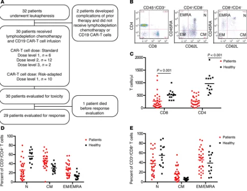
Prior to leukapheresis, we evaluated the absolute counts and proportions of TN, TCM, and TEM/EMRA (where TEMRA indicates CD45RA+ effector memory) cells within the CD4+ and CD8+ T cell
subsets in the blood of each B-ALL patient (Figure 1, B–E). B-ALL patients had lower numbers of CD4+ and CD8+ T cells compared
with healthy individuals (Figure 1C), and the CD4+:CD8+ T cell
ratio was highly variable within the patient population (median 1.19; range 0.27–8.89). Importantly, we found marked hetero-geneity in the percentages of TEM/EMRA cells within the CD4+ and
CD8+ T cell subsets in B-ALL patients (Figure 1, D and E; CD4+
TEM/EMRA, range 7.8%–57.5%; CD8+ T
EM/EMRA, range 8.5%–87.4%).
These data suggest that, particularly in B-ALL patients who have high fractions of TEM/EMRA or a disparate ratio of CD4+:CD8+ T cells
in blood, the ability to manufacture a CD19 CAR–T cell product of consistent potency may be enhanced by selection of defined T cell subsets from the leukapheresis product for CAR gene transfer and formulation of the CAR–T cell product in a defined CD4+:CD8+ CAR–T cell ratio.
CD19 CAR–T cell manufacturing. There were no serious
adverse events in the 32 patients who underwent leukapher-esis. Highly enriched CD4+ T cells were isolated for CAR–T cell
manufacturing from all patients (CD3+CD4+ T cell purity 97.2%;
range 48.5%–99.6%; n = 32). Our preclinical studies had suggest-ed that infusion of CD8+ T
CM CAR–T cells combined with CD4+
CAR–T cells might provide optimal antitumor efficacy and that an absolute CD8+ T
CM cell count of 20/μl or more on a
screen-ing assay was necessary for efficient selection of CD8+ T CM cells
using a sequential 2-step enrichment that involved depletion of CD4+CD14+CD45RA+ cells followed by selection of CD62L+ cells
(ref. 18 and Supplemental Figure 1, A and B; supplemental mate-rial available online with this article; doi:10.1172/JCI85309DS1). Enrichment of CD8+ T
CM cells using this approach was
success-ful in 18 of 19 patients. For the patients undergoing CD8+ T CM
selection, the frequency of CD3+CD8+ T cells with a CD45RA–
CD62L+ phenotype increased from 13.2% (range 1.12%–41.8%)
in the leukapheresis product to 72.2% (range 24.5%–90.2%) after the 2-step enrichment. CD3+CD8+ T cells made up 17.4% (range
0.86%–66.9%) of the CD4–CD14–CD45RA–CD62L+ product,
with the remainder being CD13+CD15+CD16+ myeloid cells and
CD13+CD123+CD16– basophils (Supplemental Figure 1, B and C).
Fourteen patients, including one who had an unsuccessful CD8+
TCM selection, had profound lymphopenia (CD4+, median 150
ation in CAR–T cell doses administered to patients, differences in the phenotypic composition of T cells isolated from patients for genetic modification and in the infused products, and differences in chemotherapy regimens administered to patients to provide lymphodepletion before CAR–T cells are infused (11).
Prior work has demonstrated that human CD4+ and CD8+ T
cells comprise functionally and transcriptionally distinct subsets that differ in their capacities to proliferate and persist in vivo after in vitro expansion and adoptive transfer (12–16). Using a preclinical model, we demonstrated that human CD19 CAR–T cells that were manufactured from purified CD8+ or CD4+ central memory T cells
(TCM cells) or naive T cells (TN cells) were more potent in elimina-tion of CD19+ tumors from immunodeficient mice compared with
CD19 CAR–T cells that were manufactured from effector memory T cells (TEM cells) (17). Synergistic enhancement in potency could be achieved by infusion of a defined ratio of CD19 CAR–T cells derived from CD8+ T
CM cells and CD4+ T cells. These results
sug-gested that selecting defined subsets of T cells from patients with B-ALL prior to transduction and formulating therapeutic CAR–T cell products of uniform composition might provide reproducible potency in clinical therapy and facilitate determining potential correlations between cell dose and efficacy or toxicity. Thus, we initiated a phase I/II clinical trial in patients with refractory B-ALL in which CD8+ and CD4+ T cell subsets were separately modified
to express a CD19-targeted CAR incorporating 4-1BB and CD3ζ signaling domains, formulated in a defined ratio of CD4+:CD8+
CAR–T cells, and administered in a dose-escalation/deescalation format after lymphodepletion with a cyclophosphamide-based (Cy-based) regimen, alone or with fludarabine (Flu).
The eligibility criteria for this study did not exclude any patient based on the absolute lymphocyte count or on a predeter-mined measurement of the ability of the patient’s T cells to expand after activation with anti-CD3/CD28 beads. Our data show that a therapeutic CAR–T cell product could be manufactured from all patients that enrolled in the study and that formulation of CAR–T cells in a defined composition was feasible in a majority of these heavily pretreated patients. We observed reproducible in vivo pro-liferation of CAR–T cells and antitumor activity, and low doses (2 × 10⁵/kg) of the defined-composition CAR–T cell product were
remarkably potent for inducing CR without a high rate of toxic-ity in patients with a high tumor burden. We demonstrate that a T cell–mediated immune response specific for epitopes encoded by the CAR transgene can develop in some patients who receive Cy lymphodepletion and limit the persistence of transferred CAR–T cells. The persistence of CAR–T cells, duration of remission, and disease-free survival (DFS) were improved by the addition of Flu to the lymphodepleting chemotherapy regimen.
Results
Patient characteristics. Thirty-two consecutive patients with
relapsed or refractory CD19+ B-ALL and a median age of 40 years
The Journal of Clinical Investigation
C l i n i C a l M e d i C i n etal Figure 2). CD8+EGFRt+ CAR–T cells that were manufactured
from bulk CD8+ T cells contained a lower fraction of CD45RA–
CD62L– cells than those manufactured from CD8+ T CM cells.
Assessment of toxicity following administration of CD19 CAR–T cells. The study was designed to evaluate the safety of 3 DLs (2 × 10⁵/
kg; 2 × 10⁶/kg; and 2 × 10⁷/kg) of CAR–T cells administered 48 to
96 hours after lymphodepleting chemotherapy. Serious acute tox-icity in the first 2 hours after CAR–T cell infusion was not observed at any CAR–T cell dose. Patients developed the toxicities expected with cytotoxic chemotherapy, including BM suppression, alopecia, mild mucositis, and neutropenic fever. The most common toxic-ity that was observed in the first 14 days after CAR–T cell infusion was CRS, characterized by fever and/or hypotension and elevated serum levels of IL-6 and IFN-γ. Two patients died due to toxicity after CAR–T cell infusion: one developed severe CRS (sCRS) and multiorgan failure that was unresponsive to tocilizumab, etan-ercept, and corticosteroids, and another patient who developed transient sCRS died 122 days after CAR–T cell infusion with irre-cells/μl, range 4–624 cells/μl; CD8+ median 76 cells/μl, range
9–458 cells/μl, n = 14) due to previous therapy and/or had circu-lating CD19+ blasts and had enrichment of CD8+ T cells without
CD62L selection. The median CD3+CD8+ T cell purity after CD8+
selection was 84% (range 36.1%–95.7%, n = 12).
The selected CD4+ and CD8+ T cells were lentivirally
trans-duced to express the CD19 CAR and a truncated human epider-mal growth factor receptor (EGFRt) that enabled identification of transduced cells by flow cytometry using the anti-EGFR mono-clonal antibody cetuximab. Transduced EGFRt+ CD4+ and CD8+
T cells were enriched during culture by a single stimulation with irradiated CD19+ lymphoblastoid cell line (LCL) (n = 27;
Supple-mental Figure 1, D and E). The median frequency of EGFRt+
CAR–T cells within the CD3+CD4+ and CD3+CD8+ subsets in the
products at release for infusion was, respectively, 79.7% (range 50.0%–95.9%) and 84.2% (range 13.0%–95.6%). The infused CD3+CD4+EGFRt+ and CD3+CD8+EGFRt+ CAR–T cells were
pre-dominantly CD45RA–CD62L+ and CD45RA–CD62L–
(Supplemen-Figure 1. Heterogeneity in distribution of TN, TCM, and TEM/EMRA cells within CD4+ and CD8+ T cell subsets in normal donors and patients with B-ALL. (A)
Study participant flow chart. (B) Representative flow cytometry plots showing the immunophenotype of T cell subsets in blood from a B-ALL patient are shown. TN (CD45RA+CD62L+), T
CM (CD45RA–CD62L+), and TEM/EMRA (CD62L–) cells can be identified in the CD3+CD4+ and CD3+CD8+ T cell populations. (C) The absolute CD4+ and CD8+ T cell counts in blood from healthy individuals (n = 14) and B-ALL patients (n = 30) are shown. Mann-Whitney U test was used for statistical analysis. (D) The percentages of TN, TCM, and TEM/EMRA cells in the CD3+CD4+ T cell population are shown. (E) The percentages of T
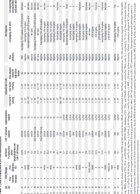
[image:4.585.48.543.60.443.2]Table 1. Clinical charac teris tics of the treated patients
Age (yr)
Prior ther apy BM blas ts (% leuk oc yt es) bef or e
lymphodepletion and CAR
–T c
ell ther
apy
CD8
+ T c
ell subset isolation Lymphodepletion re gimen Infused C AR –T c ells BM r esponse Prior int ensiv e ther apies Prior
allogeneic HCT
CD4
+EGFRt + T
cells /kg CD8 +EGFRt + T c ells /kg Risk -adapt ed dosing f or
high tumor burden
Flow cy tometr y Kar yo
typing, FISH, QPCR
IGH sequencing (copies
/million nucle at ed c ells) 1 A 37 2 N 0.016 CD8T CM CE
1 × 10
5
1 × 10
5 NA MRD Kar yot yping
, FISH neg
ativ
e, p210 QPCR positiv
e Not done 2 50 4 DUCB 0. 65 CD8T CM CE
1 × 10
6
1 × 10
6 NA Neg ativ e Kar yot yping
, FISH neg
ativ e 0 3 32 3 MR elD 1.6 CD8T CM Cy 1.9
7 × 10
6
3 × 10
4 NA MRD Kar yot yping neg ativ
e, p210 QPCR positiv
e Not done 4 71 2 N 16.5 CD8T CM Cy
1 × 10
7
1 × 10
7 NA Neg ativ e Not done Not done 5 28 4 N 4. 8 CD4/ 14/ 45RA -neg Cy
1 × 10
6
1 × 10
6 NA Neg ativ e Kar yot yping
, FISH neg
ativ
e, p210 QPCR positiv
e Not done 6 B 48 2 N 31 .8 CD8T CM Cy
1.16 × 10
7
8.
4 × 10
6 No Not done Not done Not done 7 24 3 N 97 CD4/ 14/ 45RA -neg Cy
1 × 10
5
1 × 10
5 Ye s Neg ativ e Kar yot yping
, FISH, QPCR neg
ativ e 9 8 37 4 MURD 83. 6 CD8T CM Cy
1 × 10
5
1 × 10
5 Ye s Neg ativ e Kar yot yping
, FISH neg
ativ e Not done 9 37 2 N 30 .2 CD8T CM Cy
1 × 10
5
1 × 10
5 Ye s Neg ativ e Kar yot yping
, FISH neg
ativ e 0 10 58 3 MURD 7.9 CD8T CM Cy
1 × 10
5
1 × 10
5 Ye s Neg ativ e Kar yot yping
, FISH neg
ativ e 0 11 C 62 3 N 46 CD8T CM Cy
1 × 10
5
1 × 10
5 Ye s Neg ativ e Kar yot yping
, FISH neg
ativ e 24 12 24 2 N 60 .7 CD8 Cy
1 × 10
6
1 × 10
6 No Neg ativ e Kar yot yping neg ativ e Polyclonal 13 29 1 N 10 CD8 Cy 1.4
9 × 10
6
5.1 × 10
5 NA Neg ativ e Kar yot yping neg ativ e 0 14 40 3 N 12.7 CD8T CM Cy /Flu5
1 × 10
6
1 × 10
6 NA Neg ativ e Kar yot yping
, FISH neg
ativ e 0 15 D 52 3 N 87. 2 CD8 Cy /Flu5
1 × 10
6
1 × 10
6 No Neg ativ e Abnormal kar yot ype
, FISH positiv
e 1950 7 16 22 1 N 23 .5 CD8T CM Cy /Flu5
1 × 10
6
1 × 10
6 No Neg ativ e Kar yot yping neg ativ e 0 17 E 52 5 N 49 .6 CD8 Cy /Flu3
1 × 10
6
1 × 10
6 No Neg ativ e Kar yot yping neg ativ e 0 18 A 38 4 MR elD 10 .7 CD8T CM Cy /Flu3
1 × 10
6
1 × 10
6 NA Neg ativ e Kar yot yping
, FISH neg
ativ e 5 19 39 3 MURD 28 CD8T CM Cy /Flu3
1 × 10
6
1 × 10
6 No Neg ativ e Kar yot yping neg ativ e Polyclonal 20 51 3 N 1.1 CD8T CM Cy /Flu5
1 × 10
6
1 × 10
6 NA Neg ativ e Kar yot yping neg ativ e 0 21 30 2 N 17. 4 CD8 Cy /Flu5
1 × 10
5
1 × 10
5 Ye s Neg ativ e Kar yot yping neg ativ e Polyclonal 22 52 6 MURD ; MURD 0.04 CD8 Cy /Flu3
1 × 10
5
1 × 10
5 NA Neg ativ e Not done Not done 23 23 6 MURD 0.55 CD8T CM Cy /Flu3
1 × 10
5
1 × 10
5 NA Neg ativ e FISH neg ativ e 2 24 38 2 N 0.014 CD8 Cy /Flu3
1 × 10
6
1 × 10
6 NA Neg ativ e FISH neg ativ e Polyclonal 25 40 1 N 21 CD8T CM Cy /Flu3
1 × 10
5
1 × 10
5 Ye s Neg ativ e
p190 QPCR neg
ativ e 0 26 73 4 N 0.03 CD8 Cy /Flu3
1 × 10
6
1 × 10
6 NA Neg ativ e Kar yot yping
, FISH neg
ativ e Polyclonal 27 43 4 MURD 0 CD8T CM Cy /Flu3
1 × 10
6
1 × 10
6 NA Neg ativ e Kar yot yping neg ativ e Not done 28 43 11 MR elD ; R el Haplo 28 .6 CD8T CM Cy /Flu3
1 × 10
5
1 × 10
5 Ye s Neg ativ e
p190 QPCR neg
ativ e 0 29 F 61 1 N 28 .4 CD8 Cy /Flu3
1 × 10
5
1 × 10
5 Ye s Neg ativ e Kar yot yping
, FISH neg
ativ e 178 30 20 4 MURD 74 .7 CD8 Cy /Flu3
1 × 10
5
1 × 10
5 Ye s Neg ativ e FISH neg ativ e 9
N, none; DUCB, double umbilical c
ord blood; MURD
, matched unr
elated donor
; MR
elD
, matched r
elated donor
; R
el Haplo
, r
elated haploidentical; CD4
, CD4-positive immunomagnetic selection; CD4/
14/
45R
A-neg
, PBMCs depleted of CD4
+, CD14 +, and CD4
5R
A
+ cells
; CD8T
CM, PBMCs depleted of CD4
+, CD14 +, and CD4
5R
A
+ cells and selected f
or CD6
2L
+ cells
; C
y, 2
–4 g
/msq C
y; CE, C
y 2
–4 g
/msq and et
oposide 100 mg
/
msq × 3; C
y/Flu3, C
y 30–60 mg
/kg × 1 and Flu 25 mg
/msq × 3; C
y/Flu5, C
y 30–60 mg
/kg × 1 and Flu 25 mg
/msq × 5; MRD
, < 5% blasts by flow c
yt
ometr
y.
APatient 1 under
went allogeneic HCT after r
ec eiving CAR –T c ells. A fter HCT
, she r
elapsed and w
as r
eenrolled on the trial as patient 18.
BDied on day 3 after C
AR
–T c
ell infusion due t
o sCRS.
CPET indeterminate. DSubsequent r
elapse with phenot
ype switch t
o
my
eloid leuk
emia.
EDied on day 122 in CR with irr
eversible neurot
oxicit
y.
FDay 28, MRD (
0.08%
) by flow and IGH sequencing (
17
8 c
opies
/million); day 42, MRD (
0.00
7%
) by flow
; day 8
3, CR by flow
, k
ar
yot
ype,
FISH. No antileuk
emic tr
eatment w
[image:5.585.47.537.60.739.2]The Journal of Clinical Investigation
C l i n i C a l M e d i C i n ethe resolution of CRS. No patients developed grade 1 to 2 neuro-toxicity. The clinical presentation of neurotoxicity was variable and manifested as mild to severe encephalopathy, focal neuro-logic deficits, and in 3 patients, generalized seizures. Five patients with grade of 3 or higher neurotoxicity developed transient dis-seminated intravascular coagulation. With the exception of one patient who developed severe irreversible neurologic deficits and subsequently died 122 days after CAR–T cell infusion, the neuro-logic symptoms and signs completely resolved over days to weeks. Depletion of normal CD19+ B cells, consistent with the in vivo
presence of functional CD19 CAR–T cells, was noted in 29 of 29 patients on or before day 28, but hypogammaglobulinemia and late opportunistic infections were not prominent during the period versible neurologic toxicity. The first 2 patients treated at
dose-level 3 (DL3) developed severe toxicities, including one of the patients who died. DL3 was therefore deemed too toxic, and no further patients were treated at this DL. Overall, 25 of 30 patients developed CRS between 6 hours and 9 days after CAR–T cell infu-sion, and 7 of these 25 patients had sCRS requiring ICU care. Of the 28 patients treated at DL1 and DL2, dexamethasone alone (n = 3) or with tocilizumab (n = 7) was used to treat CRS, result-ing in prompt resolution of fever and hypotension in all patients (Supplemental Table 1).
[image:6.585.77.520.54.464.2]Severe neurotoxicity (National Cancer Institute [NCI] com-mon terminology criteria for adverse events [CTCAE] v4.03 grade ≥ 3) occurred in 15 of 30 patients either concurrent with or after
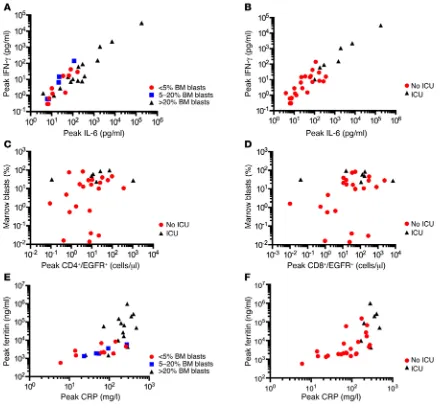
Figure 2. Serious toxicity due to CRS is mainly seen in B-ALL patients with high BM-tumor burden. (A) The peak IFN-γ and IL-6 concentrations in serum in the first 28 days after CAR–T cell infusion are shown in patients who had high (> 20% blasts; n = 15), intermediate (5%–20% blasts; n = 5), or low (≤ 5% blasts; n = 10) tumor burden before lymphodepletion chemotherapy and CAR–T cell infusion. Each point represents data from 1 patient. (B) The peak IFN-γ
of follow-up. A difference in the rate or severity of toxicity in the 11 patients with a prior allogeneic HCT was not apparent. CAR–T cells were manufactured from the engrafted donor T cells obtained from the patients after transplant, and it is notable that none of the 11 patients developed acute graft-versus-host disease (GVHD) after CAR–T cell therapy. One patient who had stage 1 acute skin GVHD before study enrollment developed chronic GVHD requir-ing corticosteroid therapy 3 months after CAR–T cell infusion.
Correlates of CRS and neurotoxicity. CRS is initiated by
activa-tion and proliferaactiva-tion of CAR–T cells after recogniactiva-tion of CD19+
target cells and is characterized by elevated serum levels of IL-6 and IFN-γ (2). We observed significantly higher peak IL-6 and IFN-γ levels after CAR–T cell infusion in patients with high BM tumor burden (Figure 2A) and in those requiring ICU care com-pared with those that did not (Figure 2B), and the need for ICU correlated with a higher percentage of BM blasts before
[image:7.585.53.530.52.498.2]The Journal of Clinical Investigation
C l i n i C a l M e d i C i n ephodepletion chemotherapy (Figure 2, C and D). We used flow cytometry to identify EGFRt+ CAR–T cells and determine their
frequency in peripheral blood obtained at prescribed intervals after infusion. With the exception of the patient who died on day 3 after CAR–T cell infusion prior to peak CAR–T cell expan-sion, no patients with a peak CD4+ EGFRt+ CAR–T cell count of
less than 9.8 cells/μl or CD8+ EGFRt+ count of less than 15 cells/
μl required ICU care, whereas 6 of 15 (40%) patients with a peak CD4+ EGFRt+ count of 9.8 or more cells/μl and 6 of 21 (29%) with
a peak CD8+ EGFRt+ count of 15 or more cells/μl required ICU.
Elevations of serum C-reactive protein (CRP) and ferritin, which
are more readily measured by clinical laboratories than serum cytokines, also correlated with BM disease burden (Figure 2E) and with the occurrence of sCRS requiring ICU care (Figure 2F). sCRS was more likely to occur in patients with peak CRP concentrations of 150 or more mg/l (7 of 13) and/or with serum ferritin concentra-tions of 10,000 or more ng/ml (5 of 10). Ferritin and CRP levels declined after tocilizumab or corticosteroid therapy.
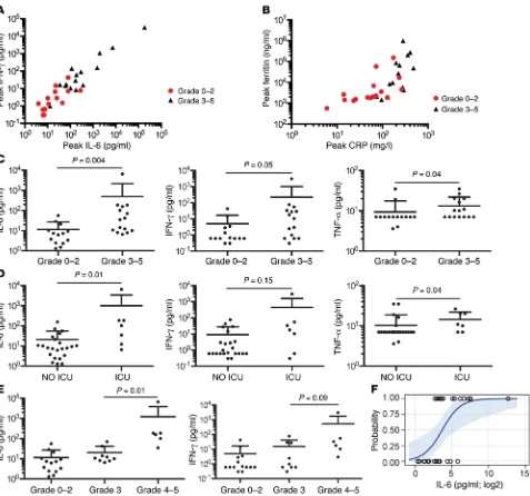
[image:8.585.60.537.53.527.2]The pathogenesis of neurotoxicity developing after CD19 CAR–T cell therapy is poorly defined. In our study, all patients who developed neurotoxicity had evidence of CRS, and peak levels of IL-6, IFN-γ, ferritin, and CRP were significantly higher
neurotoxicity compared with patients who developed grade 3 neurotoxicity (Figure 3E). Univariate logistic analysis suggested that IL-6 concentration is a predictor of patients who are likely to develop grade 3 or higher neurotoxicity (odds ratio 11.0 [95% CI: 1.4–84.3], P = 0.02) (Figure 3F). Stepwise multivariate logis-tic regression analysis incorporating day 1 serum IL-6 concen-tration, infused CAR–T cell dose, lymphodepletion chemother-apy regimen, and the fraction of BM represented by normal and abnormal CD19+ cells indicated that serum IL-6 concentration
of more than 30 pg/ml on day 1 (P = 0.02) and the total number of CD19+ cells in BM before therapy (P = 0.06) are independent
predictors of subsequent development of grade 3 or higher neu-rotoxicity. Of note, serum IL-6 of more than 30 pg/ml on day 1 identified all patients in our study who subsequently developed grade 4 or higher neurotoxicity. Thus, evaluation of serum IL-6 in patients who developed grade 3 or higher neurotoxicity
[image:9.585.48.534.58.428.2](Fig-ure 3, A and B). The fever and hypotension that accompany CRS were responsive to administration of corticosteroids and/or tocilizumab, but neurotoxicity was less responsive to these inter-ventions. We evaluated serum levels of IL-6, IFN-γ, and TNF-α on the first day after CAR–T cell infusion to determine wheth-er these might swheth-erve as biomarkwheth-ers to identify patients at high risk for subsequent neurotoxicity and ICU care. We found that IL-6, IFN-γ, and TNF-α levels on the first day after CAR–T cell infusion were significantly higher in patients who subsequently developed severe (grade ≥ 3) neurotoxicity compared with those who did not (Figure 3C) and were also higher in patients who required ICU care (Figure 3D). Moreover, serum IL-6 and IFN-γ concentrations on day 1 after CAR–T cell infusion were signifi-cantly higher in patients who subsequently developed grade 4
The Journal of Clinical Investigation
C l i n i C a l M e d i C i n eIn vivo expansion and persistence of CD19 CAR–T cells. The
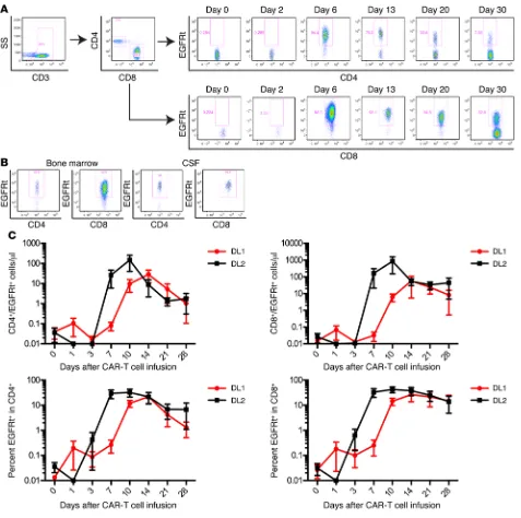
pro-liferation, migration to sites of tumor, and persistence of CAR–T cells in vivo after adoptive transfer are likely to be critical deter-minants of antitumor efficacy, and the kinetics of CAR–T cell expansion may in part dictate the risk of serious toxicity. CAR–T cells were detected by flow cytometry in the blood at 1 or more time points after infusion in all patients (Figure 4A). CAR–T cells were also present in BM obtained at response evaluation in 22 of 27 patients from whom the BM was examined for the presence of CAR–T cells and in the CSF of 6 of the 8 patients from whom sam-ples were obtained for clinical indications (Figure 4B).
Several factors could contribute to CAR–T cell expansion and persistence in vivo, including cell product composition, cell dose, tumor burden, and the conditioning regimen used to pro-vide lymphodepletion. Previous studies at other centers in which CD19 CAR–T cells were manufactured from T cells of undefined composition did not identify a clear correlation between the concentration early after CAR–T cell infusion might be used to
identify patients at high risk of severe neurotoxicity to evaluate early intervention approaches.
Because the occurrence of toxicity correlated with BM tumor burden and sCRS was noted in patients treated at a higher CAR–T cell dose, we initiated risk-adapted CAR–T cell dosing during the course of the study, in which patients with greater than 20% BM blasts received a low dose of CAR–T cells (2 × 10⁵ EGFRt+ cells/
kg) and those with 20% or less BM blasts received a higher dose of CAR–T cells (2 × 10⁶ EGFRt+ cells/kg). Prior to incorporation of
risk-adapted CAR–T cell dosing, 6 of 6 patients with high–tumor burden B-ALL who were treated at DL2 or DL3 (≥ 2 × 10⁶ EGFRt+
[image:10.585.86.508.55.457.2]cells/kg) required ICU care after CAR–T cell therapy, and all 6 patients developed severe neurotoxicity (median grade 4). In con-trast, only 1 of 10 patients with high–tumor burden B-ALL who were treated with risk-adapted dosing at DL1 required ICU care, and only 5 of 10 developed neurotoxicity (median grade 3).
The Journal of Clinical Investigation
C l i n i C a l M e d i C i n eCD8+ T cells selected from this patient to expand in culture. The
other patient had 0.016% abnormal BM blasts by flow cytometry before CAR–T cell infusion, received CAR–T cells at DL1, and had poor in vivo CAR–T cell expansion (peak CD8+ EGFRt+ 0.97/μl,
peak CD4+ EGFRt+ 0.42/μl). This patient subsequently
under-went allogeneic HCT, but relapsed after HCT and was reenrolled in the CAR–T cell study. The patient achieved a CR after being retreated with CAR–T cells that were manufactured from T cells collected from the patient after allogeneic HCT and administered at a higher dose (DL2).
Molecular studies of BM, including quantitative PCR (QPCR), FISH, and conventional karyotyping, were negative in all patients who achieved remission by flow cytometry and had an identified B-ALL–associated genomic mutation, except for 2 patients — one with a persistent t(4;11) who subsequently relapsed with a clon-ally related CD19– acute myeloid leukemia and a second patient
with BCR-ABL transcripts identified at the limit of detection by QPCR (Table 1). Thus, 25 of 29 patients (86%) achieved CR with-out evidence of MRD by flow cytometry and conventional karyo-typing, FISH, or QPCR in those with an identified B-ALL–associ-ated genomic mutation. Deep sequencing of the immunoglobulin heavy locus (IGH) genes in BM samples from 17 patients in whom we identified a neoplastic clonal IGH sequence showed that 10 (59%) had no detectable disease using an assay with a sensitivity to detect 1 malignant blast in 1,000,000 nucleated cells. In addi-tion to the efficacy in eliminating BM- and blood-based leukemic blasts, extramedullary disease identified by PET-CT scan was eliminated in 6 of 7 patients after CAR–T cell therapy (Supple-mental Figure 3). We did not observe a difference in response rate between patients who had or had not previously undergone allo-geneic HCT (HCT group, 10/11 CR, 1/11 BM MRD; no HCT group, 15/19 CR, 3/19 BM MRD, 1/19 died before evaluation), demon-strating that CD19 CAR–T cells of defined composition can be successfully manufactured and administered to patients following allogeneic HCT. Thus, the data show a high rate of CR with CAR–T cells of defined CD4+:CD8+ T cell ratio in patients with refractory
B-ALL, including those with extramedullary disease or with leuke-mia relapse after allogeneic HCT.
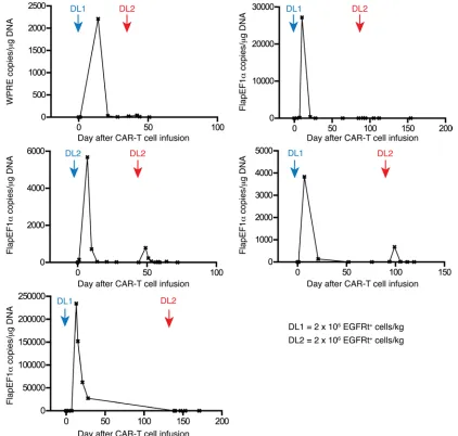
Transgene immune responses limit persistence and efficacy of sec-ond CAR–T cell infusions. Five patients treated in our study with
CAR–T cells after Cy-based lymphodepletion had persistent leu-kemia or subsequently relapsed and received a second infusion of CAR–T cells at the same (n = 1) or a 10-fold higher dose (n = 4) after no lymphodepletion (n = 1) or lymphodepletion with Cy and Flu (n = 3) or clofarabine (n = 1). Despite evidence of CAR–T cell proliferation after the first infusion and the same or higher BM blast count at the time of second infusion, there was no expansion or persistence of CAR–T cells or demonstrable antitumor activ-ity in any of these 5 patients (Figure 6). We considered that these patients might have mounted a T cell immune response directed at epitopes encoded by the CAR transgene after the first infusion, since the CD19-specific single-chain variable fragment (scFv) is of murine origin and there are unique fusion sites between com-ponents of the CAR. To evaluate whether a cytotoxic CD8+ T
cell response to autologous CAR–T cells was present, we used a modification of an in vitro assay we previously reported to analyze transgene product immunogenicity (19, 20). We detected CAR-infused CAR–T cell dose and the absolute number of CAR–T cells
detected in vivo in the recipient (2–4). We found that administer-ing a consistent CAR–T cell product of a defined CD4+:CD8+ T
cell ratio to all patients revealed a relationship between cell dose and the peak number of CD4+ and CD8+ CAR–T cells. Prior to
commencing risk-adapted CAR–T cell dosing based on BM tumor burden, we found that patients who received CAR–T cells at DL2 had an earlier and higher peak in absolute CD4+ and CD8+
EGFRt+ CAR–T cell counts in blood compared with those who
received DL1 (Figure 4C). At both DLs, the absolute number of CD8+ CAR–T cells was higher than that of CD4+ CAR–T cells at
the peak of expansion, despite CD4+ and CD8+ CAR–T cells
hav-ing been infused at the same dose.
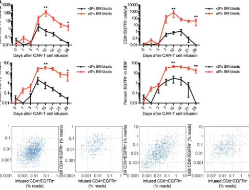
The burden of CD19+ cells in the patient could also affect
CAR–T cell numbers either by inducing their proliferation or resulting in activation-induced cell death. Stratifying patients treated prior to risk-adapted dosing based on the proportion of CD19+ leukemia blasts in the BM showed that patients with a
higher percentage of blasts in the BM had significantly higher CD4+ and CD8+ CAR–T cell counts in the blood at the peak of
expansion and better persistence at day 28 after infusion (Figure 5A). The accumulation of CD4+ and CD8+ CAR–T cells in vivo
could occur as a result of expansion of a diverse repertoire of poly-clonal T cells derived from the transduced T cell population or the expansion of few highly proliferative T cell clonotypes with a proliferative and/or survival advantage. To distinguish these pos-sibilities, we performed high-throughput sequencing of the T cell receptor β (TCRB) genes utilized by flow-sorted CD4+ and CD8+
EGFRt+ CAR–T cells obtained from the infused CAR–T cell
prod-ucts of 5 patients, at the peak of CAR–T cell expansion in vivo, and at one or more later time points. Both at the peak of expan-sion and at later times after elimination of detectable leukemia, the CD4+ and CD8+ CAR–T cells present in vivo were polyclonal,
and multiple distinct TCRB clonotypes that were identified in the infusion product were observed in the CAR–T cell population in the patient (Figure 5B). Together, these results demonstrate that the infusion of CAR–T cell products comprising CD8+ and CD4+ T
cells in a defined ratio results in predictable polyclonal prolifera-tion of both CD4+ and CD8+ T cells with kinetics that are affected
by both CAR–T cell dose and tumor burden.
Clinical response. Twenty-nine of 30 patients who received
We enrolled 32 patients and, importantly, did not exclude any patient based on a low absolute lymphocyte count, the presence of circulating blasts, or the results of a screening test to ensure the patient had sufficient T cells capable of proliferating to CD3/CD28 stimulation. Therefore, our study was not biased due to exclusion of patients with poor lymphocyte numbers and function. A clini-cal trial of CD19 CAR–T cell therapy for pediatric B-ALL excluded 24% of patients due to the failure of T cells to meet predefined cri-teria for in vitro proliferation (21).
A primary objective of the study was to assess the feasibility of selecting CD4+ and CD8+ subsets for manufacturing of CAR–T
cells and formulating the cell product in a consistent 1:1 ratio of CD4+ and CD8+ CAR–T cells. CD8+ T
CM cells and their progeny
have been shown to have stemness and to exhibit superior per-sistence compared with TEM cells after adoptive transfer, suggest-ing a potential advantage of this subset for providsuggest-ing long-term persistence (12, 22). Our data show that in heavily pretreated B-ALL patients, the absolute CD8+ T
CM counts were sufficient for
selection and manufacturing of a CAR–T cell product in 16 of 30 patients and that selection of bulk CD8+ T cells was feasible in the
remaining 14 patients. Thus, a cell product comprising CD4+ and
CD8+ CAR–T cells was successfully manufactured and infused to
all patients with refractory B-ALL, and the 1:1 ratio of CD4+:CD8+
CAR–T cells was achieved in 27 of 30 patients.
The high overall rate of BM remission of 93% by flow cytome-try in this study and differences in lymphodepletion regimens and infused cell doses do not allow comparison of the efficacy of CAR– T cell products manufactured from CD8+ T
CM cells or from bulk
CD8+ T cells. Analysis of differences in long-term persistence of
cell products that were selected for CD8+ T
CM or bulk CD8+ T cells
in our study was further complicated by our findings that immune-mediated rejection of CAR–T cells occurs in some patients, which may provide an explanation for the loss of CAR–T cells observed in a subset of patients in other studies (1–4). Nevertheless, our data demonstrate that performing cell selections from heavily pretreated B-ALL patients is feasible and that formulating CD19 CAR–T cells in a defined CD4+:CD8+ CAR–T cell ratio, either from
a starting population of isolated CD8+ T
CM or bulk CD8+ cells, can
provide a remarkably effective CAR–T cell product at cell doses that are 5- to 100-fold lower than those used to treat a majority of patients in other trials (1–4). The 93% remission rate by flow cytometry and 86% MRD-negative CR rate in our study compares very favorably to that reported by others in which CAR–T cells of undefined composition were manufactured using CD19 CARs that incorporate either a 4-1BB costimulatory domain (children and young adults, 79%) or a CD28 costimulatory domain (adults, 75%; children and young adults, 60%) (1–4).
An important predictor of the efficacy of CD19 CAR–T cells is their ability to expand in vivo in response to recognition of CD19+
target cells, and the patients that failed to respond in prior stud-ies typically had poor accumulation of CAR–T cells in vivo. The infusion of CAR–T cell products comprising a uniform ratio of CD4+:CD8+ CAR–T cells demonstrated a correlation between cell
dose and earlier and higher peak expansion of clonally diverse CAR–T cells, a finding that has not been reported in studies in which CAR–T cells were manufactured and infused without consideration of the CD4+:CD8+ ratio. Independent of cell dose,
specific T cell responses in all 5 patients in whom CAR–T cells failed to persist after the second infusion, and epitope mapping in 1 patient identified immunogenic epitopes within the murine FMC63 scFv (Supplemental Figure 4).
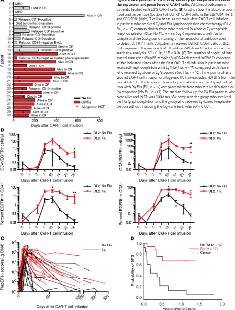
Addition of Flu to Cy lymphodepletion improves persistence of CAR–T cells. Ten of 12 patients (83%) who received
lymphodeple-tion with Cy alone or with etoposide followed by CAR–T cell infu-sion and who survived until disease restaging achieved BM CR by flow cytometry. We observed recovery of endogenous B cells in 8 of these patients by day 90 after CAR–T cells, and 7 patients sub-sequently relapsed (Figure 7A). One of the patients relapsed with CD19– leukemia, but the remaining 6 relapses were CD19+,
sug-gesting a loss of CAR–T cell immunosurveillance. Indeed, in 5 of the 6 patients with CD19+ relapse, a loss of CAR–T cell
engraft-ment in blood was shown by PCR analysis for transgene sequenc-es, and an immune response to the CAR transgene product was detected in all of these patients.
To determine whether the development of an immune response to CAR–T cells might be prevented or delayed by inten-sifying the lymphodepletion regimen, we administered Cy/Flu to 17 subsequent patients prior to CAR–T cell infusion and compared CAR–T cell expansion and persistence to those of prior patients who received Cy without Flu. Patients with similar BM blast per-centage who received Cy/Flu and DL2 of CAR–T cells had a sig-nificantly higher peak of expansion and numbers of CD4+ and
CD8+ EGFRt+ CAR–T cells at day 28 than those treated at DL2 who
did not receive Flu (Figure 7, B and C). Sixteen of 17 patients who received Cy/Flu lymphodepletion and DL1 or DL2 of CAR–T cells had a CR. Three of 4 patients who received lymphodepletion with Cy or Cy/etoposide and had no detectable disease by IGH sequenc-ing ultimately relapsed, whereas 0 of 6 patients who received Cy/ Flu lymphodepletion and eliminated the malignant clone by IGH sequencing relapsed. Although follow-up of the cohort of patients who received Cy/Flu lymphodepletion is short, the DFS of this group is superior to those that did not receive Flu (Figure 7D).
Discussion
Previous clinical trials have reported a high rate of morphologic remission in small numbers of adult and pediatric patients with B-ALL treated with CD19 CAR–T cells (2–4). This approach repre-sents an important therapeutic advance; however, much remains to be learned regarding the optimal lymphodepletion regimens and cell dose(s) to eradicate MRD, the prediction and manage-ment of toxicities, and the mechanisms of relapse. Systematically studying these issues has been challenging in part because of the differences in the phenotypic composition of the CAR–T cell products that were administered to individual patients in prior trials (2–4). Our data show that heavily pretreated adult B-ALL patients have highly variable numbers and proportions of CD4+
and CD8+ T cell subsets in peripheral blood. Preclinical
stud-ies provide support for improving potency and predictability of therapeutic efficacy by manufacturing CD19 CAR–T cell prod-ucts of defined CD4+ and CD8+ subset composition (17). Here,
The Journal of Clinical Investigation
C l i n i C a l M e d i C i n ewithin hours and discharge from the ICU within 1 to 2 days in most patients. Neurotoxicity was manifest by signs that included encephalopathy and seizures and focal deficits; in some patients, it followed a time course that was distinct from that of CRS. Neu-rologic signs often reached maximal severity after resolution of CRS and were not clearly responsive to intervention with tocili-zumab or corticosteroids at either low or high doses.
We investigated whether surrogate markers of systemic inflammation could identify patients with sCRS and potentially be used to guide intervention to either suppress cytokine release or eliminate CAR–T cells in the event of severe toxicity. Serum fer-ritin and CRP concentrations correlated with the severity of acute toxicity and declined in response to tocilizumab or corticosteroids; however, additional data are needed to determine their utility in monitoring the response to CRS therapy. Severe hyperferritinemia (> 20,000 ng/ml) is not typically observed in infections even in heavily transfused patients (24) and could serve as a useful tool in distinguishing sCRS from other causes of fever after CAR–T cell infusion. Serum concentrations of IL-6 and IFN-γ also correlated with the severity of toxicity and had predictive value, with high concentrations within 1 day after CAR–T cell infusion identify-ing patients who subsequently developed severe neurotoxicity. If these findings are confirmed in additional patients, analysis of serum IL-6 and IFN-γ concentrations early after CAR–T cell infu-sion might be employed to identify patients at risk of life-threaten-ing toxicity in whom early and aggressive intervention to mitigate immune cell cytokine production might be evaluated.
Although CAR–T cells induced CR in a majority of patients in our study, disease relapse occurred in a subset of patients. Leu-kemia relapse after CAR–T cells could be classified into 2 distinct phenotypes: those with loss of the CD19 target antigen as observed by others (25) and those that remained CD19+ and relapsed in a
manner associated with loss of CD19 CAR–T cells in blood. Two patients in our study relapsed with CD19– disease, 1 with B-ALL
and 1 with a myeloid phenotype switch. CD19+ relapse occurred
exclusively in patients who did not have persisting CAR–T cells in blood; retreatment of these patients with a second CAR–T cell infusion did not result in CAR–T cell proliferation, persistence, or antitumor activity. We demonstrated that the loss of CAR–T cells was due to development of CD8+ T cell immunity to the CAR
trans-gene product. Because all CD19 CARs used in reported clinical tri-als of CD19 CAR–T cell therapy incorporate a murine scFv, these data provide one potential mechanism for the loss of CAR–T cells observed in other trials (2–8, 23, 26). A solution that may reduce immunogenicity of the construct would be to incorporate a human rather than murine CD19-specific scFv into the CAR construct.
Our trial investigated whether the addition of Flu to the lym-phodepletion regimen could delay or abrogate development of anti-CAR immune responses and improve CAR–T cell expansion and persistence. We noted remarkably higher CD4+ and CD8+
CAR–T cell proliferation and persistence in patients who received Flu compared with those who did not receive Flu. The mechanisms by which Flu increased CAR–T cell accumulation and persistence could include greater lymphodepletion, resulting in increased lev-els of homeostatic cytokines that support T cell proliferation and survival, delaying or preventing the anti-CAR immune respons-es and potentially modifying the tumor microenvironment to tumor burden was an important factor in driving CAR–T cell
expansion, and patients with 5% or greater BM blasts had on aver-age a 100-fold greater expansion of both CD8+ and CD4+ CAR–T
cells than patients with less than 5% blasts. Administering a sin-gle low dose of 2 × 10⁵ CAR–T cells/kg of a defined CD4+:CD8+ T
cell ratio was effective in inducing a CR in all patients with high BM tumor burden. The high potency of the CAR–T cell products was not limited to elimination of BM disease, and bulky extra-medullary disease identified by PET/CT scan was also eradicated in all but 1 patient. Seven of 17 patients (41%) who achieved BM CR by flow cytometry had detectable copies of the neoplastic IGH sequence in BM early after CAR–T cell infusion; however, the detection of nonviable leukemic tissue renders the significance of this finding uncertain. Only 4 of these 7 patients subsequently relapsed; 2 of these had CD19-negative leukemia. Three patients had persistent BM disease detected by flow cytometry at the first evaluation after receiving CAR–T cells; 1 of these subsequently achieved CR in the absence of further therapy. Of the 2 patients who never achieved BM CR by flow cytometry, 1 received a pre-dominantly CD4+ CAR–T cell product because we were unable
to propagate sufficient CD8+ CAR–T cells in culture. The clinical
outcome in this patient is consistent with our preclinical observa-tions in Raji tumor–bearing immunodeficient mice that received CAR–T cells manufactured from distinct T cell subsets, showing reduced potency when either CD8+ or CD4+ CAR–T cells were
omitted from the formulated product (17). Both of the remain-ing patients had low tumor burden in the BM before CAR–T cell therapy, raising the additional possibility that a higher dose of CAR–T cells may be necessary to effectively treat patients with low tumor burden, perhaps due to a stochastic failure to receive sufficient antigenic stimulation to drive CAR–T cell expansion in vivo. Given the high rate of CR in our phase I clinical trial, a larger patient cohort will need to be studied to understand the mecha-nisms responsible for failure to achieve remission.
Although CD19 CAR–T cells are therapeutically effective in patients with relapsed and refractory B-ALL, significant toxici-ties have occurred in all studies, highlighting the need to improve the therapeutic index (2–4, 23). In our study, severe toxicity due to CAR–T cells was predominantly seen in patients with 20% or more BM blasts and occurred more often after infusion of higher CAR–T cell doses. The severity of toxicity correlated with higher peak levels of CD4+ and CD8+ CAR–T cells in blood and higher
peak IFN-γ and IL-6 concentrations in serum. Taken together, these data imply that a variable cell-dosing strategy may prove optimal for treating B-ALL with CAR–T cells. Patients with higher tumor burden may be best treated initially with a low CAR–T cell dose to minimize toxicity, whereas those with lower tumor burden may require higher or repeated doses of CAR–T cells to ensure rec-ognition of a minimal tumor antigen load.
enriched CD4+ and CD8+ T cell subsets were separately stimulated
with Dynabeads CD3CD28 CTS paramagnetic beads, transduced with the lentivirus encoding the CD19-specific CAR and EGFRt, and then cultured in 50 U/ml IL-2. After removal of CD3CD28 beads, the CD4+ and CD8+ T cells were separately stimulated with
an irradiated CD19+ EBV LCL and cultured in media
supplement-ed with 50 U/ml IL-2. CAR–T cell manufacturing was completsupplement-ed within 15 to 20 days after initial bead stimulation. Quality assess-ments were performed separately on the cultured CD4+ and CD8+
CAR–T cells, and the 2 fractions were formulated in a 1:1 ratio of CD3+CD4+EGFRt+:CD3+CD8+EGFRt+ cells for infusion.
Lymphodepletion chemotherapy. To deplete endogenous lympho-cytes prior to adoptive transfer of CAR–T cells, patients received 1 of 4 chemotherapy regimens: 2–4 g/m2 Cy i.v. on day 1 (n = 11); 2–3
g/m2 Cy i.v. on day 1 and 100 mg/m2/d etoposide i.v. on days 1 to 3
(n = 2); and 60 mg/kg Cy i.v. on day 1 and 25 mg/m2/d Flu i.v. on either
days 2 to 4 or days 2 to 6 for patients who had previously received an allogeneic HCT and/or were considered at increased risk of toxicity (n = 17; Table 1).
CAR–T cell infusion. CD19 CAR–T cells were administered i.v. to each patient at or as close as possible to 1 of 3 cell DLs (DL1, 2 × 10⁵
EGFRt+ cells/kg, n = 13; DL2, 2 × 10⁶ EGFRt+ cells/kg, n = 15; DL3, 2 ×
107 EGFRt+ cells/kg, n = 2) 48 to 96 hours after completing
chemother-apy. CD4+ and CD8+ CAR–T cells were successfully manufactured for
all patients; however, CAR–T cells from 3 patients were not formulated at the defined 1:1 ratio of CD4+:CD8+ T cells due to insufficient growth
of CD4+ or CD8+ CAR–T cells in culture (DL2, n = 2; DL3, n = 1). Two
patients received cryopreserved CAR–T cells, whereas the remainder were formulated and infused without cryopreservation.
Clinical response assessment. BM aspirates and biopsies were obtained from each patient before lymphodepletion chemotherapy and approximately 2 to 4 weeks after CAR–T cell infusion. CR was defined as absence of immunophenotypically abnormal blasts in the BM by mor-phology and flow cytometry (limit of detection 1:10,000) and, in those patients with a known molecular marker of their leukemia, the absence of abnormalities by conventional karyotyping, FISH, or QPCR. When available, an aliquot of each BM aspirate was submitted for IGH deep sequencing (Adaptive Biotechnologies) from patients who attained CR. Patients with CNS leukemia identified before lymphodepletion che-motherapy underwent CSF sampling for flow cytometry analysis after CAR–T cell therapy. Patients with extramedullary disease underwent whole-body PET-CT imaging before and after CAR–T cell therapy. Toxicity was graded using the NCI CTCAE (v4.03).
Assessment of CAR–T cell persistence. Blood samples were obtained from patients before and at intervals after CAR–T cell infusion, and flow cytometry was performed to identify CD4+ and CD8+ CAR–T
cells as viable CD45+CD3+CD4+CD8–EGFRt+ or CD45+CD3+CD4-
CD8+EGFRt+ events, respectively. Absolute CAR–T cell counts were
determined by multiplying the percentage of CAR–T cells identified by flow cytometry in a CD45+ forward scatter–side scatter (FS-SS)
lymphocyte gate by the absolute lymphocyte count established by a complete blood count (CBC) performed on the same day. To establish loss of CAR–T cell persistence due to an anti-CAR transgene immune response, we used QPCR to detect integrated transgene sequence.
Evaluation of serum cytokines. Serum concentrations of IFN-γ, IL-6, and TNF-α were evaluated by Luminex assay, according to the manufacturer’s instructions.
improve accessibility and stimulation of CAR–T cells. Consis-tent with the increased CAR–T cell expansion and persistence in patients who received Flu, we noted an improvement in DFS, albe-it walbe-ith a relatively short duration of follow-up. Only 1 of 17 patients (6%) who received Flu-containing lymphodepletion followed by CAR–T cells of defined composition developed CD19+ relapse,
and this occurred in a patient who, subsequent to achieving remis-sion after CAR–T cell therapy, had an allogeneic HCT that resulted in elimination of the CAR–T cells. While these data are encourag-ing, additional patient accrual and longer follow-up are required.
We believe this study is the largest reported series of adult B-ALL patients treated with CD19 CAR–T cells and the first to demonstrate the feasibility of selecting defined T cell subsets for CAR engineering and formulation of defined therapeutic products for adoptive therapy. Data from this phase I/II trial demonstrate potent antitumor activity and reveal dose/response and dose/ toxicity relationships that have not been clearly evident in CAR–T cell trials using unselected cells. The development of novel clini-cal cell isolation technologies that utilize flow cytometry cell sort-ing or serial column-based selections after labelsort-ing with reversibly binding reagents may further refine the selection of T cells with a complex immunophenotype and facilitate manufacturing defined CAR–T cell products for clinical evaluation (18, 27, 28).
Methods
Study design and patient selection. We performed a phase I/II open-label trial to evaluate the feasibility of manufacturing CD19 CAR–T cells formulated in a defined ratio of CD8+ and CD4+ T cell subsets and to
assess the safety and efficacy of this therapy in patients with relapsed or refractory CD19+ B cell malignancies. This manuscript reports the
data from B-ALL patients in the study. There were no exclusion crite-ria for leukapheresis, CD19 CAR–T cell manufacturing, lymphodeple-tion chemotherapy, or CD19 CAR–T cell infusion based on the abso-lute lymphocyte count, the results of a test in vitro stimulation, or the presence or number of leukemia blasts in the peripheral blood. The study has been registered at ClinicalTrials.gov (NCT01865617).
T cell subset selection and CD19 CAR–T cell manufacturing. Periph-eral blood mononuclear cells (PBMCs) were collected by leukapher-esis, and the product was divided into 2 aliquots for enrichment of CD4+ and CD8+ T cell subsets for CAR–T cell manufacturing
(Supple-mental Figure 1). CD4+ T cells were selected from 1 aliquot by
posi-tive immunomagnetic selection using the CliniMACS CD4 Reagent System (Miltenyi Biotec). CD8+ T
CM cells were enriched from the
sec-ond aliquot using a 2-step CliniMACS selection procedure involving depletion of CD4+, CD14+, and CD45RA+ cells, followed by selection
of CD62L+ cells from the CD4–CD14–CD45RA- fraction as described
(18). In patients with an absolute CD8+ T
CM cell count of less than 20/
μl, either the CD62L+ selection was omitted or bulk CD8+ T cells were
selected using the CliniMACS CD8 Reagent System.
We designed a CAR comprising an FMC63-derived CD19-spe-cific scFv fused to a modified IgG4-hinge spacer, a CD28 transmem-brane domain, a 4-1BB costimulatory domain, and a CD3ζ signaling domain (29). To enable precise measurement of transduced CD4+
and CD8+ CAR–T cells, we incorporated EGFRt separated from the
The Journal of Clinical Investigation
C l i n i C a l M e d i C i n eFHCRC Institutional Review Board. Written, informed consent was obtained from all patients after a discussion of the possible risks and adverse effects of the therapy.
Author contributions
CJT, SRR, and DGM designed the trial and experiments and wrote the paper. LAH, CB, KM, BP, TMB, ER, and NS performed experiments. LAH, MH, DS, SC, LS, XC, CY, BW, SH, JC, and MCJ designed experiments. CC collected research data. TAG and DL analyzed statistical data.
Acknowledgments
The authors would like to thank the FHCRC Cell Processing Facil-ity and Seattle Cancer Care Alliance (SCCA) Cell Therapy Labora-tory for assistance in CAR–T cell manufacturing; Kari Blankenship, Anna-Marie Kane, Franziska Sommermeyer, Sheila Gelder, Jen-nifer Lindquist, and the staff of the SCCA Immunotherapy Clinic for assistance with clinical care and data management; and the FHCRC Immune Monitoring Lab. C.J. Turtle is a Damon Runyon Clinical Investigator. Funding was provided by the following: NCI R01 CA136551; Life Science Development Fund; Juno Therapeu-tics; Bezos Family Immunotherapy Initiative.
Address correspondence to: Cameron J. Turtle, Clinical Research Division, Fred Hutchinson Cancer Research Center, 1100 Fairview Ave N, Seattle, Washington 98109, USA. Phone: 206.667.7073; E-mail: [email protected].
Michael Hudecek’s present address is: Department of Medicine II, University of Wuerzburg, Wuerzburg, Germany.
Evaluation of transgene immunogenicity. We evaluated CD8+ T cell
immune responses to the CAR transgene using a modification of an assay we previously developed in our lab (19). Cryopreserved PBMCs collected from patients before lymphodepletion chemotherapy and approximately 4 weeks after CAR–T cell infusion were stimulated twice at 7-day intervals with autologous irradiated CAR–T cells and IL-2. The preinfusion and postinfusion PBMC cultures were assayed for lysis of autologous CAR–T cells and autologous nontransduced T cells in a
51chromium release assay. An immune response against the CAR
trans-gene was defined as the presence of specific lysis of autologous CAR–T cells by postinfusion PBMC cultures, but not of autologous nontrans-duced T cells or autologous CAR–T cell by preinfusion PBMC cultures. Statistics. Comparisons of continuous variables between groups were made using the Wilcoxon rank-sum test. The correlation between 2 continuous variables was assessed using linear regression, with the values of some variables replaced by their ranks due to relatively large values for some of the parameters (ferritin, CRP, IFN-γ, IL-6). Uni-variate and stepwise multiUni-variate logistic regression were performed to assess predictors for the occurrence of severe neurotoxicity. No adjustments were made for multiple comparisons. The absolute CD4+
and CD8+ EGFRt+ CAR–T cell counts and their percentages within the
parental subset were compared using the Mann-Whitney U test. DFS was determined as the time from CAR–T cell infusion until failure, defined as death or the detection of relapsed or persistent disease by morphologic, flow cytometry, karyotyping, FISH, and/or QPCR stud-ies. We compared the group who received Cy/Flu lymphodepletion and the group who received Cy-based lymphodepletion without Flu using the log-rank test, where P = 0.001. Significant was defined as P < 0.05.
Study approval. This study was conducted according to the prin-ciples of the Declaration of Helsinki and with the approval of the
1. Brentjens RJ, et al. Safety and persistence of adoptively transferred autologous CD19-targeted T cells in patients with relapsed or che-motherapy refractory B-cell leukemias. Blood. 2011;118(18):4817–4828.
2. Lee DW, et al. T cells expressing CD19 chimeric antigen receptors for acute lymphoblastic leukae-mia in children and young adults: a phase 1 dose-escalation trial. Lancet. 2015;385(9967):517–528. 3. Maude SL, et al. Chimeric antigen receptor T
cells for sustained remissions in leukemia. N Engl
J Med. 2014;371(16):1507–1517.
4. Davila ML, et al. Efficacy and toxicity manage-ment of 19-28z CAR T cell therapy in B cell acute lymphoblastic leukemia. Sci Transl Med. 2014;6(224):224ra25.
5. Kochenderfer JN, et al. B-cell depletion and remissions of malignancy along with cytokine-associated toxicity in a clinical trial of anti-CD19 chimeric-antigen-receptor-transduced T cells.
Blood. 2012;119(12):2709–2720.
6. Kochenderfer JN, et al. Donor-derived CD19-tar-geted T cells cause regression of malignancy per-sisting after allogeneic hematopoietic stem cell transplantation. Blood. 2013;122(25):4129–4139. 7. Kochenderfer JN, et al. Chemotherapy-refractory
diffuse large B-cell lymphoma and indolent B-cell malignancies can be effectively treated with autologous T cells expressing an anti-CD19 chimeric antigen receptor. J Clin Oncol.
2015;33(6):540–549.
8. Porter DL, Levine BL, Kalos M, Bagg A, June CH. Chimeric antigen receptor-modified T cells in chronic lymphoid leukemia. N Engl J Med. 2011;365(8):725–733.
9. Porter DL, et al. Chimeric antigen receptor T cells persist and induce sustained remissions in relapsed refractory chronic lymphocytic leuke-mia. Sci Transl Med. 2015;7(303):303ra139. 10. Lee DW, et al. Current concepts in the diagnosis
and management of cytokine release syndrome.
Blood. 2014;124(2):188–195.
11. Riddell SR, et al. Adoptive therapy with chimeric antigen receptor-modified T cells of defined sub-set composition. Cancer J. 2014;20(2):141–144. 12. Berger C, Jensen MC, Lansdorp PM, Gough M, Elliott C, Riddell SR. Adoptive transfer of effec-tor CD8+ T cells derived from central memory cells establishes persistent T cell memory in pri-mates. J Clin Invest. 2008;118(1):294–305. 13. Gattinoni L, et al. A human memory T cell
subset with stem cell-like properties. Nat Med. 2011;17(10):1290–1297.
14. Stemberger C, Neuenhahn M, Gebhardt FE, Schiemann M, Buchholz VR, Busch DH. Stem cell-like plasticity of naïve and distinct memory CD8+ T cell subsets. Semin Immunol. 2009;21(2):62–68.
15. Turtle CJ, et al. Innate signals overcome acquired TCR signaling pathway regulation and govern
the fate of human CD161hi CD8α+ semi-invariant T cells. Blood. 2011;118(10):2752–2762. 16. Wang X, Berger C, Wong CW, Forman SJ, Riddell
SR, Jensen MC. Engraftment of human central memory-derived effector CD8+ T cells in immu-nodeficient mice. Blood. 2011;117(6):1888–1898. 17. Sommermeyer D, et al. Chimeric antigen
recep-tor-modified T cells derived from defined CD8+ and CD4+ subsets confer superior antitumor reac-tivity in vivo. Leukemia. 2016;30(2):492–500. 18. Terakura S, Yamamoto TN, Gardner RA, Turtle
CJ, Jensen MC, Riddell SR. Generation of CD19-chimeric antigen receptor modified CD8+ T cells derived from virus-specific central memory T cells. Blood. 2012;119(1):72–82.
19. Berger C, Flowers ME, Warren EH, Riddell SR. Analysis of transgene-specific immune responses that limit the in vivo persistence of adoptively transferred HSV-TK-modified donor T cells after allogeneic hematopoietic cell transplantation.
Blood. 2006;107(6):2294–2302.
20. Riddell SR, et al. T-cell mediated rejection of gene-modified HIV-specific cytotoxic T lym-phocytes in HIV-infected patients. Nat Med. 1996;2(2):216–223.
21. Singh N, Perazzelli J, Grupp SA, Barrett DM. Early memory phenotypes drive T cell proliferation in patients with pediatric malignancies. Sci Transl
Med. 2016;8(320):320ra3.
single-cell-derived immunocompetence reveals stemness of CD8+ central memory T cells. Immunity. 2014;41(1):116–126.
23. Brentjens RJ, et al. CD19-targeted T cells rapidly induce molecular remissions in adults with chemotherapy-refractory acute lymphoblastic leukemia. Sci Transl Med. 2013;5(177):177ra38. 24. Wormsbecker AJ, Sweet DD, Mann SL, Wang SY,
Pudek MR, Chen LY. Conditions associated with extreme hyperferritinaemia (>3000 μg/L) in adults. Intern Med J. 2015;45(8):828–833. 25. Sotillo E, et al. Convergence of acquired
muta-tions and alternative splicing of CD19 enables resistance to CART-19 immunotherapy. Cancer
Discov. 2015;5(12):1282–1295.
26. Kalos M, et al. T cells with chimeric antigen receptors have potent antitumor effects and can establish memory in patients with advanced leu-kemia. Sci Transl Med. 2011;3(95):95ra73. 27. Odendahl M, et al. Clinical-scale isolation of
‘minimally manipulated’ cytomegalovirus-specific donor lymphocytes for the treatment of refractory cytomegalovirus disease. Cytotherapy. 2014;16(9):1245–1256.
28. Pollack SM, et al. Tetramer guided, cell sorter assisted production of clinical grade autologous NY-ESO-1 specific CD8+ T cells. J Immunother
Cancer. 2014;2(1):36.
29. Hudecek M, et al. The nonsignaling extracellular spacer domain of chimeric antigen receptors is decisive for in vivo antitumor activity. Cancer
Immunol Res. 2015;3(2):125–135.