ScholarWorks @ Georgia State University
ScholarWorks @ Georgia State University
Biology Dissertations Department of Biology
Spring 5-5-2012
Regulation of Interferon Stimulated Genes in West Nile Virus
Regulation of Interferon Stimulated Genes in West Nile Virus
Infected Mouse Embryofibroblasts
Infected Mouse Embryofibroblasts
Joanna A. Pulit-Penaloza
Georgia State University
Follow this and additional works at: https://scholarworks.gsu.edu/biology_diss
Recommended Citation Recommended Citation
Pulit-Penaloza, Joanna A., "Regulation of Interferon Stimulated Genes in West Nile Virus Infected Mouse Embryofibroblasts." Dissertation, Georgia State University, 2012.
https://scholarworks.gsu.edu/biology_diss/110
REGULATION OF INTERFERON STIMULATED GENES IN WEST NILE VIRUS
INFECTED MOUSE EMBRYOFIBROBLASTS
by
JOANNA ALICJA PULIT-PENALOZA
Under the direction of Margo A. Brinton
ABSTRACT
The induction of type I interferon (IFN) and subsequent activation of interferon stimulated genes
(ISGs) represent a first line of defense against viral infection. Typically type I IFN signaling
leads to the phosphorylation of the STAT1 and STAT2 transcription factors (TFs) which then
form a trimetric complex with IRF-9 and translocate to the nucleus to induce ISG expression.
However, the results of this study showed that IFN-mediated upregulation of the ISG Oas1b, the
product of which confers resistance to flavivirus induced disease, can be induced in a
STAT1-independent manner. Since numerous ISGs have antiviral functions, many viruses have evolved
strategies to disrupt the type I IFN-signaling pathway. In cases when STAT1 activation is
blocked by a viral infection, STAT1-independent upregulation of ISGs provides an additional
strategy for the cell to mount an effective antiviral response. Infection of mouse
embryofibroblasts (MEFs) with West Nile virus (WNV) induced the production of IFN beta and
STAT1 and STAT2phosphorylation but blocked nuclear translocation and binding of these TFs
to the promoters of the ISGs, Oas1a, Oas1a, Irf7 and Irf1. However, each of these antiviral ISGs
upregulation of Oas1a, Oas1b and Irf-7. IRF-3 or IRF-7 was needed to maintain the upregulation
of these genes at later times of infection. In contrast, the upregulation of Irf1 by WNV infection
did not depend on the tested IRFs but was reduced by inhibition of the p38 or NF-kappa B
pathways. Although Irf1 mRNA was efficiently upregulated in WNV-infected cells IRF-1
protein synthesis was blocked. The precise mechanism of the IRF-1 translational suppression is
not yet known, but the suppression was shown not to be due to increased proteasomal
degradation of IRF-1 nor to alternative splicing of Irf1 mRNA. Preliminary results suggest
miRNAs may play an indirect role in regulating IRF-1 translation.
The results of this study expand knowledge about the strategies evolved by viruses to evade host
cell antiviral responses and also provide valuable insights about alternative mechanisms utilized
by the host cell to counteract viral infections.
INDEX WORDS: West Nile virus, Type I IFN, ISG, 2'-5' Oligoadenylate synthetase, Oas1a,
REGULATION OF INTERFERON STIMULATED GENES IN WEST NILE VIRUS
INFECTED MOUSE EMBRYOFIBROBLASTS
by
JOANNA ALICJA PULIT-PENALOZA
A Dissertation Submitted in Partial Fulfillment of the Requirements for the Degree of Doctor of
Philosophy
in the College of Arts and Sciences
Georgia State University
Copyright by
Joanna Alicja Pulit-Penaloza
REGULATION OF INTERFERON STIMULATED GENES IN WEST NILE VIRUS
INFECTED MOUSE EMBRYOFIBROBLASTS
by
JOANNA ALICJA PULIT-PENALOZA
Committee Chair: Margo A. Brinton
Committee: Susanna F. Greer
Casonya M. Johnson
Electronic Version Approved:
Office of Graduate Studies
College of Arts and Sciences
Georgia State University
DEDICATION
I would like to dedicate this dissertation to my wonderful family and friends without whom this
dissertation would not be possible. To my husband, Said, who gave me the support and
encouragement to pursue my educational dreams. Thank you for your unconditional love and for
always helping me forget my troubles and stay positive. To my daughters, Isabella and Natalia,
who came into my life during this pursuit. You brought into my life more than you will ever
imagine. You filled my heart with pure love and happiness. To my parents, Monika and Marian,
for always believing in me and instilling the importance of hard work and higher education. I am
truly blessed with parents like you. Lastly, I would like to thank my sister Malgorzata, my
brother Krzysztof, my friends and my sweet and loving mother-in-law Patricia for their help
ACKNOWLEDGEMENTS
My deepest gratitude goes to my advisor Dr. Margo Brinton for her years of patience and
guidance of this work. I thank her for giving me the opportunity to work in her lab and receive
great training and knowledge that prepared me very well for my future career as a scientist. I
would also like to thank my committee members Dr. Susanna Greer and Dr. Casonya Johnson
for keeping me on track and giving me valuable advice. I would like to express my sincere
thanks to Dr. Svetlana Scherbik for her expert assistance, words of encouragement and great
friendship. Lastly, I would like to thank all the members of the Brinton lab, past and present, for
TABLE OF CONTENTS
ACKNOWLEDGEMENTS ... v
LIST OF TABLES ... xi
LIST OF FIGURES ... xii
LIST OF ACRONYMS ... xiv
CHAPTER 1 ... 1
1 INTRODUCTION ... 1
1.1 Classification and medical importance of flaviviruses ... 1
1.2 General characteristics of WNV. ... 1
1.3 Flavivirus replication cycle. ... 3
1.4 Host cell response to viral infection. ... 5
1.5 IFN system ... 7
1.6 Type I IFN signaling pathway. ... 9
1.7 Type II IFN and type III IFN signaling pathways. ... 9
1.8 Interferon regulatory factor family. ... 11
1.9 Strategies utilized by viruses to evade host immune antiviral responses ... 15
1.9.1 Inhibition of IFN production by viruses... 15
1.9.2 Inhibition of IFN signaling by viruses ... 16
1.9.3 Mechanisms of blockage of the synthesis of antiviral proteins by viruses. ... 17
CHAPTER 2 ... 22
2 AIM 1: Functional analysis of the induction of Oas1a and Oas1b promoters by type I IFN... 22
INTRODUCTION ... 22
RESULTS ... 25
2.1 Mapping the Oas1a and Oas1b gene promoter regions required for basal promoter expression and induction by IFN beta. ... 25
2.2 Analysis of the core promoter elements in the Oas1a and Oas1b promoters. ... 29
2.3 Comparison of the sequences of the Oas1a and Oas1b promoters in resistant C3H/RV and susceptible C3H/He MEFs. ... 34
2.4 Analysis of transcription factors binding to the Oas1a and Oas1b promoters. ... 34
2.5 Functional analysis of TFBSs predicted in the Oas1a and Oas1b promoters. ... 37
2.6 IFN beta-induction of the Oas1a and Oas1b genes in the absence of STAT1 or STAT2... 40
DISCUSSION ... 42
MATERIALS AND METHODS ... 47
2.7 Cells. ... 47
2.8 Quantification of mRNA levels. ... 47
2.9 Cloning the Oas1a and Oas1b gene promoter regions. ... 48
2.10 Prediction and mutation of TFBSs. ... 49
2.12 EMSA ... 50
2.13 ChIP. ... 50
CHAPTER 3 ... 53
3 AIM 2: Investigation of the activation of ISGs in MEFs infected with WNV Eg101. ... 53
INTRODUCTION ... 53
RESULTS ... 57
3.1 The kinetics of IFN beta expression, secretion and signaling in WNV Eg101-infected MEFs. ... 57
3.2 Comparison of Oas1a, Oas1b, Irf7 and Irf1 expression levels in primary and transformed C3H/He MEFs infected with WNV Eg101. ... 60
3.3 Association of STAT1 and STAT2 with ISG promoters in WNV Eg101-infected MEFs. ... 61
3.4 Expression of ISGs in WNV Eg101-infected tSTAT1-/- and tSTAT2-/- MEFs. ... 64
3.5 Analysis of the dependence of Oas1a, Oas1b, Irf7 and Irf1 expression in WNV Eg101-infected MEFs on alternative signaling pathways activated by the IFN alpha/beta receptor. ... 67
3.6 Analysis of the transcription factors regulating ISG expression during WNV Eg101 infection. ... 70
DISCUSSION ... 75
MATERIALS AND METHODS ... 80
3.8 ELISA. ... 81
3.9 ChIP. ... 81
3.10 Quantification of mRNA levels. ... 82
3.11 Confocal immunofluorescence microscopy. ... 83
3.12 Western blotting. ... 84
ADDITIONAL DATA... 86
CHAPTER 4 ... 95
4 AIM 3. Translational suppression of IRF-1 expression in WNV Eg101-infected cells. ... 95
INTRODUCTION ... 95
RESULTS ... 97
4.1 Analysis of the expression of IRF-1 in WNV Eg101-infected tC3H/He MEFs. ... 97
4.2 The effect of a proteasomal inhibitor on IRF-1 protein levels in MEFs infected with WNV Eg101... 100
4.3 WNV infection does not lead to production of alternatively spliced variants of Irf1 mRNAs. ... 100
4.4 The effect of the Irf1 3'UTR on luciferase reporter expression in WNV infected cells. ... 102
4.5 Analysis of the effect of dicer knock-down on IRF-1 protein levels in WNV Eg101-infected. ... 105
DISCUSSION ... 105
4.6 Cells and viruses. ... 108
4.7 Cloning of the Irf1 3'UTR. ... 108
4.8 Luciferase assay. ... 109
4.9 Quantification of mRNA levels. ... 109
4.10 RT-PCR analysis of Irf1 mRNA splice variants. ... 110
4.11 Western blotting. ... 110
4.12 Dicer knock-down using RNAi. ... 111
CHAPTER 5 ... 112
5 CONCLUSIONS AND FUTURE DIRECTIONS ... 112
5.1 Type I IFN mediated induction of a subset of ISGs in a STAT1 independent manner. ... 112
5.2 WNV Eg101 mediated blockage of STAT1 and STAT2 translocation to the nucleus of infected MEFs... 113
5.3 IFN-independent upregulation of ISGs in MEFs infected with WNV Eg101. ... 115
5.4 WNV Eg101-mediated translational suppression of a subset of ISGs ... 117
SIGNIFICANCE ... 118
LIST OF TABLES
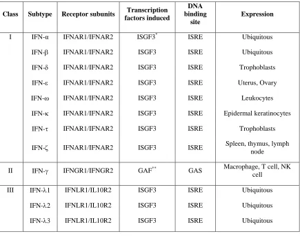
Table 1.1. Classification of IFNs, subtypes, their receptors and expression patterns... 8
Table 1.2. Comparison of IRF family members. ... 12
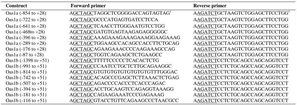
Table 2.1. Primers used to generate the Oas1a and Oas1b promoter deletion reporter constructs.
... 26
Table 2.2. Primers used for site directed mutagenesis. ... 32
LIST OF FIGURES
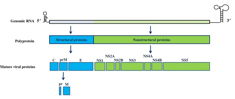
Figure 1.1. Schematic representation of the WNV genome and polyprotein processing. ... 2
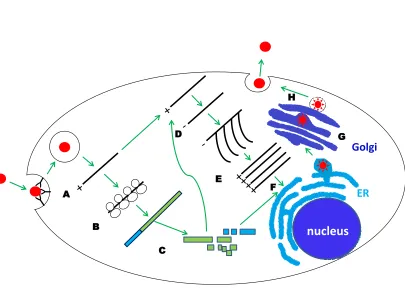
Figure 1.2. WNV replication cycle. ... 4
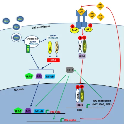
Figure 1.3. Type I IFN induction in MEFs in response to viral dsRNA signaling in MEFs. ... 10
Figure 1.4. miRNA maturation. ... 19
Figure 2.1. IFN beta-induced upregulation f Oas1a and Oas1b in vivo and of promoter reporter constructs in vitro. ... 27
Figure 2.2. Predicted TFBSs in the Oas1a and Oas1b promoters. ... 30
Figure 2.3. Functional analysis of the predicted Oas1a and Oas1b ICEs. ... 33
Figure 2.4. Binding of STAT1 and STAT2 to the Oas1a and Oas1b promoters in in viro and in vivo. ... 35
Figure 2.5. Effect of mutation of the ISRE or overlapping/adjacent TFBSs on Oas1a and Oas1b promoter activity. ... 38
Figure 2.6. Analysis of the inductionof Oas1a and Oas1b by IFN beta in wild type, STAT1-/- and STAT2-/- MEFs. ... 41
Figure 3.1. IFN beta is produced by WNV Eg101-infected MEFs and induces phosphorylation of STAT1 and STAT2. ... 59
Figure 3.2. STAT1 and STAT2 translocation to the nucleus is inhibited in WNV Eg101-infected MEFs. ... 63
Figure 3.3. Oas1a, Oas1b, Irf7 and Irf1 gene expression is induced in WNV Eg101-infcted tSTAT1-/- and tSTAT2-/- MEFs. ... 65
Figure 3.5. The expression of Oas1a and Oas1b but not Irf1 and Irf9 is reduced in IRF-3/7-/-
MEFs. ... 71
Figure 3.6. Irf1 but not Oas1a, Oas1b and Irf7, is upregulated in IRF-3/9-/- MEFs infected with
WNV Eg101. ... 72
Figure 3.7. IPS-1 is involved in the upregulation of Oas1a, Oas1b, Irf7 and Irf1 in MEFs infected
with WNV Eg101. ... 74
Figure 3.8. Analysis of the involvement of the ERK and JNK MAPK pathways in the
upregulation of ISG mRNAs in WNV Eg101-infected MEFs. ... 88
Figure 3.9. Analysis of the activation and involvement of the p38 MAPK pathway in the
upregulation of ISG mRNAs in WNV Eg101-infected MEFs. ... 89
Figure 3.10. Analysis of the involvement of NF-kappa B in the upregulation of ISG mRNAs in
WNV Eg101-infected MEFs. ... 91
Figure 3.11. Analysis of the involvement of the p65 and p50 NF-kappa B subunits in the
upregulation of ISG mRNAs in WNV Eg101-infected MEFs. ... 93
Figure 4.1. Expression of IRF-1 in WNV Eg101-infected tC3H/He MEFs. ... 98
Figure 4.2. The effect of the proteasomal inhibitor MG132 or IRF-1 protein levels in tC3H/He
MEFs. ... 99
Figure 4.3. Comparison of the sizes of Irf1 mRNA transcripts in mock- or WNV Eg101-infected
or IFN beta-treated MEFs. ... 101
Figure 4.4. Analysis of the effect of the Irf1 3’UTR on firefly luciferase reporter gene
expression. ... 103
Figure 4.5. Effect of dicer knock-down on the protein levels of IRF-1 in cells infected with WNV
LIST OF ACRONYMS
AP1- ATF2/c-Jun
ARE - AU-rich element
CARD - caspase activation and recruitment domain
ChIP – chromatin immunoprecipitation
C protein – capsid protein
dsRNA – double stranded RNA
E protein –envelope protein
GAF – IFN-alpha factor
GAS – IFN-gamma activated sequence
EBV - Epstein-Barr virus
EMSA – electrophoretic mobility shift assay
ELISA – enzyme-linked immunoabsorbent assay
ER –endoplasmic reticulum
ERK – extracellular signal-regulated kinase 2
GCN2 - general control non-derepressible-2 kinase
HIV - human immunodeficiency virus
HRI – heme-regulated inhibitor kinase
IFN – interferon
IFA – immunofluorescence assay
IPS-1 - IFN beta promoter stimulator-1
IRF – IFN regulatory factor
ISG - IFN stimulated gene
ISGF3 - IFN stimulated gene factor 3
ISRE - IFN stimulated response element
JEV - Japanese encephalitis virus
JNK - c-Jun N-terminal kinase
MAPK - serine-threonine mitogen-activated kinases
MDA5 – melanoma differentiation antigen 5
MEFs – mouse embryofibroblasts
MHC – major histocompatibility complex
MOI – multiplicity of infection
miRNA - micro RNA
miRISC - miRNA-induced silencing complex
M protein –membrane protein
MVEV - Murray Valley encephalitis virus
MyD88 - myeloid differentiation factor 88
NK - natural killer cell
NS – nonstructural protein
nt - nucleotide
OAS - 2'-5'-oligoadenylate synthetase
ORF – open reading frame
PAMP - pathogen-associated molecular pattern
PABP - poly(A) binding protein
P-bodies – processing bodies
pC3H/He MEFs – primary C3H/He MEFs
PERK - PKR-like ER kinase
PI3K - phosphatidylinositol 3-kinase
PKR - double-stranded RNA-dependent protein kinase
pre-miRNA – precursor miRNA
pri-miRNA – primary miRNA
qPCR - quantitative polymerase chain reaction
qRT-PCR – quantitative reverse transcription polymerase chain reaction
RIG-I –retinoic acid inducible gene - I
RLR - RIG-I-like receptor
RT PCR - reverse transcription polymerase chain reaction
SLEV -St. Louis encephalitis virus
ssRNA –single stranded RNA
STAT - signal transducer and activator of transcription
TBEV - tick-borne encephalitis
tC3H/He MEFs – transformed C3H/He MEFs
TF – transcription factor
TFBS – transcription factor binding site
TLR – Toll-like receptor
TRIF - TIR domain containing adaptor-inducing IFN beta
UTR – untranslated region
VSV - vesicular stomatitis virus
WNV – West Nile virus
CHAPTER 1
1 INTRODUCTION
1.1 Classification and medical importance of flaviviruses
The genus Flavivirus in the family Flaviviridae contains over 70 viruses including several
human pathogens such as dengue, Japanese encephalitis virus (JEV), yellow fever virus (YFV),
West Nile virus (WNV), St. Louis encephalitis virus (SLEV), Murray Valley encephalitis virus
(MVEV) and tick-borne encephalitis virus (TBEV). Many flaviviruses are transmitted by
arthropods and cause a variety of diseases including fever, encephalitis, meningitis and
hemorrhagic fever (Lindenbach, 2007). A secondary Dengue infection with a different serotype
can lead to a life threatening hemorrhagic fever with a mortality rate of about 5%. Vaccines are
available for only a few flaviviruses, namely YFV, TBEV and JEV. WNV is transmitted in
nature via a mosquito-bird cycle. Although WNV primarily infects birds, it occasionally infects
mammals including humans and horses. The majority of WNV infections in humans are
asymptomatic; however, infection induces a mild febrile illness in about 20% of infected
individuals and encephalitis or poliomyelitis-like disease in less than 1% of infected individuals
(Petersen et al., 2003). WNV isolates have been divided into two lineages. Lineage I strains are
often associated with outbreaks of human disease, while the majority of lineage II strains are
non-emerging and cause zoonotic infections in Africa (Brinton, 2002). No antiviral therapies or
human vaccines have been developed so far to treat or prevent WNV infections.
1.2 General characteristics of WNV.
The WN virion is spherical and has a diameter of 40-60 nm. It contains a positive sense, single
M
C prM E NS1
NS2A
NS2B NS3 NS4A
NS4B NS5
pr
5' Structural Nonstructural 3'
C prM E 1 2A 2B 3 4A 4B 5
M
pr Protein Position (nts)
C (97-465) pr (466-741) M (742-966) E (967-2469) NS1 (2470-3525) NS2A (3526-4218) NS2B (4219-4611) NS3 (4612-6468) NS4A (6469-6915) NS4B (6916-7680) NS5 (7681-10395)
5' Structural Nonstructural 3'
C prM E 1 2A 2B 3 4A 4B 5
M
pr Protein Position (nts)
C (97-465) pr (466-741) M (742-966) E (967-2469) NS1 (2470-3525) NS2A (3526-4218) NS2B (4219-4611) NS3 (4612-6468) NS4A (6469-6915) NS4B (6916-7680) NS5 (7681-10395)
5’ 3’
Structural proteins Nonstructural proteins Genomic RNA
Polyprotein
Mature viral proteins
[image:21.612.100.508.255.428.2]is capped at the 5' end; however, it lacks a poly A at the 3' end. The single open reading frame
of WNV genome encodes a polyprotein that when processed by viral and cell proteases produces
ten mature viral proteins (Fig. 1.1). Viral structural proteins [capsid (C), membrane (prM/M) and
the envelope (E)] are encoded by the 5' portion of the genome open reading frame (ORF) and the
nonstructural proteins (NS1, NS2A, NS2B, NS3, NS4A, NS4B and NS5) are encoded by the 3’
portion. The genome is surrounded by a capsid layer composed of dimers of the C protein. A
host-derived lipid envelope surrounds the capsid layer and is spanned by the transmembrane
regions of viral E and M proteins (Lindenbach, 2007).
1.3 Flavivirus replication cycle.
The flavivirus E protein binds to an unknown receptor(s) on the cell surface and this leads to
clathrin-mediated endocytosis (Fig. 1.2). Upon acidification of the virion-containing endocytic
vesicle, the E protein undergoes a low pH mediated conformational change that leads to the
fusion of the viral envelope with the late endosome (Chu and Ng, 2004). The viral genome is
released into the cytoplasm and is translated into a single polyprotein that is then processed by
viral and cellular proteases to produce the mature viral proteins (Lindenbach, 2007). Viral
replication occurs entirely in the cytoplasm of the infected cell. The replication of flavivirus
RNA is associated with extensive membrane biogenesis and rearrangements and viral RNA
replication complexes are formed in invaginations of the endoplasmic reticulum (ER). Each of
these endoplasmic vesicles is connected with the cytoplasm via a narrow “neck” (Gillespie et al.,
2010). Accumulation of viral nonstructural proteins including the NS5 RNA dependent RNA
polymerase, results in the synthesis of negative sense RNA which in turn functions as a template
for the synthesis of genomic plus strand RNA (Lindenbach, 2007). Initially the synthesis of
Nucleus
Golgi
ER
nucleus
A
B
C D
E
F
[image:23.612.103.508.185.473.2]G H
asymmetric with about 10 to 100 times more plus strand synthesis occurring (Chu and
Westaway, 1985). The newly synthesized genomic RNA moves through the neck of the vesicles
into the cytoplasm where it either associates with ribosomes and is translated or associates with
capsid proteins associated with ER membranes that also contain E and M proteins and buds into
the lumen of the ER forming immature virions (Gillespie et al., 2010). Immature virions are then
transported through the trans-Golgi to the plasma membrane. During transport, prM is cleaved
by the Golgi-resident protease furin into pr and M. As a result the prM-E complexes dissociate
allowing the formation of E protein homodimers and maturation to “smooth surface” viral
particles (Stadler et al., 1997). Mature virions are released from infected cells by the fusion of
the virus-containing vesicle with the cell plasma membrane (Mackenzie and Westaway, 2001).
1.4 Host cell response to viral infection.
Viral infection is sensed through pattern recognition receptors (PRR) that recognize specific
pathogen-associated molecular patterns (PAMPs) within viral structures. PRRs that recognize
viral infections include Toll-like receptors (TLRs) and RIG-I-like receptors (RLRs). The TLR
family includes twelve members that bind to a variety of PAMPs (Akira, 2009). Viral ribonucleic
acids are recognized by three of the TLRs. TLR-3 is expressed at the cell surface or in
endosomes (Matsumoto et al., 2003) and recognizes double stranded dsRNA (dsRNA)
(Alexopoulou et al., 2001). Upon binding dsRNA, TLR-3 binds the TIR domain containing
adaptor-inducing IFN beta (TRIF) adaptor molecule and activates the IRF-3, NF-kappa B and
ATF2/c-Jun (AP1) transcription factors (TFs) which activate IFN beta expression (Akira, 2009).
TLR-7 and TLR-8 recognize single stranded RNAs (ssRNAs) (Diebold et al., 2004; Heil et al.,
2004; Triantafilou et al., 2005) and signal through the adaptor molecule myeloid differentiation
2009). The involvement of TLR-3 in the antiviral response of mice to WNV infection is not
clear. Although a previous study reported that blood-barrier permeability and WNV entry into
the brain was decreased in TLR-3-/- mice, suggesting that TLR-3 promotes WNV entry into the
brain (Wang et al., 2004), a second study showed that TLR-3 plays a protective role against
WNV infection by restricting virus replication in neurons (Daffis et al., 2008). TLR-7 was
reported to be important in promoting effective WNV clearance in mice (Town et al., 2009;
Welte et al., 2009).
Viral RNA structures can also be detected by retinoic acid inducible gene-I (RIG-I) and
melanoma differentiation antigen 5 (MDA5). These cytoplasmic sensors are DExD/H helicases
containing two caspase activation and recruitment domains (CARDs) at their N termini.
Although the RIG-I like receptors share about 25% identity within the CARD domains and about
40% identity within the helicase domains (Barral et al., 2009), they recognize different RNA
species. RIG-I binds ssRNA molecules containing a 5' triphosphate, short dsRNAs, and
adenosine/uridine rich RNAs while MDA5 has been shown to be activated by long dsRNAs
(Hornung et al., 2006; Marques et al., 2006; Kato et al., 2008; Saito et al., 2008; Takahasi et al.,
2008). Activated RIG-I and MDA5 bind the IFN beta promoter stimulator (IPS)-1 protein that
serves as a downstream adaptor molecule. IPS-1 then induces downstream signaling cascades
leading to the activation of TFs mediating upregulation of IFN (Barral et al., 2009). Recent
studies suggest that both RIG-I and MDA5 play a role in sensing WNV infection and the
induction of IFN (Fredericksen et al., 2008; Loo et al., 2008). The IFN response to WNV
infection was completely abolished in cells lacking IPS-1 (Fredericksen et al., 2008) confirming
1.5 IFN system
IFNs are secreted cytokines that play a pivotal role in host defense against intracellular
pathogens. IFNs can directly inhibit a virus infection by the induction of IFN stimulated genes,
(ISGs) many of which have antiviral functions, or indirectly through the induction of major
histocompatibility complex (MHC) antigens and the activation of immune cells (Paun and Pitha,
2007). IFNs are grouped into three classes based on their amino acid sequence and recognition
by specific receptors. Type I IFNs consist of IFN- alpha (α), -beta () and the lesser known IFNs,
-omega (), -epsilon (), -kappa (), - delta (), -tau () and – zeta (). Only IFN alpha, beta,
omega, epsilon and kappa are found in humans. IFN alpha and beta are secreted by a wide
variety of cells including macrophages, lymphocytes, fibroblasts, endothelial cells, osteoblasts
and others (Pestka et al., 2004) while the other type I IFNs are expressed in specific cell types
(Table 1.1). IFN alpha and IFN-beta have been extensively studied and were shown to play a
major role in the innate response to viral infections (Randall and Goodbourn, 2008). Since
IFN-alpha and IFN-beta are secreted by fibroblasts, this dissertation will concentrate on these two
subtypes of type I IFNs. The type II IFN class consists of only IFN-gamma (γ), which is critical
for both the innate and adaptive immune responses to virus infection, intracellular bacterial
infections as well as tumor control. IFN-gamma is produced by macrophages, natural killer cells
(NK) and by CD4 and CD8 cytotoxic T-cells (Takaoka and Yanai, 2006; Schoenborn and
Wilson, 2007). Type III IFNs consist of IFN-lambda 1 (1), -lambda 2 (2) and –lambda 3 (3)
which are also referred to as IL-29, IL-28A and IL-28B, respectively. These IFNs are closely
Table 1.1. Classification of IFNs, subtypes, their receptors and expression patterns.
Class Subtype Receptor subunits Transcription
factors induced
DNA binding
site
Expression
I IFN-α IFNAR1/IFNAR2 ISGF3* ISRE Ubiquitous
IFN- IFNAR1/IFNAR2 ISGF3 ISRE Ubiquitous
IFN- IFNAR1/IFNAR2 ISGF3 ISRE Trophoblasts
IFN- IFNAR1/IFNAR2 ISGF3 ISRE Uterus, Ovary
IFN- IFNAR1/IFNAR2 ISGF3 ISRE Leukocytes
IFN- IFNAR1/IFNAR2 ISGF3 ISRE Epidermal keratinocytes
IFN- IFNAR1/IFNAR2 ISGF3 ISRE Trophoblasts
IFN- IFNAR1/IFNAR2 ISGF3 ISRE Spleen, thymus, lymph node
II IFN- IFNGR1/IFNGR2 GAF** GAS Macrophage, T cell, NK cell
III IFN-1 IFNLR1/IL10R2 ISGF3 ISRE Ubiquitous IFN-2 IFNLR1/IL10R2 ISGF3 ISRE Ubiquitous IFN-3 IFNLR1/IL10R2 ISGF3 ISRE Ubiquitous
*
ISGF3 (STAT1, STAT2 and IRF9) **GAF (STAT1 homodimer)
1.6 Type I IFN signaling pathway.
During a viral infection, activation of a PRR leads to the induction of signaling pathways
resulting in the activation of TFs including IRF-3, NF-kappa B and AP1 which translocate to the
nucleus and cooperatively activate IFN-beta gene expression (Barral et al., 2009). Secreted IFN
beta binds to a cellular receptor complex composed of two subunits, IFNAR1 and IFNAR2 (Fig.
1.3), that are associated with Janus kinases (JAKs). The cytoplasmic tail of IFNAR1 is
associated with the tyrosine kinase (Tyk2) while the cytoplasmic tail of IFNAR2 is associated
with Jak1. The binding of IFN to the receptor complex induces cross-activation of these kinases.
IFNAR1 is then phosphorylated by Tyk2 which allows recruitment of signal transducer and
activator of transcription 2 (STAT2) to the receptor and its phosphorylation by Tyk2. Next
STAT1 binds to STAT2 and is phopshorylated by Jak1. Phosphorylated STAT1 and STAT2
form a trimeric transcription factor complex referred to as IFN stimulated gene factor 3 (ISGF3)
with IFN regulatory factor-9 (IRF-9). This complex translocates to the nucleus and binds to IFN
stimulated response elements (ISREs) in the promoters of ISGs (Paun and Pitha, 2007; Randall
and Goodbourn, 2008). One of the ISGs induced by IFN-beta is IRF-7. It is a transcription factor
involved in the induction of the IFN-alpha genes. In addition IRF-7 forms a complex with IRF-3
that binds to the IFN-beta promoter and induces its expression (Wathelet et al., 1998). Secreted
IFN-alpha utilizes the same signaling pathway as IFN beta resulting in the amplification of the
type I IFN response via a positive feedback loop (Marie et al., 1998; Sato et al., 2000).
1.7 Type II IFN and type III IFN signaling pathways.
In the case of type II IFN signaling, IFN-gamma binds to the IFNGR1 and IFNGR2 receptor
complex which leads to the formation of a STAT1 homodimer that is referred to as IFN-alpha
IFN-beta R IG -I MD A5 Virus Virus Virus TL R -3 Endosome Virus dsRNA dsRNA IPS-I
IRF-3 AP1 NF-B
IRF-3 AP1 NF-B
IFN α/ IFN
α/ IFN α/ Jak1 Tyk2 P P ISRE ISRE ISG expression (Irf7, OAS, PKR)
[image:29.612.97.516.103.521.2]ST A T2 IRF-9 ST A T1 P P ST A T2 IRF-9 ST A T1 P P IFN α/ IRF-7 IFN-alpha
sequences (GAS) in the promoters of IFN-gamma responsive genes. Although IFN-gamma
primarily activates the GAF complex, it can also activate the formation of the ISGF3 complex.
Likewise IFN-alpha/beta signaling mainly activates the formation of ISGF3 but can also activate
GAF (Kessler et al., 1990; Decker et al., 1991; Lew et al., 1991; Williams, 1991). Type III IFNs
bind to a receptor complex composed of a specific receptor subunit IFNLR1and IL10R2. Similar
to the type I IFN signaling pathway, the induction of IFN-lambda leads to the formation of the
ISGF3 complex (Kotenko et al., 2003). However, the mechanism of IFN-lambda downstream
signaling remains to be elucidated in detail.
1.8 Interferon regulatory factor family.
ISGs are typically induced by type I IFN through the binding of ISGF3 to ISREs with a
consensus sequence 5' A/GNGAAANNGAAACT 3' located in the promoters of ISGs (Darnell et
al., 1994). However, increasing evidence shows that some ISGs can be upregulated by viral
infections in an IFN-independent manner. Besides ISGF3, additional complexes: ISGF1 (IRF-2)
and ISGF2 (IRF-1) were identified to bind an ISRE (Kessler et al., 1988; Levy et al., 1988). The
mammalian IRF family comprises of nine members, IRF-1 through IRF-9 (Table 1.2). Each of
these proteins has a conserved DNA binding domain, and is a transcription factor and can bind to
IRF-E binding sites in the promoters of some genes. The sequences of IRF-E sites are similar to
that of the ISRE and contain tandem repeats of GAAA motifs separated by two bases (Honda
and Taniguchi, 2006).
IRF-1 is a very unstable protein with a half-life of about 30 m (Watanabe et al., 1991). It is
usually undetectable or expressed at low levels in a variety of cell types; however, it can be
Table 1.2. Comparison of IRF family members.
Factor Expression pattern Role in host defense
IRF-1 Ubiquitous Type I IFN and ISG induction IRF-2 Ubiquitous Repression or induction of ISGs IRF-3 Ubiquitous Type I IFN and ISG induction
IRF-4 Hematopoietic cells Immune cell differentiation and cytokine expression IRF-5 Ubiquitous Type I IFN and cytokine induction
IRF-6 High in skin cells Unknown
IRF-7 Ubiquitous Type I IFN and ISG induction
IRF-8 Hematopoietic cells Cell differentiation and cytokine expression IRF-9 Ubiquitous ISG induction
controlled by phosphorylation (Taniguchi et al., 2001). IRF-1 can also be SUMOylated, which
was shown to increase its stability, or ubiquitylated, which leads to its degradation by the
proteasome (Park et al., 2007). Although, IRF-1 was initially described as a transcriptional
activator of IFN-beta expression (Miyamoto et al., 1988); subsequently, it was also shown to
upregulate some ISGs in an IFN-independent manner (Schoggins et al., 2011). One of the IRF-1
target genes, viperin (Stirnweiss et al., 2010) was reported to inhibit both WNV and dengue
infections (Jiang et al., 2010). In addition, a recent study showed that overexpression of IRF-1
leads to the inhibition of the replication of not only WNV and other flaviviruses such as dengue
virus, YFV, and hepatitis C virus, but also other types of viruses including Chikungunya virus,
Venezuelan encephalitis virus and human immunodeficiency virus 1 (HIV) (Schoggins et al.,
2011). In addition to the induction of antiviral genes, IRF-1 also activates the expression of
genes regulating innate and adaptive immune responses, the cell cycle, apoptosis, DNA damage
repairs and tumor-suppression (Taniguchi et al., 2001). Because IRF-1 functions as a tumor
suppressor, its expression is suppressed in many tumor cells to prevent apoptosis and cell cycle
arrest from occurring (Taniguchi et al., 1997). MEFs and hepatocytes lacking IRF-1 were unable
to undergo growth arrest. In addition, IRF-1 together with p53 was required for optimal
induction of the cyclin-dependent kinase inhibitor p21 after treatment with ionizing radiation
(Tanaka et al., 1996).
In contrast to IRF-1, IRF-2 is a relatively stable protein (half-life of about 8 h) (Watanabe et al.,
1991) and it was shown to function as a transcriptional repressor of the type I IFN gene and some
showed that IRF-2 can also act as a transcriptional activator of some genes (Vaughan et al.,
1995; Jesse et al., 1998).
Similar to IRF-1, IRF-3 and IRF-7 are TFs that play distinct and nonredundant roles in the
induction of type I IFN (Sato et al., 2000). IRF-3 is a constitutively expressed protein that is
found in the cytoplasm of all cells in an inactive form. During a viral infection, it undergoes
phosphorylation which induces dimerization followed by translocation to the nucleus in a
complex with the histone acetyltransferases p300 and CREB-binding protein (CBP) (Yoneyama
et al., 1998; Suhara et al., 2002; Fitzgerald et al., 2003; Sharma et al., 2003). Although IRF-3
alone is not sufficient to induce IFN beta expression, it cooperates with NF-kappa B and AP1
TFs to enhance IFN beta expression. IRF-3 was also identified as a component of the
virus-inducible dsRNA-activated factor 1 complex (DRAF1) (Weaver et al., 1998) that was shown to
play a role in IFN-independent upregulation of a subset of ISGs (Grandvaux et al., 2002;
Andersen et al., 2008). In contrast to the constitutive expression of IRF-3, IRF-7 expression is
typically induced by type I IFN signaling. IRF-7 plays a role in the later phase of the type I IFN
response to viral infection through the amplification of the response via the activation of various
IFN-alpha species and IFN-alpha dependent genes (Marie et al., 1998; Sato et al., 1998) as well
as by enhancing IFN-beta production (Wathelet et al., 1998). Several ISGs can also be
upregulated directly by IRF-7 (Barnes et al., 2004).
IRF-4 and IRF-8 expression is restricted to particular types of immune cells. These IRFs play a
role in the regulation of cell differentiation, DNA repair and cytokine production (Ozato et al.,
2007). IRF-5 is ubiquitously expressed and was shown to be involved in virus-mediated
2005; Takaoka et al., 2005; Paun et al., 2008). The role of IRF-6 in the immune response has not
been studied. IRF-6 is expressed to high levels in skin cells and is important for regulation of
skin cell development (Ozato et al., 2007).
IRF-9 plays a crucial role in type IFN-signaling and the activation of ISGs. It associates with
STAT1 and STAT2 to form the trimeric ISGF3 transcription factor complex that binds to ISREs
in the promoters of ISGs and activates their expression (Paun and Pitha, 2007) (Fig.1.3). One
previous study reported that although IRF-9 translocated to the nucleus, it could not activate
gene expression alone (Kraus et al., 2003). However, both a 9/STAT2 complex and a
IRF-9/STAT2 fusion protein were shown to efficiently activate ISRE-driven transcription of a
reporter gene (Kraus et al., 2003; Lou et al., 2009) suggesting that IRF-9 may play a role in
ISGF3-independent gene expression when complexed with other factor(s).
1.9 Strategies utilized by viruses to evade host immune antiviral responses
The establishment of an antiviral state largely depends on both the induction of IFN and the
functions of antiviral ISGs and many viruses have evolved mechanisms to suppress the
expression of ISGs. Three main strategies utilized by viruses to suppress the induction of ISGs
are: inhibition of IFN production, inhibition of the IFN signaling pathway and blockage of the
synthesis of antiviral proteins. Within each of these strategies, different viruses have evolved
strain, cell type and species specific mechanisms to evade the induction of the antiviral state.
1.9.1 Inhibition of IFN production by viruses
Because IFN induction is a result of the recognition of viral structures by PRRs, many viruses
have developed replication strategies that minimize the production of PAMPs. For example,
2005; Welsch et al., 2009; Gillespie et al., 2010) that likely limit the detection of viral nucleic
acid structures during the exponential replication phase. A recent report showed that although
PKR, a dsRNA sensor, is not actively suppressed by WNV Eg101 infection, it is not activated in
infected rodent cells (Elbahesh et al., 2011). However, during the initial stages of WNV infection
when the replication complexes are not yet well formed the small amounts of RNA species
including dsRNA produced may be detected by PRR. RIG-I and MDA5 cytoplasmic sensors
were previously shown to mediate the activation of the IRF-3 (Fredericksen and Gale, 2006;
Scherbik et al., 2007; Fredericksen et al., 2008) and NF-kappa B (Scherbik, Pulit-Penaloza and
Brinton, unpublished data) TFs and the subsequent induction of type I IFN in WNV infected
cells.
Many viruses encode proteins that were experimentally shown to antagonize IFN induction by
binding dsRNA and limiting activation of host dsRNA binding sensors or by direct inhibition of
RIG-I, MDA-5 or TLR-3 signaling or by blocking the activation of IRF-3 or NF-kappa B TFs
[Reviewed in (Randall and Goodbourn, 2008)]. In the case of WNV, the NS2A protein of the
attenuated lineage I WNV strain, Kunjin was shown to inhibit IFN beta transcription by an
unknown mechanism (Liu et al., 2004; Liu et al., 2006).
1.9.2 Inhibition of IFN signaling by viruses
Numerous reports showed that viruses can block IFN signaling at virtually each step of that
pathway [reviewed in (Randall and Goodbourn, 2008)]. Some of the strategies reported for
different strains of WNV include downregulation of IFN alpha/beta R expression (Evans et al.,
2011), suppression of type I IFN signaling through the redistribution of cholesterol (Mackenzie
STAT1 and STAT2 phosphorylation and translocation to the nucleus (Laurent-Rolle et al.; Liu et
al., 2005; Keller et al., 2006).
1.9.3 Mechanisms of blockage of the synthesis of antiviral proteins by viruses.
One of the strategies utilized by viruses to antagonize the host innate response to viral infection
is the inhibition of protein translation. The process of translation is divided into the initiation,
elongation, termination and ribosome recycling phases. Although viruses can inhibit translation
at each of these steps, most reports show that translation is mainly regulated at the level of
initiation (Walsh and Mohr, 2011). Induction of global inhibition of host mRNA translation is
beneficial for viruses that have evolved alternative translational mechanisms for viral protein
translation. For example, both caliciviruses, which encode proteins that serve as cap-analogues,
and picornaviruses, which can initiate translation from an internal ribosome entry site (IRES),
can initiate translation of viral proteins when cap-dependent translation is blocked (Lopez-Lastra
et al., 2010). However, many viruses including flaviviruses depend entirely on the canonical host
translation machinery and so are susceptible to global translation inhibition. Hence, inhibition of
host translation is not observed in flavivirus infected cells (Lindenbach, 2007).
An alternative means of inhibiting protein translation by viruses is blocking the export of cellular
mRNAs from the nucleus as has been observed for adenovirus, influenza virus and vesicular
stomatitis virus (VSV) infections (Faria et al., 2005; Satterly et al., 2007; Yatherajam et al.,
2011). Some herpesviruses have also been shown to suppress host protein translation through the
induction of increased cell mRNA turnover (Glaunsinger and Ganem, 2004; Feng et al., 2005;
Several known mechanisms of transcript specific suppression of translation have been reported.
One of these involves the binding of host RNA binding proteins to cis-acting elements in the
3'UTRs of target mRNAs which inhibits translation initiation (Abaza and Gebauer, 2008). For
example, translational repression by trans-acting proteins binding to the 3'UTR was reported for
the 15-lipoxygenase (Ostareck et al., 1997), MEF-2A (Black et al., 1997) and p53 (Fu and
Benchimol, 1997) mRNAs. Proteins binding to AU-rich elements (AREs) in the 3'UTRs of target
mRNAs can cause either destabilization or stabilization mRNA or suppress translation (Zhang et
al., 2002). In rare instances, translation initiation can be blocked by the binding of a RNA
binding protein to a cis-element in the 5'UTR (Gray and Hentze, 1994). It is possible that a viral
infection may induce this type of transcript regulation of protein translation but no examples
have yet been reported.
Transcript specific protein synthesis can also be suppressed by specific micro RNAs (miRNAs).
RNA interference was first described as an antiviral mechanism in plants (Lindbo et al., 1993;
Ratcliff et al., 1997; Anandalakshmi et al., 1998; Brigneti et al., 1998; Kasschau and Carrington,
1998; Hamilton and Baulcombe, 1999). Hundreds of miRNAs have been identified in humans
with each of the miRNAs having the potential to regulate multiple genes (Bentwich, 2005).
Many miRNA genes are transcribed under strict developmental-stage and tissue-specific control.
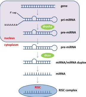
Primary miRNA transcripts (pri-miRNAs) form stem-loop structures which are cleaved by the
RNase III enzyme drosha to precursor miRNAs (pre-miRNAs) that are transported from the
nucleus into the cytoplasm by exportin 5. In the cytoplasm, pre-miRNAs are cleaved by the
RNase III enzyme dicer into mature miRNAs (Murchison and Hannon, 2004). The guide strand
of a mature ~22 nucleotide miRNA associates with argonaute proteins and other components to
5’ cap
AAAAAAA
RISC
gene
pre-miRNA
miRNA/miRNA duplex
nucleus
cytoplasm
pri-miRNA
pre-miRNA
miRNA
RISC complex
drosha
[image:38.612.155.445.145.471.2]dicer
the target mRNAs and the interaction of usually multiple miRNAs with a target RNAs leads to
translation suppression or inhibition or alteration of target mRNA stability (Eulalio et al., 2008;
Hutvagner and Simard, 2008). miRNAs most often bind to the 3'UTR of a target message.
Occasionally, miRNAs bind to 5'UTR or ORF regions but they are usually less effective in
silencing translation at these sites (Lytle et al., 2007). Additionally, it has been shown that the
binding of miRNAs can lead to sequestering target mRNAs in processing bodies (P-bodies)
(Parker and Sheth, 2007).
Some viruses including herpesviruses, adenoviruses and polyomaviruses encode miRNAs which
regulate viral and/or host cell gene translation (Gottwein and Cullen, 2008; Grundhoff and
Sullivan, 2011). Other viruses such as Epstein-Barr virus (Motsch et al., 2007; Cameron et al.,
2008; Ho et al., 2011) and enteroviruses (Ho et al., 2011) induce the expression of cellular
miRNAs that benefit the virus, while HIV infections downregulate cellular miRNAs that inhibit
viral replication (Triboulet et al., 2007). The majority of known viral miRNAs are encoded by
DNA viruses due to the fact that the maturation of miRNAs involves cleavage by drosha in the
nucleus (Rouha et al., 2010). However, a recent study showed that TBEV containing a EBV
pre-miRNA in its genome can express mature pre-miRNA in the cytoplasm without the involvement of
drosha processing (Rouha et al., 2010). Although none of the so far identified viral miRNAs are
encoded by RNA viruses which replicate in the cytoplasm this study indicates that the generation
of miRNAs by cytoplasmic viruses is mechanistically feasible. Interestingly, several flaviviruses
produce a highly structured, nuclease resistant, noncoding RNA derived from the 3' UTR of the
viral genome that was shown to be crucial for virus-induced cytopathicity and pathogenicity
encode a miRNA-like small RNA that is important for efficient replication of this virus in
mosquito cells (Hussain et al., 2011).
GOALS OF THE DISSERATION
WNV-host cell interactions are not fully understood and the overall goal of this study is to
investigate mechanisms by which WNV modulates the expression of host antiviral genes.
Although WNV strains have the ability to inhibit IFN signaling, efficient upregulation of many
ISGs including Oas1a, Oas1b and Irf7 was still observed in cells infected with these viruses
(Fredericksen et al., 2004; Scherbik et al., 2006; Daffis et al., 2007; Scherbik et al., 2007; Daffis
et al., 2008; Fredericksen et al., 2008). Although it was previously reported that the expression of
some ISGs can be upregulated in an IFN-independent manner, many ISGs including Irf7, Oas1a
were thought to be activated only in a type I IFN-dependent-manner. In addition, our previous
results showed that the upregulation of a subset of ISGs including Irf1 is delayed in
WNV-infected cells. Also protein expression of the delayed subset of ISGs was suppressed in WNV
Eg101-infected MEFs (Scherbik et al., 2007). The mechanisms of ISG transcription regulation in
IFN beta-treated and WNV Eg101-infected MEFs will be investigated under Aims 1 and 2, while
the mechanism of WNV Eg101 induced suppression of ISG protein levels will be analyzed under
Aim 3.
AIM 1. Functional analysis of the induction of Oas1a and Oas1b promoters by type I IFN.
AIM 2. Investigation of the activation of ISGs in MEFs infected with WNV Eg101.
AIM 3. Investigation of transcriptional and translational regulation of IRF-1 expression in
CHAPTER 2
2 AIM 1: Functional analysis of the induction of Oas1a and Oas1b promoters by type I
IFN.
The data from this aim has been accepted for publication in Virology, 2012.
INTRODUCTION
The recognition of viral dsRNA by cellular sensors in infected cells leads to activation of the
NF-kappa B, IRF-3 and ATF2/c-Jun transcription factors that bind cooperatively to the interferon
(IFN) beta promoter and activate its transcription (Merika and Thanos, 2001). IFN beta secreted
by infected cells binds to IFN alpha/beta receptors on the surfaces of both infected and
uninfected cells resulting in activation of JAK1 and Tyk2 kinases that phosphorylate STAT1 and
STAT2 transcription factors. Phosphorylated STAT1 and STAT2 and IFN regulatory factor 9
(IRF-9) form the IFN stimulated gene factor 3 complex (ISGF3) that translocates to the nucleus
where it binds to IFN stimulated response elements (ISREs) in the promoters of IFN stimulated
genes (ISGs) and upregulates their expression (Stark et al., 1998).
Three oligoadenylate synthetase genes (OAS1, OAS2 and OAS3) and one OAS-like (OASL)
gene have been identified in the human genome (Hovnanian et al., 1998; Rebouillat et al., 1998).
The transcripts of three of these genes are alternatively spliced and polymorphisms have been
identified that alter splicing sites. Five isoforms have been reported for OAS1 (p42, p44, p46/p48
and p52), two for OAS2 (p69 and p71), one for OAS3 (p100) and two for OASL (p30 and p59)
(Hartmann et al., 1998; Rebouillat et al., 1998; Hovnanian et al., 1999; Justesen et al., 2000;
(Oas1a-Oas1h), two OASL (Oasl1 and Oasl2), one OAS2 (Oas2) and one OAS3 (Oas3) gene orthologs
(Rutherford et al., 1991; Justesen et al., 2000; Shibata et al., 2001; Kakuta et al., 2002). Gene
duplication, rather than alternative splicing, is responsible for the multiple murine Oas1 isoforms
(Perelygin et al., 2006). Enzymatically active OAS proteins play an important antiviral role.
When activated by viral dsRNA, they catalyze the synthesis of short 2'-5'-oligoadenylates (2-5A)
from ATP. 2-5A activates latent endonuclease RNase L which degrades both cellular and viral
single-stranded RNAs (Samuel, 2001). Among the eight murine Oas1 proteins, only Oas1a and
Oas1g have been reported to be active 2'-5'-oligoadenylate synthetases (Kakuta et al., 2002;
Elbahesh et al., 2011). However, the inactive synthetase Oas1b mediates resistance to
flavivirus-induced disease through an unknown mechanism that is independent of RNase L (Scherbik et al.,
2006). The flavivirus-induced disease resistant mouse strain C3H/RV is homozygous for the
dominant Oas1br allele encoding a full length Oas1b protein, while the congenic susceptible
mouse strain C3H/He is homozygous for the recessive Oas1bs allele that encodes a C-terminally
truncated Oas1btr protein (Perelygin et al., 2002). A previous study showed that the full length
Oas1b protein, which is an inactive synthetase, can inhibit in vitro Oas1a synthetase activity in a
dose-dependent manner and reduce 2-5A production in vivo in response to poly(I:C) (Elbahesh et
al., 2011). A similar function was reported for Oas1d, another of the inactive Oas1 proteins (Yan
et al., 2005).
Each of the human OAS genes contains an ISRE in its promoter and is induced by type I IFN
(Wang and Floyd-Smith, 1997; Hartmann et al., 1998; Floyd-Smith et al., 1999; Yu et al., 1999;
Rebouillat et al., 2000). Similarly, murine Oas gene expression with the exception of Oas1f was
TFSEARCH analysis predicted transcription factor binding sites (TFBSs) in 500 bp promoter
fragments of the murine Oas1a-h, Oas2 and Oas3 genes and identified an ISRE in only the
promoters of the Oas1a, Oas1b, Oas1g and Oas2 genes (Mashimo et al., 2003). Only the ISRE in
the Oas1b promoter was predicted to overlap GAS and NF-kappa B sites. These results
suggested the possibility of differential regulation of Oas1a and Oas1b expression by IFN and/or
viral infection but this prediction was not functionally tested.
In the present study, the Oas1a and Oas1b promoters from both C3H/RV and C3H/He mouse
embryofibroblasts (MEFs) were cloned and sequenced. A GENOMATIX search of the Oas1a
and Oas1b promoter sequences predicted that neither had a TATA box but that both had a
canonical initiator element (INR). An inverted CCAAT element (ICE) was predicted and
functional analysis showed that it may be important for the basal activities of the C3H/He and
C3H/RV Oas1b promoters and the C3H/RV Oas1a promoter. The C3H/He Oas1a promoter
contained a mutation in this site that was predicted to make it nonfunctional. A single ISRE as
well as overlapping STAT and IRF sites were predicted in both promoters. Functional mapping
of the Oas1a and Oas1b promoters by sequential 5' deletion and TFBS mutagenesis indicated
that the ISRE as well as the overlapping STAT site are required for Oas1a promoter induction by
IFN beta while Oas1b expression requires only the ISRE. Also, both STAT1 and STAT2 are
required for Oas1a upregulation by IFN beta, while only STAT2 is required for Oas1b
upregulation. A single nucleotide difference between the STAT sites of the Oas1b and Oas1a
promoters appeared to be responsible for the differential STAT1-dependence of Oas1a and
RESULTS
2.1 Mapping the Oas1a and Oas1b gene promoter regions required for basal promoter
expression and induction by IFN beta.
The Oas1a and Oas1b genes are ISGs and after treatment of C3H/He MEFs with 1000 U/ml of
murine IFN beta for 3 h, Oas1a mRNA was upregulated by about 9 fold while the Oas1b mRNA
was upregulated by about 7 fold (Fig. 2.1A). The time and the dose of IFN treatment was
selected based on a previous study showing that the highest level of ISG induction was at 3h
after IFN treatment of MEFs and that the levels of ISG mRNA upregulation were similar with
10, 100 and 1000 U/ml of IFN beta (Scherbik et al., 2007). The regions of the Oas1a and Oas1b
proximal promoters required for basal gene expression and activation by IFN beta were then
mapped using luciferase reporter assays. Two firefly luciferase reporter gene constructs, one
containing a C3H/RV Oas1a gene promoter fragment [Oas1a (-1768, +28)] and the other
containing a C3H/RV Oas1b gene promoter fragment [Oas1b (-1398, +51)], were first generated
and then a set of 5' sequentially deleted constructs was made for each promoter. C3H/RV MEFs
were transfected with construct DNA, and 24 h later cell lysates were harvested and used to
analyze basal promoter activity. To analyze the effect of IFN on Oas1 promoter activity, cells
transfected with a luciferase reporter for 24 h were incubated with murine IFN beta. Initial pilot
experiments showed that although luciferase activity was the highest at both 3 and 6 h after
treatment, the 3h peak level was the most reproducible. At 24 and 48 h, luciferase activity was
significantly decreased. Based on these preliminary results and the maximum half-life of the
firefly luciferase protein of 4 h (Thompson et al., 1991; Brandes et al., 1996), assays were done
in all subsequent luciferase reporter experiments after 3 h of IFN beta treatment. Low basal
Table 2.1. Primers used to generate the Oas1a and Oas1b promoter deletion reporter constructs.
Construct Forward primer Reverse primer
and the longest Oas1b (-1398, +51) construct (Fig. 2.1C). These constructs showed a modest but
significant increase in promoter activity after IFN treatment. The observation that the longest
Oas1a and Oas1b promoter fragments tested produced the lowest reporter activities suggested the
presence of upstream transcriptional repressor elements that negatively affected the basal and
IFN-induced expression levels of both promoters.
Promoter constructs with sequential 5' deletions were used to determine the shortest Oas1a and
Oas1b promoter fragments able to produce increased luciferase activity upon stimulation with
IFN beta. Each of the 5' deleted Oas1a promoter constructs tested produced similar significantly
increased basal luciferase activities compared to the Oas1a (1768, +28) construct and the
activities for each of these constructs increased upon stimulation with IFN beta (Fig. 2.1B). For
instance, the Oas1a (-87, +28) and Oas1a (-854, +28) constructs had basal activities that were 7.1
and 8.6 fold higher, respectively, than that of the Oas1a (-1768, +28) construct and both showed
a 1.75 fold increase in luciferase activity after stimulation with IFN beta. These results indicated
that the TFBSs required for IFN induction of the Oas1a are located between -87 and +28.
In contrast to what was observed with the truncated Oas1a constructs, the activities of each of the
5' deleted Oas1b constructs varied. The Oas1b (-576, +51) construct showed a 21 fold increase
over the activity produced by the Oas1b (-1398, +51) construct (Fig. 2.1C). Constructs that were
either shorter or longer than Oas1b (-576, +51) produced lower basal activities. The observation
that the basal activity of the Oas1b (-116 to +51) construct was 2.5 fold lower than that of Oas1b
(-576, +51) suggested that the region between -576 and -116 contains elements that enhance
basal activity decreased suggesting the presence of repressor elements in this region. The activity
of each of the truncated Oas1b promoter constructs was induced to a similar extent above its
basal level by IFN beta treatment (Fig. 2.1C) indicating that the elements required for the
induction of the Oas1b promoter by IFN beta are located between -116 and +51 bp.
Although the results obtained were reproducible and statistically significant, basal levels were
high and the maximal induction observed with IFN beta was about 2 fold. The low levels of
induction by IFN are likely to be due to the use of promoter fragments with only a single ISRE in
the context of a natural promoter sequence which also contains multiple additional TF binding
sites that modulate both basal and IFN-induced luciferase activity. Previous studies using natural
TLR9 (Guo et al., 2005) and RIG-I (Su et al., 2007) promoter sequences with single copies of an
ISRE also reported a 2 fold or lower activation of the reporter gene after stimulation with IFN
beta.
2.2 Analysis of the core promoter elements in the Oas1a and Oas1b promoters.
Elements in the -87 to +28 C3H/RV Oas1a promoter fragment and in the -116 to +51 C3H/RV
Oas1b promoter fragment were predicted with GENOMATIX and TFSEARCH programs.
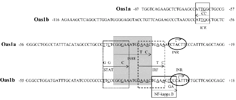
Neither promoter contained a TATA box but each contained an INR (Fig. 2.2), an element
commonly found in TATA-less promoters and known to be able to direct transcriptional
initiation (Smale and Baltimore, 1989; Smale, 1997). Within the INR consensus
C
/TC/TA+1NA/TC/TC/T, the adenine is the transcription start site (TSS) and this nucleotide was used
to predict the location of the Oas1a and Oas1b TSSs. The GENOMATIX program also predicted
an inverted CCAAT element (ICE) at -63 to -68 in the Oas1a promoter and at -62 to -67 in the
orientation are typically located about 60 to 100 nucleotides upstream of the TSS (Mantovani,
1999; Dolfini et al., 2009). To determine whether the predicted ICE plays a functional role in the
Oas1a and Oas1b promoters, two nucleotides (ATTGG → ATCCG) were substituted in this
element in the Oas1a (-854, +28) and Oas1b (-576, +51) constructs. A GENOMATIX search
confirmed that the introduced mutations eliminated the targeted binding site and did not create a
new TFBS. Mutation of ICE reduced the basal luciferase activity of the Oas1a and Oas1b
promoters by 43% and 40%, respectively, but did not negatively affect induction of either
promoter by IFN beta (Fig.2.3A). IFN beta induced the wild type and ICE-mutated Oas1a
promoters by 1.7- and 1.9 fold, respectively, and the wild type and ICE-mutated Oas1b promoter
by 1.4 and 1.5 fold, respectively.
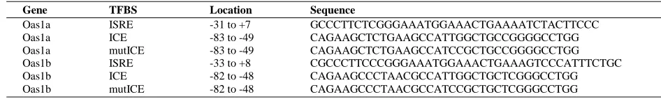
DNA probes consisting of the -88 to -49 bp for Oas1a or -82 to -48 bp for Oas1b (Table 2.3)
were next used in an electrophoretic mobility shift assay (EMSA) to determine whether TFs bind
to the ICE sequence. Oas1a and Oas1b probes with a wild type ICE bound to two complexes in
nuclear extracts from untreated C3H/RV MEFs (Fig. 2.3B). With both probes, the upper band
was much darker than the lower band. These bands were either not detected or were much fainter
when a specific unlabeled competitor DNA was included in the reaction suggesting that the
binding detected was specific. Neither of the shift bands was observed when an Oas1a probe
containing a mutated ICE was tested. An Oas1b probe with a mutated ICE detected a faint lower
complex band but did not detect an upper complex band. The results suggest that nuclear extracts
from untreated control cells contain TFs that bind to the ICE. Although the TFs binding to the
ICE in the Oas1a and Oas1b promoters were not identified, both the reporter assay (Fig. 2.3A)
and the EMSA (Fig. 3B) results suggest that the ICE element plays a role in the regulation of
Table 2.2. Primers used for site directed mutagenesis.
Construct Primer Primer sequence
Oas1a (-854, +28) mIRF F1 GCCCTTCTCGGGAAATGGAAACTGTACATCTACTTCCC2 R GGGAAGTAGATGTACAGTTTCCATTTCCCGAGAAGGGC Oas1a (-854, +28) mISRE F GCCCTTCTCGGGAAATGGTACCTGAAAATCTACTTCCC
R GGGAAGTAGATTTTCAGGTACCATTTCCCGAGAAGGGC Oas1a (-854, +28) mSTAT F CATAGCCCTGCCCGTGTCGGCAAATGGAAACTG
R CAGTTTCCATTTGCCGACACGGGCAGGGCTATG Oas1a (-854, +28) mICE F CAGAAGCTCTGAAGCCATCCGCTGCCGGGGCCTGG
R CCAGGCCCCGGCAGCGGATGGCTTCAGAGCTTCTG Oas1b (-576, +51) mNF-kappa B F CCCGGGAAATGGAAACTGAAAGGACCATTTCTGCTTCAGCG
R CGCTGAAGCAGAAATGGTCCTTTCAGTTTCCATTTCCCGGG Oas1b (-576, +51) mIRF F CCCGGGAAATGGAAACTGTACGTCCCATTTCTGCTTCAGCG R CGCTGAAGCAGAAATGGGACGTACAGTTTCCATTTCCCGGG Oas1b (-576, +51) mISRE F GCCCTTCCCGGGAAATGGTACCTGAAAGTCCC
R GGGACTTTCAGGTACCATTTCCCGGGAAGGGC
Oas1b (-576, +51) mSTAT F GCATATCCGCGCCCGTGCCGGCAAATGGAAACTGAAAGTCCC R GGGACTTTCAGTTTCCATTTGCCGGCACGGGCGCGGATATGC Oas1b (-576, +51) mICE F CAGAAGCCCTAACGCCATCCGCTGCTCGGGCCTGG
R CCAGGCCCGAGCAGCGGATGGCGTTAGGGCTTCTG 1
F, forward primer; R, reverse primer 2 Underlined letters indicate mutated nts.
Table 2.3. Sequences of DNA probes used in EMSA.
Gene TFBS Location Sequence
[image:51.612.66.539.499.570.2]2.3 Comparison of the sequences of the Oas1a and Oas1b promoters in resistant C3H/RV
and susceptible C3H/He MEFs.
To determine whether the sequences of the Oas1a or Oas1b promoters differ between congenic
flavivirus disease susceptible C3H/He and flavivirus disease resistant C3H/RV mice, Oas1a
(-1768, +28) and Oas1b (-1398, +51) DNA fragments were amplified by PCR from MEFs from
each mouse strain, cloned and sequenced. Although complete identity was observed between the
C3H/RV and C57BL/6J (NT_078458.6) Oas1a and Oas1b promoter sequences, the C3H/He
Oas1a promoter sequence differed by T to C substitutions at positions -66 and -298 and the
C3H/He Oas1b promoter sequence differed by a G to C substitution at position +15 and a 4 nt
deletion between -327 and -323. GENOMATIX and TFSEARCH TFBS searches of the C3H/He
promoter sequences predicted that only the T to C substitution at -66 in the Oas1a promoter was
located in a TFBS. This substitution changed the ICE from ATTGG to ATCGG. Based on the
observation that mutation of the ICE from ATTGG to ATCCG significantly decreased the basal
activities of both the C3H/RV Oas1a and Oas1b promoters, the single nucleotide substitution in
the C3H/He Oas1a promoter producing a ATCGG ICE sequence would be expected to reduce
the basal expression level of the Oas1a gene in C3H/He MEFs.
2.4 Analysis of transcription factors binding to the Oas1a and Oas1b promoters.
Both the previously published search (Mashimo et al., 2003) and the TFSEARCH and
GENOMATX TFBS searches done in this study predicted canonical ISREs with identical
sequences (5' GGGAAATGGAAACT 3') between -22 to -9 bp in the Oas1a promoter and
Figure 2.4. Binding of STAT1 and STAT2 to the Oas1a and Oas1b promoters in in viro and
in vivo. DIG-labeled Oas1b and Oas1a DNA probes and nuclear extracts from untreated or