D
E
V
E
LO
P
M
E
N
T
INTRODUCTION
In Drosophila, early embryonic development is supported by maternal deposition of the contents of the oocyte into the fertilized egg (Spradling, 1993). The oocyte nucleus is transcriptionally inactive during most of oogenesis (King and Burnett, 1959). Consequently, the majority of the contents of the growing oocyte originates from the polyploid nurse cells, with the exception of yolk granules and some specific signals that are secreted from the overlying follicle cells (Spradling, 1993). Such long-distance transport of cellular components, including mRNA, between interconnected cells or within very large cells is a common and important process in many biological systems (Pollard and Earnshaw, 2002). Nurse cell-to-oocyte transport occurs through actin-rich ring canals and can be divided into four distinct phases (Mahajan-Miklos and Cooley, 1994). First, in early oogenesis, oocyte specification in the germarium depends on the selective transport of Bicaudal D (BicD) and Egalitarian (Egl) (Mach and Lehmann, 1997; Suter and Steward, 1991), which, together with microtubules (MTs), are required for oocyte specification (Koch and Spitzer, 1983; Theurkauf et al., 1993). Second, during a period of 2 days in mid-oogenesis, from stages 2 to 7, both the nurse cells and the oocyte grow at the same rate (Spradling, 1993), and a variety of important transcripts that encode axis specification factors are transported into the oocyte. During the third phase, at stages 8-10A of oogenesis, this process continues, except the relative size of the oocyte increases in relation to the nurse cells because of the transport of yolk from the follicle cells and cytoplasm from the nurse cells. The fourth and last transport phase involves the ‘dumping’ of the entire nurse cell contents into the oocyte at stage 10B, leading to the doubling of the oocyte volume within 30 minutes (Mahajan-Miklos and Cooley, 1994).
It has been proposed that MTs provide the tracks along which minus-end-directed motors, such as Dynein, could transport proteins, organelles and mRNAs into the oocyte (Theurkauf et al., 1993). This hypothesis is supported by the fact that strong mutations in components of the Dynein complex, including Dhc, Lis-1, BicD and Egl, result in egg chambers devoid of oocytes (Gepner et al., 1996; Liu et al., 1999; McGrail and Hays, 1997; Swan et al., 1999). However, no direct evidence for these proposals has been obtained and the exact mechanism by which these proteins affect this process is unclear. Moreover, the specificity of a putative Dynein-dependent nurse cell-to-oocyte transport system for different cargoes, such as organelles and mRNAs, remains elusive, as does the exact path of transport.
Key transcripts that must be transported from the nurse cells to the oocyte during mid- to late-oogenesis include: gurken(grk) mRNA, which encodes a TGF␣homologue responsible for setting up the primary axes (Gonzalez-Reyes et al., 1995; Neuman-Silberberg and Schüpbach, 1993); bicoid (bcd) mRNA, encoding the anterior morphogen (Driever and Nüsslein-Volhard, 1988); and oskar (osk) mRNA, encoding a key factor responsible for posterior patterning and germ cell determination (Ephrussi et al., 1991). Once in the oocyte, the minus-end-directed MT-dependent molecular motor, cytoplasmic Dynein (Dynein), is required for the localization of grk(MacDougall et al., 2003) and bcd(Januschke et al., 2002b) mRNA. By contrast, the plus-end-directed molecular motor Kinesin I is required for osk mRNA localization to the posterior of the oocyte. The transport of each of these mRNAs into the oocyte has been suggested to involve the same mechanism as that of apical transport of pair-rule transcripts in the blastoderm embryo (Bullock and Ish-Horowicz, 2001), which was previously shown to involve MTs (Lall et al., 1999) and Dynein (Wilkie and Davis, 2001). The former study showed the signal-dependent nurse cell-to-oocyte transport of ectopically expressed pair-rule transcripts, which are ordinarily not expressed during oogenesis. Taking these observations together, they suggest the existence of a similar Dynein-dependent process that could mediate nurse cell-to-oocyte mRNA transport during oogenesis. However, the role of Dynein in the movement of transcripts, such as bcd, oskand grkmRNA, that are expressed in oogenesis, remains unclear.
A Dynein-dependent shortcut rapidly delivers axis
determination transcripts into the
Drosophila
oocyte
Alejandra Clark*, Carine Meignin and Ilan Davis†
The primary axes of Drosophila are set up by the localization of transcripts within the oocyte. These mRNAs originate in the nurse cells, but how they move into the oocyte remains poorly understood. Here, we study the path and mechanism of movement of gurkenRNA within the nurse cells and towards and through ring canals connecting them to the oocyte. gurkentranscripts, but not control transcripts, recruit the cytoplasmic Dynein-associated co-factors Bicaudal D (BicD) and Egalitarian in the nurse cells. gurken RNA requires BicD and Dynein for its transport towards the ring canals, where it accumulates before moving into the oocyte. Our results suggest that bicoidand oskartranscripts are also delivered to the oocyte by the same mechanism, which is distinct from cytoplasmic flow. We propose that Dynein-mediated transport of specific RNAs along specialized networks of microtubules increases the efficiency of their delivery, over the flow of general cytoplasmic components, into the oocyte.
KEY WORDS: Drosophila, Oogenesis, Oocyte, Nurse cells, Intracellular mRNA localization, Microtubules, Actin, Cytoplasmic Dynein, Bicaudal D, Egalitarian, Ring canals
Development 134, 1955-1965 (2007) doi:10.1242/dev.02832
The Wellcome Trust Centre for Cell Biology, Michael Swann building, The University of Edinburgh, Mayfield Road, Edinburgh EH9 3JR, UK.
*Present address: The Wellcome Trust/Cancer Research UK Gurdon Institute, The University of Cambridge, Tennis Court Road, Cambridge CB2 1QN, UK
†Author for correspondence (e-mail: [email protected])
D
E
V
E
LO
P
M
E
N
T
The transport ofbcdand osk mRNA into the oocyte has been known for some time to be MT dependent (Pokrywka and Stephenson, 1991). Furthermore, bcdmRNA forms particles that move into the oocyte at speeds suggestive of an active-transport process (Cha et al., 2001). Exu, a factor required genetically for bcd mRNA localization (St Johnston et al., 1989), was shown to be required within the nurse cells for injected bcdRNA to localize correctly once in the oocyte (Cha et al., 2001) and to move as particles from the nurse cells to the oocyte (Theurkauf and Hazelrigg, 1998). However, the role of motor proteins in nurse cell-to-oocyte transport was not addressed directly, although a Dynein-binding protein, Swallow (Schnorrer et al., 2000), and the Dynein co-factor Dynactin (Januschke et al., 2002a) were shown to be required for bcdmRNA localization in the oocyte. Thus, although the requirement for motor proteins in mRNA localization in the oocyte has been demonstrated conclusively, the mechanism and path of the nurse cell-to-oocyte transport of specific mRNAs remains poorly studied. It is also not known whether mRNAs are simply swept along by the flow of cytoplasm from nurse cell to oocyte. Although some selectivity of cytoplasmic transport has been observed (Bohrmann and Biber, 1994), it is also possible that cellular components are transported into the oocyte via the pressure created by the synthesis of cytoplasmic components in the nurse cells.
Here, we define the path of movement and mechanism of grk mRNA transport from nurse cells to oocyte. By injecting fluorescently labelled grkRNA into nurse cells and assaying its movement, we show that grk RNA is first transported rapidly and directly towards the ring canals, accumulating in a discrete zone in front of the ring canals, followed by its rate-limiting transport through the ring canals. We show that this transport is MT-, BicD-and Dynein-dependent, BicD-and that grkRNA recruits the Dynein-associated proteins Egl and BicD in the nurse cells. We show further that bcdand osk RNA are likely to be transported by a similar mechanism. We propose that Dynein-dependent transport of grk, bcd and osktranscripts towards the ring canal allows the more-efficient transport of key axis specification transcripts than would the slower transport of cytoplasmic components, which occurs throughout much of oogenesis.
MATERIALS AND METHODS Fly strains
Stocks were raised on standard cornmeal-agar medium at 25°C. The strains used include: wild type (Oregon-R, OrR); Tau-GFP (D. St Johnston, Wellcome Trust/Cancer Research UK Gurdon Institute, Cambridge, UK), to visualize MTs; w;p[UASp-GFP-actin] driven by ‘triple driver’ (w;pCOGGal4VP16; NGT40; nosGal4VP16) (L. Cooley, Yale University Medical School, New Haven, CT) to visualize actin; and dhc6-6/dhc6-12
trans-heterozygotes (T. Hays, University of Minnesota, Minneapolis, MN), which are female-sterile. To examine the role of BicD in grkRNA nurse cell-to-oocyte transport in mid-oogenesis,w; Df(2L)TW119/CyO; P [w+hsBic-D]-94 andw; BicDr8/CyO (B. Suter, University of Bern, Bern, Switzerland)
were crossed to generate w; Df(2L)TW119/ BicDr8; P [w+hsBic-D]-94/+
(Bic-Dmom) flies, as described by Swan and Suter (Swan and Suter, 1996).
For controls, siblings of the cross (w, Df(2L)TW119/CyO orw, BicDr8/CyO;
P [w+hsBic-D]-94/+) andOrRflies were used. To confirm the absence of BicD protein in Bic-Dmomovaries, each fly had one ovary dissected and fixed
for BicD immunostaining, whereas the other ovary was used for the in vivo study of grkRNA nurse cell-to-oocyte transport.
RNA in situ hybridization
In situ hybridization was performed as previously described (MacDougall et al., 2003; MacDougall et al., 2001). Digoxigenin anti-sense RNA probes were transcribed using full-length grk (G. Schupbach, Princeton
University, Princeton, NJ), osk (A. Ephrussi, EMBL, Heidelberg, Germany) and bcd(C. Nusslein-Volhard, Max Planck Institute, Tübingen, Germany) cDNAs. In each experiment, at least 40 ovarioles (stage 2-9) and ten egg chambers (stage 10A and older) were examined from OrRor dhc6-6/dhc6-12females.
Synthesis of fluorescent and biotinylated RNA
RNAs were in vitro transcribed using T3, T7 and SP6 polymerases and UTP-AlexaFluor546, UTP-AlexaFluor488, UTP-Cy3 or UTP-Biotin as previously described (Wilkie and Davis, 2001). Plasmids templates were: full-lengthgrk(G. Schupbach); bcd(C. Nusslein-Volhard); K10 3⬘UTR (S. Bullock, MRC LMB, Cambridge, UK); I factor (also known as I-element; V. Van de Bor, University of Nice, Nice, France); and a 3 kb fragment of lacZamplified from pG5-L-G3 (Thio et al., 2000) and cloned into pGEM-T.
Injection of RNAs and inhibitors
Ovaries were dissected in Series 95 halocarbon oil (KMZ Chemicals) and egg chambers were adhered to coverslips using tungsten needles. Fluorescently labelled RNA (250-500 ng/l) was injected into nurse cell cytoplasm using Femtotip needles (Eppendorf). At least two batches of RNA were injected for each experiment, on separate occasions. Colcemid was co-injected with fluorescently labelled RNA at 100 g/ml into Tau-GFPegg chambers. To study the requirement of MTs in grk RNA ring canal accumulation, colcemid (100 g/ml) was pre-injected over each Tau-GFP egg chamber followed by grkRNA injection into the nurse cells 10 minutes later. AlexaFluor568-coupled Phalloidin was injected at a concentration of 4 units/ml (0.13 M) into wild-type nurse cells to visualize ring canals and to facilitate imaging of cytoplasmic movements at the same plane of focus as a ring canal.
Immunostaining of injected egg chambers
Biotinylated RNAs were injected into wild-type nurse cells and fixed after 15-20 minutes. Egg chambers were fixed for 20 minutes by placing 3.7% formaldehyde in PBT (0.1% Tween) over the halocarbon oil. All subsequent staining steps were performed in a watchglass under a dissecting microscope to avoid losing the injected egg chambers during the washes. Biotin-labelled RNA was detected with Avidin-conjugated AlexaFluor488 or AlexaFluor546 (Molecular Probes). BicD and Egl were visualized with anti-BicD (B. Suter) and anti-Egl (R. Lehmann, New York University School of Medicine, NY), which were used at dilutions of 1/20 and 1/4000, respectively. Secondary antibodies were AlexaFluor488- and AlexaFluor568-coupled (Molecular Probes). In all recruitment experiments, at least three egg chambers were analyzed for each condition. Alexa568-coupled Phalloidin (1/50) was used to visualize actin.
Four-dimensional imaging and deconvolution
Fixed egg chambers were mounted in Vectashield (Vector Laboratories). Imaging was performed on a wide-field DeltaVision microscope (Applied Precision, Olympus IX70 microscope, Coolsnap HQ camera from Roper). Images were acquired with a 20⫻/0.75NA or 100⫻/1.35NA objective lens and subsequently deconvolved (Davis, 2000) using Sedat/Agard algorithms with Applied Precision software. Several egg chambers were imaged in parallel depending on the time resolution needed. For high-power imaging of MTs, Tau-GFPand Tau-GFP/+; dhc6-6/dhc6-12 egg chambers were
sectioned through the ring canal at 1 m intervals with a 100⫻/1.35NA objective. Live imaging of RNA particles at 100⫻/1.35NA was performed at 1 second intervals for approximately 3 minutes, after which time photobleaching prevented the identification of particles for tracking.
Particle tracking
D
E
V
E
LO
P
M
E
N
T
RNA particles that move in directed tracks, because stationary particles are difficult to distinguish from autofluorescent cytoplasmic bodies. By contrast, rapidly moving RNA particles of a similar intensity of fluorescence are easily identified.
RESULTS
InjectedgrkRNA moves from the nurse cells through ring canals into the oocyte and localizes correctly
To investigate the mechanism by which grkmoves from the nurse cells to the oocyte, we established an injection assay based on previous assays described for bcdRNA in the nurse cells (Cha et al., 2001) and grkRNA in the oocyte (MacDougall et al., 2003). Full-length grkRNA was injected into the nurse cells at different stages of oogenesis. At all stages tested, ranging from 6 to 10A, injected grk RNA was able to move into the oocyte from the nurse cells and then localize correctly within the oocyte (Fig. 1A,C,E, and see Movies 1, 2, and Table S1 in the supplementary material) in a manner similar to the endogenous transcript (Fig. 1B,D,F). At early stages, before the reorganization of MTs and the migration of the oocyte nucleus, injected grkRNA localized correctly in a posterior crescent near to the oocyte nucleus (Fig. 1A,B), where MT minus ends are thought to be concentrated (Micklem et al., 1997). By contrast, when grkRNA was injected into the nurse cells of stage 8-10A egg chambers (Fig. 1C-F), it became localized in a dorso-anterior crescent near the oocyte nucleus in a very similar pattern to its localization when injected into the oocyte, and to the endogenous transcript at the same stage of oogenesis (MacDougall et al., 2003). We found that the efficiency of transport of injected grkRNA into the oocyte was higher in the earlier stages and that some injected RNA remained in the nurse cells, within the time scale of the experiment, which was limited, by egg chamber survival in halocarbon oil, to 1-2 hours.
To determine whether the movement of injected grktranscript from the nurse cells to the oocyte is specific, we injected a number of other transcripts, including control RNAs. We first injected bcd RNA, which had previously been shown to move efficiently into the oocyte (Cha et al., 2001). bcdRNA injected into the nurse cells moved into the oocyte at an equivalent rate to grkRNA (Fig. 1G,H and see Table S1 in the supplementary material) and localized to the anterior of the oocyte, in the same pattern as the endogenous transcript. We then injected fluorescently labelled lacZ RNA and found that these control transcripts failed to accumulate substantially in the oocyte, and that the small amount of RNA that entered the oocyte was evenly distributed in the cytoplasm (Fig. 1I and see Table S1 in the supplementary material). We conclude that injected grkand bcdRNA are specifically transported efficiently from the nurse cells into the oocyte.
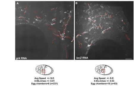
To elucidate the path and details of the movement of grkand bcd RNA from the nurse cells, we studied the movement at higher magnification and time resolution. We found that, in nurse cells, injected grkRNA forms particles that move in directed paths to a specific location near the oocyte, which appeared to be ring canals (Fig. 2A and see Movie 3 in the supplementary material). By contrast, injected lacZRNA did not accumulate in similar locations. To test directly whether grkRNA localized at ring canals, we fixed the injected egg chambers 20 minutes after the injection of Biotin-labelled grkRNA and stained them with AlexaFluor488-Phalloidin to co-visualize the ring canals with RNA. We found that, in 88% of egg chambers (n=8), grkRNA accumulated at a region in front of the ring canals (Fig. 3A, and see Movie 4 and Table S2 in the supplementary material). By contrast, injected lacZRNA failed to accumulate in this manner (n=12).
[image:3.612.316.560.60.438.2]D
E
V
E
LO
P
M
E
N
T
3), suggesting that they involve distinct mechanisms of RNA transport. These speed differences are consistent with previous observations of Exu-GFP particles (Theurkauf and Hazelrigg, 1998). We found that K10(MacDougall et al., 2003) and Ifactor (also known as I-element– FlyBase) (Van De Bor et al., 2005) mRNA behave similarly when injected into a nurse cell (see Fig. S1A-C and Tables S1, S2 in the supplementary material).
We then determined whether the movement of grkand bcdRNA within the nurse cells was specific and could explain their more-efficient accumulation in the oocyte compared with control RNA. To address this issue, we injected lacZRNA. We found that, although lacZRNA formed some particles that move within the nurse cells, fewer particles than withgrkRNA injection were observed moving directionally towards the ring canals (Fig. 2B). Most significantly, lacZRNA did not concentrate at the ring canal in any of the egg chambers examined, resulting in a more homogenous distribution than injected grkRNA (Fig. 2B, Fig. 3B and see Table S2 in the supplementary material). We conclude that, within nurse cells, grk and bcdtranscripts are specifically and actively transported towards ring canals that link these cells to the oocyte. We interpret the accumulation of grkRNA in front of the ring canals as indicating that movement through the ring canals is a rate-limiting step.
grkmRNA transport within nurse cells is Dynein and MT dependent
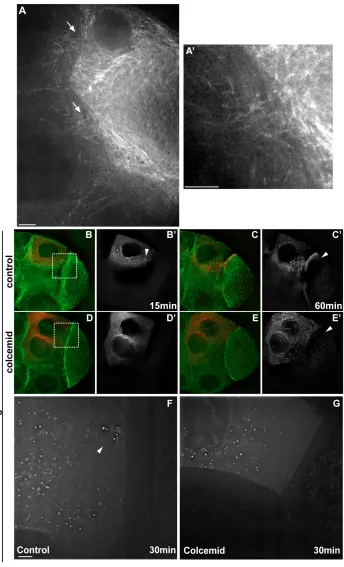
To determine whether grkmRNA transport within nurse cells and its accumulation at the ring canals requires MTs, we injected grkRNA into egg chambers expressing transgenic Tau-GFP (Micklem et al.,
1997), which decorates the MTs (Fig. 4B,C). We found that MTs are organized with a focus of high concentration at the ring canal (Fig. 4A) at the region where injected grkRNA accumulates (Fig. 4B,C). We then co-injected the MT depolymerizing drug Colcemid with fluorescent grkRNA and found that the accumulation near the ring canals and the movement into the oocyte was inhibited rapidly (Fig. 4D,E). Tau-GFP imaging showed that, 10 minutes after Colcemid injection, MTs were mostly depolymerized (Fig. 4D). We found that MT depolymerization also abolished the directionality of grk-particle movement towards the ring canals (Fig. 4F,G). We conclude that the specific movement of grkRNA particles in the nurse cells along straight paths to the ring canals is MT dependent.
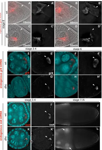
[image:4.612.56.507.382.671.2]To test whether cytoplasmic Dynein, the major minus-end MT motor in the cell, is required for grkRNA movement towards the ring canals, we injected grkRNA into hypomorphic allelic combinations of Dynein heavy chain (Dhc64C), the large force-generating ATPase subunit of the Dynein motor. These viable mutant combinations do not show dorso-ventral polarity defects, but the females are sterile (McGrail and Hays, 1997). We found that, in dhc6-6/dhc6-12mutant egg chambers, injected fluorescent grkRNA moved towards the ring canals in a directed fashion, but at slower rates than in wild type (n=14). Injected grkRNA also moved into the oocyte and localized, but, again, at a slower rate (Fig. 5A-D and see Movies 5, 6 in the supplementary material). To determine whether endogenous grk mRNA transport and/or localization is impaired in dhchypomorphic mutants, we carried out in situ hybridization. Endogenous grkmRNA was weakly localized in the oocyte of early (stage 2-7) dhcmutant egg chambers (Fig. 5E-H). To test whether the MT distribution was
D
E
V
E
LO
P
M
E
N
T
affected in dhcmutants, we imaged the distribution of MTs in living nurse cells of dhc6-6/dhc6-12mutant egg chambers, using Tau-GFP, and found that it was indistinguishable from wild-type controls (see Fig. S2 in the supplementary material), consistent with our previous work in the oocyte (MacDougall et al., 2001). We conclude that the transport of grkRNA within the nurse cells towards ring canals and from the nurse cell to the oocyte is Dynein dependent.
To test whether other localized transcripts require Dynein for their transport from the nurse cells to the oocyte, we carried out in situ hybridization with anti-sense probes against bcdand osk. We found
that, in dhc mutants from stage 10 onwards, more bcdmRNA accumulated in the nurse cells than in wild type (see Fig. S3A,B in the supplementary material). In addition, oskmRNA was also less ‘focused’ at the posterior of the oocyte in early stage (2-7) dhc6-6/dhc6-12mutants (Fig. 5K). From stage 10A onwards, both bcd and oskmRNA showed sites of nurse cell cytoplasmic accumulation in dhc6-6/dhc6-12mutants that were not evident in wild-type egg chambers (Fig. 5J,L).
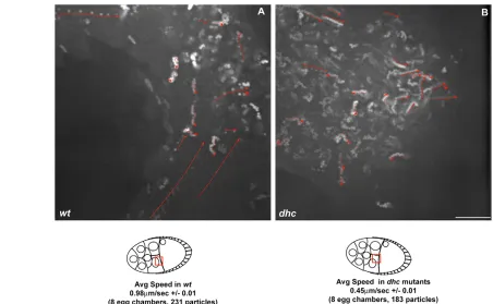
These results could be explained by either a defect in nurse cell-to-oocyte transport or by a defect in RNA localization within the oocyte. To distinguish between these possibilities and determine directly whether the movement of grkRNA towards the ring canals is Dynein dependent, we studied the movement of RNA particles at high resolution in dhc6-6/dhc6-12mutant egg chambers. We found that the speed of movement of grkRNA particles within the nurse cells towards the ring canals was substantially reduced (Fig. 6A,B). We conclude that the transport of grkRNA within the nurse cells towards the ring canals joining them to the oocyte occurs by Dynein along MTs and that Dynein is also required for the efficient transport of bcdand oskRNA from the nurse cell to the oocyte.
grkandbcdRNA recruits BicD and Egl within the nurse cells, and BicD is required for the
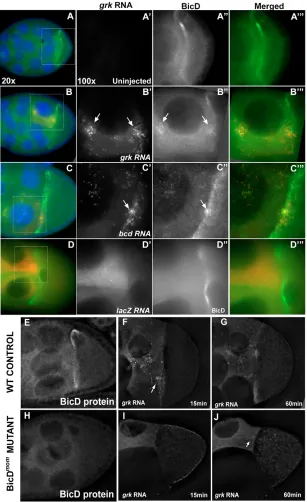
[image:5.612.52.297.58.601.2]accumulation of grkRNA in front of the ring canal To investigate the basis of the specificity of grktransport compared with that of control lacZ, we first determined whether these RNAs recruit the Dynein co-factors BicD and Egl upon injection into the nurse cells. BicD and Egl have previously been shown to be required for oocyte specification (Mach and Lehmann, 1997) and for the efficient Dynein-dependent transport of mRNA in the embryo (Bullock and Ish-Horowicz, 2001). We injected biotinylated grkand bcdRNA into wild-type egg chambers, fixed them after 20 minutes and detected BicD and Egl protein. Our results show that both BicD and Egl were recruited by injected grkand bcdRNA at the site of injection in the nurse cell cytoplasm and at the ring canals, whereas injected lacZRNA failed to recruit BicD and Egl (Fig. 7A-D and data not shown, respectively). To test whether BicD is required for grkRNA transport to the ring canals, we injected grkRNA into nurse cells of BicDmutant egg chambers. We used the BicDmommutant combination, in which the early lethality of a strong allele of BicD is rescued with the early expression of BicDfrom the heat shock Fig. 3. grkRNA particles move in directed paths towards ring canals.(A,B) Injected and subsequently fixed egg chambers show that grkRNA (A) but not lacZRNA (B) accumulates at the ring canal (arrows). (B) Arrowhead points to a ring canal itself. (C) Frames from a time-lapse movie of a nurse cell injected withgrkRNA (red) in an Actin-GFP (green) egg chamber (also see Movie 4 in the supplementary material). Panels correspond to successive time points, at 5 second intervals. Arrows indicate two grkRNA particles moving on the same path towards a ring canal that is connected to the oocyte. (D) grkRNA (red) accumulation at the ring canal at approximately 3 minutes after injection. (E) grkRNA particle (red) moving through a ring canal shown as a trail, successive time-points (at 5 second intervals) being
D
E
V
E
LO
P
M
E
N
T
promoter (Swan and Suter, 1996). In this way, later-stage egg chambers that lack BicD protein are obtained (Fig. 7E,H). We injected fluorescent grkRNA into the nurse cells of BicDmomegg chambers and found that the RNA failed to accumulate in front of the ring canals, as it does in controls (Fig. 7F,G,I,J) (n=10). We conclude that BicD, and probably also Egl, is required for grkRNA to move along directed paths within the nurse cells towards the ring canals and that lacZRNA is not able to move along these paths because it fails to recruit BicD and Egl.
grkRNA-particle movements are distinct from the general flow of cytoplasm from nurse cell to oocyte
[image:6.612.60.404.177.744.2]There are considerable cytoplasmic flows in the oocyte and nurse cells. Therefore, we tested whether the movements of grkRNA that we observed could be caused indirectly by cytoplasmic flow. To address this, we imaged the movement of cytoplasm in the nurse cells using differential interference contrast (DIC), as previously described (Bohrmann and Biber, 1994), and plotted trails of nurse
Fig. 4. Microtubules are required for grkRNA movement towards the ring canals and for transport into the oocyte.(A) Tau-GFP egg chambers show an enrichment of microtubules (MTs) at the ring canals (arrows). (A’) Higher-magnification of ring canal in A. (B-E) Tau-GFP (green) living egg chambers injected with grkRNA (red) at 15 (B,D) and 60 (C,E) minutes after injection. (B,C) grkcontrol; (D,E) grk-colcemid co-injection. (B⬘,C⬘,D⬘,E⬘) Only injected RNA is shown, at the
D
E
V
E
LO
P
M
E
N
T
cell cytoplasmic movements. Our results show that the path and direction of cytoplasmic flow in the nurse cells is significantly different to that of the injected grk RNA (see Fig. S4 in the supplementary material). We found that cytoplasmic particles did not move in directed paths towards the ring canals. By contrast, grk, and probably bcdand osk, RNAs are specifically transported by Dynein along direct paths that lead to ring canals, and are therefore transported more efficiently into the oocyte. We propose that these distinct routes to the ring canals are along a subset of MTs that we observe as a particularly high concentration of MTs near the ring canals (Fig. 4A and see Fig. S2 in the supplementary material).
DISCUSSION
Within nurse cells, we have identified a new path of Dynein-dependent transport to the ring canals that link the nurse cells to the oocyte. We show that grkRNA moves along this route, and our data
suggest that bcdand osktranscripts also follow the same path. This intracellular shortcut requires BicD and is distinct from the route taken by general cytoplasmic components and control RNAs, which move into the oocyte less effectively. Our data suggest that the distinction between RNA components that follow this direct path and those that do not is the ability to recruit the Dynein-associated co-factors BicD and Egl. We propose that Dynein-dependent transport of grk,bcd and osktranscripts towards the ring canals follows a MT network, which is distinct from other networks in the nurse cells (see Fig. S5 in the supplementary material).
[image:7.612.54.384.255.738.2]The movement of macromolecules, including RNA, within and between cells is an important process in the biology of most organisms. Although diffusion is, in principle, fast enough to distribute macromolecules over relatively large cellular distances, active transport can provide more-efficient, directed movements to specific locations as well as overcome effective diffusion barriers
D
E
V
E
LO
P
M
E
N
T
provided by cytoskeletal networks in the cell (Luby-Phelps, 2000). Moreover, diffusion of mRNA within a dense cytoplasm is very slow, because transcripts are decorated with many proteins to form very large ribonucleoprotein particles with a large hydration volume (Shav-Tal et al., 2004). It has therefore been proposed that all mRNAs, both localized and unlocalized, are associated with molecular motors to varying degrees to allow them to be rapidly distributed within the cytoplasm, and the presence of a localization signal increases the efficiency of association with the motors (Fusco et al., 2003) and perhaps even influences the activity of the motors and their co-factors (Bullock et al., 2003). Our study highlights that the presence of localization signals and recruitment of specific motor co-factors affects the choice of intracellular routes adopted by such cargo in the oocyte.
We were not able to determine the proportion of grk RNA particles that move compared with ones that were stationary because it is hard to distinguish stationary particles from autofluorescence. By contrast, rapidly moving RNA particles of the same intensity are easy to distinguish from background. By showing directly that Dynein is required for the transport of axis specification transcripts from the nurse cells to the oocyte, our work explains previous work on this topic that did not directly address the mechanism of transport from the nurse cells into the oocyte (see Introduction). Our results also explain why the movement of bcdand oskmRNA into the oocyte is MT dependent (Pokrywka and Stephenson, 1991), and why pair-rule transcripts, which are transported in the blastoderm embryo in a MT-dependent manner (Lall et al., 1999), by Dynein
(Wilkie and Davis, 2001), are also transported into the oocyte when exogenously expressed in the nurse cells (Bullock and Ish-Horowicz, 2001). We suggest that nurse cell-to-oocyte transport is likely to be a fairly promiscuous transport system that can deliver any transcript that has the capacity for transport by the Dynein motor complex along MTs to their minus ends. It is therefore likely that the Dynein-dependent shortcut is deployed by many other transcripts that are localized in the oocyte during mid-oogenesis, such as orb, K10and nanos (nos). In fact, given that the oocyte nucleus is largely transcriptionally inactive, it is possible that up to 10% of all transcripts thought to be localized in the oocyte (Dubowy and Macdonald, 1998) could first be transported by the same Dynein-dependent mechanism into the oocyte.
[image:8.612.55.507.58.337.2]D
E
V
E
LO
P
M
E
N
T
Filardo and Ephrussi, 2003; Kim-Ha et al., 1995) and Hrp48 (also known as Hrb27C – FlyBase) (Goodrich et al., 2004; Huynh et al., 2004; Norvell et al., 2005; Yano et al., 2004); however, it is unclear whether they are transported within the same complexes into the oocyte. At stage 10B, the mechanism we have described is not required, because the rapid dumping of all of the cytoplasmic contents of the nurse cells into the oocyte occurs. However, by stage 10B, most of the major patterning transcripts have probably been localized in the oocyte.
Our work does not address directly the speed of passive diffusion of RNA into the oocyte or the mechanism of cytoplasmic flow and dumping in stage 10B. Although Kinesin 1 is required for cytoplasmic movements within the oocyte (Palacios and Johnston, 2002), it is not required for the general growth of the oocyte or for
the presence of mRNAs in the oocyte (Brendza et al., 2000). These observations suggest that Kinesin 1 is not important for cytoplasmic transport or for specific mRNA transport into the oocyte.
[image:9.612.52.359.60.562.2]The existence of a specific intracellular route for the transport of transcripts in nurse cells adds to existing evidence that there are various minus-end destinations to which different cargos are delivered by Dynein within the same cell. For example, within the oocyte, bcdRNA is transported to the cortex if injected into the oocyte, but to the anterior, after transport into the oocyte, following injection into the nurse cells (Cha et al., 2001). grk RNA is transported in two steps, both of which depend on Dynein. The second step is towards the oocyte nucleus, and is unique to grkand I factorRNA (Van De Bor et al., 2005), but is not shared with bcd and K10transcripts (MacDougall et al., 2003), despite the fact that
Fig. 7. BicD protein is recruited by grkand bcd RNA and is necessary for grkRNA accumulation at ring canals.(A-D) Stage 8-9 egg chambers injected with RNA (red); BicD protein (green) and DNA (blue) are also shown (acquired at 20⫻). Outlined region represents the area shown (acquired at 100⫻) in the panels to the right (RNA, BicD and merged). (A-A) Uninjected egg chamber showing the BicD localization pattern around the oocyte nucleus. (B-C) Injected grkRNA (B⬘) and bcdRNA (C⬘) colocalize (arrows) with BicD (B⬙and C⬙, respectively). (D-D) Injected lacZRNA forms particles that do not accumulate (D⬘) and that do not colocalize with BicD (D⬙). (E,H) Immunofluorescence showing BicD localization in wild-type controls (E) and BicDmom(H) egg chambers. Notice the absence
of BicD protein in BicDmomegg chambers (H).
(F,G,I,J) Wild-type control (F,G) and BicDmom(I,J) egg
D
E
V
E
LO
P
M
E
N
T
all of these transcripts are probably being transported by Dynein. There are, therefore, likely to be several distinct MT routes along which Dynein can transport cargos within egg chambers. In neurons, choices between distinct MT routes are made by Kinesin-dependent vesicle transport depending on the presence of a specific neurotransmitter-receptor-interacting protein, GRIP1 (Setou et al., 2002). How Dynein chooses between distinct MT networks is less clear, but could be based on distinct isoforms of the motor complex, on distinct kinds of MTs with different tubulin isoforms, or on their decoration with different MT-associated proteins. In addition, there is evidence that cargos can influence the behaviour of their motor (Bullock et al., 2003), raising the interesting possibility that cargos could also influence the choice of MT route adopted by their motors. Our work suggests that the presence of BicD and Egl could also influence the choice of MT route adopted by motors. Future work, including new approaches for co-visualizing MTs and RNAs in living cells, will be required to distinguish between all of these possible ways of selecting intracellular routes. Whatever the basis of such distinct routes, they are likely to exist for various kinds of molecular motors and to be functionally important for a wide range of tissues and cargos.
We thank Nina MacDougall for originally establishing the injection assay for
grkRNA movement from the nurse cell to the oocyte. We are grateful to Simon Bullock, David Finnegan, Catherine Rabouille, Hildegard Tekotte and Georgia Vendra for their incisive comments on the manuscript. This work was supported by a Wellcome Trust Senior Research Fellowship (067413) to I.D., Edinburgh University Studentship to A.C. and a Marie Curie Postdoctoral Fellowship to C.M.
Supplementary material
Supplementary material for this article is available at http://dev.biologists.org/cgi/content/full/134/10/1955/DC1
References
Bohrmann, J. and Biber, K.(1994). Cytoskeleton-dependent transport of cytoplasmic particles in previtellogenic to mid-vitellogenic ovarian follicles of
Drosophila: time-lapse analysis using video-enhanced contrast microscopy. J. Cell Sci. 107, 849-858.
Brendza, R. P., Serbus, L. R., Duffy, J. B. and Saxton, W. M.(2000). A function for kinesin I in the posterior transport of oskarmRNA and Staufen protein.
Science 289, 2120-2122.
Bullock, S. L. and Ish-Horowicz, D.(2001). Conserved signals and machinery for RNA transport in Drosophilaoogenesis and embryogenesis. Nature 414, 611-616.
Bullock, S. L., Zicha, D. and Ish-Horowicz, D.(2003). The Drosophila hairyRNA localization signal modulates the kinetics of cytoplasmic mRNA transport. EMBO J. 22, 2484-2494.
Castagnetti, S., Hentze, M. W., Ephrussi, A. and Gebauer, F.(2000). Control of
oskarmRNA translation by Bruno in a novel cell-free system from Drosophila
ovaries. Development 127, 1063-1068.
Cha, B., Koppetsch, B. S. and Theurkauf, W. E.(2001). In vivo analysis of
Drosophila bicoid mRNA localization reveals a novel microtubule-dependent axis specification pathway. Cell 106, 35-46.
Chekulaeva, M., Hentze, M. W. and Ephrussi, A.(2006). Bruno acts as a dual repressor of oskar translation, promoting mRNA oligomerization and formation of silencing particles. Cell 124, 521-533.
Davis, I.(2000). Visualising fluorescence in Drosophila – optimal detection in thick specimens. In Protein Localisation by Fluorescence Microscopy: A Practical Approach(ed. V. J. Allan), pp. 131-162. Oxford: Oxford University Press.
Driever, W. and Nüsslein-Volhard, C.(1988). A gradient of bicoid protein in
Drosophilaembryos. Cell 54, 83-93.
Dubowy, J. and Macdonald, P. M.(1998). Localization of mRNAs to the oocyte is common inDrosophila ovaries. Mech. Dev.70, 193-195.
Ephrussi, A., Dickinson, L. K. and Lehmann, R.(1991). Oskarorganizes the germ plasm and directs localization of the posterior determinant nanos. Cell 66, 37-50.
Ferrandon, D., Koch, I., Westhof, E. and Nusslein-Volhard, C.(1997). RNA-RNA interaction is required for the formation of specific bicoidmRNA 3⬘ UTR-STAUFEN ribonucleoprotein particles. EMBO J. 16, 1751-1758.
Filardo, P. and Ephrussi, A.(2003). Bruno regulates gurkenduring Drosophila
oogenesis. Mech. Dev.120, 289-297.
Fusco, D., Accornero, N., Lavoie, B., Shenoy, S. M., Blanchard, J. M., Singer,
R. H. and Bertrand, E.(2003). Single mRNA molecules demonstrate probabilistic movement in living mammalian cells.Curr. Biol. 13, 161-167.
Gepner, J., Li, M., Ludmann, S., Kortas, C., Boylan, K., Iyadurai, S. J., McGrail, M. and Hays, T. S.(1996). Cytoplasmic Dynein function is essential in
Drosophila melanogaster. Genetics 142, 865-878.
Gonzalez-Reyes, A., Elliott, H. and St Johnston, D.(1995). Polarization of both major body axes in Drosophilaby Gurken-Torpedo signaling. Nature 375, 654-658.
Goodrich, J. S., Clouse, K. N. and Schupbach, T.(2004). Hrb27C, Sqd and Otu cooperatively regulate gurken RNA localization and mediate nurse cell chromosome dispersion in Drosophilaoogenesis. Development 131, 1949-1958.
Huynh, J. R., Munro, T. P., Smith-Litiere, K., Lepesant, J. A. and St Johnston, D.(2004). The Drosophila hnRNPA/B homolog, Hrp48, is specifically required for a distinct step in oskmRNA localization. Dev. Cell 6, 625-635.
Januschke, J., Gervais, L., Dass, S., Kaltschmidt, J. A., Lopez-Schier, H., Johnston, D. S., Brand, A. H., Roth, S. and Guichet, A.(2002a). Polar transport in the Drosophilaoocyte requires Dynein and Kinesin I cooperation.
Curr. Biol. 12, 1971-1981.
Januschke, J., Gervais, L., Dass, S., Kaltschmidt, J. A., Lopez-Schier, H., St Johnston, D. S., Brand, A. H., Roth, S. and Guichet, A.(2002b). Polar transport in the Drosophilaoocyte requires Dynein and Kinesin I cooperation.
Curr. Biol. 12, 1971-1981.
Kim-Ha, J., Kerr, K. and Macdonald, P. M.(1995). Translational regulation of
oskarmRNA by Bruno, an ovarian RNA-binding protein, is essential. Cell 81, 403-412.
King, R. C. and Burnett, R. G.(1959). Autoradiographic study of uptake of tritiated glycine, thymidine, and uridine by fruit fly ovaries. Science 129, 1674-1675.
Koch, E. A. and Spitzer, R. H.(1983). Multiple effects of colchicine on oogenesis in Drosophila: induced sterility and switch of potential oocyte to nurse-cell developmental pathway. Cell Tissue Res.228, 21-32.
Lall, S., Francis-Lang, H., Flament, A., Norvell, A., Schupbach, T. and Ish-Horowicz, D.(1999). Squid hnRNP protein promotes apical cytoplasmic transport and localization of Drosophilapair-rule transcripts. Cell 98, 171-180.
Liu, Z., Xie, T. and Steward, R.(1999). Lis1, the Drosophilahomolog of a human lissencephaly disease gene, is required for germline cell division and oocyte differentiation. Development 126, 4477-4488.
Luby-Phelps, K.(2000). Cytoarchitecture and physical properties of cytoplasm: volume, viscosity, diffusion, intracellular surface area. Int. Rev. Cytol.192, 189-221.
MacDougall, N., Lad, Y., Wilkie, G., Francis-Lang, H., Sullivan, W. and Davis, I.(2001). Merlin, theDrosophilahomologue of Neurofibromatosis-2, is specifically required in posterior follicle cells for axis formation in the oocyte.
Development 128, 665-673.
MacDougall, N., Clark, A., MacDougall, E. and Davis, I.(2003). Drosophila gurken(TGFalpha) mRNA localizes as particles that move within the oocyte in two dynein-dependent steps. Dev. Cell 4, 307-319.
Mach, J. M. and Lehmann, R.(1997). An Egalitarian-BicaudalD complex is essential for oocyte specification and axis determination in Drosophila. Genes Dev.11, 423-435.
Mahajan-Miklos, S. and Cooley, L.(1994). Intercellular cytoplasm transport during Drosophila oogenesis. Dev. Biol. 165, 336-351.
McGrail, M. and Hays, T. S.(1997). The microtubule motor cytoplasmic Dynein is required for spindle orientation during germline cell divisions and oocyte differentiation in Drosophila. Development 124, 2409-2419.
Micklem, D. R., Dasgupta, R., Elliott, H., Gergely, F., Davidson, C., Brand, A., Gonzalez-Reyes, A. and St Johnston, D.(1997). The mago nashigene is required for the polarisation of the oocyte and the formation of perpendicular axes in Drosophila. Curr. Biol. 7, 468-478.
Neuman-Silberberg, F. S. and Schüpbach, T.(1993). The Drosophila
dorsoventral patterning gene gurkenproduces a dorsally localized RNA and encodes a TGF alpha-like protein. Cell 75, 165-174.
Norvell, A., Debec, A., Finch, D., Gibson, L. and Thoma, B.(2005). Squid is required for efficient posterior localization of oskarmRNA during Drosophila
oogenesis. Dev. Genes Evol.215, 340-349.
Palacios, I. M. and Johnston, D. S.(2002). Kinesin light chain-independent function of the Kinesin heavy chain in cytoplasmic streaming and posterior localisation in the Drosophilaoocyte. Development 129, 5473-5485.
Pokrywka, N. J. and Stephenson, E. C.(1991). Microtubules mediate the localization of bicoidRNA during Drosophilaoogenesis. Development 113, 55-66.
Pollard, T. and Earnshaw, W.(2002). Intracellular motility. In Cell Biology, pp. 619-632. Philadelphia: Saunders.
Schnorrer, F., Bohmann, K. and Nüsslein-Volhard, C.(2000). The molecular motor Dynein is involved in targeting swallow and bicoidRNA to the anterior pole of Drosophilaoocytes. Nat. Cell Biol. 2, 185-190.
D
E
V
E
LO
P
M
E
N
T
Shav-Tal, Y., Darzacq, X., Shenoy, S. M., Fusco, D., Janicki, S. M., Spector, D. L. and Singer, R. H.(2004). Dynamics of single mRNPs in nuclei of living cells.
Science 304, 1797-1800.
Spradling, A. C.(1993). Developmental genetics of oogenesis. In The Development of Drosophila melanogaster(ed. M. Bate and A. M. Martinez Arias), pp. 1-70. Cold Spring Harbour, NY: Cold Spring Harbour Laboratory Press.
St Johnston, D., Driever, W., Berleth, T., Richstein, S. and Nüsslein-Volhard, C.(1989). Multiple steps in the localization of bicoidRNA to the anterior pole of the Drosophilaoocyte. Development 107, 13-19.
Suter, B. and Steward, R.(1991). Requirement for phosphorylation and localization of the Bicaudal-D protein in Drosophilaoocyte differentiation. Cell 67, 917-926.
Swan, A. and Suter, B.(1996). Role for Bicaudal-D in patterning the Drosophila
egg chamber in mid-oogenesis. Development122, 3577-3586.
Swan, A., Nguyen, T. and Suter, B.(1999). DrosophilaLissencephaly-1 functions with Bic-D and Dynein in oocyte determination and nuclear positioning.Nat. Cell Biol.1, 444-449.
Tekotte, H. and Davis, I.(2006). Bruno: a double turn-off for Oskar. Dev. Cell 10, 280-281.
Theurkauf, W. E. and Hazelrigg, T. I.(1998). In vivo analyses of cytoplasmic
transport and cytoskeletal organization during Drosophilaoogenesis: Characterization of a multi-step anterior localization pathway. Development 125, 3655-3666.
Theurkauf, W. E., Alberts, B. M., Jan, Y. N. and Jongens, T. A.(1993). A central role for microtubules in the differentiation of Drosophilaoocytes.
Development 118, 1169-1180.
Thio, G. L., Ray, R. P., Barcelo, G. and Schüpbach, T.(2000). Localization of
gurkenRNA in Drosophilaoogenesis requires elements in the 5⬘and 3⬘regions of the transcript. Dev. Biol. 221, 435-446.
Van De Bor, V., Hartswood, E., Jones, C., Finnegan, D. and Davis, I.(2005).
gurkenand the Ifactor retrotransposon RNAs share common localization signals and machinery. Dev. Cell 9, 51-62.
Wagner, C., Palacios, I., Jaeger, L., St Johnston, D., Ehresmann, B., Ehresmann, C. and Brunel, C.(2001). Dimerization of the 3⬘UTR of bicoid
mRNA involves a two-step mechanism.J. Mol. Biol. 313, 511-524.
Wilkie, G. S. and Davis, I.(2001). Drosophila winglessand pair-rule transcripts localize apically by Dynein-mediated transport of RNA particles. Cell 105, 209-219.