ORIGINAL RESEARCH
PATIENT SAFETY
Radiation Dose Reduction in CT-Guided Spine Biopsies Does
Not Reduce Diagnostic Yield
K.A. Shpilberg, B.N. Delman, L.N. Tanenbaum, S.J. Esses, R. Subramaniam, and A.H. Doshi
ABSTRACT
BACKGROUND AND PURPOSE: CT-guided biopsy is the most commonly used method to obtain tissue for diagnosis in suspected cases of malignancy involving the spine. The purpose of this study was to demonstrate that a low-dose CT-guided spine biopsy protocol is as effective in tissue sampling as a regular-dose protocol, without adversely affecting procedural time or complication rates.
MATERIALS AND METHODS: We retrospectively reviewed all patients who underwent CT-guided spine procedures at our institution between May 2010 and October 2013. Biopsy duration, total number of scans, total volume CT dose index, total dose-length product, and diagnostic tissue yield of low-dose and regular-dose groups were compared.
RESULTS:Sixty-four patients were included, of whom 31 underwent low-dose and 33 regular-dose spine biopsies. There was a statistically significant difference in total volume CT dose index and total dose-length product between the low-dose and regular-dose groups (P⬍ .0001). There was no significant difference in the total number of scans obtained (P⫽.3385), duration of procedure (P⫽.149), or diagnostic tissue yield (P⫽.6017).
CONCLUSIONS: Use of a low-dose CT-guided spine biopsy protocol is a practical alternative to regular-dose approaches, maintaining overall quality and efficiency at reduced ionizing radiation dose.
ABBREVIATIONS:CTDIvol⫽volume CT dose index; DLP⫽dose-length product; kVp⫽peak kilovoltage; mGy⫽milligray
I
maging-guided biopsy is a commonly used method to obtain a tissue diagnosis in suspected cases of malignancy. In particular, CT guidance is often used for precise localization of a lesion be-fore and during biopsy. It provides the operator with great ana-tomic detail for biopsy planning and execution and allows for confirmation of needle placement into the area of concern. CT guidance is the preferred method of biopsy for osseous lesions within the vertebrae.1-4Even though CT guidance has becomeincreasingly used for various procedures, there is concern over the amount of radiation exposure to the patient.5-8
Radiation dose reduction is commonly used in routine diag-nostic CT scanning. Pediatric patients and patients who receive
multiple scans for acute disease follow-up, chronic conditions, and screening purposes often undergo CT with modified scan-ning protocols to reduce dose.9-14This type of protocol
modifi-cation has also been used in CT-guided interventions to limit radiation dose when performing multiple scans during the proce-dure.8,15-18Given the increased desire to reduce radiation dose to
patients, we transitioned our protocols for CT-guided spine biop-sies to use a lower dose.
The purpose of this study was to demonstrate that a low-dose protocol for CT-guided spine biopsies is as effective in tissue sampling without an increase in procedural time or an increase in complication rates compared with our legacy higher-dose approaches.
MATERIALS AND METHODS
After obtaining Institutional Review Board approval, we retro-spectively reviewed all patients who underwent CT-guided spine procedures at our institution between May 2010 and October 2013. The total number of charts reviewed was 132.
Patients who underwent disk space aspirations and biopsies for suspected diskitis/osteomyelitis were excluded because of lim-ited availability of surgical pathology data as most specimens were
Received April 29, 2014; accepted after revision June 5.
From the Department of Radiology, Icahn School of Medicine, Mount Sinai Health System, New York, New York.
Paper previously presented as an oral presentation at: American Society of Spine Radiology Annual Symposium, February 23–26, 2014; Miami Beach, Florida and An-nual Meeting of the American Society of Neuroradiology, May 17–22, 2014; Mon-treal, Que´bec, Canada.
Please address correspondence to Amish Doshi, MD, Mount Sinai Hospital, Depart-ment of Radiology, Box 1234, 1 Gustave L Levy Pl, New York, NY 10029; e-mail: [email protected], [email protected]
only submitted for microbiology analysis. CT-guided pain man-agement procedures such as facet cyst ruptures and epidural in-jections were also excluded. Patients for whom dose reports were not available in our institution’s PACS were excluded.
Ultimately, 64 patients were included in this analysis. Two lesions were biopsied in 2 patients and 1 lesion in the remaining 62 patients, yielding a total of 66 lesions. All the biopsies were per-formed by 1 Certificate of Added Qualification– certified neuro-radiologist (A.H.D.) with 6 years of experience. The low-dose protocol was initiated in February 2012 and has been almost ex-clusively used since November 2012.
Procedure
All CT-guided spine biopsies were performed on a 4-channel CT scanner (Volume Zoom; Siemens, Erlangen, Germany) or 8-sec-tion CT scanner (LightSpeed Ultra; GE Healthcare, Milwaukee, Wisconsin) in helical mode based on availability. The 8-section scanner was used to guide 50 biopsies (78.1%) and 4-section scan-ner for the remaining 14 biopsies (21.9%). CT fluoroscopy was not available. Patients all followed a standard course for these biopsies. Each was positioned prone for the procedures. Vital signs were monitored. Mild to moderate conscious sedation was used in 60 patients (93.8%), monitored anesthesia care in 2 pa-tients (3.1%), and local anesthetic only in 2 papa-tients (3.1%). A Fast Find Grid (Webb Manufacturing, Philadelphia, Pennsylva-nia) was placed over the general biopsy site for localization. In each patient, 1 preprocedure CT scan was obtained using a regu-lar-dose protocol (120 peak kilovoltage [kVp]) and 200 mAs) for planning. Skin was prepped and draped in normal sterile fashion. One percent lidocaine was infiltrated into tissues for local and deep anesthesia. An 11-, 12-, or 13-gauge bone biopsy needle set (Osteo-Site; Cook, Bloomington, Indiana, or Bonopty; AprioMed, Londonderry, New Hampshire) was advanced into the lesion
with CT images obtained after each nee-dle advancement. Once the neenee-dle was confirmed within the lesion, CT scans were performed after each biopsy pass. In each patient, 1 final postbiopsy scan was obtained after the needle was removed us-ing regular-dose parameters to assess for postprocedural complications. Patients were then transferred to a recovery area to be monitored before discharge or return to their hospital room.
Data Collection and Scanning Parameters
Data from PACS and dose reports were collected, including age, sex, location, and characteristics of lesion biopsied, kVp, mAs, pitch, volume CT dose index (CTDIvol) per series (milligray [mGy]), CTDIvol total (mGy), scan range (mm), dose-length product (DLP) per series (mGy䡠cm), total DLP (mGy䡠cm), number of biopsy-guiding scans, number of pre-and postbiopsy diagnostic scans, number of needle passes, total number of scans, duration of each biopsy (defined as time from the first prebiopsy scan to last postbiopsy scan), and complications. Pathology re-sults were obtained for each patient from electronic medical records.
Low-dose biopsies were defined as those with a kVp of 80 and mAs of 40 – 60. Regular-dose biopsies were defined as those with a kVp of 120 and mAs⬎200. Scans performed at kVp and mAs parameters outside the above-mentioned criteria of low-dose or regular-dose biopsies were classified based on average CTDIvol (CTDIvol⬍10 mGy for low dose; CTDIvol⬎10 mGy for regular dose) as previously described by Kro¨pil et al.19They defined
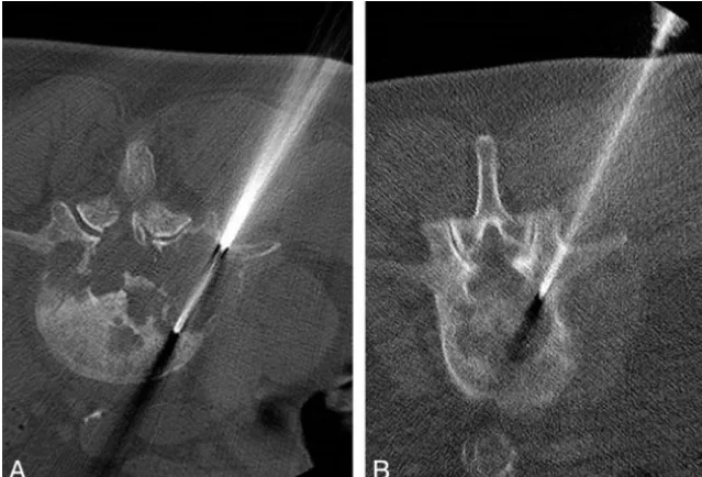
low-dose CTs as having a CT low-dose index⬍10 mGy. For example, 2 patients whose biopsies were started as low-dose protocol were switched to regular-dose protocol at the operator’s discretion be-cause of insufficient conspicuity of subtle lesions and were classi-fied as “regular-dose” because the average CTDIvol was 17.1 mGy in one and 20.3 mGy in the other. Figure 1 demonstrates repre-sentative images from regular-dose and low-dose CT-guided spine biopsies.
Diagnostic tissue yield was classified as “positive for malig-nancy,” “specific benign diagnosis,” and “negative for malignancy without a specific benign diagnosis.” Lesions were classified as lytic, sclerotic, or mixed. The location of lesions was recorded as cervical, thoracic, lumbar, or sacral.
Age, biopsy duration, total number of scans (including prebi-opsy and postbiprebi-opsy scans), total CTDIvol (including that used for prebiopsy and postbiopsy scans), and total DLP (including that used for prebiopsy and postbiopsy scans) of low-dose and regular-dose groups were compared using an unpaired t test (GraphPad Prism software; GraphPad Software, San Diego, Cal-ifornia). Diagnostic tissue yield and the distribution of lesions by type and location of low-dose and regular-dose biopsies were FIG 1. A, Axial CT performed with the regular-dose technique (kVp 120, mAs 250) demonstrates
[image:2.594.53.373.48.265.2]compared using Fisher exact test (GraphPad Software).Pvalue⬍ .05 was considered statistically significant.
RESULTS
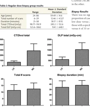
Of the 64 patients who underwent CT-guided spine biopsies from 2010 to 2013, 29 patients (45.3%) underwent the procedure using a low-dose protocol and 35 patients (54.7%) using a regular-dose protocol. Table 1 demonstrates the mean and ranges for age, number of scans, duration of procedure, total CTDIvol, and total DLP for low-dose protocol; Table 2 denotes the same for regular-dose protocol.
Demographics
There was no significant difference between the 2 groups in pa-tient age (63.86⫾13.67 years for low dose versus 59.49⫾14.6
years for regular dose;P⫽.2239) or in sex distribution (14 of 29 or 48.3% women for low dose versus 19 of 35 or 54.3% women for regular dose;P⫽.802).
Dose and Scanning Time
There was a statistically significant difference between low-dose and regular-dose groups in total CTDIvol (69.47⫾24.76 mGy for low dose versus 285.2⫾132.6 mGy for regular dose;P⬍.0001) and total DLP (601.5⫾237.7 mGy䡠cm for low dose versus 1541⫾ 648.1 mGy䡠cm for regular dose;P⬍.0001) (Fig 2).
There was no significant difference in total number of scans obtained (11.38⫾4.354 for low dose versus 12.46⫾4.527 for regular dose;P⫽ .3385) and duration of procedure (34.31⫾ 12.19 minutes for low dose versus 38.17⫾8.92 minutes for reg-ular dose;P⫽.149) between the 2 groups (Fig 2).
Several outliers were noted, falling greater or less than 2 stan-dard deviations from the mean. One patient in the low-dose group who had 2 lesions (one mixed and one sclerotic) biopsied had significantly more scans, longer duration of the procedure and higher total DLP than average. Two patients in the regular-dose group had significantly more scans than average (25 and 29) because of difficulty in accessibility of small vertebral body le-sions, which resulted in significantly higher than average total CTDIvol (761.86 mGy) in one and total DLP (3062.46 mGy䡠cm) in the other.
Biopsy Results
There was no significant difference between the 2 groups in the proportion of cases positive for malignancy (20 of 29 or 69.0% for low dose versus 21 of 35 or 60% for regular-dose;P⫽.6017), those with a specific benign diagnosis (2 of 29 or 6.9% for low dose versus 6 of 35 or 17.1% for regular dose;P⫽.2754), and those whose pathology was negative for malig-nancy without a specific benign diagnosis (7 of 29 or 24.1% for low dose versus 8 of 35 or 22.9% for regular dose;P⫽1.00).
Of the 66 lesions that were biopsied, 39 (59.1%) were lytic, 15 (22.7%) were scle-rotic, and 12 (18.2%) were mixed. There was no statistically significant difference in lesion type between the low-dose and regular-dose groups (P values ranging from .1174 to .7694).
Most of the lesions that underwent bi-opsy were located within the lumbar spine (29 of 66; 44%). This was followed by the thoracic spine (28 of 66; 42.4%), sacrum (7 of 66; 10.6%) and cervical spine (2 of 66; 3%). There was no statistically signif-icant difference in the location of lesions between the low-dose and regular-dose groups (P values ranging from .4334 to 1.00).
There were sufficient specimens for diagnosis in all patients in both biopsy groups. Overall, there was only 1 minor complication characterized by bleeding from the cannula, which was successfully FIG 2. Graphs of means with standard deviations comparing radiation dose (total CTDIvol and
[image:3.594.53.284.245.323.2]DLP), total number of scans, and biopsy duration between low-dose and regular-dose groups. Table 1: Low-dose biopsy group results
Range
MeanⴞStandard Deviation
Age (years) 29–87 63.86⫾13.67
Total number of scans 5–22 11.38⫾4.354
Duration (minutes) 19–76 34.31⫾12.19
[image:3.594.55.369.328.703.2]Total CTDIvol (mGy) 29.98–147.9 69.47⫾24.76 Total DLP (mGy䡠cm) 239.3–1206 601.5⫾237.7
Table 2: Regular dose biopsy group results
Range
MeanⴞStandard Deviation
Age (years) 24–86 59.49⫾14.6
Total number of scans 6–29 12.46⫾4.527
Duration (minutes) 21–58 38.17⫾8.92
treated with Gelfoam (Pfizer, New York, New York), in a low-dose group patient whose biopsy yielded metastatic renal cell car-cinoma. No major complications were reported.
DISCUSSION
Imaging guidance for biopsy is a commonly used procedure in patients with imaging findings concerning for malignancy. In particular, CT guidance has been used for biopsy of a variety of sites within the body.1-4,8,15,16,20-23This is largely attributed to an
improved ability of the operator to identify the lesion and plan a trajectory for biopsy. CT-guided biopsy has been shown to be an effective tool in identifying pathology with relatively low risk and cost compared with open biopsy.4,22,24 However, a frequently
cited concern with CT scanning is the potential consequences of ionizing radiation, and there is much emphasis on limiting radi-ation to as low as reasonably achievable to obtain the necessary results whenever possible.8,25
Previous studies have demonstrated the utility of a low-dose CT technique for a variety of interventional procedures. Meng et al15performed biopsies of lung lesions at lower doses and found
that a reduction in the measure of radiation dose, CT dose index, and DLP were possible without sacrificing diagnostic yield. Smith et al8were able to reduce the radiation dose to the chest during
CT-guided percutaneous lung biopsies by greater than 95% (from DLP of 677.5 mGy䡠cm to 18.3 mGy䡠cm) without decreasing tech-nical success or patient safety. Pediatric CT-guided bone biopsies have been performed using lower mAs and kVp techniques pro-ducing acceptable image quality and providing similar diagnostic yield compared with standard techniques.16A low-dose CT
pro-tocol has also been used in spinal pain interventions. One study found that a change in CT parameters to lower radiation dose resulted in an 86% reduction in total DLP (from 1458 mGy䡠cm to 199 mGy䡠cm) for CT-guided spine injection procedures for pain.17Artner et al18demonstrated that the dose related to
CT-guided sacroiliac joint injections can be significantly reduced to levels of pulsed fluoroscopy without compromising needle place-ment into the joint.
In this study, we found a significantly reduced radiation dose as expressed by CTDIvol and DLP in patients undergoing CT-guided spine biopsies using a low-dose protocol compared with a regular-dose protocol. There was no significant difference in the total number of biopsy scans, procedure time, or in the diagnostic yield between the groups. To our knowledge, this is the first study demonstrating a significantly reduced patient exposure to ioniz-ing radiation durioniz-ing CT-guided spine biopsies without sacrificioniz-ing the quality, efficiency, and diagnostic yield of the procedure.
Although there was a significantly lower radiation exposure in the low-dose biopsy group compared with the regular-dose group, we predict that a DLP might be lowered even further by reducing tube voltage (mAs) and/or current (kVp), increasing the pitch and decreasing the scan range in the z-axis.20,26-29
Intermit-tent axial scanning mode rather than helical mode and the use of a stationary CT table may further contribute to radiation dose reduction.24,30
A substantial proportion of radiation exposure comes from pre- and postbiopsy scans because they are designed to optimize soft tissue visualization for needle guidance and to exclude
post-biopsy traumatic sequelae. In fact, 1 study showed that up to 90% of the total patient dose during biopsies was administered during the helical planning stage.29Therefore, prebiopsy diagnostic
im-aging should be carefully reviewed beforehand to determine whether repeat conventional dose scanning may be avoided dur-ing the procedure.31If a prebiopsy scan is necessary, a grid can be
placed over the spinal level of interest before the first series of scans based on known anatomic landmarks.31Chintapalli et al31
suggest that in low-risk CT-guided interventions, which may in-clude some spine biopsies, regular-dose postbiopsy scans can be eliminated at the discretion of the radiologist. A low-dose proto-col, as well as techniques to further reduce dose, should be famil-iar to the radiologist performing the procedures and technologist acquiring the images.31
Newer techniques have recently emerged to address image quality when reducing CT dose. These include iterative recon-struction models such as adaptive statistical iterative reconstruc-tion and model-based iterative reconstrucreconstruc-tion.9-14,32-40Although
these imaging algorithms provide an additional method for dose reduction in CT-guided procedures, their availability is currently limited to newer CT scanners for routine diagnostic CT imaging. The greater availability of the iterative reconstruction software over time may allow for increased operator comfort when evalu-ating low-dose images during CT-guided procedures, potentially further reducing the radiation dose.
This retrospective study did have some limitations. It was not randomized, and a single operator performed most of the CT-guided spine biopsies. Therefore, the reproducibility of the results using the low-dose protocol cannot be fully assessed in this study. In addition, it would be difficult to determine whether increasing comfort with the procedure may have contributed to a slightly greater efficiency of the procedure using a low-dose protocol. The retrospective nature of this study also limits assessment of factors related to operator scanning protocol adjustments in challenging biopsy cases.
CONCLUSIONS
Radiation exposure to patients undergoing CT-guided spinal bi-opsies can be optimized to reduce the overall dose during the examination. Low-dose CT-guided spine biopsies have a signifi-cantly lower total cumulative radiation exposure compared with regular-dose CT biopsies without significantly affecting proce-dural time or diagnostic tissue yield. A simple dose-reduction protocol can use reduction in mAs and kVp during the procedure. A number of additional modifications to image acquisitions can be made to reduce the dose. Our data show that a low-dose tocol should be considered as an alternative to regular-dose pro-tocol when performing CT-guided spinal biopsies, allowing the operator to reduce ionizing radiation dose while maintaining overall quality and efficiency of the procedure.
ACKNOWLEDGMENTS
The authors thank Idoia Corcuera-Solano, MD, for help with sta-tistical analysis.
UNRELATED: Payment for Lectures (including service on Speakers Bureaus):GE, Siemens.*Money paid to the institution.
REFERENCES
1. Brugieres P, Revel MP, Dumas JL, et al.CT-guided vertebral biopsy.
A report of 89 cases.J Neuroradiol1991;18:351–59
2. Lis E, Bilsky MH, Pisinski L, et al.Percutaneous CT-guided biopsy of osseous lesion of the spine in patients with known or suspected
malignancy.AJNR Am J Neuroradiol2004;25:1583– 88
3. Kornblum MB, Wesolowski DP, Fischgrund JS, et al.Computed to-mography-guided biopsy of the spine. A review of 103 patients.
Spine (Phila Pa 1976)1998;23:81– 85
4. Rimondi E, Staals EL, Errani C, et al.Percutaneous CT-guided bi-opsy of the spine: results of 430 biopsies.Eur Spine J2008;17:975– 81 5. Coursey C, Frush D.CT and radiation: what radiologists should
know.Appl Radiol2008;37:22–29
6. Brenner DJ, Hall EJ.Computed tomography—an increasing source
of radiation exposure.N Engl J Med2007;357:2277– 84
7. Huda W, Mettler FA.Volume CT dose index and dose-length
prod-uct displayed during CT: what good are they? Radiology
2011;258:236 – 42
8. Smith JC, Jin DH, Watkins GE, et al.Ultra-low-dose protocol for
CT-guided lung biopsies.J Vasc Interv Radiol2011;22:431–36
9. Corcuera-Solano I, Doshi AH, Noor A, et al.Repeated head CT in the neurosurgical intensive care unit: feasibility of sinogram-affirmed iterative reconstruction-based ultra-low-dose CT for surveillance.
AJNR Am J Neuroradiol2014;35:1281– 87
10. Mathieu KB, Ali H, Fox PS, et al.Radiation dose reduction for CT lung cancer screening using ASIR and MBIR: a phantom study.
J Appl Clin Med Phys2014;15:4515
11. Flicek KT, Hara AK, Silva AC, et al.Reducing the radiation dose for CT colonography using adaptive statistical iterative reconstruction: a
pi-lot study.AJR Am J Roentgenol2010;195:126 –31
12. Kambadakone AR, Chaudhary NA, Desai GS, et al.Low-dose MDCT and CT enterography of patients with Crohn disease: feasibility of
adaptive statistical iterative reconstruction.AJR Am J Roentgenol
2011;196:W743–52
13. Shuman WP, Green DE, Busey JM, et al.Model-based iterative re-construction versus adaptive statistical iterative rere-construction and filtered back projection in liver 64-MDCT: focal lesion
detec-tion, lesion conspicuity, and image noise.AJR Am J Roentgenol
2013;200:1071–76
14. Becce F, Ben Salah Y, Verdun FR, et al.Computed tomography of the cervical spine: comparison of image quality between a standard-dose and a low-standard-dose protocol using filtered back-projection and
iterative reconstruction.Skeletal Radiol2013;42:937– 45
15. Meng XX, Kuai XP, Dong WH, et al.Comparison of lung lesion biopsies between low-dose CT-guided and conventional CT-guided
techniques.Acta Radiol2013;54:909 –15
16. Patel AS, Soares B, Courtier J, et al.Radiation dose reduction in
pediatric CT-guided musculoskeletal procedures.Pediatr Radiol
2013;43:1303– 08
17. Shepherd TM, Hess CP, Chin CT, et al.Reducing patient radiation dose during CT-guided procedures: demonstration in spinal
injec-tions for pain.AJNR Am J Neuroradiol2011;32:1776 – 82
18. Artner J, Cakir B, Reichel H, et al.Radiation dose reduction in CT-guided sacroiliac joint injections to levels of pulsed fluoroscopy: a
comparative study with technical considerations. J Pain Res
2012;5:265– 69
19. Kro¨pil P, Lanzman RS, Walther C, et al.Dose reduction and image quality in MDCT of the upper abdomen: potential of an adaptive
post-processing filter[in German].Rofo2010;182:248 –53
20. Kloeckner R, dos Santos DP, Schneider J, et al.Radiation exposure in
CT-guided interventions.Eur J Radiol2013;82:2253–57
21. Monfardini L, Preda L, Aurilio G, et al.CT-guided bone biopsy in cancer patients with suspected bone metastases: retrospective
re-view of 308 procedures.Radiol Med2014;119:852– 60
22. Gogna A, Peh WC, Munk PL.Image-guided musculoskeletal
bi-opsy.Radiol Clin North Am2008;46:455–73, v
23. Hau A, Kim I, Kattapuram S, et al.Accuracy of CT-guided biopsies
in 359 patients with musculoskeletal lesions. Skeletal Radiol
2002;31:349 –53
24. Leng S, Christner JA, Carlson SK, et al.Radiation dose levels for
interventional CT procedures. AJR Am J Roentgenol 2011;197:
W97–103
25. Lucey BC, Varghese JC, Hochberg A, et al.CT-guided intervention
with low radiation dose: feasibility and experience.AJR Am J
Roent-genol2007;188:1187–94
26. Kalra MK, Maher MM, Toth TL, et al.Strategies for CT radiation
dose optimization.Radiology2004;230:619 –28
27. Tsalafoutas IA, Tsapaki V, Triantopoulou C, et al.CT-guided inter-ventional procedures without CT fluoroscopy assistance: patient
effective dose and absorbed dose considerations.AJR Am J
Roent-genol2007;188:1479 – 84
28. Smith AB, Dillon WP, Lau BC, et al.Radiation dose reduction strat-egy for CT protocols: successful implementation in neuroradiology
section.Radiology2008;247:499 –506
29. Sarti M, Brehmer WP, Gay SB.Low-dose techniques in CT-guided
interventions.Radiographics2012;32:1109 –19; discussion 1119 –20
30. Chang AL, Schoenfeld AH, Brook AL, et al.Radiation dose for 345
CT-guided interlaminar lumbar epidural steroid injections.AJNR
Am J Neuroradiol2013;34:1882– 86
31. Chintapalli KN, Montgomery RS, Hatab M, et al.Radiation dose management: part 1, minimizing radiation dose in CT-guided
pro-cedures.AJR Am J Roentgenol2012;198:W347–51
32. McKnight CD, Watcharotone K, Ibrahim M, et al.Adaptive statisti-cal iterative reconstruction: reducing dose while preserving image
quality in the pediatric head CT examination. Pediatr Radiol
2014;44:997–1003
33. Hara AK, Paden RG, Silva AC, et al.Iterative reconstruction
tech-nique for reducing body radiation dose at CT: feasibility study.AJR
Am J Roentgenol2009;193:764 –71
34. Silva AC, Lawder HJ, Hara A, et al.Innovations in CT dose reduction strategy: application of the adaptive statistical iterative
reconstruc-tion algorithm.AJR Am J Roentgenol2010;194:191–99
35. Yanagawa M, Gyobu T, Leung AN, et al.Ultra-low-dose CT of the lung: effect of iterative reconstruction techniques on image quality.
Acad Radiol2014;21:695–703
36. Dea´k Z, Grimm JM, Treitl M, et al.Filtered back projection, adaptive statistical iterative reconstruction, and a model-based iterative
re-construction in abdominal CT: an experimental clinical study.
Ra-diology2013;266:197–206
37. Pickhardt PJ, Lubner MG, Kim DH, et al.Abdominal CT with mod-el-based iterative reconstruction (MBIR): initial results of a pro-spective trial comparing ultralow-dose with standard-dose imag-ing.AJR Am J Roentgenol2012;199:1266 –74
38. Sagara Y, Hara AK, Pavlicek W, et al.Abdominal CT: comparison of low-dose CT with adaptive statistical iterative reconstruction and
routine-dose CT with filtered back projection in 53 patients.AJR
Am J Roentgenol2010;195:713–19
39. Prakash P, Kalra MK, Kambadakone AK, et al.Reducing abdominal CT radiation dose with adaptive statistical iterative reconstruction
technique.Invest Radiol2010;45:202–10
40. Mitsumori LM, Shuman WP, Busey JM, et al.Adaptive statistical iterative reconstruction versus filtered back projection in the same patient: 64 channel liver CT image quality and patient radiation