Single-molecule characterization of Fen1 and
Fen1/PCNA complexes acting on flap substrates
Timothy D. Craggs
1, Richard D. Hutton
2, Alfonso Brenlla
1, Malcolm F. White
2,* and
J. Carlos Penedo
1,2,*
1
SUPA, School of Physics and Astronomy, University of St Andrews, St Andrews, Fife, KY16 9SS, UK and 2
Biomedical Sciences Research Complex, University of St Andrews, St Andrews, Fife KY16 9SS, UK
Received August 10, 2013; Revised October 21, 2013; Accepted October 22, 2013
ABSTRACT
Flap endonuclease 1 (Fen1) is a highly conserved structure-specific nuclease that catalyses a specific incision to remove 50 flaps in double-stranded DNA substrates. Fen1 plays an essential role in key cellular processes, such as DNA replica-tion and repair, and mutareplica-tions that compromise Fen1 expression levels or activity have severe health implications in humans. The nuclease activity of Fen1 and other FEN family members can be stimulated by processivity clamps such as proliferating cell nuclear antigen (PCNA); however, the exact mechanism of PCNA activation is currently unknown. Here, we have used a combination of ensemble and single-molecule Fo¨rster resonance energy transfer together with protein-induced fluor-escence enhancement to uncouple and investigate the substrate recognition and catalytic steps of Fen1 and Fen1/PCNA complexes. We propose a model in which upon Fen1 binding, a highly dynamic substrate is bent and locked into an open flap conformation where specific Fen1/DNA interactions can be estab-lished. PCNA enhances Fen1 recognition of the DNA substrate by further promoting the open flap con-formation in a step that may involve facilitated threading of the 50 ssDNA flap. Merging our data with existing crystallographic and molecular dynamics simulations we provide a solution-based model for the Fen1/PCNA/DNA ternary complex.
INTRODUCTION
The activity of Flap Endonuclease 1 (Fen1) as a divalent metal ion-dependent phosphodiesterase is essential to
maintain genome integrity in all domains of life (1,2). As
a central component of the DNA replication and repair mechanisms, Fen1 recognizes and removes bifurcated
RNA or DNA junctions known as 50 flaps in a
sequence-independent manner (3,4). In humans, 50 flaps
are generated 5 million times per cell cycle during lagging strand DNA replication and failure to eliminate
them would compromise cell viability (5,6). In DNA
repair processes, Fen1 is required for non-homologous end joining of double-stranded DNA breaks and for
long-patch base-excision repair (lpBER) (1,2,7).
Consistent with this critical role of Fen1 preventing genome instability, mutations that decrease expression levels or alter biochemical activity predispose humans and mouse models to a number of genetic diseases and
cancer (5,6). Biochemical and structural studies of Fen1
proteins from phage to humans have shown that members of the FEN family have activity on a variety of branched
DNA structures (1–4); however, the optimal substrate
leading to efficient catalysis differs among species
(1–4,7). For instance, a 50double flap containing a 30
un-paired nucleotide is the optimal substrate for Fen1
endo-nucleases from archaeal and eukaryotic organisms (8),
whereas phage Fen1s are known to prefer pseudo-Y
struc-tures (7). The mechanism by which the presence of this
30-extrahelical nucleotide enhances the catalytic rate and
promotes Fen1 cleavage exactly 1 nt into the downstream
duplex has received considerable attention (1–4,6,9–11).
Recent crystal structures of archaeal Fen1 in complex
with dsDNA carrying a 30-overhang (11) and human
Fen1 in complex with a double-flap substrate (12)
provided a general model to rationalize the FEN
family’s specificity (1–3). Structure-specific recognition of
double-flap substrates arises from a combination of sharp bending of the flexible junction using two separate DNA
binding sites and specific interactions of the 30 unpaired
nucleotide with a cleft adjacent to the upstream dsDNA
*To whom correspondence should be addressed. Tel: +44 1334 463106; Fax: +44 1334 463104; Email: [email protected]
Correspondence may also be addressed to Malcolm F. White. Tel: +44 1334 463106; Fax: +44 1334 463104; Email: [email protected] Present addresses:
Timothy D. Craggs, Department of Physics, Clarendon Laboratory, Oxford University, Oxford OX1 3PU, UK. Alfonso Brenlla, Department of Chemistry, Wayne State University, Detroit, MI 48202, USA.
doi:10.1093/nar/gkt1116
ßThe Author(s) 2013. Published by Oxford University Press.
This is an Open Access article distributed under the terms of the Creative Commons Attribution License (http://creativecommons.org/licenses/by/3.0/), which permits unrestricted reuse, distribution, and reproduction in any medium, provided the original work is properly cited.
at St Andrews University Library on December 5, 2013
http://nar.oxfordjournals.org/
binding site (12,13). In fact, Fen1 enclosing a single 30 nucleotide ensures the cleavage product is ready for
ligation and also directs the 50-ssDNA flap through a
conserved helical arch using a threading mechanism, thus solving a highly debated question regarding Fen1
engagement with 50flaps (3,13).
In addition to the enhancement of Fen1 activity by the
presence of the 30 unpaired nucleotide (12,13), it is also
known that Fen1 association with the proliferating cell
nuclear antigen (PCNA) stimulates Fen1 function
in vitroby up to 50-fold, depending on the experimental
conditions (14). The archaeal/eukaryotic PCNA, the
prokaryotic b-clamp and the Rad9-Hus1-Rad1 (9-1-1
complex) are some examples of these multimeric toroidal structures that encircle duplex DNA and coordinate DNA
processing (15). The role of sliding clamps as coordinators
of cellular machineries that act in DNA replication, DNA repair and DNA modification together with their ability to enhance the activity of a variety of DNA-processing
enzymes has long been recognized (16–18). However,
whether sliding clamps act only as landing platforms where proteins can dynamically exchange during DNA
processing (18), or whether they play a more active role
remains poorly understood. Potentially, enhancement of protein function by sliding clamps can take place at several steps of the DNA-processing cycle. Protein activa-tion may involve facilitating recruitment to the DNA-editing site, enhancing recognition of the DNA substrate or by directly participating in the catalytically competent
complex, as recently found for theSulfolobus solfataricus
Xeroderma Pigmentosum Group F endonuclease (XPF)
(19,20). Remarkably, despite the ever-increasing number
of proteins that have been shown to directly interact with PCNA and for which such interaction is known to have
functional consequences (15–19), there is currently very
little information regarding PCNA-activation mechan-isms. Despite PCNA’s moderate effect on Fen1 activity in vitro, disruption of this interaction in a mouse model resulted in slow cell proliferation and embryonic lethality
(21,22). In fact, PCNA accompanies Fen1 in most
Fen1-involved cellular pathways suggesting a crucial role for the
PCNA/Fen1 complex (1,14–18,22). In general, the
inter-action between PCNA and its client proteins, including Fen1, is highly conserved and takes place between the PCNA-interacting motif (PIP-box) in the client protein and the interdomain connector loop (IDC) of a PCNA
subunit (14–18). The trimeric architecture of the PCNA
ring can accommodate distinct replication and repair partners simultaneously competing for PCNA subunit
association (19,22,23). Indeed, the crystal structure of
human Fen1 with PCNA revealed three nuclease
proteins bound to the sliding clamp (22) and recent
in vitroreconstitution of the Okazaki fragment maturation
complex from crenarchaeon S. solfataricus supports a
model in which a single-PCNA ring acts as the assembly platform for Fen1, the DNA polymerase PolB1 and the
ATP-dependent DNA ligase Lig1 (18). Although the
crystal structures of S. solfataricus PCNA on its own
(23) and in a complex with Fen1 (24) are available, no
solution-based model of the Fen1/PCNA/DNA ternary complex has been reported.
In this study, we have used a combination of Fo¨rster resonance energy transfer (FRET) and protein-induced
fluorescence enhancement (PIFE) at ensemble and
single-molecule level to investigate the activation
mechan-ism of S. solfataricus Fen1 by the sliding clamp PCNA
and propose a nuclease-reaction profile. Our data suggest a model in which in the absence of Fen1, the double-flap
DNA exhibits a Mg2+-dependent fluctuation between a
Y-shape structure (in the absence of Mg2+) and a more
extended duplex conformation. Fen1 binding significantly distorts the overall structure of the DNA flap, inducing a pronounced opening of the flap structure at the branch point, as observed in the x-ray crystal structure. PCNA association increases Fen1 affinity for the flap structure without altering the catalytic step and additionally cooper-ates with Fen1 to promote the flap opening/threading step. Finally, we use our solution-based FRET and PIFE data to refine current models of the Fen1/PCNA/ DNA ternary complex based on existing crystal structures and molecular dynamics (MD) simulations.
MATERIALS AND METHODS
Oligonucleotide labelling and purification
Oligonucleotides were purchased from Integrated DNA Technologies either unlabelled, or labelled with Cy3/ Fluorescein and/or an internal amino modifier C6-dT. Succinimidyl ester derivatives of the fluorophores Cy3, Cy5 (GE Healthcare) and Alexa 488 (Invitrogen) were used according to the manufacturer’s protocol for the specific labelling of DNA oligonucleotides. The various double-flap substrates were assembled using 0.1 OD of
each of the relevant strands (Supplementary Materials
and Methods section and Supplementary Table S1) and mixed with hybridization buffer (20 mM Tris–HCl pH 7.8,
25 mM NaCl). Samples were then heated at 93C for 2 min
followed by slow overnight cooling to 4C.
Protein expression and purification
Sulfolobus solfataricus Fen1 was expressed as described
previously (20) and purified using a HiLoadÕ 26/60
SuperdexÕ 200 gel filtration column (GE Healthcare, see
Supplementary Material for details). Sulfolobus solfataricus PCNA heterotrimer subunits were expressed
and purified as described previously (25).
PCNA labelling
The point mutation N131C (seeSupplementary Figure S1)
was introduced to the PCNA1 expression plasmid by QuickChange (Agilent Technologies), and the mutant protein was expressed and purified as per the wild type. Pure protein was conjugated to Cy5-maleimide (GE Healthcare) according to the manufacturer instructions. Briefly, the protein was incubated in 50 mM Tris–HCl buffer, pH 7.2, 500 mM NaCl, 10-fold molar excess of Cy5 maleimide (GE healthcare) for 1 h at room
temperature. Reaction was stopped with 10 mM
dithiothreitol (DTT). Cy5 labelled enzyme was separated
at St Andrews University Library on December 5, 2013
http://nar.oxfordjournals.org/
from the free dye using two successive PD-10 columns.
Labelling yield (80%) was assessed by UV-VIS
absorbance spectra, correcting for the dye absorbance at 280 nm.
Molecular modelling of the DNA/Fen1/PCNA complex
Molecular models were built in Pymol starting from a MD simulation derived model of the human DNA/Fen1/
PCNA complex (26) and mean dye positions were
modelled by the accessible volume (AV) approach using
software from the Seidel lab (27). See ‘Supplementary
Materials and Methods section’for detailed description.
Ensemble fluorescence experiments
Ensemble experiments were performed in 30 mM HEPES, pH 7.6, 40 mM KCl, 5% glycerol, 0.1 mg/ml bovine serum albumin with 5–50 nM DNA substrate. For experiments performed in the presence of PCNA, addition of the clamp loader RFC was not required as PCNA can readily diffuse on to the short synthetic DNA substrates used in this study. Experiments were performed using a Cary Eclipse spectrofluorimeter (Varian Inc., Palo Alto, CA, USA), equipped with a Peltier temperature controller set to
20C as described in ‘Supplementary Materials and
Methods’ section.
sm-FRET measurements
Single-molecule FRET (sm-FRET) trajectories were acquired from immobilized single-molecules using a prism-type total-internal reflection setup based on an
inverted microscope as described in the Supplementary
Methods section.
Measurement of cleavage activity
A flow cell was constructed by affixing plastic tubing to a drilled polyethylenglycol (PEG) passivated quartz slide
(28). The unattached end of one piece of tubing was
placed in an eppendorf containing buffer. The open end of the other tubing was attached to a syringe, allowing buffer to be drawn through the flow cell by suction. Control experiments to confirm that the loss of Cy5 emission was not due to photobleaching were performed
using the Cy5 direct excitation method (See
Supplementary ‘Materials and Methods’section).
RESULTS
Global structure and dynamics of 50-flap DNA substrates
Despite the fact that crucial cellular process including
DNA replication and lpBER generate 50-flap DNA
substrates as intermediate products (1,11,12,29), the
global structure and dynamics of these bifurcated moieties in the absence of processing proteins is largely unknown. Here, we used intra-molecular FRET to
investigate the structure of unbound 50-flap DNA
substrates as a function of Mg2+ ions. Three FRET
constructs were engineered carrying the donor and the
acceptor dye at different positions (Figure 1 and
Supplementary ‘Materials and Methods’ section). The
double-flap substrate contains two duplex regions that
we have termed the 50-flap-duplex (5F-duplex which
refers to the duplex containing the 50-flap strand) and
the 30-flap-duplex (3F-duplex, which refers to the duplex
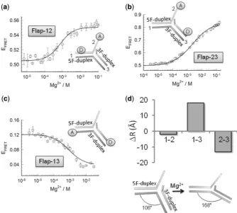
region containing the 30-flap strand, seeFigure 1). Flap-12
and Flap-23 report structural distortions involving the 9-nt single-strand flap and the 5F-duplex (Flap-12) or the 3F-duplex (Flap-23), while Flap-13 reports changes in the kink angle between the two duplex regions. On addition of magnesium ions, all three flap constructs exhibited significant variations in the efficiency of energy
transfer (EFRET), indicative of a global reorganization of
the flap structure (Figure 1a–c). All conformational
changes took place in the mM range of Mg2+ ion
concentration and were well fitted by a two-state model (Supplementary Equation S1). Indeed, a global fit of the three flap vectors yielded a good fit of all the experimental
data with a value ofn= 0.96 for the Hill coefficient and a
half-point of [Mg2+]1/2= 1.14 mM (R= 0.998)
(Supplementary Table S2).
We next analysed in detail the relative variation in FRET efficiencies and inter-dye distances induced by
the addition of Mg2+ ions (Figure 1d, Supplementary
Table S3 andSupplementary Methods section for details regarding the calculation of FRET-derived distances). For Flap-12 and Flap-23, the FRET efficiency increased from
0.51 ± 0.04 and 0.50 ± 0.01, in the absence of Mg2+, to
values of 0.56 ± 0.02 and 0.84 ± 0.01 at 20 mM
concentration of Mg2+ions, respectively. This implies a
moderate 2 A˚ decrease in distance for Flap-12 and13 A˚
for Flap-23 (Figure 1d, upper panel, Supplementary
Table S3). In contrast, Flap-13 exhibited a decrease in FRET efficiency from a value of 0.13 ± 0.01 with no
Mg2+ions to a value of 0.034 ± 0.004 in the presence of
30 mM Mg2+ ions concentration, which represents a
18 A˚ decrease in the inter-dye distance (Figure 1d
upper panel, Supplementary Table S2). A significant
shortening of the end-to-end distance associated with to Flap-13 can be explained by a change in the kink angle between the upstream and downstream complexes centred at the phosphate opposite the ss/dsDNA junction. Using the calculated inter-dye distance for Flap-13 and taking into account the length of the fluorescein linker and that Cy3 stacks at 5 A˚ distance from the terminal base pair of
duplex DNA (30), we estimated a decrease in the kink
angle from 106 in the absence of Mg2+
ions to 168 at
30 mM Mg2+(Figure 1d, bottom panel).
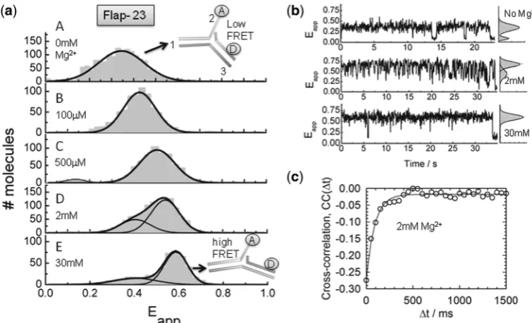
In view of the potential for structural heterogeneity in solution and to get insights into the dynamics of flap substrates, we carried out sm-FRET experiments using immobilized Flap-23 constructs. Flap-23 is well-suited for sm-FRET studies because of its high variation in
FRET efficiency as a function of Mg2+ions (Figure 1b).
Single-molecule time traces and apparent FRET efficiency
(Eapp) histograms were obtained for different Mg
2+ ion
concentrations (Figure 2a and b and Supplementary
Figure S2). In the absence of Mg2+ions, the flap substrate
remained in a low-FRET state (Eapp 0.37) until
photobleaching occurred (Figure 2a, panel A and
Figure 2b, upper trace). As the concentration of Mg2+ ions is increased, the single-molecule histograms showed
at St Andrews University Library on December 5, 2013
http://nar.oxfordjournals.org/
a predominant Gaussian peak that progressively shifted to
higher FRET (Figure 2a, panels B–D), reaching a value of
Eapp0.6 at 30 mM concentration of Mg2+(Figure 2a,
panel E). Interestingly, at concentrations of Mg2+ ions
between 1 and 30 mM, the single-molecule histograms also showed a minor contribution of an additional
Gaussian peak with a similar Eapp (0.37) to that
obtained in the absence of Mg2+. At these conditions,
the single-molecule trajectories exhibited fast, but clear transitions between these two FRET states. Because the observed fluctuations were too fast to be clearly resolved using Hidden Markov modelling methods, we performed
cross-correlation analysis to determine their rates
(Figure 2c) (31). The correlation time (t) at 2 mM Mg2+ ions was extracted from the single-exponential fitting of the cross-correlation curve providing a value of
11.3 ± 2 s1
for the rate of fluctuations on the Flap-23
substrate. Taken together, our data suggest a Mg2+
-induced conformational change taking place on the flap substrate from a bent to an extended structure.
Interestingly, at concentrations of Mg2+ions where Fen1
exhibits maximal catalytic activity (10 mM) (32), we
observed the flap substrate alternating rapidly between
both structures, whereas at concentrations of Mg2+ions
known to inhibit Fen1 function (>30 mM), the flap
substrate remained locked in the extended conformation. The fast inter-conversion observed here for the flap substrate using sm-FRET is in good agreement with recent studies on the structure of nicked, gapped and
bulged structures, which have shown that these
intermediates are highly dynamic and able to adopt a
broad range of conformations (33–35).
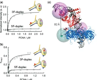
Fen1 directs PCNA loading to the 3F-duplex region of the double flap
To shed light on the organization of the Fen1/PCNA complex bound to a double-flap substrate, we determined the position of the PCNA ring relative to the double flap in the presence of Fen1. We previously demonstrated that
PCNA on its own encircles the 3F-duplex (KD= 8.5mM)
and 5F-duplex (KD= 6.95mM) regions with equal
probability and with a 1:1 stoichiometry (20). Here, we
used a different approach based on PIFE, in which a fluorescent dye becomes brighter when a protein binds
in close proximity (36). This photophysical property,
exhibited by Cy3 and other dyes has been previously employed to explore the positioning of sequence-specific
BamHI restriction enzymes along duplex DNA (36). We
[image:4.612.131.466.68.369.2]designed double-flap DNA substrates with a 9-nt
Figure 1. Conformational changes on the double-flap DNA structure induced by the addition of Mg2+ions. Intra-molecular FRET assay to monitor the effect of Mg2+ions with flap substrates labelled with donor and acceptor at the indicated positions (insets). Variation in FRET efficiency as a function of Mg2+ions at 20C on Flap-12 (a), Flap-23 (b) and Flap-13 (c). Individual fitting of each FRET isotherm for each flap substrate using a
two-state model described by Supplementary Equation S1(see Supplementary ‘Materials and Methods’section) are shown as continuous lines. Experiments were performed in triplicate and error bars represent the SEM. (d) (Upper panel) Comparison of the relative changes in inter-dye distance (A˚) observed for the three 50-flap substrates upon addition of Mg2+
ions. (Lower panel) Diagram showing the overall angle changes in flap structure induced by Mg2+ions.
at St Andrews University Library on December 5, 2013
http://nar.oxfordjournals.org/
50-ssDNA flap carrying an internal Cy3 fluorophore positioned either 10-nt downstream or upstream of the
50-flap junction (Figure 3a) and performed PCNA
titrations in a 5-mM background of Fen1 and in the
presence of 10 mM concentration of Ca2+ ions to
prevent cleavage. As demonstrated for other metal
ion-dependent nucleases, Ca2+ions efficiently stabilized the
nucleic acid–protein interaction but did not support
catalysis (20,23). For the 5F-duplex Cy3-labelled flap
substrate pre-incubated with Fen1, only a small increase
in Cy3 fluorescence emission (10%) was detected. In
contrast, the 3F-duplex labelled substrate showed a 2-fold relative increase in fluorescence emission and
a KD of 160 ± 14 nM (Figure 3a). This KD-value for
PCNA interacting with the Fen1/DNA complex
represents a 50-fold decrease in dissociation constant when compared to DNA alone and compares favourably
with previous Fen1/PCNA dissociation constants
(210 nM) (38). These data confirm that in the Fen1/
PCNA/DNA ternary complex, PCNA loads onto the
3F-duplex dsDNA region that is below the 50 ssDNA
flap as previously predicted using a streptavidin-biotin
blocking assay (39) and recently proposed from MD
simulations (26). For comparison, and given that PCNA
is an heterotrimer in which PCNA1 and PCNA2 subunits form an stable heterodimer (PCNA12) to which PCNA3 binds; we carried out similar experiments with the tightly associated PCNA12 complex. Interestingly, a substantial 45% increase in fluorescence emission was still observed using the 3F-duplex Cy3-labelled substrate and we
obtained a KD of 360 ± 30 nM for the PCNA12/Fen1
interaction. A moderate decrease (2-fold) in the affinity
of PCNA12 when compared to the heterotrimeric PCNA provides strong experimental evidence for selective recruitment of Fen1 to PCNA12 but not PCNA3 as observed from the crystal structure of the PCNA12/Fen1
complex (40). Also, the additional contacts lost in the
absence of PCNA3 may account for the observed decrease in affinity.
Organization of the Fen1/PCNA/DNA ternary complex
FRET studies to investigate the structural organization of the eukaryotic Fen1/PCNA/DNA complex in solution are challenging due to the homotrimeric nature of the sliding clamp, which facilitates binding of up to three copies of the same client protein, as observed in the crystal structure
of Fen1 with PCNA (24). Here, we took advantage of the
heterotrimeric nature of theS. solfataricusPCNA (23,25)
to circumvent these problems and obtain sliding clamps singly labelled at the PCNA1 subunit. For this, a PCNA1 variant carrying a single-cysteine mutation (N131C) was labelled with a thiol-reactive maleimide derivative of Cy5 and incubated with unlabelled subunits PCNA2 and PCNA3 to reconstitute the sliding clamp (See
Supplementary ‘Materials and Methods’ section). Titration of acceptor-labelled PCNA (PCNA-Cy5) with upstream or downstream donor-labelled DNA substrates, identical to those used for the PIFE experiments, and
pre-equilibrated with 5mM Fen1, revealed a significantly
higher intermolecular FRET efficiency (Supplementary
Equation S2) for the 3F-duplex labelled substrate (Eapp
0.48) corresponding to a distance of 63 ± 5 A˚,
[image:5.612.121.499.68.297.2]compared to the 5F-duplex labelled complex (Eapp
Figure 2. Structural transitions in the Flap-23 substrate studied by sm-FRET. (a) Histograms of FRET efficiency summed over multiple single molecules in the absence (panel A) and presence of increasing Mg2+ion concentrations (panels B–D). Individual traces were filtered to remove contributions from fluorophore blinking and photobleaching. (b) Representative sm-FRET trajectories (33 ms integration time) as function of Mg2+ ion concentration. Corresponding FRET histograms for each trajectory are shown as panels on the right. (c) Donor and acceptor intensities were obtained with 16 ms integration time at 2 mM Mg2+ion concentration and the cross-correlation signal was calculated and averaged for 15 traces. Solid line represents the fitting to a single-exponential decay to extract the sum of the backward and forward rates.
at St Andrews University Library on December 5, 2013
http://nar.oxfordjournals.org/
0.12, 82 ± 7 A˚) (Figure 3b). A shorter distance from PCNA to the 3F-duplex dsDNA region agrees with our
previous observations using PIFE (Figure 3a) and
confirms PCNA loading to the 3F-duplex region of the flap substrate in the presence of Fen1. The dissociation
constant recovered from these experiments (KD of
117 ± 34 nM) also matches that obtained by fluorescence enhancement using non-labelled PCNA, indicating that the presence of Cy5 acceptor on the PCNA does not affect the formation of the ternary complex.
Recently, a structural model of the human Fen1/
PCNA/DNA ternary complex was reported (26). This
was constructed by combining all available
high-resolution crystal structures of the individual components and subassemblies, and then refined by multi-nanosecond atomistic MD simulations. Using our experimental FRET data, we were able to assess whether this model was consistent with the ternary complex structure present in solution. To compare FRET-derived inter-dye distances with those derived from atomic structures, it is important to correctly model the dye positions with respect to their attachment points on the biomolecules. To do this, we used an AV approach which takes into account the various dimensions of the dye and linker (37,41; see ‘Materials and methods’ section for details), to model the mean dye positions onto the ternary structure (Figure 3c). The resulting distances from PCNA to the 3F-duplex (60 A˚) and 5F-duplex regions (83 A˚) are in
excellent agreement with our FRET-derived distances, thus providing the first experimental evidence in support of this ternary complex structure. It is also interesting to note that the AV of the Cy3 dye located at the 3F-duplex
is significantly restricted by the PCNA ring (Figure 3c),
which is consistent with the increased PIFE effect seen for
this labelling position (Figure 3a).
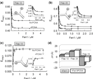
Affinity of Fen1 and Fen1/PCNA complexes for flap DNA substrates
Once the structures and dynamics of the flap DNA substrate in the unbound state and the organization of the ternary complex were determined, we investigated
Fen1 association to the 9-nt ssDNA 50 flap in the
absence and presence of PCNA (2mM). FRET-binding
curves were obtained at room temperature by titrating each vector with increasing concentrations of Fen1 in a
background of 10 mM Ca2+ ions to prevent cleavage
(Figure 4a–c,Supplementary Table S4). Global fitting of
the three binding curves to Supplementary Equation S3
yielded a KD of 14 ± 3 nM that fitted accurately
(R= 0.998) all Fen1 binding isotherms (Figure 4a–c).
This value is very similar to that reported for Fen1 binding to flap substrates using electrophoretic mobility
assays (KD 5–10 nM), suggesting that the presence of
the fluorophores has no significant effect on complex
[image:6.612.134.469.68.341.2]formation (38,39). FRET titrations performed under
Figure 3. Organization of the ternary complex studied by FRET and PIFE. (a) Normalized PIFE of Cy3 emission in a background of 10 mM Ca2+ and 5mM Fen1 obtained for Cy3-labelled upstream and downstream duplexes as a function of PCNA concentration. Data were fitted to a binding model as described by Supplementary Equation S3. (b) Variation in inter-molecular FRET efficiency between an upstream or downstream Cy3 donor-labelled DNA flap and Cy5 acceptor-labelled PCNA as a function of PCNA concentration. (c) Modelling of the ternary Fen1:DNA:PCNA complex using MD simulation derived from (26) and the FRET distances extracted in (b). Pink and cyan spheres represent mean dye positions modelled using the AV approach (27,37) (See ‘Materials and Methods’ section andSupplementary Materialfor details).
at St Andrews University Library on December 5, 2013
http://nar.oxfordjournals.org/
identical conditions, but in the presence of 2mM PCNA (Figure 4a–c), revealed a decrease in the dissociation
constant (KD 3 ± 1 nM). This result is in good
agreement with the 4-fold reduction in KM observed at
room temperature under steady-state conditions using a
similar flap substrate (38). For comparison, a 50-flap DNA
substrate lacking the 30 extra-helical nucleotide yielded a
KD in excess of 10mM in the absence of PCNA, that was
only moderately rescued (KD 0.7 ± 0.1mM) in the
presence of 2mM PCNA (Supplementary Figure S3).
Reorganization of the flap substrate by Fen1 and Fen1/PCNA binding
Previous studies using FRET provided evidence that Archaeoglobus fulgidusFen1 promotes a bent conformation
between the 5F-duplex and 3F-duplex regions (11);
however, the extent to which the DNA-flap structure bound to Fen1 is affected by the presence of PCNA is still currently unknown. Therefore, we used a similar FRET approach to determine the global reorganization taking place in the flap DNA structure induced by Fen1 and Fen1/PCNA complexes. In the absence of PCNA, addition of Fen1 altered the overall structure of the flap
substrate (Figure 4d and Supplementary Table S5). At
saturating concentrations of Fen1 (>1mM), the inter-dye
distance associated with Flap-12 increased only by3 A˚,
whereas the equivalent duplex DNA to single-strand distance reported by Flap-23 exhibited a 10 A˚ increase in
the presence of Fen1 (Figure 4d). For Flap-13, the
end-to-end distance between the 5F-duplex and 3F-duplex regions
decreased by 16 A˚ upon Fen1 binding, which is consistent with earlier reports and with the X-ray crystal structure. We next repeated these experiments in a background of
2mM PCNA. Because addition of PCNA induces as a
fluorescence enhancement of the Cy3 FRET donor
located in the 3F-duplex (Figure 3a), the inter-dye distances
were corrected following procedures described in the
literature (20) (see Supplementary Equation S4). For
Flap-12 and Flap-23, addition of PCNA promoted an
additional increase in the inter-dye distance of 2 A˚
(Figure 4d). For Flap-13 the inter-dye distance increased
by8 A˚ in the presence of PCNA (Figure 4d).
Single-molecule assay to monitor Fen1/PCNA substrate recognition
Based on recent crystallographic data (3,11,12,42),
opening of the Flap-23 vector has emerged as a
characteristic feature shared by FEN superfamily
endonucleases as a way to target specific DNA structures. Experimental evidences for a Fen1-induced double-nucleotide unpairing mechanism being responsible for the observed opening of the Flap-23 vector have been recently reported in two elegant studies using
2-aminopurine fluorescence (43) and disulfide cross-linking
between base pairs either side of the scissile phosphate
(44). The pronounced variation in FRET efficiency
observed for Flap-23 (EFRET0.3) upon Fen1 and
[image:7.612.158.468.424.684.2]Fen1/PCNA binding allowed us to use sm-FRET to test this flap opening model in the presence and absence of the sliding clamp. A double-flap substrate (Flap-23) carrying
Figure 4. DNA distortion by Fen1 in the absence (squares) and presence (circles) of PCNA at 10 mM Ca2+ion concentration and 20C.
Intra-molecular FRET was used to monitor the interaction between Fen1 and different flap substrates (a) Flap-12, (b) Flap-23 and (c) Flap-13. Solid lines represent the fitting to the FRET-binding isotherm for each flap using a binding model described bySupplementary Equation S3. (d) Comparison of the relative changes in inter-dye distances (A˚) observed for the three 50-flap substrates upon association to Fen1 (grey) and Fen1/PCNA complexes
(white).
at St Andrews University Library on December 5, 2013
http://nar.oxfordjournals.org/
a 9-nt 50-ssDNA flap, similar to that used for ensemble-FRET measurements, was modified for sm-ensemble-FRET using
the donor–acceptor FRET pair Cy3-Cy5 (Ro53 A˚) (28).
A biotin moiety was also incorporated at the 50end of the
downstream duplex for surface immobilization to a PEG functionalized quartz microscope slide via a neutravidin–
biotin interaction (28).
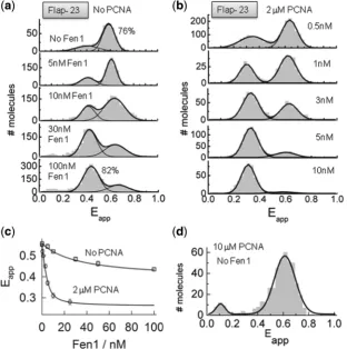
The variation in FRET efficiency of
surface-immobilized flap substrates (Flap-23) was monitored as
a function of Fen1 concentration (Figure 5a). In the
absence of Fen1 protein, the sm-FRET histogram displayed two unbound states with Gaussian peaks
centred at Eapp0.62 ± 0.1 (76%) and a minor
contribution atEapp0.38 ± 0.2 (Figure 5a, top panel).
As previously shown (Figure 1), these two FRET
populations can be assigned to the extended and bent conformations of the flap substrate, the latter being the
predominant conformer at 10 mM concentration of Ca2+
ions. As the concentration of Fen1 protein was increased, the sm-FRET histograms showed a progressive decrease in the contribution of the high-FRET population
(Eapp0.65 ± 0.2) and a concomitant increase in the
contribution of the low-FRET state (Eapp0.44 ± 0.2),
reaching a value of 82% at 100 nM concentration of
Fen1 (Figure 5a, bottom panel). Judging by the relative
similarity of their FRET values, Fen1 binding to the DNA substrate seems to stabilize Flap-23 into a conformation
(Eapp0.44 ± 0.2) very close to that observed in the
absence of Mg2+ ions (Eapp0.37 ± 0.1) (Figure 1).
However, both conformational states displayed very different dynamic properties (see later), which allowed us to assign them unambiguously. Thus, we interpreted the
single-molecule population centred at low-FRET
(Eapp0.44 ± 0.2) as arising from Fen1–DNA complexes
in which Fen1 binding to the flap substrate increases the inter-dye distance as previously found using ensemble
FRET (Figure 4b and d).
The ability of single-molecule methods to monitor with high accuracy the formation of very high affinity protein– DNA complexes prompted us to re-evaluate the values previously obtained for the dissociation constant using ensemble-averaging methods. For this, we extracted the average FRET value from the sm-FRET histograms obtained at each Fen1 concentration and the results were fitted to a similar model as previously described (see Supplementary ‘Materials and Methods’ section) (Figure 5c, No PCNA). We obtained a dissociation constant of 23 ± 2 nM in the absence of PCNA, which is in very good agreement with the value calculated using
[image:8.612.144.457.372.687.2]ensemble FRET (14 ± 3 nM). This agreement also
Figure 5. Opening of the flap substrate by Fen1 and Fen1/PCNA complexes studied using sm-FRET. sm-FRET histograms obtained for surface-immobilized Flap-23 substrates in the absence (a) and presence of 2mM PCNA (b). Gaussian fits corresponding to the unbound and bound species are also shown. (c) Variation in averaged FRET efficiency integrated from the sm-FRET histograms shown in (a) and (b) as a function of Fen1 concentration in the absence (squares) and presence of 2mM PCNA (circles). Solid lines represent the fit to a binding model as described in
Supplementary Equation S3. (d) sm-FRET histogram and corresponding Gaussian fit obtained for Flap-23 in the presence of 10mM PCNA.
at St Andrews University Library on December 5, 2013
http://nar.oxfordjournals.org/
provides additional evidence to confirm that surface immobilization of the flap substrate via its 5F-duplex does not disrupt the Fen1–DNA interaction.
In order to study the effect of PCNA on Fen1 binding to the flap substrate and the subsequent opening of the
Flap-23 vector, a sm-FRET titration of
surface-immobilized substrates at increasing concentrations of
Fen1 protein was repeated in a background of 2mM
PCNA and 10 mM Ca2+(Figure 5b). However, prior to
studying the Fen1:PCNA:DNA ternary complex at single-molecule level, we performed control experiments to
investigate the possibility of PCNA inducing a
conformational change on the flap substrate in the absence of Fen1 protein. sm-FRET histograms in the
presence of 10mM PCNA showed a single-Gaussian
peak centred at an identical FRET efficiency value to
that observed in the absence of PCNA (Eapp0.6 ± 0.2)
(Figure 5d), therefore confirming that PCNA alone does not disrupt the flap structure. As we increased the concentration of Fen1, the contribution of the
high-FRET peak corresponding to the unbound state
progressively decreased and shifted to a low-FRET value
(Eapp0.32 ± 0.08). This low-FRET population became
the predominant contribution at concentrations of Fen1
higher than3 nM (65%) and therefore can be assigned
to the formation of the PCNA:Fen1:DNA ternary complex. From these data it is clear that the presence of PCNA had two major effects in the mechanism of Fen1 recognition of the flap substrate. First, it increased Fen1’s affinity for the flap substrate. Indeed, average FRET values extracted at each Fen1 concentration from the sm-FRET histograms and fitted to the model described by Supplementary Equation S3 provided a KD-value of
1.6 ± 0.5 nM (Figure 5b) which is more than 10-fold
lower than in the absence of PCNA. This increase in affinity agrees with the proposed role of PCNA recruiting Fen1 to the flap junction. Secondly, a shift to lower FRET
(Eapp0.32 ± 0.08) in the ternary complex, compared to
Fen1 alone (Eapp0.44 ± 0.2), confirmed the moderate
increase in the inter-dye distance observed in ensemble
data (Figure 4b and d).
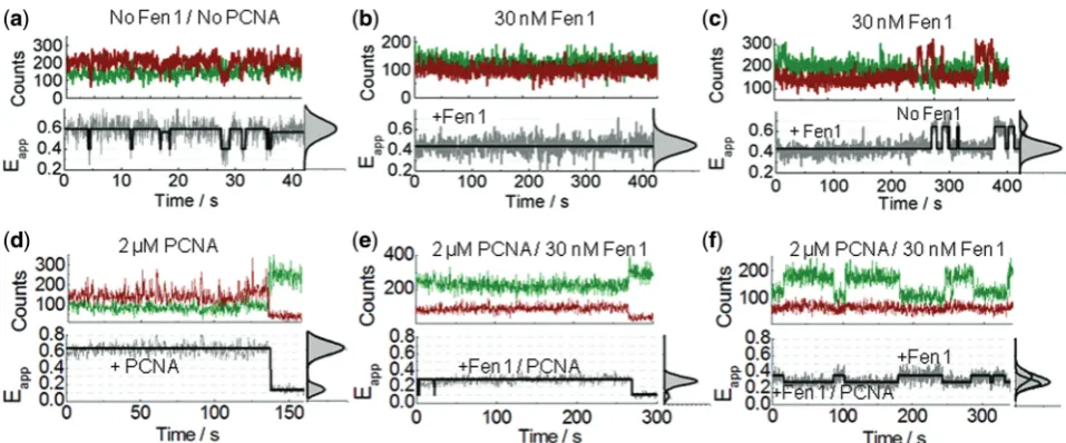
Equilibrium dynamics of Fen1 and Fen1/PCNA complexes bound to surface-immobilized substrates
To get some insights into the binding dynamics of Fen1 to flap DNA, we monitored the donor and acceptor intensities from surface-immobilized Flap-23 substrates
for extended periods in a background of 10 mM Ca2+to
avoid cleavage. Representative time-intensity traces and corresponding FRET trajectories in the absence and
presence of Fen1 are shown in Figure 6. In the absence
of Fen1, the trajectories displayed flap substrates
remaining for several tens of seconds in the extended
state (Eapp0.62 ± 0.2) with occasional short-lived
fluctuations (<1s) to the bent conformation
(Eapp0.37 ± 0.2) (Figure 6a). In contrast, when the
flap substrate was pre-incubated with progressively higher concentrations of Fen1, an increasing percentage
of traces remained in a low-FRET state
(Eapp0.44 ± 0.2) for long periods of time (5–8 min)
before photobleaching (Figure 6b and Supplementary
Figure S4). Because the probability of these long-lived low-FRET trajectories increased with the concentration of pre-incubated Fen1, we assigned them as representing Fen1/DNA bound complexes exhibiting a very low dissociation dynamics. At each Fen1 concentration analysed, a small subset of these low-FRET trajectories
(<15–20%) displayed very occasional transitions between
both FRET states that we interpreted as Fen1 binding/
unbinding events (Figure 6c and Supplementary Figure
S4). The statistical frequency of these fluctuations
remained low even when the observation time was
significantly increased (>15 min). We additionally
confirmed that the slow dissociation kinetics did not result from surface-immobilization artefacts using an
ensemble-FRET competition assay (Supplementary
Figure S5). To get additional insights into the effect of
PCNA, we next investigated the single-molecule
equilibrium dynamics of flap substrates incubated with
Fen1 in background of 2mM PCNA and 10 mM Ca2+.
Although the lack of a significant PCNA-induced effect on the predominant structure of the flap substrate was
already confirmed (Figure 5d), and before studying the
effect of PCNA/Fen1 complexes, we decided to further test whether PCNA alone could affect the dynamics of
the flap substrate (Figure 6d). We observed that addition
of 2mM concentration of PCNA has a subtle effect on the
flap dynamics with most of the trajectories lacking the occasional fast transitions to the low-FRET state
(Eapp0.37) observed for the flap alone (Figure 6a). We
propose that in the absence of Fen1 directing PCNA loading to the 3F-duplex region, PCNA can randomly assembly at each side of the flap junction and freely scan each of the duplex regions. Thus, the observed
PCNA-induced subtle stabilization of the extended flap
conformer, which is predominant at 10 mM Ca2+, may
arise from a restricted ability of the flap substrate to adopt the bent conformation, most likely due to the steric hindrance provided by the freely diffusing sliding clamp.
In the presence of Fen1/PCNA complexes, no statistically
significant transitions to a high-FRET state (Eapp0.6),
corresponding to Fen1/PCNA dissociation events, were detected during the time scale of our observation window
(5 min) (Figure 6e–f, Supplementary Figure S6).
Surprisingly, a high percentage (>70%) of the
single-molecule trajectories showed a non-anticorrelated
behaviour where the donor emission fluctuated between
two intensity levels, differing by 2-fold; while the
acceptor intensity remained practically unchanged
(Figure 6f, Supplementary Figure S6). As a result, the corresponding FRET traces displayed transitions between
efficiency values ofEapp0.3 andEapp0.44. Taking into
account that the two-state dynamics were only detected for Fen1/DNA complexes in the presence of PCNA and that a 2-fold fluorescence enhancement (PIFE) of the Cy3 donor located in the 3F-duplex was already confirmed by ensemble
measurements (Figure 3a), we interpreted this as arising
from binding events of PCNA to Fen1/DNA complexes.
An alternative explanation involving the transient
association of PCNA3 (KD1mM) to PCNA12
at St Andrews University Library on December 5, 2013
http://nar.oxfordjournals.org/
permanently bound to the Fen1/DNA complex, was ruled out because a control experiment using only PCNA12
exhibited a similar non-anticorrelated dynamics
(Supplementary Figure S7). Overall, the invariance of the Cy5 acceptor signal can be explained by the formation of the Fen1/PCNA/DNA complex leading to two competing processes that counteract one another. PCNA binding promotes an increase in quantum yield of the Cy3 donor in the 3F-duplex, which would normally lead to a concomitant increase in acceptor signal; however, this effect is almost entirely compensated for by a simultaneous PCNA-induced opening of the Flap-23 distance. Such an increase in inter-dye distance leads to a less efficient energy transfer process to the acceptor dye and therefore lower Cy5 fluorescence. The fact that this increase in distance in the ternary complex compared to Fen1/DNA
has been observed in both, ensemble (Figure 4b and d)
and single-molecule experiments (Figure 5d and6f), using
two different FRET pairs, is strong evidence that we are observing a PCNA-induced conformational change.
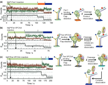
Single-molecule nuclease-reaction profiles
We used real-time injection experiments to monitor Fen1 nuclease activity at single-molecule level. Because the
acceptor dye is located at the 50 end of the single-strand
flap, cleavage of this single-stranded region in the Flap-23 substrate should lead to a complete loss of the FRET
signal, while the formation of Fen1/DNA (Eapp0.44)
or Fen1/DNA/PCNA (Eapp0.32) complexes can be
easily distinguished by their distinctive FRET levels as shown in the previous sections. Representative real-time single-molecule trajectories and corresponding FRET
traces obtained at each condition are shown in Figure 7
andSupplementary Figures S8andS9.
After injection of Fen1 and 10 mM Mg2+to a flow cell
pre-incubated with 10 mM Mg2+, most of the Flap-23
substrates remained in the high-FRET unbound state
(Eapp 0.6) for a variable period of time (25–500 s)
before the FRET signal abruptly decreased to Eapp
0.4–0.45. This change in FRET corresponds to the formation of the Fen1/DNA complex (Step 1 in
Figure 7a) with the donor and acceptor signals showing
a clearly anti-correlated behaviour (Figure 7a, left panel).
In the majority of traces (80%) this step was followed by
a sudden loss of the Cy5 fluorescent signal (Step 2 in
Figure 7a) and the simultaneous recovery of the Cy3
emission with practically the same amplitude
(Figure 7a). We interpreted this as evidence for Fen1-induced cleavage of the flap substrate and subsequent product dissociation and diffusion into the solution (Figure 7a, right panel). To confirm unambiguously that the observed loss of FRET was indeed caused by Fen1 cleavage of the flap substrate and not simply by photobleaching of the Cy5 acceptor, several control experiments were performed using direct Cy5 excitation
methods (see Supplementary ‘Materials and Methods’
section andSupplementary Figure S10).
Similar experiments injecting a pre-incubated Fen1/ PCNA complex exhibited a similar pattern to that observed with no PCNA; however, now the FRET
signal changed from Eapp 0.6 (unbound state) to Eapp
0.3, confirming the assembly of the ternary complex
(Figure 7b, left panel). The likelihood that this sudden change in FRET efficiency was caused by Fen1/PCNA
binding to the flap (Step 1 in Figure 7b) was also
[image:10.612.59.538.66.265.2]supported by the increase in total fluorescence intensity arising from the expected enhancement in Cy3 quantum yield upon PCNA binding. With our integration time (200 ms), none of the single-molecule trajectories showed
Figure 6. Equilibrium dynamics of Fen1 and Fen1/PCNA-induced flap opening using sm-FRET. (a–c) Representative single-molecule intensity traces of donor (green) and acceptor (red) intensities and corresponding FRET traces (grey) obtained for Flap-23 in a background of 10 mM Ca2+ions concentration in the absence (a) and presence (b, c) of 30 nM Fen1. (
d–f) Representative single-molecule time records of donor (green) and acceptor (red) intensities and corresponding sm-FRET traces (grey) obtained for Flap-23 in a background of 10 mM Ca2+ion concentration and 2mM PCNA, in the absence (d) and presence (e, f) of 30 nM Fen1. Solid black lines represents the estimated trajectory obtained using Hidden Markov Modelling. FRET histograms and corresponding Gaussian fits for each trace are also shown in the right panels.
at St Andrews University Library on December 5, 2013
http://nar.oxfordjournals.org/
transitions involving sequential binding of Fen1 first (Eapp
0.4), followed by PCNA assembly (Eapp 0.3).
Interestingly, the majority of reaction profiles analysed
for the Fen1/PCNA/DNA ternary complex (90%)
showed a quenching in Cy3 emission simultaneous to
the cleavage event (Step 2 in Figure 7b, left panel). This
is in marked contrast to the anti-correlated behaviour observed in Fen1/DNA complexes where cleavage of the
ssDNA 50 flap carrying the Cy5 acceptor was
simultaneously followed by the recovery of the Cy3
emission (Figure 7a, left panel). The non-anticorrelated
cleavage pattern found for these Fen1/PCNA complexes can be explained assuming all steps following the Fen1/ PCNA binding event and including cleavage, product
release and Fen1/PCNA dissociation, occurred
simultaneously (Step 2 inFigure 7b, right panel). In this
case, cleavage and break of the energy transfer to the
acceptor, which should lead to an enhanced Cy3 emission, was at least partially counteracted by PCNA dissociation and the subsequent lack of the PIFE effect on the Cy3 donor induced by PCNA. These two competing effects produced an overall increase in Cy3
signal, suggesting that in the pre-cleaved ternary
complex, the PIFE channel has a dominant contribution to the total Cy3 output. Additionally, a small fraction of
the reaction profiles (<10%) showed a non-synchronized
[image:11.612.86.541.66.423.2]cleavage and Cy3-quenching events (Steps 2 and 3 in
Figure 7c, left panel). Here, a significant decrease in Cy3 emission was only detected several seconds later than the
cleavage step (15 s inFigure 7c) suggesting that PCNA
remained bound to the nicked substrate after cleavage took place. Interestingly, for these trajectories, no increase in Cy3 emission was observed concomitant with the cleavage and loss of Cy5 emission event (Step
Figure 7. Single-molecule Fen1 nuclease-activity profiles. Dissecting binding and cleavage events was performed on surface-immobilized Flap-23 substrates using real-time injection of Fen1 or pre-incubated Fen1/PCNA complexes while monitoring the donor (green) and acceptor (red) intensity trajectories. (a) Representative single-molecule reaction profile obtained after real-time injection of Mg2+/Fen1 indicated by an arrow at 15 s. Sequential binding and cleavage events were revealed by a sharp transition (1) from high- (Eapp 0.6) to low-FRET (Eapp 0.44) followed by a loss of the Cy5 signal due to cleavage and release of the 50-flap ssDNA region into the solution (2), respectively. (b) Representative single-molecule
reaction profile obtained after real-time injection of Mg2+/Fen1/PCNA indicated by an arrow at 15 s. As before, Fen1/PCNA binding was revealed by a sudden decrease in FRET (1), but now to a lower FRET value (Eapp0.3). Subsequent loss of the Cy5 signal due to cleave of the 50-flap ssDNA region is shown in (2) together with the Cy3 signal decreasing due to PCNA-induced PIFE. (c) A small percentage of Fen1/PCNA reaction profiles (10%) revealed binding (1), cleavage and product release (2) and a third event (3) representing PCNA molecules that remained bound to the nicked product for some period of time (15 s in the trace shown). After this interval, PCNA dissociated from the nicked substrate leading to a decrease in Cy3 emission due to the lack of PIFE effect.
at St Andrews University Library on December 5, 2013
http://nar.oxfordjournals.org/
2,Figure 7c). We interpret this as evidence for a subtle rearrangement of the bound complex following product release. Such structural reorganization may change the local environment reducing the PIFE effect around the Cy3 probe and thus counteracting the expected increase in Cy3 emission. It is worth to mention that PIFE is very sensitive to small distances and it has been proposed as a molecular ruler complementary to FRET to detect
changes in distance below the 1–2 nm FRET limit (36).
Although with our current assay we cannot clarify whether Fen1 remained associated to the cleaved substrate and the statistics of these events was too low to extract any quantitative information, the observation of an stable
PCNA/DNA interaction in the nicked product
agrees with the proposed role of PCNA acting as a protein-recruitment platform during flap DNA processing (15–17).
Using the ability of sm-FRET to differentiate all the Fen1/PCNA processing steps, we have quantified the
Fen1 binding (Figure 8a and b) rates separately from
the cleavage and product dissociation rates (Figure 8c
and d), in the absence and presence of PCNA. For each single-molecule reaction profile, Fen1 and Fen1/PCNA binding rates were extracted by measuring the time interval from the injection of the protein(s) to the appearance of the change in FRET signal from the high-FRET unbound state to the low-high-FRET bound state. Similarly, the cleavage and product dissociation rates were obtained by measuring the time interval from the binding event to the loss of Cy5 emission. Single-molecule frequency histograms for each of these intervals were plotted and fitted to monoexponential decay functions to extract the corresponding rates. For Fen1 binding to Flap-23, we obtained a pseudo first-order rate
constant kobs= 0.006 ± 0.002 s
1
without PCNA and
kobs= 0.023 ± 0.003 s1 in the presence of 1mM PCNA.
For the cleavage rate that included the catalytic and product dissociation steps, we obtained similar values
of 0.040 ± 0.002 s1 and 0.038 ± 0.004 s1 with (1mM
PCNA) and without PCNA, respectively. These values
obtained at 20C are very similar to those previously
reported by us (0.022 s1) at 25C using an ensemble
fluorimetric assay to monitor cleavage (38) and confirm
that PCNA activation of Fen1 takes place exclusively at the level of substrate recognition with no direct effect on the catalytic rate.
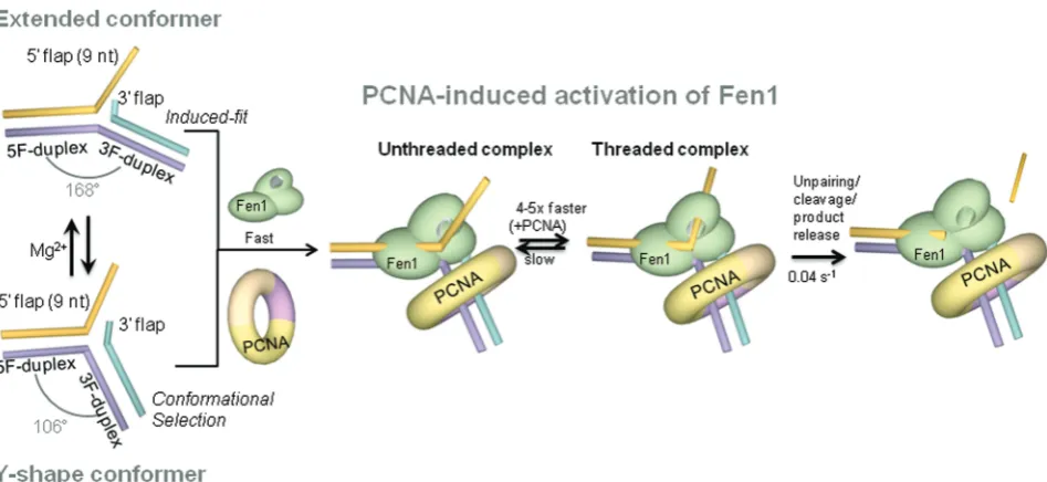
DISCUSSION
It has been suggested that Fen1 specificity for certain flap substrates may be linked to their intrinsic flexibility and
potential to become distorted by Fen1 (1–3). Despite their
prevalence in DNA replication and repair pathways, there is currently very little known about the structure and dynamics of unbound DNA flaps. To address this question, we investigated the structure and flexibility of such flaps, including the effect of divalent metal ions
such as Ca2+ and Mg2+; the latter being indispensable
cofactors for Fen1 activity (1) and also well-known
folding agents of branched DNA (33–35, 45). Based on
our combined ensemble (Figure 1) and sm-FRET data
(Figure 2), we introduce a model in which, DNA flaps
rapidly fluctuate (kbackward+kforward= 11.3 s
1
) between two conformations; a Y-shape structure and an extended
form with a 168angle between the 5F-duplex and the
3F-duplex (Figure 9); the latter being the predominant
structure at high concentrations of divalent metal ions
(>30 mM). It is also interesting to note the close
positioning of the 9-nt 50-ssDNA flap to the 3F-duplex
in the extended conformation (Eapp= 0.84, Figure 1b)
compared to the Y-shape structure (Eapp= 0.5,
Figure 1b). By analysing these observations in the context of the known interactions between Fen1 and the
DNA flap (1–3,11,12), it is possible to draw some
conclusions about the possible role of flap conformation in Fen1’s substrate-recognition step. It has been shown that the substrate binding affinity of murine flap
endonuclease reaches an optimal value at 5–10 mM
Mg2+ that rapidly decreases at higher concentrations
(32). Although Mg2+ions could affect the structure of
Fen1 and therefore its substrate binding efficiency, no reorganization of human Fen1’s local structure was
detected using FTIR and SAXS when excess Mg2+was
added to the wild-type structure and to the D181A
variant, a mutant with similar affinity (KD= 7 nM) but
unable to cleave (46). Taking these findings together, we
propose that Mg2+-induced inhibition of Fen1 activity at
relatively high concentrations may arise, at least to some extent, from the difficulty to engage with a non-flexible DNA substrate, predominantly locked in an extended conformation, that requires extensive reorganization of its structure to achieve the cleavage-competent form.
Single-molecule experiments revealed the presence of two alternative protein-free structures of the DNA
substrate (Figure 2), which also raises the important
[image:12.612.374.494.69.251.2]question of how the recognition process takes place at
Figure 8. PCNA association enhances Fen1 binding to the substrate but has no effect on the catalytic step. Dwell times for the binding (a) and cleavage (b) events were directly extracted from the single-molecule reaction profiles in the absence (light grey) and presence of PCNA (dark grey). Dwell times were fitted to single-exponential decay functions to extract the pseudo first-order association rate constants (s1) (a) and the cleavage-product dissociation rate constants (s1) (b).
at St Andrews University Library on December 5, 2013
http://nar.oxfordjournals.org/
the molecular level. In agreement with previous studies
(11,12), our ensemble-FRET data using the Flap-13
construct (Figure 4c) indicate that Fen1 binds to the
DNA substrate with high affinity (KD= 14 nM),
inducing a pronounced kink (90–100) at the flap
junction between both DNA duplex regions. Based exclusively on these ensemble data, it would be reasonable to conclude that an induced-fit binding model was in
operation (47), where protein binding drives the substrate
into a more compact conformation (47). However, there is
a remarkable similarity between the kink angle of the
protein-bound structure (100) and that calculated by us
for Flap-13 (106) in the absence of metal ions,
suggesting that the Y-shaped conformer could represent an excellent fit to the Fen1 scaffold. Based on this, and the presence of two rapidly interconverting conformers detected in the single-molecule experiments described here, it seems reasonable to speculate that Fen1 could bind the Y-shaped structure inducing a minimal distortion on the kink angle and displace the equilibrium between both conformations towards the bent structure. A conformational selection model of this type is reminiscent of the interaction mode between the L7Ae protein and
equilibrium conformers of the k-turn RNA (48). If such
conformational selection takes place for Fen1, the protein’s ability to sense the DNA structure would extend beyond detecting the presence of the required DNA binding motifs to also involve testing for their relative conformation. In this context, structure-specific recognition of Fen1 substrates may involve a complex continuum of induced-fit and conformational selection
modes (Figure 9). The balance between these recognition
mechanisms most likely will be dictated by a delicate interplay between the rate of inter-conversion between conformers of the DNA substrate and the multiple
structural and catalytic roles that Mg2+ ions seem to
hold in Fen1 function (1–3,32,46).
An emerging feature shared across the FEN superfamily of nucleases is their ability to expose the scissile phosphate by opening the junction at the base of the flap and threading the ssDNA portion through a helical gateway
that becomes ordered upon threading (1–3,11,12,42). Our
ensemble (Figure 4b) and single-molecule data (Figures5a
and6a–c) on the Flap-23 substrate provide some insights
into this mechanism and how this is influenced by PCNA (Figures 5b and6d–f). In both ensemble- and sm-FRET, we observed a decrease in FRET efficiency for Flap-23 upon addition of Fen1 or Fen1/PCNA. We estimated
that this change in FRET efficiency represents an 10 A˚
increase in dye-to-dye distance for Fen1 alone and by12
A˚ in the presence of Fen1/PCNA. Given that PCNA alone had no effect on the sm-FRET distribution for Flap-23 (Figure 6c), we are confident that the observed distance changes reflect Fen1-induced opening of the DNA junction that is slightly enhanced by PCNA. Our results broadly agree with a recent single-molecule study where a similar increase in FRET induced by Fen1 was observed
using flaps of different length (49). However, in contrast to
the high dissociation constant reported for Fen1 in that
study (1.3 ± 0.3mM), we obtained a value of 23 ± 2 nM
for surface-immobilized Flap-23 substrates (Figure 5b),
which is only 3-fold higher than Fen1’s dissociation
constant (7 nM) previously reported using ensemble
[image:13.612.77.551.66.284.2]methods (46).
Figure 9. Model of PCNA activation of Fen1-substrate recognition. In the absence of protein, the substrate exhibits an Mg2+-dependent equilibrium between a Y-shaped structure and extended conformation with a [Mg2+]1/2= 1.2 mM. Binding of Fen1 to the extended conformer induces a kink from 168to 90–100 between the upstream and downstream duplexes (induced-fit model) and promotes opening of the flap junction. At moderate Mg2+ion concentrations (<2 mM), Fen1 could associate to the already bent Y-shape structure following a conformational selection model. After Fen1 binding to the flap base by establishing interactions with the downstream and upstream duplexes, the flap substrate threads through Fen1’s helical arch. Association and dissociation rates are both slow but in the presence of PCNA, the association rate becomes 4-fold faster, suggesting a role in facilitating the flap threading step.
at St Andrews University Library on December 5, 2013
http://nar.oxfordjournals.org/
To assign the conformational change observed for
surface-immobilized Flap-23 substrates upon Fen1
binding to specific molecular-level interactions, we need to take into account the mechanism for Fen1’s recognition of the substrate. According to the threading model, Fen1 establishes contacts primarily to the 5F-duplex via the H2TH motif and then searches for DNA structures that can bend sharply by interacting with the 3F-duplex and
the unpaired 30 nucleotide (3,10–13,46,49). In principle,
this range of Fen1 interactions leading to substrate bending and Flap-23 opening could account by themselves
for the observed variation in dye-to-dye distance (10 A˚)
without the need to invoke any additional process. However, several lines of evidence suggest that the
Fen1-bound Flap-23 substrates showing an Eapp 0.4
constitute threaded Fen1/DNA complexes (Figure 9)
(3,10–13,46). First, the formation of the pre-cleavage complex was detected in real-time as a single-step FRET
transition (Figure 7a), suggesting that all significant
readjustments of the ssDNA flap occur at the time of binding, at least within our time resolution (200 ms). Secondly, dissociation of Fen1 from a complex where ssDNA flap is threaded through Fen1 may be a slow process due to the difficulty of releasing the trapped flap. This hypothesis was confirmed by the slow dissociation rate measured for Fen1/DNA complexes by
ensemble-FRET ((1.33 ± 0.06)103
s1
) and sm-FRET
experiments (Figure 6b and c,Supplementary Figure S5),
which agrees with previous studies reporting dissociation
half-times >10 min for 5-nt ssDNA flaps (39).
Interestingly, the pseudo first-order association rate
measured using real-time injection (Figure 8a) was also
slow (kobs= 0.006 ± 0.002 s
1
). It is important to note that in these experiments Fen1 association is reported by flap opening using an intra-molecular FRET assay; thus the measured rate actually represents a combination of the initial binding event, most likely involving interaction of the H2TH domain with the 5F-duplex, plus any subsequent conformational rearrangements taking place
on the flap (Figure 9). These slower conformational
changes may include Fen1 recognition of the 30-flap site
and bending of the substrate at the flap junction, threading of the ssDNA through Fen1’s cavity, or a combination of both processes. Several studies have reported significantly longer association and dissociation rates for Fen1 as the flap length increased from 2 to 12 nt
(39,47). Based on these findings, it has been suggested that
the initial Fen1 association step to the flap base is a
favourable process regardless of the 50 ssDNA flap
length. In fact, a substrate without 50flap showed only a
2-fold increase in KM, while a single-flap lacking the
30 unpaired nucleotide increased the K
M by 8-fold. In
contrast, mechanically threading the 50 flap could
present considerable entropic problems that would explain a progressively slower formation of the threaded
state as flap length increases (39).
Compared to Fen1 alone, the association rate in the
presence of PCNA increased by 4–5-fold (Figure 8b),
while the catalytic rate remained unchanged (Figure 8d).
Assuming a model as described above, in which threading represents the slowest step in the formation of the
pre-cleavage complex, we hypothesize that the observed increase in apparent association rate of Fen1 may be indicative of PCNA facilitating the threading process.
Whether the exact activation mechanism involves
repositioning of Fen1 in the proper orientation to thread the flap, or whether PCNA acts mechanically as a platform from which Fen1 can push the flap more efficiently, will require further clarification. Nevertheless, our findings confirm an active role for PCNA in the formation of the active Fen1-substrate complex that may be shared by other members of the FEN superfamily. In addition to increasing the association rate, real-time cleavage by Fen1 in the presence of PCNA is consistently observed from a ternary complex in which Flap-23
exhibits a lower FRET value (Eapp0.3) (Figure 7b and
c). This moderate increase in dye-to-dye distance for
Flap-23,12 A˚ compared to10 A˚ without PCNA, was also
detected in ensemble measurements (Figure 3c) and
a range of mechanisms may be responsible. Among these, a PCNA-induced kink of the 3F-duplex bound to Fen1 has been suggested by MD simulations of the
ternary complex (26) and further confirmed from our
FRET data (Figure 3c). Alternatively, an enhanced
threading of the ssDNA in the ternary complex may also be possible.
In summary, we demonstrate that unbound flap DNA substrates fluctuate rapidly between a Y-shaped con-former, with a structure relatively close to that observed in complex with Fen1, and an extended conformation (Figure 9). Fen1 binding to these structures could follow
either an induced-fit model (predominant at Mg2+
>2 mM) or presumably a conformational selection
model at lower Mg2+ ion concentrations. Our work
confirms that Fen1 binding to the DNA opens the substrate at the flap base, as seen in the crystal structure, in addition to promoting a kink between the 5F- and
3F-duplex regions of the extended conformer (Figure 9).
Fen1’s association and dissociation rates obtained from
single-immobilized substrates are slow due to the
threading process. We provide the first experimental evidence for PCNA activating by 4–5-fold Fen1’s apparent association rate and subsequent flap threading, without altering catalysis. Given that the interaction of Fen1 and PCNA constitutes a paradigm for PCNA-interacting proteins, our findings establish a framework to further explore PCNA’s role as an architectural organizer of the DNA-processing machinery.
SUPPLEMENTARY DATA
Supplementary Data are available at NAR Online, including [50–57].
ACKNOWLEDGEMENTS
We thank the St Andrews University Mass Spectrometry facility for analyses and Paul Talbot and Biljana Petrovic-Stojanovska for technical support. A.B. thanks the Spanish Ministry of Science and Innovation for the award of a travel and subsistence grant.
at St Andrews University Library on December 5, 2013
http://nar.oxfordjournals.org/