contributes to mRNA 3' processing and cell cycle
regulation.
CAMPBELL, Susan, LI DEL OLMO, Marcel, BEGLAN, Paul and BOND,
Ursula
Available from Sheffield Hallam University Research Archive (SHURA) at:
http://shura.shu.ac.uk/8298/
This document is the author deposited version. You are advised to consult the
publisher's version if you wish to cite from it.
Published version
CAMPBELL, Susan, LI DEL OLMO, Marcel, BEGLAN, Paul and BOND, Ursula
(2002). A sequence element downstream of the yeast HTB1 gene contributes to
mRNA 3' processing and cell cycle regulation. Molecular and cellular biology, 22
(24), 8415-8425.
Copyright and re-use policy
See
http://shura.shu.ac.uk/information.html
Sheffield Hallam University Research Archive
Copyright © 2002, American Society for Microbiology. All Rights Reserved.
A Sequence Element Downstream of the Yeast
HTB1
Gene
Contributes to mRNA 3
⬘
Processing and Cell
Cycle Regulation
Susan G. Campbell,
1Marcel li del Olmo,
2Paul Beglan,
1and Ursula Bond
1*
Microbiology Department, Moyne Institute for Preventive Medicine, Trinity College, University of Dublin, Dublin 2, Ireland,1and Departament de Bioquímica i Biologia Molecular, Facultat de Cie`ncies
Biolo`giques, Universitat de Vale`ncia, and Departamento de Biotecnología, Instituto de Agroquímica y tecnología de Alimentos (IATA), M. L. Burjassot, Spain2
Received 17 July 2002/Accepted 17 September 2002
Histone mRNAs accumulate in the S phase and are rapidly degraded as cells progress into the G2phase of
the cell cycle. InSaccharomyces cerevisiae, fusion of the 3ⴕuntranslated region and downstream sequences of the yeast histone geneHTB1to a neomycin phosphotransferase open reading frame is sufficient to confer cell cycle regulation on the resulting chimera gene (neo-HTB1). We have identified a sequence element, designated the distal downstream element (DDE), that influences both the 3ⴕ-end cleavage site selection and the cell cycle regulation of theneo-HTB1mRNA. Mutations in the DDE, which is located approximately 110 nucleotides downstream of theHTB1gene, lead to a delay in the accumulation of theneo-HTB1mRNA in the S phase and a lack of mRNA turnover in the G2phase. The DDE is transcribed as part of the primary transcript and binds
a protein factor(s). Maximum binding is observed in the S phase of the cell cycle, and mutations that affect the turnover of theHTB1mRNA alter the binding activity. While located in the same general region, mutations that affect 3ⴕ-end cleavage site selection act independently from those that alter the cell cycle regulation.
The synthesis of histone proteins is tightly regulated during the cell cycle to ensure maximum expression in the S phase coincident with DNA synthesis. Replication-dependent his-tone mRNAs accumulate in the S phase and are rapidly de-graded as cells enter the G2phase. This periodic accumulation is controlled by both transcriptional and posttranscriptional events (33). At the level of posttranscriptional processing, both mRNA 3⬘-end formation and mRNA stability play important roles in controlling this cell cycle regulation (33). The 3⬘ends of metazoan histone mRNAs differ from general mRNAs in so far as they are nonpolyadenylated. Instead of a poly(A) tail, histone mRNAs possess a highly conserved stem-loop struc-ture immediately upstream of the 3⬘end of the mRNA (10).
The formation of metazoan histone mRNA 3⬘ends is con-trolled by a unique multicomponent process (14). Evidence from studies of sea urchin, rodent, and human cells defines at least three factors required for correct 3⬘-end cleavage of these mRNAs. The snRNA component of the U7 small nuclear ribonucleoprotein (snRNP) base pairs with a loosely conserved purine-rich element (referred to as the histone downstream element [HDE]), which lies approximately 10 to 15 nucleotides (nt) immediately downstream of the cleavage site (7, 29). A second factor, the stem-loop binding protein (SLBP), also re-ferred to as the hairpin binding protein, interacts with the conserved stem-loop at the 3⬘end of the mRNA via a unique RNA binding domain (4, 15, 25). This 32-kDa protein remains associated with the mature mRNA as it relocates from the nucleus to the polysomes in the cytoplasm (19). The third
factor, an unidentified protein referred to as heat labile factor (HLF), was discovered through the ability of moderate tem-perature increases to disrupt mRNA 3⬘-end formation (17). Both SLBP (hairpin binding protein) and HLF levels fluctuate during the cell cycle; the SLBP accumulates in late G1 just prior to the accumulation of histone mRNAs, while HLF be-comes limiting in G1 (23, 36). Although the U7 snRNP is constitutively expressed during the cell cycle, conflicting data exist regarding the cell cycle regulation of the interaction of U7 with the HDE (7, 21).
Paradoxically, unlike their metazoan counterparts, all Sac-charomyces cerevisiae histone mRNAs appear to be polyade-nylated (16). While the factors influencing yeast histone mRNA 3⬘-end cleavage have not yet been defined, it is thought that the general 3⬘-end processing machinery is involved. In this regard, histone pre-mRNAs can be processed in vitro by cleavage and polyadenylation extracts capable of processing general mRNAs (10). Furthermore, degenerate sequence mo-tifs, which are similar to those of the positioning elements and efficiency elements and are located upstream of most yeast mRNAs, can be identified upstream of the 3⬘ ends of yeast histone mRNAs. Despite the difference in the structures of the 3⬘ends of metazoan and yeast histone mRNAs, posttranscrip-tional events also contribute to the cell cycle regulation of yeast histone mRNAs (24, 37).
The role of posttranscriptional events in the cell cycle reg-ulation of yeast histone mRNAs was demonstrated by experi-ments in which the 3⬘ untranslated region (UTR) and se-quences downstream of the cleavage sites of the histone gene,
HTB1, were fused to a bacterial neomycin phosphotransferase (neo) open reading frame under the control of the GAL1 promoter (37). In the presence of galactose, the resultant chi-meric mRNA accumulated in the S phase coincident with the * Corresponding author. Mailing address: Microbiology
Depart-ment, Moyne Institute for Preventive Medicine, Trinity College, Uni-versity of Dublin, Dublin 2, Ireland. Phone: 1-608-2578. Fax: 353-1-679-9294. E-mail: [email protected].
endogenousHTB1mRNA. Deletion of sequences in theHTB1
3⬘ UTR abolished the cell cycle regulation of theneo-HTB1
transcript. Since the chimera gene also contained the HTB1
3⬘-end cleavage sites and sequences downstream, it is not clear whether additional events such as 3⬘-end processing or tran-scription termination also contribute to cell cycle regulation.
The formation of nonhistone and histone mRNA 3⬘ends has been shown to be tightly coupled to transcription termination (13, 27, 30, 31). InS. cerevisiae,mutations in signals controlling 3⬘-end cleavage and polyadenylation can reduce transcription termination efficiency (5). Additionally, mutations in a number of the trans-acting cleavage and polyadenylation factors, in-cluding Pcf11p and Rna15p, result in the disruption of both 3⬘-end processing and transcription termination (5). Coupling between cleavage and termination appears to be mediated through the C-terminal domain of RNA polymerase II (3, 26). Studies of mice have also revealed a link between mRNA 3⬘-end processing and transcription termination of histone genes. Removal of the 3⬘-end processing signals (consisting of the stem-loop structure at the 3⬘end of the RNA and the U7 snRNA binding site) of the mouse H2A-614 gene leads to a disruption of transcription termination (12). Interestingly, a physical and genetic linkage between the cleavage and polyad-enylation factor CstF-64 (Rna15p) and the transcription factor Res2p, a factor involved in cell cycle regulation of a number of genes, has been demonstrated (2).
In this study, we examined the role of downstream se-quences in the 3⬘-end processing and the cell cycle regulation of the yeast histoneHTB1mRNA. Using aneo-HTB1chimera gene as a model system, we carried out a mutagenesis study of a region of DNA lying approximately 110 nt downstream of the cleavage sites of theHTB1gene. We identify a sequence ele-ment that lies in the region of transcription termination which influences, in vivo, both mRNA 3⬘-end cleavage site selection and cell cycle regulation of the upstream mRNA. We show that RNA transcribed from this region binds a protein factor(s) and that maximum binding is observed during the S phase of the cell cycle.
MATERIALS AND METHODS
Plasmids and mutagenesis.The 10-kb centromericURAshuttle vector
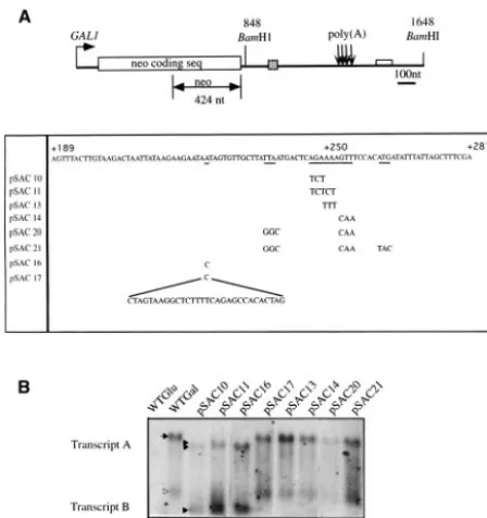
pLJ31-HTB1, containing a neomycin phosphotransferase gene fused to the 3⬘ untrans-lated and downstream sequences of the yeastHTB1gene, was constructed by Xu et al. (37). The gene is under the control of a Gal promoter. TheHTB1region consists of a 1,168-ntSphI-HindIII fragment containing the last 17 amino acids of the coding sequence ofHTB1and 1,113 nt of 3⬘ UTR and downstream sequences. This fragment was inserted at theBamHI-HindIII site immediately downstream of theneogene (Fig. 1A). The plasmid pLJ31-HTB1was kindly provided by Michael Grunstein (University of California at Los Angeles). Se-quence analysis revealed the presence of a 157-base-pair (bp) fragment of DNA immediately downstream of theBamHI restriction site at position⫹848 (Fig. 1A). This sequence represents a duplication of sequences in the 5⬘UTR of the
neogene (data not shown).
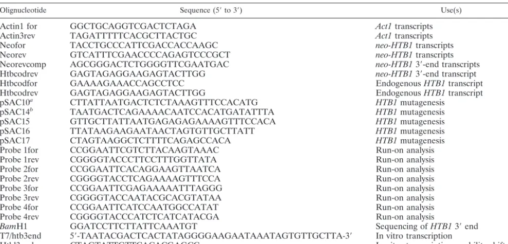
To prepare mutants in theHTB1downstream region, an 800-ntBamHI frag-ment was subcloned into the bacterial plasmid pBC (Stratagene Inc.). Mutations were created by using the QuikChange site-directed mutagenesis kit (Stratagene) in accordance with the manufacturer’s specifications. Synthetic oligonucleotides serving as mutagenic primers are listed in Table 1. Plasmids isolated from selected colonies were sequenced to confirm the mutagenesis. The mutated
BamHI fragment was subsequently subcloned back into the yeast vector,
pLJ31-HTB1.
Strains and media.TheS. cerevisiaestrain S150-2B (MATaleu2-3,112ura3-52 trp1-289 his3D) was used as the parental strain in this study. The yeast organisms were routinely cultured at 30°C in yeast extract-peptone-dextrose (YEPG)
[image:3.603.308.535.89.442.2]dium (1% [wt/vol] yeast extract, 2% [wt/vol] Bacto Peptone, 2% [wt/vol] galac-tose) to a midlogarithmic phase (107cells/ml, or an optical density at 600 nm
[OD600] of 0.6). Typically, 1g of plasmid DNA was used to transform S150-2B
by using the lithium acetate procedure (9). Transformants were selected on synthetic complete medium containing 1⫻yeast synthetic dropout medium lack-ing lysine, histidine, leucine, tryptophan, and uracil (Sigma Chemical Co.), 0.7% (wt/vol) yeast nitrogen base without amino acids (Sigma Chemical Co.), 0.5% (wt/vol) ammonium sulfate, and 2% (wt/vol) galactose and supplemented with 10 mg each of lysine, tryptophan, and histidine/ml and 20 mg of leucine/ml.
Preparation and analysis of RNA.Total RNA was isolated by the hot-phenol method (9). RNA (30g per lane) was separated on 1.0% formaldehyde agarose gels and subsequently blotted to Nytran membranes (Schleicher & Schuell). Digoxygenin-UTP (Roche Biochemicals)-labeled DNA probes were generated by PCR amplification using the primer pairs listed in Table 1, as previously described (9). The membranes were prehybridized for 1 to 3 h in high sodium dodecyl sulfate (SDS) buffer (7% sodium dodecyl sulfate [SDS], 50% formamide, 5⫻SSC [1⫻SSC is 0.15 M NaCl plus 0.015 M sodium citrate], 2% blocking reagent, 50 mM sodium phosphate [pH 7.0], 0.1%N-lauroylsarcosine) at 50°C and hybridized for 15 to 20 h at 50°C, with approximately 25 ng of labeled probe per milliliter of hybridization buffer. Hybridization was detected with a chemi-luminescent substrate, CSPD or CDP-Star (both from Roche), as previously described (9).
S1 nuclease analysis. Two probes were used to map the 3⬘ ends of the
neo-HTB1mRNAs. A 591-ntBamHI-StyI fragment was isolated from the plas-mid pLJ31-HTB1(Fig. 1A). A second 458-ntStyI-ScaI DNA fragment (see Fig. 3B) was used to determine the 3⬘end closer to theneosequences. These DNA fragments were 3⬘end labeled at theBamHI andStyI sites, respectively, by using a Klenow fragment of DNA I polymerase and incorporating [␣-32P]dATP
(spe-cific activity, 3,000 Ci/mmol).
The hybridization reactions and S1 digestions were carried out as described previously (8). For all S1 nuclease assays, the probe concentration was always in excess of that of cellular RNA. Total RNA (100g) was precipitated in 70% EtOH and 0.3 M NaOAc, pH 5.3, with the labeled DNA probes (5⫻105cpm).
The mix was then resuspended in 30l of S1 hybridization buffer [80% form-amide, 40 mM piperazine-N,N⬘-bis(2-ethanesulfonic acid) (PIPES) (pH 6.4), 10 mM EDTA, 400 mM NaCl]. The samples were heat denatured at 85°C for 10 min and hybridized at 42°C for 16 h. After the hybridization, 300l of S1 digestion buffer (50 mM sodium acetate, 4.5 mM ZnSO4, 280 mM NaCl, 20g of salmon
sperm DNA/ml) containing S1 nuclease (Promega) (100 U) was added to the RNA-probe mix. S1 digestion was carried out for 1 h at 37°C. The reaction was
stopped by the addition of 80l of S1 nuclease stop buffer (4 M ammonium acetate, 50 mM EDTA [pH 8.0], 50g of tRNA/ml). The samples were extracted with phenol-chloroform and then ethanol precipitated before electrophoresis on a 6% acrylamide–7 M urea gel in 1⫻Tris-borate-EDTA buffer.
TRO assays.Transcription run-on (TRO) assays were carried out as previ-ously described (1), with the following modifications: yeast cultures (100 ml each), transformed with the plasmid pLJ31, were grown to an OD600of
approx-imately 0.1 to 0.15. Cells were harvested and washed with ice-cold distilled water (dH2O). Cells were rendered more permeable by resuspension in 0.95 ml of
ice-cold dH2O and 50l of 10% (wt/vol) sodiumN-lauryl sarcosine sulfate. The
mix was incubated on ice for 20 min and then centrifuged for 1 min. The pellet was resuspended in 100l of transcription buffer (50 mM Tris-HCl [pH 7.9], 5 mM MgCl2, 100 mM NaCl, 1 mM MnCl2, 2 mM dithiothreitol, 0.5 mM ATP, 0.5
mM GTP, 0.5 mM CTP, 100Ci [␣-32P]UTP [3,000 Ci/mmol]). Transcription
was allowed to proceed for 5 min at 30°C, and the reaction was stopped by the addition of 1 ml of ice-cold H2O. Pelleted cells were resuspended in 500l of
TES buffer (10 mM Tris-HCl [pH 7.5], 1 mM EDTA, 0.1% [wt/vol] SDS), and total RNA was extracted as described previously. RNA was partially hydrolyzed for 5 min on ice (0.2 M NaOH), neutralized (0.2 M Tris-HCl [pH 7.2]), and used for hybridization to DNA fragments corresponding to the untranslated and downstream regions of theHTB1gene.
To generateHTB1DNA fragments, DNA in 200-bp increments corresponding to the regions downstream of theneoopen reading frame was amplified by PCR using oligonucleotides which include recognition sites forEcoRI (upstream oli-gonucleotide) orKpnI (downstream oligonucleotide) (Table 1). The amplified DNA was cloned into the M13 phage. Single-stranded M13 DNA corresponding to each 200-bp fragment was immobilized onto nylon filters. Filters were prehy-bridized for 2 h in a solution containing 5⫻SSPE (1⫻SSPE is 0.18 M NaCl, 10 mM NaH2PO4, and 1 mM EDTA [pH 7.7]), 10⫻Denhardt solution, 50%
formamide, and 0.2% SDS. Hybridization was carried out in the same solution overnight. Filters were washed four times with 2⫻SSC–0.1% SDS at room temperature and 0.2⫻SSC–0.1% SDS at 37 to 42°C for 10 min. Run-on signals were then visualized with an Instant Imager (Packard) or an Image Reader FLA 3000 (Fujifilm).
Synchronization of yeast cells.Strain S150-2BMATacells containing either the pLJ31-HTB1plasmid or its mutant derivatives were grown in selective me-dium (synthetic complete meme-dium without uracil) to early log phase and were then grown overnight in YEPG medium.␣1-mating factor
[image:4.603.45.540.81.319.2](TRP-HIS-TRP-LEU-GLN-LEU-LYS-PRO-GLY-GLN-PRO-MET-TYR) was added to the yeast cul-ture at a concentration of 2g/ml. The culture was then incubated for another TABLE 1. Olignucleotides used in this study
Olignucleotide Sequence (5⬘to 3⬘) Use(s)
Actin1 for GGCTGCAGGTCGACTCTAGA Act1transcripts
Actin3rev TAGATTTTTCACGCTTACTGC Act1transcripts
Neofor TACCTGCCCATTCGACCACCAAGC neo-HTB1transcripts
Neorev GTCATTTCGAACCCCAGAGTCCCGCT neo-HTB1transcripts
Neorevcomp AGCGGGACTCTGGGGTTCGAATGAC neo-HTB13⬘-end transcripts
Htbcodrev GAGTAGAGGAAGAGTACTTGG neo-HTB13⬘-end transcript
Htbcodfor GAAAAGAAACCAGCCTCC EndogenousHTB1transcript
Htbcodrev GAGTAGAGGAAGAGTACTTGG EndogenousHTB1transcript
pSAC10a CTTATTAATGACTCTCTAAAGTTTCCACATG HTB1mutagenesis
pSAC14b TAATGACTCAGAAAACAATCCACATGATATTTA HTB1mutagenesis
pSAC15 GTTGCTTATTAATGAGAGAGAAAAGTTTCCACA HTB1mutagenesis
pSAC16 TTATAAGAAGAATAACTAGTGTTGCTTATT HTB1mutagenesis
pSAC17 CTAGTAAGGCTCTTTTCAGAGCCACA HTB1mutagenesis
Probe 1for CCGGAATTCGTCTTACAAGTAAAC Run-on analysis
Probe 1rev CGGGGTACCCTTCCTTTGGTTATA Run-on analysis
Probe 2for CCGGAATTCACAGGAAGTTAATCA Run-on analysis
Probe 2rev CGGGGTACCTCAGAAAAGTTTCCA Run-on analysis
Probe 3for CCGGAATTCGAGAAAAATTTAGGG Run-on analysis
Probe 3rev CGGGGTACCAATACGCACGTATAA Run-on analysis
Probe 4for CCGGAATTCATCCAATGGCCATAT Run-on analysis
Probe 4rev CGGGGTACCCATCTCATCATACGA Run-on analysis
BamH1 GGATCCTTCTTATTCAAATGT Sequencing ofHTB13⬘end
T7/htb3end 5⬘-TAATACGACTCACTATAGGGGAAGAATAAATAGTGTTGCTTA-3⬘ In vitro transcription
Htbl3endrev CTAGTATTGTTCACACGAGCC In vitro transcription, mobility shift assay
3 h at 30°C until the cells were arrested in the G1phase of the cell cycle. Arrested
cells showing the characteristic peanut shape (shmooed) appearance were iden-tified by microscopic examination. When at least 90% of the cells had shmooed, the cells were centrifuged at 3,000⫻gfor 5 min at 4°C. The␣-factor was removed by washing the cells twice with 150 ml of ice-cold sterile distilled water, followed by centrifugation at the same speed as before. After the last wash, the pelleted cells were resuspended in 300 ml of prewarmed YEPG medium. Sam-ples (15 ml) were then taken at 5-min intervals, spun at 3,000⫻gfor 5 min, and frozen at⫺70°C.
In vitro transcription of RNA.RNA transcripts were generated by T7 RNA polymerase runoff transcription as previously described (8). The sequences to be transcribed were cloned downstream of the T7 promoter. Runoff transcription products were obtained by linearizing the plasmid with a restriction enzyme downstream of the sequence to be transcribed. Alternatively, templates were obtained by amplification of a DNA fragment by using a primer containing the T7 promoter fused to sequences complementary to the 5⬘start of the RNA transcript and a reverse primer complementary to the end of the transcript. Where radiolabeled in vitro-transcribed RNA was required, a molar ratio of 26:1 of cold rUTP-[␣-32P]UTP (3,000 Ci/mmol) was added.
Preparation of yeast cell extracts and protein-RNA binding assays.Cell pellets from either synchronous or asynchronous cultures were prepared as previously described (9). Various concentrations of total protein extract were added to 5⫻ 105cpm of [␣-32P]UTP-labeled RNA probe. Then, 1⫻binding buffer [10 mM
Tris-HCl (pH 7.5), 5 mM Mg(OAc)2, 100 mM KOAc, 2 mM dithiothreitol, 0.1
mM spermine, 100 ng of bovine serum albumin/l, 8U of RNasin, 0.2g of tRNA/l, 10% (vol/vol) glycerol, 5 mg of heparin/ml] was added to the protein-probe mix and the volume was adjusted to 20l with double-distilled water (ddH2O). The reaction mixture was incubated on ice for 30 min and then loaded
onto 4% (40:1 acrylamide-bisacrylamide) nondenaturing gel containing 1⫻ Tris-borate-EDTA buffer and 5% (vol/vol) glycerol. After electrophoresis, the gel was dried and exposed to X-ray film.
Quantification of mRNA transcripts.The level ofneo-HTB1mRNA tran-scripts during the cell cycle was quantified by using the densitometric program Gel Works 1D Advanced, version 3.01 (Nonlinear Dynamics Ltd.).
neo-HTB1mRNA transcripts levels were normalized with respect to the level of actin transcripts in each lane. For each data set, the value at every time point was accessed relative to the value at 20 h after the end of log-phase growth (T20)
and the results were plotted accordingly.
RESULTS
The model system we used to examine the influences of downstream sequences on the 3⬘-end formation and cell cycle regulation of yeast histone mRNAs was based on the plasmid pLJ31-HTB1, in which the coding region of the neomycin phosphotransferase gene was fused to a fragment of theHTB1
gene. TheHTB1fragment contains the last 17 amino acids of the open reading frame, the stop codon, and 1,113 bp of 3⬘
untranslated and downstream sequences. The chimera gene is under the control of a GAL1 promoter (see Materials and Methods). Using this same system, Xu et al. (37) have shown that the HTB1 sequences present in the chimera gene are sufficient to confer cell cycle regulation on the neo-coding sequence. The salient features of the plasmid are outlined in Fig. 1A.
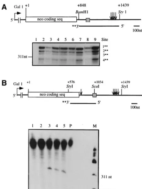
To ensure that the chimeraneo-HTB1mRNA was correctly processed, the 3⬘end of the mRNA was mapped by using S1 nuclease protection experiments (Fig. 1A). Four cleavage sites were identified and mapped to positions⫹104,⫹119,⫹126, and ⫹138 relative to the position of the stop codon of the
HTB1gene (Fig. 1B). These cleavage sites correspond closely to those mapped in the endogenous HTB1 gene (data not shown and Fig. 1B), indicating that the chimera gene is au-thentically processed at the correct cleavage sites.
To identify potentially conserved sequences required for 3⬘-end processing and/or cell cycle regulation, we initially car-ried out a bioinformatic analysis of the sequences in the region
encompassing the 3⬘ ends of yeast histone mRNAs. The 3⬘
ends of five of the eight yeast histone mRNAs have been mapped (20, 32, 35). These histone mRNAs were aligned with respect to their 3⬘-end cleavage sites by using the ClustalW multiple sequence alignment program. Using parameters that identify the features which are most conserved between mem-bers of a set of sequences, we observed that each of the histone genes contained stretches of purine-rich sequences down-stream of the 3⬘-end cleavage sites (Fig. 1B and data not shown). Based on their locations, these purine stretches can be defined as being proximal or distal to the 3⬘-end cleavage sites. While not all of the histone genes contained identifiable prox-imal purine-rich sequences, they all contained one or more purine-rich stretches in the region of nt⫹60 to⫹120 down-stream of their 3⬘ ends (data not shown). The HTB1 gene contains three such purine rich clusters at positions⫹21,⫹73, and⫹106 relative to the position of the strongest cleavage site (Fig. 1B). The presence of purine-rich stretches, or “words,” downstream of yeast genes is quite unusual. Two recent bioin-formatics studies have shown that the most common words downstream of yeast genes are U-rich stretches, while the least common words are GA-rich stretches (18, 34). Given the known role of downstream purine-rich sequences in metazoan histone mRNA processing and the unusual distribution of GA stretches downstream of the yeast histone genes, we first fo-cused our analysis on one of theHTB1 distal purine-rich se-quences.
Mutations in the downstream sequences of theHTB1mRNA alter 3ⴕ-end cleavage site selection and the steady-state levels of theneo-HTB1transcript.The distal purine-rich sequence of theHTB1gene that we have examined lies approximately 110 nt downstream of the last cleavage site of the mRNA and contains the sequence AGAAAAG (Fig. 1B). To examine the role of this sequence in the 3⬘-end processing and cell cycle regulation of theHTB1gene, mutants were prepared in which purines were replaced by pyrimidines. Since the purine-rich HDE of metazoan histone mRNAs is always found in the context of the stem-loop of the histone mRNA, we also pre-pared a mutant (pSAC17) in which a stem-loop structure was inserted upstream of the purine-rich sequence. Additional mu-tations were also prepared in the sequences surrounding the purine stretch (Fig. 2A). Following transformation of the mu-tated plasmids into yeast cells, Northern blotting was per-formed on RNA extracted from cultures grown in galactose-containing medium. The blots were hybridized with a probe containingneosequences only (Fig. 2A). As shown in Fig. 2B, a number of alterations are apparent in the neo-HTB1 tran-script.
Growth of cells in galactose-containing medium results in the induction of an mRNA of approximately 1.2 kb that hy-bridizes to a probe containing neo sequences only (Fig. 2B, lane 2). This transcript also hybridizes to a probe containing sequences downstream of the neo region and including the
only a single product by reverse transcription-PCR (data not shown), and no additional protected fragments were observed by using S1 nuclease mapping (see Fig. 3A and B).
Experiments with the mutants pSAC10, -11, and -16 resulted in the generation of a smaller transcript (transcript B; Fig. 2B, lanes 3, 4, and 5). The full-lengthneo-HTB1transcript is also altered in size in these mutants (transcript A). S1 mapping showed that transcript A contained both neo and HTB1 se-quences and had the same 3⬘-end cleavage sites as the neo-HTB1wild-type (WT) transcript (see below).
All of the other mutants showed the same transcript pattern as cells harboring the WTneo-HTB1transcript (Fig. 2B, lanes 6 to 10). We did observe some fluctuations in the steady-state levels of theneo-HTB1transcript in some of the mutants (mu-tants pSAC10 and -20; Fig. 2B, lanes 3 and 9). In most cases this fluctuation correlated with the OD600at which the cells were harvested, since increased transcript levels were observed as the OD600 of the culture increased. However, we
consis-tently observed that the steady-state levels of the neo-HTB1
transcript in mutant pSAC20 were significantly lower than those of the WT transcript (Fig. 2B, lane 9). In three indepen-dent experiments in which RNA from cultures harvested at the same OD600was used, the steady-state levels of theneo-HTB1 transcript in mutant pSAC20 were on average fivefold lower than those of the WT transcript. This mutant contains two sets of mutated triplets (Fig. 2A), including the triplet mutated in pSAC14. Since the steady-state level ofneo-HTB1mRNA in mutant pSAC14 appears normal (Fig. 2B, lane 8), we can deduce that the additional bases mutated in pSAC20 are re-sponsible for the decrease in steady-state levels. Surprisingly, the levels of theneo-HTB1transcript in mutant pSAC21 are similar to the WT levels (Fig. 2B, lane 10). This mutant con-tains the same mutated bases as mutant pSAC20 plus an ad-ditional triplet base change, suggesting that the adad-ditional changes in sequence had compensated for the decrease in transcript levels observed with the pSAC20 mutant.
Transcript B is generated by the use of an alternative 3ⴕ-end cleavage site. To determine whether the shorter transcripts (transcripts A and B) observed with mutants pSAC10, -11, and -16 resulted from the use of alternative 3⬘-end cleavage sites, S1 mapping using probes encompassing bothneo and HTB1
sequences was performed. As shown in Fig. 3A, transcript A results from cleavage at the normal 3⬘ cleavage sites down-stream of theHTB1sequences (Fig. 3A, lanes 3 and 4, and data not shown). Since no other 3⬘-end cleavage sites were identi-fied, the size difference between the full-length transcripts gen-erated by the WT and mutant genes may result from the presence of a shorter poly(A) tail or from transcription from an alternative initiation site. The full-length WT neo-HTB1
mRNA and transcripts A and B are all equally retained on oligo(dT) cellulose columns (data not shown). As with mutants pSAC10, -11, and -16, the full-length neo-HTB1 transcripts observed in all other mutants are cleaved at the normal 3⬘-end cleavage sites (Fig. 3A).
To identify the 3⬘ends of transcript B, S1 nuclease mapping was carried out by using a DNA probe encompassing the 3⬘
end of theneo gene and the 5⬘ end of theHTB1 sequences (Fig. 3B). We observed that all three mutants (pSAC10, -11, and -16) contain a new 3⬘-end cleavage site which lies approx-imately 45 nt downstream of the neo stop codon and 12 nt downstream of theBamHI site (Fig. 3B, lanes 3, 4, and 5, and C). Therefore, transcript B does not contain any HTB1 se-quences. This new cleavage site was not observed in the WT mRNA or with any of the other mutants (Fig. 3B, lanes 1 and 2, and data not shown).
Mutations downstream of theHTB1gene lie in a region of RNA polymerase II termination.The area where the mutations were made (hereafter referred to as the distal downstream element [DDE]) lies quite distal to the 3⬘-end cleavage sites, and yet these mutants affect both the steady-state levels and 3⬘-end cleavage site selection of the neo-HTB1 mRNA. We therefore wanted to test whether the region in which the mu-tations were made was in fact transcribed as part of the pri-mary pre-mRNA transcript. TRO experiments were carried out as previously described (1). This assay quantifies the den-sity of RNA polymerase molecules along a transcript by incor-porating [␣-32P]UTP into the elongating RNA transcript and is most effective for analysis of highly transcribed genes. Single-FIG. 2. Northern blot analysis ofneo-HTB1transcripts from
[image:6.603.51.275.70.308.2]stranded DNA probes, corresponding to sequences located in approximately 200-nt increments downstream of the HTB1
stop codon, were prepared by the cloning of these fragments into the replicative form of the phage M13. Single-stranded DNA from the phage was immobilized onto nylon membranes and hybridized to32P-labeled nascent RNA isolated from cells transformed with the WTneo-HTB1plasmid (Fig. 4). To dis-tinguish between transcripts emanating from the endogenous gene and theneo-HTB1gene, we compared the levels of nas-cent transcripts in cells grown in glucose and galactose. The first DNA fragment (probe 1 [nt⫹9 to ⫹196]; Fig. 1B) con-tains the entire 3⬘UTR and the 3⬘-end cleavage sites (nt⫹104,
⫹119,⫹126, and⫹138), while probe 2 (⫹235 to⫹409) con-tains the DDE, which lies close to the 5⬘end of the fragment (nt ⫹250; Fig. 1B). As shown in Fig. 4B, we observed tran-scription proceeding from the position of probe 1 to probe 2. Taking the transcription level to be 100% for probe 1 and having subtracted the background hybridization, in our analy-sis of galactose-containing medium we observed that transcrip-tion decreased to 63% in the probe 2 region, 24% in the probe 3 region, and 0% in the probe 4 region. Similarly, in glucose-containing medium, there was a decrease in transcription to 51% in the probe 2 region, 16% in the probe 3 region, and 0% in the probe 4 region. These results suggest that transcription termination was occurring somewhere in the region of probes 2 and 3. Analysis of run-on transcription in cells transformed with the mutated plasmids indicated that all mutant transcripts were transcribed as far as the region of probe 2, and there
appeared to be no significant quantitative differences in the levels of transcription of the mutant mRNAs (data not shown).
Mutations in the DDE alter the cell cycle periodicity of the neo-HTB1transcript.To test whether mutations in the DDE had any effect on cell cycle regulation of theneo-HTB1 tran-script, cells transformed with the mutated plasmids were syn-chronized at the G1 border of the cell cycle by using the
␣1-mating factor. Once the cells were synchronized, the␣ -fac-tor was removed and RNA was prepared from samples taken at 5-min intervals. The RNA was hybridized with a DNA probe specific to theneoregion of the gene (Fig. 2A). The pattern of the endogenous histone mRNA was monitored by hybridizing the RNA samples with a probe specific to the coding region of
[image:7.603.303.538.66.137.2] [image:7.603.43.277.68.379.2]detected at 55 min. The WT neo-HTB1 transcript (Fig. 5B) shows an accumulation pattern similar to that of the endoge-nous HTB1; however, the rate of decline in mRNA levels appears slower than that of the endogenous HTB1. These results confirm that the sequences in the 3⬘ UTR and down-stream of the cleavage sites can confer cell cycle periodicity on aneomRNA (37).
Of the mutants analyzed, only pSAC14 and pSAC21 show an altered cell cycle pattern ofneo-HTB1expression (Fig. 5C and E, respectively). In both cases, there appears to be a delay in the accumulation of the neo-HTB1 mRNA. Once induced, both transcripts continued to accumulate, and there appeared to be no turnover of the mRNA, with levels remaining high at 55 and 60 min. Other mutants, such as pSAC10, -11, and -16, showed accumulation patterns identical to that of the WT
neo-HTB1mRNA (data not shown). Interestingly, the pSAC20 mutant showed a normal pattern of cell cycle accumulation (Fig. 5D); however, the peak of accumulation is narrower than that observed with the endogenous HTB1 or with the WT
neo-HTB1 mRNA. This mutant contains the same base changes as mutant pSAC14 plus an additional three base changes (Fig. 2A), while mutant pSAC21 contains the same base changes as pSAC20 but with a further three base changes (Fig. 2A). Since mutant pSAC20 appeared to show normal cell cycle regulation, the additional three base changes in this mu-tant must compensate for the alterations caused by the three base changes in mutant pSAC14. This compensation appears to be overridden by the additional base changes in mutant pSAC21.
Sequences in the DDE bind protein factor(s).The results obtained from the analysis of mutations in the DDE suggest
that these sequences influence the accumulation and subse-quent turnover of HTB1 mRNA during the cell cycle. Since two of the mutants showed a delay in the accumulation of and a lack of turnover of neo-HTB1mRNA, we reasoned that a protein factor might be required to stabilize the pre-mRNA in the S phase or destabilize it immediately thereafter. To exam-ine whether this region was capable of binding protein factors, a 100-nt [32P]dUTP-labeled RNA probe, consisting of the DDE and 80 nt of surrounding sequences (Fig. 6A), was gen-erated by using T7 RNA polymerase. The RNA was incubated with cell extracts prepared from yeast cells harvested at differ-ent time points following release of cells from ␣-factor syn-chronization. In the presence of cell extracts, the labeled RNA bound to one or more protein factors, as indicated by the FIG. 4. The DDE is transcribed as part of the primary transcript.
[image:8.603.51.278.71.234.2](A) Schematic representation of theHTB1region of theneo-HTB1 gene, showing the position of the 200-nt DNA fragments to which nascent RNA was hybridized. The fragments are labeled Probe 1 to Probe 4. The values above the poly(A) sites show their positions relative to that of the stop codon ofHTB1 (⫹1). The shaded box represents the HTB1 coding sequences, while the open box down-stream of the poly(A) sites (arrows) represents the DDE. (B) Hybrid-ization of nascent transcripts from yeast cells transformed with the WT plasmid pLJ31-HTB1 grown in glucose or galactose. M13, single-stranded phage DNA with no insert. Probes 1 to 4 were as described for panel A.
FIG. 5. Cell cycle regulation of the neo-HTB1 transcript. Yeast cells transformed with the plasmid pLJ31-HTB1or mutant plasmids were synchronized to the G1phase of the cell cycle by the addition of the␣1-mating factor. RNA was extracted at 5-min intervals and hy-bridized to a probe specific for the coding region of theHTB1gene (A) (Table 1) or aneo-specific probe (B to E) (Fig. 2A and Table 1). To account for slight variations in RNA loading, the blots were stripped and rehybridized with an actin DNA probe. The levels of hybridization to the three probes were quantified, normalized to the actin mRNA levels, and expressed as mRNA levels at each time point relative to the level of hybridization at 20 min following removal of
retardation of the mobility of the RNA on nondenaturing gels (Fig. 6B). Interestingly, maximum binding was observed in extracts prepared at 35 min following␣-factor release (Fig. 6B, lanes 5 and 6), the time point corresponding to the peak of
neo-HTB1 mRNA during the cell cycle. Binding is also ob-served at 30 min (Fig. 6B, lanes 2 to 4) and 40 min (lanes 7 to 9), with a marked decrease in binding occurring at 45 min (lanes 10 to 12).
To determine whether binding was affected by the mutations made in this region, the binding experiments were repeated by using RNA incorporating the mutations. We observed that binding was slightly reduced in the pSAC14 mutant RNA (Fig. 6C, lanes 4 to 6), while no binding was observed in the pSAC21 mutant RNA (lanes 10 to 12). Mutant pSAC20 showed binding similar to that of the WT RNA; however, the kinetics of bind-ing was altered somewhat, with reduced bindbind-ing observed at the lower concentrations of extract and greater binding at the highest extract concentration (Fig. 6C, lanes 7 to 9). These results suggest that the DDE, as defined by the mutants de-scribed here, influences accumulation ofHTB1during the cell cycle through the binding of specific protein factors. The cor-relation between the continued accumulation of the mRNA in pSAC14 and -21 and the decrease in binding activity in the cell extracts suggests that the protein factors function to destabilize the pre-mRNA.
DISCUSSION
We have identified a novel sequence element lying approx-imately 110 nt downstream of the 3⬘end ofHTB1that influ-ences at least two cellular processes, namely, mRNA 3⬘-end processing and cell cycle regulation. The element is referred to as the DDE. To analyze this sequence element, we generated a series of mutations within the DDE. Experiments using three of the mutants, pSAC10, -11, and -16, resulted in the selection of an alternative 3⬘-end cleavage site just downstream of the
neo sequences of the neo-HTB1 chimera, generating a new transcript referred to here as transcript B. In addition, the full-lengthneo-HTB1transcript generated in mutants pSAC10, -11, and -16, referred to here as transcript A, is approximately 50 nt shorter than the WT transcript. Since transcript A is generated through the use of the same cleavage sites as the WT transcript, the difference in sizes between the WT and tran-script A is most likely accounted for by the presence of a shorter poly(A) tail on the mutant-derived transcripts or by the use of an alternative transcription initiation site. Further ex-periments will be required to distinguish between these two possibilities.
We observed that experimentation using one of the mutants, pSAC20, resulted in a fivefold or greater decrease in the steady-state levels of theneo-HTB1mRNA. Since the mutated sequences are not contained in the final transcript, this reduc-tion in steady-state levels ofneo-HTB1 may be a result of a decrease in efficient 3⬘-end processing or may affect the stabil-ity of the final transcript. Interestingly, Kim et al. (22) have identified a 54-nt element downstream of the distal poly(A) site of the Schizosaccharomyces pombe uv15⫹gene that can
influence the stability of the mature mRNA. Like the DDE described here, theuv15⫹element is not included in the
ma-ture mRNA. Analysis of the mutant pSAC20 RNA during the FIG. 6. A protein factor binds to the DDE. (A) Schematic
cell cycle revealed that the RNA, while induced in the S phase, appears to be more rapidly degraded as cells enter the G2 phase (Fig. 5D). Thus, the inherent instability of the mutant pSAC20 RNA may be a consequence of its rapid turnover in the G2phase of the cell cycle. Taken together, the data suggest that sequences downstream of theHTB1gene, though not part of the mature mRNA, can affect the steady-state levels of the final transcript.
It is surprising that mutant pSAC21 mRNA, which contains the same mutated bases as mutant pSAC20 (Fig. 2A) plus an additional triplet base pair change, shows steady-state tran-scription levels similar to those of the WT mRNA. Further-more, mutant pSAC14 shares three of the six base pair changes of pSAC20 and it too shows normal steady-state levels of
neo-HTB1transcript. These findings suggest that the presence of a specific sequence is not alone sufficient to determine the overall function of this region and that a secondary structure may also play a role (see below).
Of the other mutants generated, pSAC13 and -17 showed normal steady-state levels (Fig. 2A). Mutant pSAC13 (AAA to TTT) includes three of the bases changed in mutant pSAC11 (AGAAA to TCTCT) (Fig. 2A). Unlike pSAC11, this mutant does not result in the generation of transcript B. Therefore, we can conclude that transcript B is generated as a result of the introduction of bases (TCT) that are common between mu-tants pSAC10 and -11 or, alternatively, that it is the introduc-tion of C residues that accounts for the generaintroduc-tion of transcript B. The latter argument is supported by findings for mutant pSAC16 in which a single base change from A to C resulted in the generation of transcript B (Fig. 2A). Finally, it is surprising that the introduction of a stem-loop structure (pSAC17) at the same location as mutant pSAC16 caused the transcript pattern to revert to that of the WT. This may indicate that the overall sequence context in this region is important to its function.
Sequences located at a distance from the 3⬘-end cleavage sites are known to influence transcription termination. To de-termine whether the DDE was transcribed as part of the pre-mRNA, TRO experiments were carried out. While we have not defined the exact position of termination, the results of this study show that transcription does indeed proceed as far as the DDE and that the majority of transcripts terminate no more than 150 nt downstream of the element.
The processes of 3⬘-end cleavage and polyadenylation and transcription termination are intimately linked (5). A bipartite signal consisting of a functional polyadenylation element and a downstream transcription pause element are required for ef-ficient transcription termination (30). Mutations in the former element have been shown to reduce the efficiency of termina-tion. Among the components of cleavage factor 1A in yeast, the most notable proteins required for efficient termination are Rna15, Rna14, and Pcf11 (30). These proteins appear to in-fluence termination through their interaction with the C-ter-minal domain of RNA polymerase II (2, 11). A connection between transcription termination and 3⬘-end processing of the nonpolyadenylated mouse histone H2A gene has also been demonstrated (12). Removal ofcis-acting sequences that are required for 3⬘-end processing of this gene disrupts transcrip-tion terminatranscrip-tion. The DDE described here is located just up-stream of the region of transcription termination. However, according to the findings for the mutations examined in this
[image:10.603.331.490.70.592.2]study, it appears that thesecis-acting sequences are not directly required for transcription termination (data not shown). Given the location of the element and the known connection between transcription termination and 3⬘-end cleavage, further muta-tions may uncover a link between these two processes.
Cell cycle regulation is altered by mutations in the DDE.
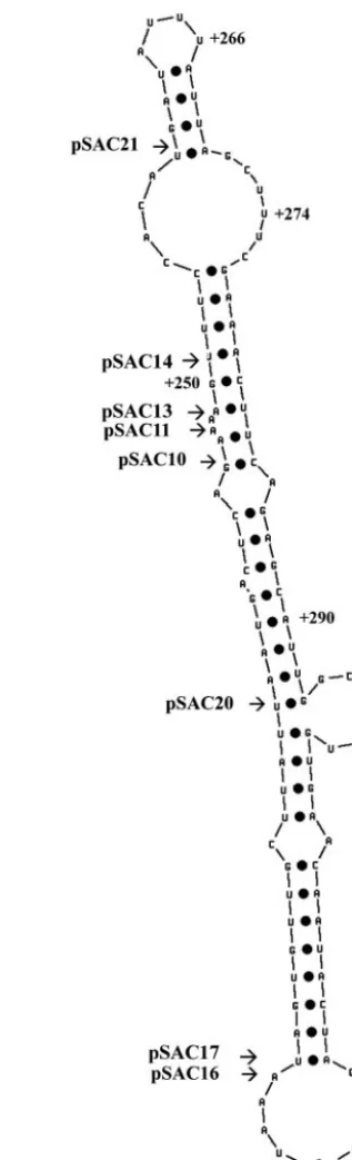
Our data also reveal that two of the mutants in the DDE, pSAC14 and pSAC21 (Fig. 5C and E), can alter the periodic accumulation ofneo-HTB1mRNA during the cell cycle. Inter-estingly, these mutations, while in the same general location as those altering 3⬘-end cleavage, are in fact distinct. Mutations altering 3⬘-end cleavage do not alter cell cycle regulation, and mutations altering cell cycle regulation do not alter 3⬘-end cleavage. The mutations in pSAC-14 and -21 lie adjacent to one another (Fig. 2A). Using the RNA folding program MFOLD (http://www.bioinfo.rpi.edu/), we found that the se-quences surrounding the DDE can be folded into a putative stem-loop structure (Fig. 7). Both mutant pSAC14 and mutant pSAC21 lie at the top end of the stem structure, while the mutants that affect 3⬘-end cleavage lie further down the stem. Using a band shift assay, we showed that an RNA molecule encompassing this stem-loop structure binds a protein factor or factors that are maximally present in the S phase of the cell cycle. The fact that maximum binding is observed in the S phase of the cell cycle may indicate the binding of a cell cycle-regulated factor. Both pSAC14 and -21 mutants showed a reduction and an absence of protein binding, respectively, and also showed a lack of turnover of the mRNA following the S phase, suggesting that the protein factor is required for destabilization of the mRNA following the S phase. Further-more, mutant pSAC20, which exhibited more rapid turnover of the RNA, showed a greater degree of binding to proteins present in extracts from the S phase of the cell cycle. Using the MFOLD program, we observed that the putative secondary structure of these RNAs, in the region of the mutations, was altered (data not shown), strengthening the argument that a specific factor is interacting with this region. The sequences in the DDE that regulate 3⬘-end processing and cell cycle peri-odicity appear to act independently of each other; however, since they are so closely linked spatially, it is possible that further mutations may reveal an overlapping link between 3⬘ -end processing and cell cycle regulation.
Given the location of the DDE and the myriad of proteins associated with RNA polymerase II, it is not surprising that this RNA fragment binds to proteins present in cell extracts. Aranda and Proudfoot (2) have uncovered a physical and ge-netic link between the C-terminal domain of CstF-64 (Rna15p), a protein required for 3⬘-end processing and tran-scription termination, and the trantran-scription factor Res2p. This protein is a component of the G1/S transcription factor com-plex MBF (Mlu1 cell cycle box-binding factor), which regulates the activation of a set of genes at the G1/S boundary of the cell cycle (38). While histone genes are not regulated by Res2p, taken together, the data suggest that 3⬘-end processing and transcription termination events play a role in the control of cell cycle-regulated genes. This argument is strengthened by recent data showing that the phosphorylation and ubiquitina-tion state of poly(A) polymerase, a component of the cleavage and polyadenylation machinery, exhibits variation during the cell cycle (6, 28). Interestingly, it has been shown that tran-scription of the human histone gene, H3.3, is regulated by a transcription termination site located within an intron in the gene. This gene belongs to the class of replication-indepen-dent, basally expressed histone genes that do not accumulate during the S phase of the cell cycle. Rather, members of this
class of histone genes are constitutively expressed in certain tissues such as spermatids and nondividing higher eukaryotic cells. Basally transcribed histone genes differ from replication-dependent histone genes, as they often possess introns and are polyadenylated. Transcription elongation of the H3.3 gene is blocked in cells arrested at the initiation of the S phase. DNA binding studies revealed the presence of protein factors in HeLa cell nuclear extracts that bind specifically to the region of the transcription elongation block. The block in transcriptional elongation was shown to correlate with the protein binding activity (33). The data presented here, showing that sequences close to the site of termination of a yeast histone gene play a role in cell cycle regulation, may provide an insight into the evolutionary process that resulted in such divergent mecha-nisms for the regulation of histone genes in lower and higher eukaryotes.
ACKNOWLEDGMENTS
We thank Tharappel C. James for helpful discussions and experi-mental advice. Thanks also to Stephan Keegan, who provided technical assistance. We thank Agustin Aranda and Aurora Marco for helpful contributions in the TRO analyses.
This work was supported by a grant to U.B. from Enterprise Ireland (SC/97/326) and as part of the National Development Plan (grant SC/01/398) and was partly funded by the European Community-Euro-pean Regional Development Plan. M. del Olmo is funded by Gener-alitat Valenciana (GV99-105-1-13) and the Ministerio de Educacio´n y Ciencia (CICYT ALI99-1224-C02-02).
REFERENCES
1. Aranda, A., J. E. Perez-Ortin, C. Moore, and M. del Olmo.1998. Transcrip-tion terminaTranscrip-tion downstream of theSaccharomyces cerevisiae FBP1poly(A) site does not depend on efficient 3⬘end processing. RNA4:303–318. 2. Aranda, A., and N. Proudfoot.2001. Transcriptional termination factors for
RNA polymerase II in yeast. Mol. Cell7:1003–1011.
3. Barilla, D., B. A. Lee, and N. J. Proudfoot.2001. Cleavage/polyadenylation factor IA associates with the carboxyl-terminal domain of RNA polymerase II inSaccharomyces cerevisiae.Proc. Natl. Acad. Sci. USA98:445–450. 4. Battle, D. J., and J. A. Doudna.2001. The stem-loop binding protein forms
a highly stable and specific complex with the 3⬘stem-loop of histone mRNAs. RNA7:123–132.
5. Birse, C. E., L. Minvielle-Sebastia, B. A. Lee, K. Keller, and N. Proudfoot.
1998. Coupling transcriptional termination to messenger RNA maturation in yeast. Science280:298–301.
6. Bond, G., C. Prives, and J. L. Manley.2000. Poly(A) polymerase phosphor-ylation is dependent on novel interactions with cyclins. Mol. Cell. Biol.
20:5310–5320.
7. Bond, U., and T. Yario.1994. The steady-state levels and structure of the U7 snRNP are constant during the human cell cycle: lack of cell cycle regulation of histone mRNA 3⬘end formation. Cell. Mol. Biol. Res.40:27–34. 8. Bond, U. M., T. A. Yario, and J. A. Steitz.1991. Multiple processing-defective
mutations in a mammalian histone pre-mRNA are suppressed by compen-satory changes in U7 RNA both in vivo and in vitro.Genes Dev.5:1709– 1722.
9. Bracken, A., and U. Bond.1999. Reassembly and protection of small nuclear ribonucleoprotein particles by heat shock proteins in yeast cells. RNA
5:1586–1596.
10. Butler, J. S., P. P. Sadhale, and T. Platt.1990. RNA processing in vitro produces mature 3⬘ends of a variety ofSaccharomyces cerevisiaemRNAs. Mol. Cell. Biol.10:2599–2605.
11. Calvo, O., and J. Manley.2001. Evolutionarily conserved interaction be-tween CstF-64 and PC4 links transcription, polyadenylation and termination. Mol. Cell7:1013–1023.
12. Chodchoy, N., N. B. Pandey, and W. F. Marzluff.1991. An intact histone 3⬘-end processing site is required for transcription termination in a mouse histone H2A gene. Mol. Cell. Biol.11:497–509.
13. Cramer, P., A. Srebrow, S. Kadener, S. Werbajh, M. de la Mata, G. Melen, G. Nogues, and A. R. Kornblihtt.2001. Co-ordination between transcription and pre-mRNA processing. FEBS Lett.498:179–182.
14. Dominski, Z., and W. F. Marzluff.1999. Formation of the 3⬘ends of histone mRNA. Gene239:1–14.
16. Fahrner, K., J. Yarger, and L. Hereford.1980. Yeast histone mRNA is polyadenylated. Nucleic Acids Res.8:5725–5737.
17. Gick, O., A. Kramer, A. Vasserot, and M. L. Birnstiel.1987. Heat-labile regulatory factor is required for 3⬘ end processing of histone precursor mRNAs. Proc. Natl. Acad. Sci. USA84:8937–8940.
18. Graber, J. H., C. R. Cantor, S. C. Mohr, and T. F. Smith.1999. Genomic detection of new yeast pre-mRNA 3⬘end processing signals. Nucleic Acids Res.27:888–894.
19. Hanson, R. J., J. Sun, D. G. Willis, and W. F. Marzluff.1996. Efficient extraction and partial purification of the polyribosome-associated stem-loop binding protein bound to the 3⬘end of histone mRNA. Biochemistry35:
2146–2156.
20. Hereford, L., K. Fahrner, T. Woolford, M. Rosbash, and D. B. Kaback.1979. Isolation of yeast histone genes H2A and H2B. Cell18:1261–1271. 21. Hoffman, I., and M. L. Birnstiel.1990. Cell cycle-dependent regulation of
histone precursor mRNA processing by modulation of U7 snRNA accessi-bility. Nature346:665–668.
22. Kim, M., W. Lee, J. Park, J. B. Kim, Y. K. Jang, R. H. Seong, S. Y. Choe, and S. D. Park.2000. The stress-activated MAP kinase sty1/spc1 and a 3⬘ regu-latory element mediate UV-induced expression of the gene uv15⫹at the
post-transcriptional level. Nucleic Acids Res.28:3392–3402.
23. Luscher, B., and D. Schumperli.1987. RNA 3⬘ end processing regulates histone mRNA levels in a mammalian cell cycle mutant. A processing factor becomes limiting in G1-arrested cells. EMBO J.6:1721–1726.
24. Lycan, D. E., M. Osley, and L. M. Hereford.1987. Role of transcriptional and post-transcriptional regulation in the expression of histone genes in
Saccharomyces cerevisiae. Mol. Cell. Biol.7:614–621.
25. Martin, F., A. Schaller, S. Eglite, D. Schumperli, and B. Muller.1997. The gene for histone RNA hairpin binding protein is located on human chromo-some 4 and encodes a novel type of RNA binding protein. EMBO J.16:
769–778.
26. McCracken, S., N. Fong, K. Yankulov, S. Ballantyne, G. Pan, J. Greenblatt, S. Patterson, M. Wickens, and D. Bentley.1997. The C-terminal domain of RNA polymerase II couples mRNA processing to transcription. Nature
385:357–361.
27. Minvielle-Sebastia, L., and W. Keller.1999. mRNA polyadenylation and its coupling to other RNA processing reactions and to transcription. Curr. Opin. Cell Biol.11:352–357.
28. Mizrahi, N., and C. Moore.2000. Posttranslational phosphorylation and ubiquitination of theSaccharomyces cerevisiaepoly(A) polymerase at the S/G2stage of the cell cycle. Mol. Cell. Biol.20:2794–2802.
29. Mowry, K. L., and J. A. Steitz.1987. Identification of the human U7 snRNP as one of several factors involved in the 3⬘end maturation of histone premes-senger RNAs. Science238:1682–1687.
30. Proudfoot, N. J.2000. Connecting transcription to messenger RNA process-ing. Trends Biochem. Sci.25:290–293.
31. Proudfoot, N. J., A. Furger, and M. J. Dye.2002. Integrating mRNA pro-cessing with transcription. Cell108:501–512.
32. Smith, M. M., and K. Murray.1983. Yeast H3 and H4 histone messenger RNAs are transcribed from two non-allelic gene sets. J. Mol. Biol.69:641– 661.
33. Taylor, A., L. Zhang, J. Herrmann, B. Wu, L. Kedes, and D. Wells.1997. Cell-cycle-specific transcription termination within the human histone H3.3 gene is correlated with specific protein-DNA interactions. Genet. Res.69:
101–110.
34. van Helden, J., M. del Olmo, and J. E. Perez-Ortin.2000. Statistical analysis of yeast genomic downstream sequences reveals putative polyadenylation signals. Nucleic Acids Res.28:1000–1010.
35. Wells, D., and D. Brown.1991. Histone and histone gene compilation: an alignment update. Nucleic Acids Res.19(Suppl.):2173–2188.
36. Whitfield, M. L., L.-X. Zheng, A. Baldwin, T. Ohta, M. Hurt, and W. Marz-luff.2000. Stem-loop binding protein, the protein that binds the 3⬘end of histone mRNA, is cell cycle regulated by both translational and posttrans-lational mechanisms. Mol. Cell. Biol.20:4188–4198.
37. Xu, H., L. Johnson, and M. Grunstein.1990. Coding and noncoding se-quences at the 3⬘end of yeast histone H2B mRNA confer cell cycle regu-lation. Mol. Cell. Biol.10:2687–2694.