Immobilization of Nanoscale Sunscreening Agents
onto Natural Halloysite Micropowder
Yong Jae Suh
1,2and Kuk Cho
3,+1Mineral Resources Research Division, Korea Institute of Geoscience and Mineral Resources,
124 Gwahang-no, Yuseong-gu, Daejeon 305-350, Republic of Korea
2Nanomaterials Science and Engineering, Korea University of Science and Technology,
217 Gajeong-ro, Yuseong-gu, Daejeon 305-350, Republic of Korea 3Department of Environmental Engineering, Pusan National University,
2 Busandaehak-ro 63beon-gil, Geumjeong-gu, Busan 609-735, Republic of Korea
Titanium dioxide (TiO2) nanoparticles contained in sunscreen products are typically smaller than 30 nm in order to enhance the efficacy of the sun protection. TiO2nanoparticles of this size range raise toxicity concerns because of the generation of reactive oxygen species (ROS) and the ability to penetrate into the skin tissue. To solve these problems, we immobilized the nanoparticles on a natural tubular powder, halloysite. The nanoparticle-halloysite hybrid powders are prepared by two synthetic methods: anex situimmobilization of TiO2nanoparticles (after separate synthesis of a nanoparticle batch), and anin situgeneration of TiO2nanoparticles inside halloysite. Notably, TiO2nanoparticles of pure rutile phase are synthesized so that the ROS generation is suppressed before immobilization. The ultraviolet-visible (UV-vis) light extinction results show that the sunscreening efficacy of the hybrid powder producedviatheex situroute appears to be 22%higher than that of the hybrid powder fabricated by thein situmethod. An increase in the hybrid powder concentration results in a sunscreening efficiency equivalent to that of the bare TiO2nanoparticles. Our results can be used to produce nanoparticle-halloysite hybrid powders suitable for UV light screening while reducing the potential toxicity concerns. [doi:10.2320/matertrans.M2015086]
(Received March 2, 2015; Accepted April 3, 2015; Published May 15, 2015)
Keywords: colloid, nanomaterials, penetration, absorption, scattering, functional cosmetics
1. Introduction
The use of inorganic ultraviolet (UV)filters, such as TiO2
and ZnO nanoparticles, has been increasing significantly,
especially in sunscreen products for children and people with sensitive skin.1)The inorganic UV-screening ingredients are
contained in commercially available skin care creams and lotions, makeup foundations, eye shadows, and even in lip balms. Among the inorganic ingredients, titanium dioxide (TiO2) nanoparticles are typically adopted as the main
UV-screening agent.
The sunscreen is required to block the UV solar radiation
over the whole UVB (290320 nm) and UVA (320400 nm)
ranges, while being transparent to visible light for cosmetic
aesthetics. To satisfy the cosmetic efficacy, TiO2
nano-particles in the range of 2050 nm have been considered as an optimal material.24)On the other hand, TiO
2nanoparticles of
this size range have also been used as catalysts because of their high photoactive efficiency; in particular, TiO2particles
with a size of approximately 33 nm show the highest activity.35) The photoactive surfaces of the TiO
2
nano-particles produce reactive oxygen species (ROS), such as
•OH, O
2¹•, and H2O2,68) which are known to be cytotoxic.
For example, hydroxyl radicals (•OH) generated from
photoactive TiO2 nanoparticles cause damage to DNA
plasmids in vitro and to the whole human skin cells in
cultures.1,9) Accordingly, the generation of ROS has been
suppressed by modifying the TiO2nanoparticles surface with
other materials, e.g., Al2O3, SiO2, and ZrO2, which form
hydrated oxides that capture the hydroxyl radicals and thus reduce the photoactivity.1) For the same reason, the rutile
phase is more advantageous than the anatase phase, as the
latter shows the highest activity among the TiO2
poly-morphs.6,1014)The most effective phase at generating ROS is
likely to exhibit the highest cytotoxicity.12)
Moreover, as the dermal exposure to TiO2 nanoparticles
occurs regularly during the use of sunscreen products, nanoparticles penetration through the skin has become a
growing concern.8,1522) For example, the normal flexing
movement of the skin facilitates the penetration of 0.5 and 1.0 µm particles into the dermis.22)Besides, when a sunscreen lotion containing TiO2nanoparticles is rubbed on a skin area
burned by the sun or damaged by acne, the TiO2
nano-particles may enter the human body through the skin and have harmful effects.
The toxicity of the sunscreen nanoparticles can be prevented by encapsulating them in a micro-container, reducing the chance of direct exposure of the skin to the nanoparticles themselves. As a result, the ROS generated by the UV light remain in the proximity of the nanoparticles inside the container without entering in contact with the skin. In addition, the nanoparticles are located inside the container and cannot infiltrate through the container walls, excluding the possibility of their penetration. Even the nanoparticles strongly adsorbed onto the outer surface of the container cannot penetrate through the damaged skin. A good candidate for the fabrication of such containers is a naturally occurring mineral: halloysite.
Halloysite is an aluminosilicate clay belonging to the kaolin group and with a composition of Al2Si2O5(OH)4·
2H2O; the ratio of aluminum to silicon is 1 : 1.23)Halloysite
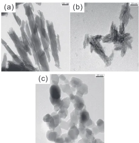
is mostly found in the form of a tubular structure with a 15
125 nm internal lumen, a 30250 nm outer diameter, and a
length of less than a few microns (Fig. 1(a)). The outer surface of the halloysite comprises a silicate SiO2¹ layer,
whereas the inner surface comprises an alumina Al2O3+
+Corresponding author, E-mail: kukcho@pusan.ac.kr
layer. Inside the tube, inorganic or organic materials can be encapsulated to provide new functionalities.24)Furthermore,
the cytotoxicity testing of halloysite shows no toxic effect on
fibroblast cells and human breast cancer cells for over 48 h.25)
Thus, the micron-sized tubular halloysite is considered a benign carrier suitable for delivering active substances such as fragrances, cosmetics, and drugs.2529)
To date, a hybrid powder, in which the light-scattering nanoparticles are loaded into natural clay minerals similar to halloysite, has not yet been developed for sunscreen purposes. Presumably, immobilizing the light-scattering nanoparticles into halloysite and using them as UV-screening agents may reduce or eliminate the unwanted side effects on the skin. Here, to fabricate hybrid sunscreen powders, we
provide two synthetic methods in which TiO2 nanoparticles
are encapsulated into or coated on halloysite. An ex situ
immobilization of the nanoparticles both on the inner and outer surfaces of halloysite is achieved by vacuum pulling of
TiO2nanoparticles prepared in advance. On the other hand,
anin situgeneration of the nanoparticles inside halloysite is conducted by loading the reactants first, and then initiating the particle formation. The nanoparticle synthetic process is controlled to produce pure rutile phase, which generates a smaller amount of ROS compared to the anatase phase. The
sunscreening efficacy is examined by comparing the
UV-visible (UV-vis) extinction efficiency with that of the bare
TiO2 nanoparticles at the same concentration. The hybrid
powder produced by theex situmethod shows higher UV-vis
extinction than that fabricated by the in situ method. An
increase in hybrid powder concentration makes the extinction efficiency equivalent to that of the bare TiO2 nanoparticles.
Our study enables the synthesis of nanoparticle-halloysite hybrids suitable for use as UV-screening agents while mitigating the toxicity concerns.
2. Experimental Procedure
A sample of halloysite was donated by Atlas Mining Company, USA. Titanium tetraisopropoxide (TTIP, Aldrich,
97%), HCl (Junsei, GR, 35.037.0%), NaOH (Kanto, Cica
reagent, min. 97%), anhydrous ethanol (Sigma-Aldrich,
>99.5%), and T805 TiO2 nanoparticles (AEROXIDEμ
TiO2 T 805, Evonik, Germany) were used as received. All
the chemical solutions not specifically mentioned were
prepared with ultra-high-purity deionized water.
The hybrid powder of TiO2 nanoparticles and halloysite
was prepared by either of the following two methods. The
first method is anex situTiO2colloidal method based on the
direct loading of TiO2 nanoparticles into halloysite. The
second entails the in situ TTIP-HCl solution method that
generates TiO2 nanoparticles inside halloysite by
vacuum-pulling the nanoparticle precursor into the halloysite lumen. The TiO2colloidal method is used to immobilize the TiO2
nanoparticles, which are prepared separately, directly into halloysite. The synthetic process for the rutile phase TiO2
nanoparticles is developed based on the study conducted by
Han’s group.30) In detail, a 1 M HCl (80 mL) solution is
placed in a 250 mL beaker and stirred using a magnetic bar; TTIP (20 mL) is then slowly added and stirred at 60°C for 3 h. When the TiO2 nanoparticles are produced, the solution
is left to cool to room temperature, and the TiO2nanoparticles
are collected by centrifugation at 10000 rpm (=15260©g)
for 3 min. The collected TiO2 nanoparticles are added to
distilled water (20 mL) and dispersed with ultrasonic waves for 30 min to prepare a colloidal solution. Then, halloysite powder (3 g) is added to the colloidal solution and the resulting mixture is vacuum pulled. Subsequently, the hybrid powder is collected by centrifugation, washed with a pH 1 solution twice, with distilled water once, and then dried at 60°C.
Thein situTTIP-HCl solution method is used to load the TiO2nanoparticles into halloysite by adding halloysite in the
early formation stages of the TiO2 nanoparticles. In detail,
a TTIP solution (20 mL) is added to a 1 M HCl solution (80 mL) and stirred at 60°C for 30 min. Next, the halloysite powder (3 g) is added to the resulting solution and then vacuum pulled. Subsequently, the powder is collected by centrifugation and washed with anhydrous ethanol once. Then, the collected powder is dispersed in a 1 M HCl solution (80 mL), and the resulting mixture is stirred at 60°C for 3 h. After cooling the resulting solution to room temperature, the hybrid powder is collected by centrifugation and washed with a solution with pH 1 once, with distilled water once, and then dried at 60°C.
The size and shape of the nanoparticles and hybrid powders were characterized by transmission electron
mi-croscopy (TEM, Philips, CM12, Netherlands) and field
emission scanning electron microscopy (FE-SEM, FEI, Quanta650F, Netherlands). The crystalline structure of the nanoparticles was characterized by X-ray diffractometry (XRD, Rigaku Denki Co. Ltd., RU-200B, Japan). The UV-screening efficiencies of the TiO2 nanoparticles, halloysite,
and hybrid powders are measured as follows. T805 and
synthesized TiO2 nanoparticles are dispersed in anhydrous
ethanol, whereas raw halloysite and the hybrid powders are
[image:2.595.54.283.67.302.2]dispersed in a pH 10 aqueous solution. All the samples with a
concentration of 0.0025 mass% are sonicated for 10 min to
disperse uniformly the particles. The measurement sample is injected into a 1 cm path length quartz cuvette and mounted in a UV-vis spectrophotometer (Scinco, S3100, Korea). Then, the light transmittance of the sample is measured in the wavelength () range of 250500 nm.
3. Results and Discussion
To obtain the rutile phase TiO2nanoparticles, we modified
the synthetic process reported by Han’s group.30) The
reaction time was shortened from 48 to 3 h, the reaction temperature was raised from room temperature to 60°C, and no stabilizing agents were utilized. On the other hand, hydrochloric acid was used to control the hydrolysis and condensation of the TTIP precursor. The use of stabilizing agents, such as the Pluronic P-123 polymer, should be avoided, as their bulky structure prevents the movement of the TiO2nanoparticles into the lumen of halloysite; besides,
they may be unsuitable for cosmetics. As a result, oblong
TiO2 nanoparticles with an average size of 15 nm©70 nm
were fabricated (Fig. 1(b)). This size appears to be similar to that of T805, which exhibits an average size of 21 nm (Fig. 1(c)). T805 is a fumed TiO2 nanoparticles treated with
octylsilane to form a hydrophobic surface and widely used as a physical UV-filter in sunscreen and daily care products.
The crystalline structures of the synthesized TiO2
nano-particles were confirmed by the XRD patterns (Fig. 2). The
synthesized particles show sharp peaks at the characteristic angles of rutile phase and no peaks at those of anatase phase, confirming the formation of a pure rutile phase. On the other hand, T805 shows a mixed pattern revealing the presence of both the rutile and anatase phases, and exhibiting sharper peaks at the characteristic angles of the anatase phase.
The UV-vis extinction of the nanoparticle suspensions can be determined by measuring the transmittance of the
radiation between 250 and 500 nm.31)The absorbance, A=
¹log(I/I0), is calculated from the BeerLambert law:32)
A¼¾cl ð1Þ
where I/I0, I, I0, ¾, c, and l represent the transmittance,
intensity of the light transmitted through the sample, intensity of the incident light, extinction coefficient, concentration of the nanoparticles, and path length of the light passing through the sample, respectively.
First, the UV extinctions of the raw materials before
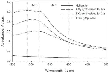
hybridization were examined (Fig. 3). All the TiO2
nano-particles show a strong extinction tendency across most of the UVB and UVA spectra, whereas halloysite is nearly trans-parent. This result indicates the dependence of the light scattering efficiency on the material optical properties, such as the refractive index.2,32) The broad-spectrum protection
shown by the TiO2 nanoparticles can impart high values of
sun protection factor (SPF) at relatively low concentra-tions.33)The highest absorbance of T805 may be attributed to
the large presence of the anatase crystalline phase, as anatase
phase is more photoactive than rutile phase.6,10,11,13,14)
The TiO2 nanoparticles synthesized in this study show a
dependence of the absorbance on the reaction time. This
variation is due to their size difference, as the particle size is the key parameter that determines the light scattering characteristics.32) By prolonging the particle growth time,
the average size of rutile nanoparticles increased from
12 nm©50 nm (data not shown) to 15 nm©70 nm
(Fig. 1(b)) as the reaction time increased from 2 to 3 h. Next, the UV extinctions of the hybrid powders were examined and compared with that of halloysite and bare TiO2
nanoparticles synthesized in this study (Fig. 4). The hybrid powders show an intermediate absorbance between halloysite and bare TiO2nanoparticles, indicating the extent of the TiO2
nanoparticle immobilization on halloysite, as the contribution of halloysite is very low. Between the two hybrid powders, that prepared by following the TiO2colloidal method exhibits
a higher UV extinction efficiency. The amounts of TiO2
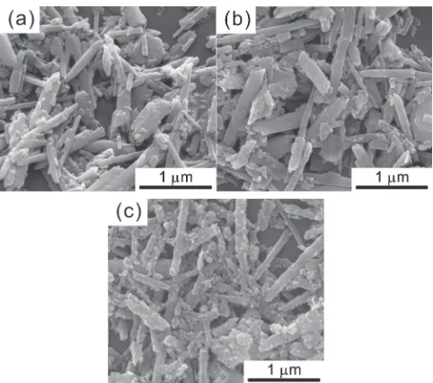
nanoparticles immobilized on halloysite in the hybrid powders can be qualitatively estimated by inspecting the SEM micrographs (Fig. 5). The outer surface of halloysite in
the hybrid powder prepared by thein situTTIP-HCl solution
method is covered by a small number of nanoparticles (Fig. 5(b)). Conversely, the surface of halloysite in the hybrid
Fig. 2 X-ray diffraction (XRD) patterns of TiO2nanoparticles prepared at 60°C in this study and Degussa T805. The XRD data of the indexed bars in grey and black are obtained from JCPSD#21-1276 and#21-1272 for the rutile and anatase crystalline phases, respectively.
[image:3.595.313.542.67.251.2] [image:3.595.311.540.320.472.2]powder prepared by the ex situ TiO2 colloidal method is
covered by a large number of nanoparticles (Fig. 5(c)). Considering a rather narrow halloysite inner lumen and the
oblong TiO2 particle shape, a higher amount of TiO2
nanoparticles can be immobilized in the case of the TiO2
colloidal method. Despite the lower sunscreening efficiency, however, the TTIP-HCl solution method would be consid-ered more advantageous if the contact between the photo-catalytic particles and the skin has to be avoided.
It is noted that raw halloysite is a natural clay mineral exhibiting a broad spectrum of size distributions. Although the SEM micrograph of Fig. 5(a) shows many smaller particles, not shown in the TEM micrograph of Fig. 1(a), all the particles are natural. In the cosmetic applications, if needed, these small particles can be easily removed by a simple separation technique such as centrifugation.
To compare the overall sunscreening performance, we defined a representative UV light extinction efficiency by integrating the UV light absorbance with respect to
wave-length over the UVB (290320 nm) and UVA (320400 nm)
ranges. The order of the representative extinction perform-ance is TiO2nanoparticles>hybrid powder produced by the
TiO2 colloidal method>hybrid powder produced by the
TTIP-HCl solution method (Fig. 6). This order is the same for both the UVB and UVA wavelength ranges. The representative extinctions of the hybrid powders produced
by the colloidal method and solution method are 31.3%and
25.6% of that of the bare TiO2 nanoparticles, respectively.
According to the BeerLambert law, the light extinction is proportional to the concentration, and thus it is expected that a four times higher concentration of the hybrid powders will exhibit a greater extinction performance than the bare TiO2
nanoparticles. To demonstrate this assumption, the
depend-ence of the UV extinction efficiency on the particle
concentration was investigated using the hybrid powders
produced by the TiO2colloidal method. In this experiment,
to optimize the synthetic process, the hybrid powder was prepared in different conditions from the previous experi-ment. The volume ratio between TTIP and 1 M HCl, i.e., TTIP:HCl was reduced to 1 : 10 from 1 : 4 and the reaction was conducted at 80°C for 30 min. Therefore a lower amount of TTIP was used and the reaction time was shortened. The shape and crystalline structure were examined by TEM
(Energy Filtering TEM, Carl Zeiss, 200 kV, Germany). TiO2
nanoparticles located in the halloysite can be hardly seen; most TiO2nanoparticles appear to cover the outer surface of
halloysite (Fig. 7). This implies that the majority of the TiO2
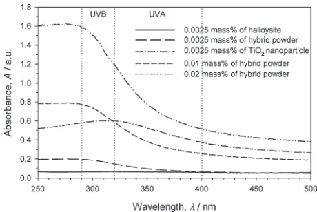
nanoparticles lie on the outer surface of halloysite when synthesized by the colloidal method. The selected area diffraction (SAD) pattern clearly shows a highly crystallized rutile phase TiO2 (Inset of Fig. 7). At 0.0025 mass%, the
absorbance of the hybrid powder synthesized at a lower TTIP concentration (Fig. 8) is lower than that of the previous sample powder (Fig. 4). This decrease in the absorbance is attributed to a lower TiO2nanoparticle proportion in the same
total solid concentration of 0.0025 mass%. Nevertheless, the
hybrid powder at 0.01 mass%shows higher absorbance in the
UVB wavelength range than the bare TiO2 nanoparticles at
0.0025 mass%. The hybrid powder at 0.02 mass% shows
a significantly higher absorbance over the whole UV light
wavelength range. Notably, the increase of the hybrid powder concentration may not cause any problems in the cosmetic
Fig. 4 Ultraviolet (UV) extinction efficiency of the hybrid powders prepared by the two processes compared with raw halloysite and bare TiO2nanoparticles. The particle concentration is 0.0025 mass%.
Fig. 5 Scanning electron microscopy (SEM) micrographs of (a) raw halloysite, (b) hybrid powders produced by the titanium tetraisopropoxide (TTIP)-HCl solution method, and (c) hybrid powders produced by the TiO2colloidal method.
[image:4.595.312.540.69.213.2] [image:4.595.55.284.71.222.2] [image:4.595.49.290.278.491.2]formulation, as the allowance on the concentration of sunscreening agents are based on the effective ingredients, i.e., TiO2. The sunscreen active concentration allowed by the
US Food and Drug Administration (FDA) is 25 mass% for
TiO2, and a typical formulation of a sunscreen product
contains approximately 5 mass%of TiO2nanoparticles.33)
4. Conclusion
As TiO2nanoparticles smaller than 30 nm are being widely
used in sunscreen products, toxicity concerns such as ROS generation and penetration into skin tissue are being raised. Here, we have developed two synthetic routes to immobilize the TiO2 nanoparticles on halloysite, a naturally occurring
benign clay mineral. Theex situTiO2 nanoparticle
immobi-lization, following the synthesis of a separate batch of nanoparticles, resulted in the adsorption of the nanoparticles
on the halloysite surface. On the other hand, the in situ
generation of TiO2nanoparticles inside halloysite resulted in
a clean surface, implying that the largest portion of TiO2
is located inside the halloysite lumen. The former TiO2
colloidal method showed higher UV extinction efficiency
than the latter TTIP-HCl solution method, probably because
of the higher TiO2 proportion in the same total solid
concentration. On the other hand, the latter method provides better results if the direct contact between the sunscreening agent and the skin tissue needs to be avoided, e.g., for
damaged or sensitive skins. It should be noted that TiO2
nanoparticles of pure rutile phase were synthesized to prevent the ROS generation. The nanoparticles were synthesized in the absence of any surfactants, eliminating any possibility
of inclusion of harmful ingredients in the final sunscreen
products. An increase in the hybrid powder concentration led to a sunscreening efficiency equivalent to that of the bare TiO2nanoparticles. Although this increase in the total solid
concentration can limit the usability, the adoption of a natural clay mineral in cosmetics is preferable for some applications. We conclude that this study provides two methods for producing nanoparticle-halloysite hybrid powders that are suitable for UV light screening while reducing the potential toxicity concerns.
Acknowledgments
This work was supported in part by the Brain Korea 21 PLUS project in the Division of Creative Low Impact
Development and Management for Ocean Port City
Infrastructures, the PM2.5 research center supported by Ministry of Science, ICT, and Future Planning (MSIP) and National Research Foundation (NRF) of Korea (NRF-2014M3C8A5030890), and the Basic Research Project of the Korea Institute of Geoscience and Mineral Resources.
REFERENCES
1) N. Serpone, D. Dondi and A. Albini:Inorg. Chim. Acta360(2007) 794802.
2) G. Murphy:Photodermatol. Photoimmunol. Photomed.15(1999) 34 36.
3) A. S. Barnard:Nat. Nanotechnol.5(2010) 271274.
4) A. S. Barnard:Energy Environ. Sci.4(2011) 439443.
5) J. Jiang, G. Oberdörster, A. Elder, R. Gelein, P. Mercer and P. Biswas:
Nanotoxicol.2(2008) 3342.
6) T. Hirakawa, K. Yawata and Y. Nosaka:Appl. Catal. A325(2007) 105111.
7) T. Hirakawa and Y. Nosaka:Langmuir18(2002) 32473254.
8) A. Nel, T. Xia, L. Mädler and N. Li:Science311(2006) 622627.
9) R. Dunford, A. Salinaro, L. Cai, N. Serpone, S. Horikoshi, H. Hidaka and J. Knowland:FEBS Lett.418(1997) 8790.
10) A. Sclafani and J. Herrmann:J. Phys. Chem.100(1996) 1365513661.
11) Z. Ding, G. Lu and P. Greenfield:J. Phys. Chem. B104(2000) 4815 4820.
12) C. M. Sayes, R. Wahi, P. A. Kurian, Y. Liu, J. L. West, K. D. Ausman, D. B. Warheit and V. L. Colvin:Toxicol. Sci.92(2006) 174185.
13) T. van der Meulen, A. Mattson and L. Österlund:J. Catal.251(2007) 131144.
14) J. Augustynski:Electrochim. Acta38(1993) 4346.
15) M. D. Newman, M. Stotland and J. I. Ellis:J. Am. Academy Dermatol. 61(2009) 685692.
16) G. Oberdörster, A. Maynard, K. Donaldson, V. Castranova, J. Fitzpatrick, K. Ausman, J. Carter, B. Karn, W. Kreyling and D. Lai:
Part. Fibre Toxicol.2(2005) 8.
17) B. W. Barry:Eur. J. Pharm. Sci.14(2001) 101114.
18) C. Bennat and C. Muller-Goymann: Int. J. Cosmetic Sci.22(2000) 271283.
19) J. Lademann, H. Richter, S. Schanzer, F. Knorr, M. Meinke, W. Sterry Fig. 7 Transmission electron microscopy (TEM) micrograph of the hybrid
powder prepared by the TiO2 colloidal method at a lower precursor concentration. The inset shows a selected area diffraction (SAD) pattern obtained for the entire sample shown in the TEM micrograph.
[image:5.595.48.290.69.252.2] [image:5.595.53.283.318.472.2]and A. Patzelt:Eur. J. Pharm. Biopharm.77(2011) 465468.
20) J. Lademann, H.-J. Weigmann, C. Rickmeyer, H. Barthelmes, H. Schaefer, G. Mueller and W. Sterry: Skin Pharmacol. Physiol. 12 (1999) 247256.
21) F. Menzel, T. Reinert, J. Vogt and T. Butz: Nucl. Instrum. Methods Phys. Res., Sect. B219220(2004) 8286.
22) S. S. Tinkle, J. M. Antonini, B. A. Rich, J. R. Roberts, R. Salmen, K. DePree and E. J. Adkins:Environ. Health Perspect.111(2003) 1202 1208.
23) E. Joussein, S. Petit, J. Churchman, B. Theng, D. Righi and B. Delvaux:Clay Miner.40(2005) 383426.
24) D. G. Shchukin, G. B. Sukhorukov, R. R. Price and Y. M. Lvov:Small 1(2005) 510513.
25) Y. J. Suh, D. S. Kil, K. S. Chung, E. Abdullayev, Y. M. Lvov and D. Mongayt:J. Nanosci. Nanotechnol.11(2011) 661665.
26) R. R. Price, B. P. Gaber and Y. Lvov:J. Microencapsul.18(2001) 713 722.
27) S. R. Levis and P. B. Deasy:Int. J. Pharm.243(2002) 125134.
28) S. R. Levis and P. B. Deasy:Int. J. Pharm.253(2003) 145157.
29) Y. M. Lvov, D. G. Shchukin, H. Möhwald and R. R. Price:ACS Nano 2(2008) 814820.
30) S. Han, S. H. Choi, S. S. Kim, M. Cho, B. Jang, D. Y. Kim, J. Yoon and T. Hyeon:Small1(2005) 812816.
31) T. F. Tadros (ed.):Colloids in Cosmetics and Personal Care, (WILEY-VCH Verlag GmbH & Co. KGaA, Weinheim, 2008).
32) H. C. van de Hulst: Light Scattering by Small Particles, (Dover Publications, Mineola, 1981).