Second
Edition
F
orensic
CRC SERIES IN
PRACTICAL ASPECTS OF CRIMINAL AND FORENSIC INVESTIGATIONS
VERNON J. GEBERTH, BBA, MPS, FBINA Series Editor
Practical Homicide Investigation: Tactics, Procedures, and Forensic Techniques, Third Edition
Vernon J. Geberth
The Counter-Terrorism Handbook: Tactics, Procedures, and Techniques, Second Edition
Frank Bolz, Jr., Kenneth J. Dudonis, and David P. Schulz
Forensic Pathology, Second Edition
Dominick J. Di Maio and Vincent J. M. Di Maio
Interpretation of Bloodstain Evidence at Crime Scenes, Second Edition
William G. Eckert and Stuart H. James
Tire Imprint Evidence
Peter McDonald
Practical Drug Enforcement: Procedures and Administration
Michael D. Lyman
Practical Aspects of Rape Investigation: A Multidisciplinary Approach, Third Edition
Robert R. Hazelwood and Ann Wolbert Burgess
The Sexual Exploitation of Children: A Practical Guide to Assessment, Investigation, and Intervention, Second Edition
Seth L. Goldstein
Gunshot Wounds: Practical Aspects of Firearms, Ballistics, and Forensic Techniques, Second Edition
Vincent J. M. Di Maio
Friction Ridge Skin: Comparison and Identification of Fingerprints
James F. Cowger
Footwear Impression Evidence, Second Edition
William J. Bodziak
Principles of Kinesic Interview and Interrogation
Stan Walters
Practical Fire and Arson Investigation, Second Edition
David R. Redsicker and John J. O’Connor
The Practical Methodology of Forensic Photography, Second Edition
David R. Redsicker
Practical Gambling Investigation Techniques
Kevin B. Kinnee
Practical Aspects of Interview and Interrogation
David E. Zulawski and Douglas E. Wicklander
Investigating Computer Crime
Franklin Clark and Ken Diliberto
Practical Homicide Investigation Checklist and Field Guide
Vernon J. Geberth
Bloodstain Pattern Analysis: With an Introduction to Crime Scene Reconstruction
Tom Bevel and Ross M. Gardner
Practical Aspects of Munchausen by Proxy and Munchausen Syndrome Investigation
Kathryn Artingstall
Quantitative-Qualitative Friction Ridge Analysis: An Introduction to Basic and Advanced Ridgeology
Vincent J. DiMaio
Dominick DiMaio
Second
Edition
F
orensic
P
athology
This book contains information obtained from authentic and highly regarded sources. Reprinted material is quoted with permission, and sources are indicated. A wide variety of references are listed. Reasonable efforts have been made to publish reliable data and information, but the author and the publisher cannot assume responsibility for the validity of all materials or for the consequences of their use.
Neither this book nor any part may be reproduced or transmitted in any form or by any means, electronic or mechanical, including photocopying, microfilming, and recording, or by any information storage or retrieval system, without prior permission in writing from the publisher.
The consent of CRC Press LLC does not extend to copying for general distribution, for promotion, for creating new works, or for resale. Specific permission must be obtained in writing from CRC Press LLC for such copying.
Direct all inquiries to CRC Press LLC, 2000 N.W. Corporate Blvd., Boca Raton, Florida 33431.
Trademark Notice: Product or corporate names may be trademarks or registered trademarks, and are used only for identification and explanation, without intent to infringe.
Visit the CRC Press Web site at www.crcpress.com
© 2001 by CRC Press LLC
No claim to original U.S. Government works International Standard Book Number 0-8493-0072-X
Library of Congress Card Number 2001025798 Printed in the United States of America 1 2 3 4 5 6 7 8 9 0
Printed on acid-free paper
Library of Congress Cataloging-in-Publication Data
Di Maio, Dominick J.
Forensic pathology / Dominick J. Di Maio, Vincent J.M. Di Maio --2nd ed. p. cm. (Practical aspects of criminal and forensic investigation) Includes bibliographical references and index.
ISBN 0-8493-0072-X
1. Forensic pathology. I. Di Maio, Vincent J.M., 1941- II. Title .III. CRC series in practical aspects of criminal and forensic investigations.
[DNLM: 1. Forensic Medicine. 2. Pathology. W 700 D582f2001] RA1063.4 .D5 2001
Dedication
Foreword
The medicolegal investigation of death is the most crucial and significant function of the medical examiner within the criminal justice system. The medical examiner’s office is primarily concerned with the investigation of violent, sudden, unexpected, and suspicious deaths.
Forensic pathology is the branch of medicine that applies the principles and knowledge of the medical sciences to the many legal issues within the field of law. The medical examiner is responsible for determining the cause and manner of death, identifying the deceased if unknown, determining the approximate time of death and injury, collecting evidence from the body that can be used to prove or disprove an individual’s guilt or innocence and to confirm or deny the account of how the death occurred, documenting inju-ries or lack of them, deducing how the injuinju-ries occurred, documenting any natural disease present, determining or excluding other contributory or caus-ative factors of death, issuing the death certificate, and documenting these events through an official autopsy report. This autopsy protocol is a complete medical record based on a thorough and conclusive review of all the facts and information.
Forensic pathology, the science of recognizing and interpreting diseases and injuries in the human body, is the basis of the medicolegal investigation. The medical examiner provides the expert testimony if the case goes to trial.
Forensic Pathology Second Edition, written by Dominick J. DiMaio, M.D. and Vincent J. DiMaio, M.D., provides the reader with more than 75 years of practical, hands-on experience in the essentials of forensic medicine. The material presented in this revised edition continues to be based on the per-sonal experience of two forensic pathologists who are nationally renowned experts in their respective fields. Dominick J. DiMaio, M.D., a retired Chief Medical Examiner of New York City, is currently a forensic consultant. He served as the professorial lecturer in pathology for the State University of New York Health Science Center at Brooklyn.
The new and revised Forensic Pathology Second Edition begins with an overview of the medicolegal investigative systems and then delves into the substance and mechanics of forensic pathology. The authors present the essentials of forensic medicine in a concise, lucid, and comprehensive manner. They omit superflu-ous, confusing medical terminology and present medicolegal facts pertinent to the many and varied cases discussed, and have added new case histories and current information, bringing this remarkable text into the 21st century. Foren-sic Pathology Second Edition is geared toward medical practitioners, medical students, homicide detectives, medicolegal investigators, prosecuting and defense attorneys, and others interested in forensic pathology.
The first edition of Forensic Pathology addressed and corrected the insuf-ficiencies that existed between criminal investigations textbooks and medicole-gal investigations textbooks and established a recognized reference standard for medical examiners and homicide investigators. The second edition has been completely revised and expanded to include current statistical information. It has been updated and new photographs have been added, as well as two additional chapters that address the concerns of the medicolegal profession.
The new chapter dealing with “Deaths In Nursing Homes” addresses issues of improper health care resulting in drug overdoses, accidents not involving medication, homicide, and gross negligence. The revised informa-tion presents the dynamics involved in evaluating signs of neglect such as contractures, malnutrition, dehydration and the various stages of decubitus ulcers, which provide the practitioner with a frame of reference in a medi-colegal determination. The chapter also addresses deaths caused by hospital-bed rails and medical restraints.
The new chapter on “Sudden Deaths During or Immediately after a Violent Struggle Unassociated with an Anatomical Cause of Death” is, with-out a doubt, an extremely important addition to this text. Deaths involving police or medical personnel who were attempting to restrain a violent or irrational individual are fraught with controversy. The medical examiner and investigator must be cognizant of the necessity for a complete and extensive medicolegal examination. The circumstances leading up to and surrounding the death should be obtained, and any medical records of the deceased should be reviewed. A complete autopsy with microscopic survey of all organs — especially the heart — as well as a complete toxicological screen should be performed. This chapter discusses the evaluation of excited delirium, cate-cholamine release, potassium and their effects on the heart. Drug actions, alcohol and acute psychotic episodes are covered, along with deaths ascribed to positional asphyxia. The authors present important information on the proper certification of death in these circumstances.
detective in the New York City Police Department. When I retired as command-ing officer of The Bronx Homicide, I became a homicide and forensic consultant. I have continued my professional affiliation with Dr. DiMaio and we have been involved in many consultative investigations over the last 15 years. I have also known his son, Vincent J. DiMaio, M.D. for more than 21 years and have had the privileges of his professional affiliation in the sphere of medicolegal investigations and of editing his excellent edition in this series,
Gunshot Wounds: Practical Aspects of Firearms, Ballistics, and Forensic Tech-niques, which is the recognized standard for the interpretation and evaluation of gunshot wounds.
According to the latter, the success of the medical examination and the homicide investigation is assured when a mutual cooperation exists between the forensic pathologist and the homicide investigator. Teamwork is essential. This teamwork is based upon a recognition and appreciation of each other’s duties and responsibilities so that all parties can benefit from their contributions and expertise in the professional investigation of sudden death and homicide.
In my professional opinion as an expert in the sphere of practical homi-cide investigation, Forensic Pathology Second Edition is the must-have com-panion text to Practical Homicide Investigation: Tactics, Procedures, and Forensic TechniquesThird Edition.
Forensic Pathology Second Edition provides an outstanding insight into the investigation of death and serves as a brilliant guide for any pathologist, investigator, or attorney who lacks the formal training of a board-certified forensic pathologist. This text provides practitioners with a foundation upon which they can build an excellent medicolegal investigation. The authors have provided graphic and descriptive photographs throughout the text that high-light for the reader exactly what to look for and how to handle specific situations. This revision carries this textbook well into the 21st century.
Forensic Pathology Second Edition by Dominick J. DiMaio, M.D. and Vincent J. DiMaio, M.D. continues to be the most comprehensive, definitive, and practical medicolegal textbook on forensic pathology today
Vernon J. Geberth, M.S., M.P.S.
Acknowledgments
The authors thank Wanda Austin and Gloria Delgado for fending off indi-viduals so that we would have time to think and write. We also thank Suzanna E. Dana, M.D., for reviewing the manuscript, and Vernon E. Geberth, the editor of this series, for giving us the opportunity to write this book. We must also, of course, acknowledge Cain, who initiated this field of endeavor.
From lightning and tempest; from earthquake, fire and flood; from plague, pestilence and famine; from battle and murder and from sudden death, Good Lord deliver us.
Table of Contents
1
Medicolegal Investigative Systems
Definition of Death Delayed Deaths
Cause, Manner, and Mechanism of Death Sudden, Unexpected Natural Death The Coroner System
The Medical Examiner System
Operation of a Medical Examiner System NAME Accreditation
References
2
Time of Death
Livor Mortis Rigor Mortis Body Temperature Decomposition
Chemical Changes in Body Fluids Flow-Cytometry
Gastric Emptying and Digestion Insect Activity
Scene Markers References
3
Deaths Due to Natural Disease
Cardiovascular Disease Coronary Atherosclerosis Bridging
Dissecting Coronary Aneurysm Coronary Artery Spasm
Valvular Disease
Coronary Artery Anomalies Myocarditis
Aortic Dissection
Sudden Death on a Physiological Basis Deaths Due to Intracranial Lesions Epilepsy
Nontraumatic Subarachnoid Hemorrhage Berry Aneurysms
Intracerebral Hemorrhage Primary Brain Tumors Meningitis
Reyes Syndrome Hydrocephalus Psychiatric Patients Respiratory System Epiglottitis
Pulmonary Thromboemboli Asthma
Pneumonia Hemoptysis
Spontaneous Pneumothorax of Newborns Urogenital and Gastrointestinal Tracts Spleen
Pancreas Liver Adrenals Miscellaneous Tumor and Trauma References
4
Blunt Trauma Wounds
Abrasions
Dating of Abrasions Contusions
Dating of Contusions Postmortem Bruising The Eyes and Eyelids Decomposed Bodies Lacerations
Defense Wounds Due to Blunt Force
Fractures of the Face
Fractures of the the Extremities
Fractures from Direct Application of Force Fractures Caused by Indirect Application of Force Pelvic Fractures
Healing of Fractures References
5
Blunt Trauma Injuries of the Trunk
and Extremities
Blunt Force Injuries of the Chest Injury to the Ribs
The Sternum The Heart The Aorta Diaphragm Lungs
Blunt Force Injuries of the Abdominal Viscera Liver
Pancreas Spleen
Gastrointestinal Tract Kidneys
Urinary Bladder Internal Genitalia
Blunt Force Injuries of the Extremities References
6
Trauma to the Skull and Brain:
Craniocerebral Injuries
Impact Injuries Soft Tissue Injuries Fractures of the Skull Contusions of the Brain
Lacerating, Penetrating, and Perforating Wounds of the Brain Epidural Hemorrhages
Acceleration/Deceleration Injuries Subdural Hematomas
Diffuse Axonal Injury
Vertebral Artery Injury (Laceration) Traumatic Injury of the Carotid Artery Traumatic Dissection of Intracranial Arteries Traumatic Brain Swelling and Edema
Boxing Injuries
Occipito-Cervical Injuries Lacerations of the Brainstem Traumatic Intracranial Aneurysms Cerebral Palsy
References
7
Wounds Caused by Pointed and
Sharp-Edged Weapons
Stab Wounds
Depth of Stab Wounds
Appearance of Stab Wounds in Skin
Stab Wounds from Weapons Other than Knives Identifying the Weapon
Manner of Death
Stab Wounds by Anatomical Location Incised-Stab Wounds
Physical Activity Following a Fatal Stab Wound Force Necessary to Inflict Fatal Stab Wounds Postmortem Bleeding
Incised Wounds
Incised Wounds of the Neck Miscellaneous
Chop Wounds
Therapeutic or Diagnostic Wounds References
8
Asphyxia
Suffocation
Entrapment / Environmental Suffocation Smothering
Choking
Mechanical Asphyxia Suffocating Gases Strangulation
Chemical Asphyxiants
Sexual Asphyxia (Autoerotic Asphyxia, Autoerotic Deaths) References
9
Deaths Caused by Motor Vehicle Accidents
Causes of Motor Vehicle Accidents
Natural Disease as a Cause of Motor Vehicle Accidents Categories of Motor Vehicle Accidents
Front Impact Crashes Side Impact Crashes Rollovers
Rear Impact Crashes Seat Belts and Air Bags Air Bags
Motor Vehicle Fires Motorcycle Accidents Suicide by Motor Vehicles
Determination of Who Was Driving Motor Vehicle–Train Accidents
Toxicology in Motor Vehicle Accidents Pedestrian Deaths
Relationship between Speed at Impact and Injuries Child Pedestrians
Adult Pedestrians References
10
Airplane Crashes
Crashes Involving Light Planes Crashes of Commercial Aircraft Causes of Crashes
Crash Scene Crash Patterns In-Flight Fire Helicopter Crashes Hot-Air Balloons Lightning Strikes References
11
Sudden Infant Death Syndrome
Non-Hereditary Nature of SIDS Diagnosis of SIDS
Etiology of SIDS
Investigation of the Death Communication with the Parents References
12
Neonaticide, Infanticide, and Child Homicide
Neonaticide
Infanticide and Child Murder Battered Baby Syndrome
The “Impulse” or “Angry” Homicide
“Gentle” Homicides and the Lethal Variant of Munchausen’s Syndrome by Proxy
Miscellaneous Causes of Child Deaths Radiological Evidence of Child Abuse The Shaken Baby Syndrome
Unintentional Causes of Intracranial Hemorrhage References
13
Fire Deaths
Burns
Severity of Burn Injuries Deaths Caused by Fire The Burned Body Smoke Inhalation Inhalation Injuries
Identification of the Deceased Causes of Fires
Homicide by Fire Self Immolation Scalding Burns Chemical Burns Microwave Burns Miscellaneous
Flash Fires: Fires Involving Flammable Hydrocarbon Liquids
14
Carbon Monoxide Poisoning
Mechanism of Action
Deaths from Motor Vehicle Exhaust Survival Time
Outdoor Deaths Caused by Carbon Monoxide
Carbon Monoxide Deaths from Sources Other than Exhaust Decomposition and Carbon Monoxide
Signs and Symptoms of Carbon Monoxide Poisoning Fatal Levels of Carbon Monoxide
Suicide or Accident Autopsy Findings References
15
Death by Drowning
Physiology of Drowning Autopsy Findings Tests for Drowning Drownings in Bathtubs Scuba Divers
References
16
Electrocution
Mechanism of Death
Fractures Caused by Electrocution
Involuntary Movements Caused by Electricity-Induced Contraction of Muscle
Autopsy Findings Manner of Death Lightning
References
17
Hyperthermia and Hypothermia: the Effects
of Heat and Cold
Heat Stroke
Malignant Hyperthermia
Miscellaneous Causes of Hyperthermia Saunas
Hypothermia
Hypothermia in Water Hypothermia on Land References
18
Rape
The Living Rape Victim Rape-Homicides Transport of the Body Examination of the Body
Trace Evidence Recovery from the Hands Examination of the Clothing
The Body Hair
Evidence of Sexual Intercourse Bite Marks
Homosexual Rape DNA Analysis
Short Tandem Repeats
Combined DNA Index System Mitochondrial DNA Analysis References
19
Emboli
Gas Embolism
Fat Embolism Syndrome Amniotic Fluid Emboli References
20
Topics in Forensic Pathology
Primary Cardiac Arrest during Exercise Starvation
Anaphylactic Deaths
Injury of the Eye Caused by Acids and Alkalis Death in the Dental Chair
Maternal Mortality: Criminal Abortion Abortifacient Drugs
Stillborn Infants
Traumatic Lesions of Birth Water Intoxication
Vitreous
Electrolyte Disorders Blast Injuries
Intra-Operative Deaths References
21
Nursing Home Deaths
Drug Overdoses
Accidents Not Involving Medications Homicides
Suicides
Gross Neglect of Patients Signs of Neglect
Contractures Malnutrition Dehydration Decubitus Ulcers
Homicide by Decubitus Ulcers
Deaths Caused by Hospital Bed Side-Rails Deaths Caused by Medical Restraints References
22
Sudden Death During or Immediately
after a Violent Struggle
Excited Delirium Catecholamine Release Potassium
Effects of Catecholamines on the Heart Actions of Drugs
Alcohol
Endogenous Mental Disease: Acute Psychotic Episodes Natural Disease without Anatomical Manifestations Deaths Ascribed to Positional Asphyxia
Certification of Death Pepper Spray
23
Interpretive Toxicology: Drug Abuse
and Drug Deaths
Collection of Tissue for Analysis Tissue Analyses
Analysis
Toxicology Screens Deaths
Ethyl Alcohol Methyl Alcohol Isopropanol Ethylene Glycol Phencyclidine (PCP) Heroin
Propoxyphene Fentanyl Cocaine
Methamphetamine and Amphetamine Miscellaneous Narcotics
Inhalants Lead Iron
Lye (NaOH) Arsenic Cyanide Strychnine
Digoxin, Succinylcholine, and Insulin Antidepressants
Mixed Drug Overdose Barbiturates
References
Appendix
Medicolegal
Investigative
Systems
Forensic pathology is a branch of medicine that applies the principles and knowledge of the medical sciences to problems in the field of law
The major duties of a medicolegal system in handling deaths falling under its jurisdiction are:
• To determine the cause and manner of death • To identify the deceased if unknown
• To determine the time of death and injury
• To collect evidence from the body that can be used to prove or disprove an individual’s guilt or innocence and to confirm or deny the account of how the death occurred.
• To document injuries or lack of them • To deduce how the injuries occurred • To document any natural disease present
• To determine or exclude other contributory or causative factors to the death
• To provide expert testimony if the case goes to trial
Deaths that come to the attention of the office of the medical examiner or the coroner generally fall into the following categories: violent deaths (accidents, suicides, and homicides), suspicious deaths, sudden and unexpected deaths, deaths without a physician in attendance, and deaths in an institution. There are variations to these categories, depending on the local jurisdiction.
Definition of Death
Because of advances in medical science, what was formerly not a problem has now become one—the definition of death. In simpler times, death was defined as the permanent cessation of cardiac and/or respiratory function. Today, instrumentation can keep a heart beating and an individual breathing
in spite of the fact that if this machinery were turned off, heart and respiratory activity would cease. This has brought about the concept of brain death. There is extensive literature on this subject, and the definition of brain death in adults and children is not necessarily the same.1
The declaration of an individual as brain dead, with all the requirements to be met, is mostly of academic interest to the medicolegal system in that a case is not reported until some clinician has pronounced the individual dead. The only time that difficulty might arise is in the harvesting of organs and the moving of brain dead individuals. Thus, in most jurisdictions, if harvest-ing of organs is intended and family permission has been obtained, and if the case is to be a medical examiner’s or coroner’s case, prior to removal of the organs, permission must also be obtained from the medical examiner or coroner. This is because, once the individual is “dead,” he or she becomes a medicolegal case. Harvesting of organs at that time could then be interpreted as interfering with the duties of the medicolegal system and therefore could constitute a crime. Permission to harvest the organs after pronouncement of death is, for the most part, automatic in most medicolegal systems, because the importance of organ harvesting is recognized by medical examiner/cor-oner offices. If properly coordinated, the harvesting of organs can be per-formed without any interference to a subsequent medicolegal examination of the body, including homicides.
The only time the authors have had problems has been when it was decided to pronounce an individual dead, to maintain the person on life support systems, and to transport the body outside the jurisdiction of the medical examiner’s office. Once the organs are harvested and the machines turned off, who then will perform the examination of the body? Because the body has been moved out of the legal jurisdiction where it was pronounced dead, does it have to be moved back to that jurisdiction or does the medi-colegal agency in the area where the organs are harvested take jurisdiction? Does this medicolegal agency have the legal right, since the individual “died” in another jurisdiction? Fortunately, such problems can usually be settled beforehand with conferences involving the agency harvesting the organs and other medicolegal entities.
One other minor point should be mentioned. An individual may be pronounced dead, yet be maintained on a life support system for 2 to 3 days after pronouncement. This has sometimes resulted in confusion in the doc-umentation of the date of death. This is more a problem of bureaucracy than science, however.
Delayed Deaths
realize is that this jurisdiction is retained even if there is a long delay between injury and death, as long as the death was a result of injuries. Thus, if an individual suffers a head injury resulting in irreversible coma, is put in a nursing home, and dies 2 or 3 years later of pneumonia, this is still a medical examiner’s case because the medical condition was the result of trauma. In one case, an individual died of chronic renal failure within a few hours of admission to a hospital. The renal failure was due to chronic pyelonephritis, complicating paraplegia, which had in turn been caused by a gunshot wound to the spine 25 years prior. This case was not only still a medical examiner’s case, but was a homicide, since the event that started the chain of events that resulted in the death was a gunshot wound. In this case, there were no legal problems, because the perpetrator had died 10 years prior to the victim.
Cause, Manner, and Mechanism of Death
Two of the most important functions of the medical examiner’s or coroner’s office are the determination of the cause and manner of death. Clinicians, lawyers, and the lay public often have difficulty understanding the difference between cause of death, mechanism of death, and manner of death. Simply put, the cause of death is any injury or disease that produces a physiological derangement in the body that results in the death of the individual. Thus, although differing widely, the following are causes of death: a gunshot wound to the head, a stab wound to the chest, adenocarcinoma of the lung, and coronary atherosclerosis.
The mechanism of death is the physiological derangement produced by the cause of death that results in death. Examples of mechanism of death would be hemorrhage, septicemia, and cardiac arrhythmia. One must realize that a particular mechanism of death can be produced by multiple causes of death and vice versa. Thus, if an individual dies of massive hemorrhage, it can be produced by a gunshot wound, a stab wound, a malignant tumor of the lung eroding into a blood vessel and so forth. The reverse of this is that a cause of death, for example, a gunshot wound of the abdomen, can result in many possible mechanisms of death, e.g. hemorrhage or peritonitis.
Medical examiners often have to review death certificates produced by clinicians. Not infrequently, the cause of death is listed as “cardiac arrest” or “cardiopulmonary arrest.” Simply stated, this means that the heart stopped or the heart and lungs stopped. Experience tells us, however, that when any individual dies, the heart and lungs stop. These are not causes of death and, to a degree, are not even mechanisms of death. Yet, clinicians continue to list these diagnoses on the death certificate, and some government organizations accept them as causes of death.
accident, or undetermined. The authors also use the category “unclassified.” Just as a mechanism of death can have many causes and a cause many mechanisms, a cause of death can have multiple manners. An individual can die of massive hemorrhage (the mechanism of death) due to a gunshot wound to the heart (the cause of death), with the manner of death being homicide (somebody shot the individual), suicide (they shot themselves), accident (the weapon fell and discharged), or undetermined (one is not sure what occurred).
The manner of death as determined by the forensic pathologist is an opinion based on the known facts concerning the circumstances leading up to and surrounding the death, in conjunction with the findings at autopsy and the laboratory tests. The autopsy findings may contradict or agree with the account of how the death occurred. Thus, if the story is that an individual shot himself and the autopsy reveals a gunshot wound to the back inflicted from a distance, obviously the account is incorrect. If, however, it is a contact gunshot wound of the temple, then the autopsy findings are consistent with the account. It must be realized that the manner of death can be changed if subsequent information that alters the circumstances surrounding the death is discovered. Thus, if we have an individual found with a contact gunshot wound to the temple, with no weapon present and no history of any suicidal threats, one might then conclude that this case is a homicide. If, subsequently, it turns out that the individual had embezzled half a million dollars from his company and was about to be indicted by the grand jury, and his body was found by his wife, who removed a gun and suicide note from the scene, the cause of death would be changed to suicide.
Just because a forensic pathologist makes a ruling as to the manner of death does not mean that it will be accepted by either families or other agencies. The author has ruled homicide in a number of cases that police agencies have written off as accidents. Sometimes, families will challenge a ruling and go to court to have the manner of death overturned. In most instances, the court will support the medical examiner. The medical examiner should not be upset if the court comes to a different ruling as to manner of death, because juries, especially in cases of suicide, are notoriously not impar-tial or objective. Thus, if a widow, challenging a medical examiner’s ruling of suicide so that she can collect insurance, brings two or three young children to the trial, it would not be surprising if the jury should decide to rule the death an accident, no matter how much objective evidence had been pre-sented to the contrary. Their reasoning is that the widow needs money and the insurance company has plenty of it.
collapses and dies of a heart attack. The mechanism of death is a cardiac arrhythmia and the cause of death is severe coronary atherosclerosis, but the manner of death is homicide, in that the arrhythmia was brought on or precipitated by the struggle. Some individuals will rule a case a homicide even if there is no physical struggle, just sufficient psychological stress to have precipitated the arrhythmia and death. This is very controversial.
In one case, what would ordinarily be considered a natural death based on cause of death was classified as a suicide. An elderly woman attempted to commit suicide by stabbing herself. She used a dull kitchen knife and could not break the skin. She then picked up a hammer and struck herself two or three times on the head, producing some minor contusions of the scalp. The stress of the attempted suicide precipitated a fatal cardiac arrhythmia due to severe coronary atherosclerosis. One of the authors ruled the cause of death to be coronary atherosclerosis and the manner suicide. Her family initially challenged this ruling. When the reasoning for the ruling was explained to them, much to the author’s surprise, the family agreed that it was suicide. In another case, a young woman stood at the end of a pier, placed a gun to her chest, and pulled the trigger. The bullet struck her in the chest and she fell backward into the harbor. Her body was subsequently pulled out of the water by a police boat. At autopsy, she had a through-and-through gunshot wound of the left breast, with the bullet producing only soft tissue injury and not entering the chest cavity. The actual cause of death was drowning. The manner of death was ruled as suicide.
A manner of death is ruled undetermined when there is insufficient information about the circumstances surrounding the death to make a ruling, or, in some instances, when the cause of death is unknown. Thus, if one finds the skeletonized remains of a young adult male without evidence of trauma, one cannot say whether the manner of death was accident, homicide, or suicide, because the cause of death is not known. In other instances, there may be insufficient information concerning the circumstances surrounding the death to explain the manner of death. This situation often occurs in deaths due to drug overdoses. Thus, an individual dies as a result of an overdose of a central nervous system depressant drug. The individual has a long history of overmedicating, but, at the same time, has a history of attempted suicide. Is this a case of suicide or did the individual just take too much medication inadvertently, because that was his or her normal habit? Sometimes it is possible to differentiate on the basis of the metabolites of the drug present. Sometimes it is not.
head, making the case a homicide? Or was there a fight in which the indi-vidual was not injured, walked away, and, while intoxicated, fell backward, striking his head, making the death an accident?
In some instances, based on the circumstances surrounding a death, a ruling as to the manner of death can be made without a cause of death. Thus, the decomposing body of a 32-year-old female was found in a ditch two miles from her burglarized house. She was clad in pajamas, barefoot (with the soles of her feet clean), and her hands were bound behind her back. An autopsy failed to reveal a cause of death. The cause of death was ruled undetermined; the manner homicide. The ruling as to manner was based on the circum-stances surrounding the death and not the autopsy findings. In the autopsy report, it was suggested that she had either been strangled or smothered. The perpetrator was subsequently arrested and confessed to smothering her.
In addition to the usual classifications of manners of death, some forensic pathologists, ourselves included, use the term “unclassified.” This refers to a death in which the cause and circumstances are known, but the death does not readily fall into any of the aforementioned categories. An example is the case of a woman who came into the hospital for an abortion. A hypertonic saline solution was injected; the woman went into labor and delivered a live 450-g infant. There were chemical bums of the skin due to the hypertonic saline solution. The child survived an hour and a half without mechanical assistance, then died. The death was obviously not a suicide, but was it a natural death, an accident, or a homicide? You can propose valid arguments for all three rulings. The manner of death was ruled unclassified. The authors also place in the unclassified category cases that some individuals call medical misadventure. Thus, a case of a perforated heart due to an intravascular catheter, and an air embolism complicating spinal fusion are classified as unclassified.
One must also understand that sometimes the classification of manner of death is based on tradition. Thus, if two people are “kidding around” with a gun and one individual points the gun at another and pulls the trigger, in some localities, this is classified as an accident, in others, as a homicide. An individual walking down the street is hit by an automobile; the driver stops. This is an accident. If the driver continues on his way, in a number of jurisdictions, this is classified as a homicide. If one drinks too much alcohol and dies of acute alcohol intoxication, this is an accident. If one drinks too much alcohol every day for 15 years and develops cirrhosis of the liver and chronic liver failure due to the alcohol, then the manner of death is classified as natural.
Sudden, Unexpected Natural Death
These are individuals out functioning in the community, whose deaths are not expected, who suddenly collapse and die. Stress should be put on the sudden nature of these deaths, as many of these individuals may actually have a history of a serious disease.
The medical examiner’s office will also see individuals who die as a result of a chronic or terminal disease, but who have elected to die at home. These individuals may be in a hospice or under hospice care. Some elderly individ-uals with chronic end-stage disease may have been cared for at home for years without seeing a physician. The absence of medical supervision means that these deaths become medical examiner’s cases. In the case of hospice individuals, one of the authors (VJMD) has adopted a policy of pre-registra-tion in his office. The hospice personnel send the office information on patients while they are still alive. This includes the name of the attending physician who has already agreed to sign the death certificate as well as the expected cause of death. When the individual eventually dies, the hospice just informs the office of the death; the time of death, and who made the pronouncement.
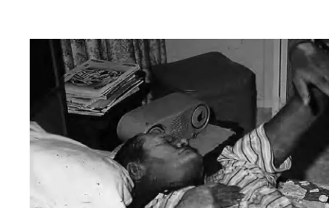
Sudden deaths can be instantaneous; sudden but not instantaneous, or cases where the individual is found dead. Most people, when talking about sudden death, envision instantaneous deaths. The best illustration of this is an individual walking along who suddenly collapses and is dead upon hitting the ground. The most common cause for this is a ventricular arrhythmia due to coronary artery disease. The individual will often show impact abrasions
of the face, indicating that as he was going down, he was unconscious and was not even able to put his arm up in front of his face to prevent impacting the ground (Figure 1.1).
The sudden, but not instantaneous, death is illustrated by the individual who begins to complain of chest pain, difficulty in breathing, weakness, sweating, nausea, and vomiting, and then collapses. He is then transported to the hospital. On the way to the hospital, he goes into cardiac arrest and by the time he reaches the emergency room he is not resuscitatable. Another individual with the same initial symptoms may arrive conscious at the hos-pital only to experience his fatal cardiac arrhythmia 2 h after admission. Is this still a sudden death? This depends upon one’s definition of sudden death. Many, if not most, medical examiners limit classification of sudden deaths as those occurring instantaneously or within 1 h of the onset of symptoms.
Table 1.1 Breakdown of Medical Examiner Cases as to Manner of Death: Bexar County Texas (1983 –1998)
If the individual complaining of chest pain and difficulty breathing survives long enough to get to the emergency room of a hospital, where an EKG shows an acute myocardial infarct and laboratory tests reveal elevated enzymes, then a diagnosis of myocardial infarct can be made and the case is not a medical examiner’s case.
[image:28.666.109.291.49.314.2]There is a third category of sudden unexpected deaths. These are the individuals in whom the death was unexpected, but was found dead in what may or may not have been an instantaneous manner. Sometimes, one can tell how rapid the death was by the how the individual was found. Someone found sprawled on the kitchen floor with impact-type abrasions of the face is most likely an instantaneous death. In the case of a person found dead in bed, death may have been sudden but not instantaneous. The great majority of sudden, unexpected natural deaths seen at a medical examiner’s office are due to cardiovascular disease. Less common are deaths due to central nervous system lesions, pulmonary disease, and sepsis. The whole spectrum of natural disease associated with sudden death is discussed in Chapter 3.
The Coroner System
There are two general types of medicolegal investigative systems in the United States: coroner systems and medical examiner systems. As of 2000, 12 states had coroner systems; 19 states had state medical examiner systems; 3 states had county or regional medical examiner’s offices but no coroner’s offices; and 16 had a mixture of medical examiner and coroner systems.2 Over the years, there has been a gradual decrease in the number of coroner systems, with replacement by medical examiner systems, though this seems to have slowed down recently. Coroner systems, however, still make up a significant proportion of the medicolegal coverage of the American population.
The coroner system, dating back to feudal England, is the older of the two medicolegal systems. The earliest reference appears in the Articles of Eyre (1194).3 In the pure form of this system, an individual who is not a physician is elected the coroner. He then makes rulings as to the cause and manner of death in cases that fall under the coroner law. As a general rule, these cases constitute violent deaths, sudden and/or unexpected deaths, suspicious deaths, and cases in which a physician is not in attendance at the time of death. In making a ruling, the coroner is not required to consult a physician for advice, may or may not order an autopsy, and may or may not rule in agreement with autopsy findings if one is performed. The training the cor-oner receives for the position can range from absolutely none to a few hours or to 1–2 weeks. Based on this training — or lack of it — the coroner makes decisions as to cause and manner of death that may have significant criminal and civil consequences.
In some areas of the country, this system has been modified such that the coroner must be a physician, though not necessarily a pathologist. This gives a scientific veneer to the system. We now have physicians making decisions in a medical field usually having absolutely nothing to do with their areas of expertise. Thus, we have the obstetrician-coroner, the general prac-titioner-coroner, and so on. Occasionally, by chance, the coroner is a pathol-ogist, though almost never a forensic pathologist.
works for a governmental organization that either does not care or know about qualifications for this work.
California often exemplifies the extremes of this country. Thus, in a number of counties in California, the coroner is also the sheriff. Thus, a deputy sheriff might kill a civilian and his boss, the sheriff, rules as to the cause and manner of death. Sheriffs also operate jails. The sheriff acting as the coroner thus makes rulings as to the cause of death of inmates dying in his jail. Obviously, to anyone but the California legislature, there is a conflict between having a single organization whose duties are to both enforce laws and make arrests and to conduct objective investigation of death in which rulings may impeach or conflict with the other half of the organization.
In many areas of the country, the coroner is also a funeral director. Here, again, there is at least the appearance of a conflict of interest. The coroner-funeral director makes a livelihood by conducting coroner-funerals, not by being a coroner. Some unscrupulous coroners are more interested in obtaining a family’s permission to conduct the funeral than to make a ruling as to cause and manner of death. They may take great care not to make a ruling as to cause and manner of death that might offend a family and thus cost them business or potential votes in the next election.
The coroner system was developed at a time when the lay public knew as much about the science of medicine as the physicians practicing it. Times have changed. Medicine has become an extremely complicated, specialized, scientific field. Specialized knowledge is necessary not only to practice med-icine in general, but to practice any of its numerous subspecialties. Thus, a dermatologist would not consider doing neurosurgery, nor would a neuro-surgeon practice obstetrics/gynecology. The practice of forensic medicine has also become a specialty. Neither average hospital pathologists nor physicians who are not pathologists can adequately practice in this field no matter how well intentioned they are — and they are often very well intentioned.
Some non-forensic pathologists claim that any anatomical pathologist with a basic knowledge of pathology can handle 85% of medical examiner cases, with the remaining 15% needing a forensic pathologist in a fully equipped medicolegal facility. Therefore, only a small corps of experi-enced forensic pathologists is needed, to which the difficult 15% of cases can be referred.
suicide in a garage eventually ended up with a million-dollar lawsuit and involved the flying characteristics of light planes. A simple case of sudden death in infancy ended up as the final death in a decades-long chain of infanticide.
The Medical Examiner System
The medical examiner system was first introduced in the United States in 1877 in Massachusetts. The state was divided into a number of sectors, within each of which was designated a physician who functioned as a “medical examiner” to determine the cause and manner of death. Originally, medical examiners did not have the right to order autopsies. This was not corrected until the 1940s. Neither was a central laboratory for toxicological analysis available. It was only in the 1980s that a true State Medical Examiner System was established in Massachusetts.
The first true medical examiner system came into existence in 1918 in New York City.4 A medicolegal system was established in which the individual designated as Chief Medical Examiner was to be a physician experienced in the field of pathology (forensic pathology did not become a subspecialty until 1959). The system described the type of cases that fell under the Medical Examiner Law; it stated that the medical examiner could perform autopsies in cases that he felt needed them, and it established a laboratory for his use. The cases that fell under the medical examiner system were violent deaths (accidents, suicides, homicides), suspicious deaths, sudden, unexpected deaths, and deaths occurring without the attendance of a physician. Most medical examiner systems in this country are variations of the original New York concept. Some of the newer systems specify that the chief medical examiner must be a forensic pathologist.
The creation of a medical examiner system does not necessarily mean that a community actually has a functioning or effective medical examiner system, nor does the fact that it once had one guarantee that it will continue to operate in an effective manner. Thus, by the mid 1980s, the New York City Medical Examiner System had been seriously damaged in its functioning by a change in the law that allowed families to prevent the performance of autopsies in cases in which the manner of death did not appear to be homi-cide; that is, the forensic pathologist had authority to perform an autopsy only in cases that were obviously homicide.
produced by them, and knowledge of whether there was any pain or suffering involved in an injury (an important question in civil cases) become sheer speculation.
Some legislatures have created medical examiner’s offices and have not funded them adequately. In other instances, the offices are placed under state government agencies that should not be supervising the medical examiner’s office. No medical examiner’s office should function under a police agency. There is a direct conflict in values, goals, and philosophies. The police want to make an arrest and clear a case. The medical examiner’s office wants to determine the cause and manner of death, independent of who did what. While these functions usually coincide, in some cases, they do not. One of the most controversial types of death is that of a civilian killed by police. By virtue of being a subdivision of a police agency, the impartiality of the medical examiner’s office in such cases is open to serious question.
In some areas, the medical examiner’s office functions under the public health department. This may or may not work out. Public health departments often have only a vague concept of the duties and functions of a medical examiner’s office, which is a medicolegal agency rather than a pure medical agency. The contribution of the medical examiner’s office to the public health is of a tenuous nature. Placing it under a department of public health tends to increase the bureaucracy between the office and the authority to which it is responsible. In addition, public health departments are often under funded and there is always the human tendency to dip into one section of a depart-ment for monies for another section. Just as police agencies should be sep-arate from the district attorney’s office, so, ideally, the medical examiner’s office should be reportable to only the highest authority, for example, the mayor, county commissioners, or governor.
one that they often do not even recognize as difficult, can result in the imprisonment of innocent individuals and the release of the guilty. Just as we are guaranteed certain basic rights by our legal system, we should also have the right to a competent scientific medical investigation following a death, especially if there is the potential for civil or criminal litigation.
Some coroners argue that they can and do produce excellent work because they retain competent physicians and rely on them to make decisions. This is true. But, if coroners rely that much on their physicians, then what purposes do the coroners serve? They are just elected administrators subject to political influence who, if they desire, can ignore their experts. In addition, a conscientious, competent coroner may be defeated at the next election to be replaced by a venial incompetent.
A modern, well-organized medical examiner system is relatively cheap to operate, considering its benefits once the population serviced exceeds approximately 250,000. Cost to the community for a good system is approx-imately one quarter to one third the cost of a movie ticket per person per year. This money will fund a fully staffed medical examiner’s office including an investigative staff and a toxicology laboratory. Some states or counties claim to be too big, with a population too scattered, for a state system. This can be handled by establishing either regional offices or a central state office to which bodies can be transported.
The main problem with establishing a quality medical examiner system is ignorance — ignorance not only by the general public, but, more impor-tantly, by courts, judges, and attorneys. The courts permit untrained and sometimes incompetent individuals to routinely testify on the basis of a medical degree often coupled with vague forensic experience. While no judge would take his pregnant wife to a dermatologist for obstetrical care, he does permit an individual with no forensic training at all to testify in a case that may involve a sentence of lengthy imprisonment or even execution.
There is ignorance also on the part of politicians. They have very little idea of what a medical examiner’s office does, do not visit the facilities, and show very little interest in the office — after all, the dead do not vote. The only time one hears from politicians is when there are lawsuits against the government because of incompetence in the medicolegal system. The public is often ignorant of the poor quality of the medicolegal system in their area because they assume what they see on television crime shows is also true in their own community.
characteristics of the unqualified expert in forensic pathology is an ability to interpret a case in exquisite detail. This “expert” sets the time of death, plus or minus a few minutes, accurately positions the deceased, and gives detailed analysis of the events surrounding the death and precise deductions about the assault. If the police have expressed prior opinions, it is not uncommon for the opinions of the “expert” to agree in almost complete detail with the police hypotheses. The experienced forensic pathologist tends to hedge, knows there can be more than one interpretation of a set of facts, and is more “wishy-washy” than the charlatan.
Because of the poor quality of forensic medicine in many parts of this country, there are individuals languishing in jail for homicides that were suicides and murderers walking the street after having committed a homicide that was interpreted as an accident or a natural death.
Operation of a Medical Examiner System
To perform its duties, a medical examiner system requires a number of basic essentials. First is an adequate law under which to operate. Under such a law, violent deaths (accidents, suicides, and homicides), suspicious deaths, sudden and unexpected deaths, deaths without a physician in atten-dance, and deaths in jails and penal institutions should fall under the medical examiner’s jurisdiction. In many areas, there is also a broad “24-hour death” report law. That is, the death of any individual dying within 24 h of admission to a hospital must be reported to the medical examiner as a possible medical examiner case. This law is useful in picking up cases that might otherwise be missed.
After indicating the type of cases that fall under the medical examiner’s jurisdiction, the law should then state that medical examiners have the right to perform an autopsy on any cases that they feel need one to accurately determine the cause and manner of death or to document injuries or disease processes. The law should also give medical examiners the right to subpoena records and individuals, if necessary, to help in making such determinations. The law should state that deaths are to be reported to medical examiners immediately after they occur or are discovered and that, at any crime scene, medical examiners have jurisdiction over the body. Overall jurisdiction of the scene, of course, lies with the police agency involved. The law should also provide medical examiners with a toxicology laboratory.
children dying of blunt traumatic injuries show no evidence of injury exter-nally. If one could not perform an autopsy in such cases, these homicides would be missed and death ascribed to natural causes. Obvious homicide cases are less of a problem than the more subtle ones that initially appear to be natural deaths or accidents.
It is also highly desirable that the medical examiner have some civil service protection. This is because medical examiners make unpopular deci-sions, decisions that politicians, police agencies, and sometimes the public may not want to hear. There is always the natural tendency of humanity to want to “kill” the bearer of bad news.
The second requirement for an adequate medical examiner system is qualified personnel. The chief medical examiner should be a board certified forensic pathologist with a number of years of experience. Under the chief medical examiner, there should be assistant medical examiners who are also board-certified forensic pathologists. If initially not certified, the indi-viduals should be given a certain time limit (2–3 years) to obtain certifi-cation. To acquire and retain such qualified personnel, they must command competitive salaries.
What is a board certified forensic pathologist? A board certified forensic pathologist is a physician who has successfully completed a graduate medical education program in either anatomical or anatomical and clinical pathology approved by the Residency Review Committee and accredited by the Accred-itation Council for Graduate Medical Education (ACGME) or The Royal College of Physicians and Surgeons of Canada; been endorsed by the training program director and successfully passed a written and practical examination designed and administered by the American Board of Pathology in this (these) field(s) of medicine, following which they have taken 1 full year of additional supervised training in forensic pathology in a program accredited for such training by the ACGME, and passed a written and practical exam-ination designed and administered by the American Board of Pathology in this field.
Third, the medical examiner’s office needs adequate staffing. Medical examiners alone do not constitute an office. There must be competent inves-tigative, administrative, secretarial, and technical support staff.
Fourth, there must be an adequate facility. One cannot practice forensic medicine in the sub-basement of a county hospital or in the back of a funeral home. The facility must have sufficient space, an appropriate floor plan, electrical, plumbing, and cooling capabilities, and furnishings.
presence of drugs. The equipment should be of high quality and be in suf-ficient quantity to handle the caseload. Computerization of an office has now become mandatory.
Last, there must be consistent and adequate funding of the institution. Without this, a qualified staff is not possible, nor are adequate facilities or equipment.
How do medical examiners (forensic pathologists) approach a case? They approach it just like any other physician approaches a patient. In medical school, one is taught that, to make a correct diagnosis, one must take a history, perform a physical examination, and order relevant laboratory tests. Based on this, a diagnosis is made. The forensic pathologist performs all of these functions but with some variance. Thus, the history is not obtained from the patient, but from witnesses, relatives of the deceased, police agen-cies, treating physicians, and/or records (medical, nonmedical, police, gov-ernmental, etc.). It is an account of the events leading up to and surrounding the death.
In most major medical examiner systems in this country, reports of deaths do not come directly to medical examiners, but to lay investigators employed by the medical examiner’s office, who are trained to screen the cases and make a determination as to whether a death is a medical examiner’s case. If it is not, it is then released back to the reporting physician. If the case falls under the Medical Examiner’s Law or if there is no physician to sign the death certificate, the case is accepted. Whether a case is accepted or not, a detailed report should be written up. In cases that are not accepted, the report should be subsequently reviewed by a medical examiner as soon as possible. If there is any disagreement with the conclusion of the investigator, the body will then be brought in from the funeral home to which it was sent. With well-trained, highly motivated investigators, this is an extremely rare occurrence.
The reason that physicians are not used to screen calls is both economical and logistic. In a community of a million people, one might get anywhere from 4000 to 6000 death calls a year. To have a physician personally screen each one of these calls — calling other physicians, double-checking records, talking to police agencies, and going to the scene of death — is uneconomical and a waste of professional time. The U.S Department of Justice has published national guidelines for death investigation.5 In addition, there is now in exist-ence the American Board of Medicolegal Death Investigators, which certifies death investigators based on a combination of experience and testing.6
to obtain a detailed history of the circumstances leading up to and surround-ing the death. Investigators will document the scene with diagrams and photographs. In some communities, investigators videotape the scene. This material is then brought back to the office, where a detailed report is prepared for the medical examiner. At this time, the investigation report may be supplemented by telephone calls to other agencies and individuals. The lay investigator goes to hospitals to obtain medical records that may be of impor-tance in making a determination of the cause of death. Based on the inves-tigator’s report, the medical examiner will then decide what to do with the case, whether to perform an autopsy, as well as what types of tests are to be performed.
In some jurisdictions, physicians routinely make all scene investigations or at least all violent death scenes. While this is satisfactory in small, low-volume offices, it is generally a waste of personnel, time, and money in large urban areas. In some areas of the country, a physician who visits a scene has the right to sign out the case without bringing it into the medical examiner’s office. This practice should be condemned. There is no way to adequately examine a body at a scene as well as in a morgue. If a body is a medical examiner’s case, it should come into the office, where, at the minimum, a complete external examination can be performed in an environment with adequate lighting, equipment, and support personnel. In addition, at the same time, body fluids can be obtained for toxicological analysis. If it is a medical examiner’s case, it should come in. If it is not a medical examiner’s case, then the medical examiner should refuse jurisdiction and let the patient’s attending physician handle the case. When in doubt, however, bring the case in.
When the body comes in, the medical examiner then performs a physical examination and laboratory tests. The procedures may range from an external examination of the body to a complete autopsy. This will be determined by the information that has been provided to the medical examiner by the investigator, the type of case, the medical examiner’s expertise, and local or regional differences. Performing autopsies on every case coming into a med-ical examiner’s office is a waste of time and energy. The practice is common in areas where there are contract pathologists paid by the case. Thus, the more cases they autopsy, the more money they make.
screens on apparent natural deaths have, with regular monotony, revealed deaths from suicidal and accidental overdoses of drugs. Such extensive screen-ing is not possible in most areas of the country because of limitations on the toxicology laboratory.
If an autopsy is performed, what specialized tests are done at the time of autopsy are determined by the type of case. Thus, a rape examination may be conducted in a suspected rape case, or hair obtained in a death with blunt-force injuries to the head.
For all cases coming to autopsy, it is recommended that, at the minimum, blood, vitreous, urine, and bile be obtained for toxicological analysis. Blood should be obtained from the femoral vessels. If this is not possible, other sites, in descending order of preference, are the subclavian vessels, the root of the aorta, the pulmonary artery, the superior vena cava and the heart. The blood should be collected with a clean needle and new syringe. Blood should never be obtained by incising a vessel or the heart and attempting to capture the fluid as it escapes. All body fluids should be placed in glass tubes or bottles, not plastic. If the body is decomposed, liver, kidney, and thigh muscle are retained. When suicidal overdose of oral medications is suspected, the stomach contents should be kept. Some laboratories retain portions of liver and kidney in all suspected drug overdose deaths — whether decomposed or not. With modern instrumentation and analytical methods, however, it is rarely necessary to analyze these materials.
The urine is generally of use only in screening for certain drugs. The detection of a drug in the urine indicates only that the individual has taken that drug at some time in the past, not that they were under its influence at time of death. It is the presence of the drug in the blood that is important. Because of this, toxicological procedures should be concentrated on the blood. Absence of a drug in the urine also does not necessarily indicate that it will not be found in the blood. Thus, an intravenous injection of heroin could cause death before any metabolites appear in the urine.
At the time of autopsy, tissue should be retained for possible micro-scopic examination, although this is not necessary in every case. Thus, in traumatic deaths, such as a shooting or motor vehicle accident, while a medical examiner may elect to do microscopic examination of the tissue, it is rarely necessary. Even if microscopic slides are not made, tissue should still be retained for this possibility. It is the opinion of the authors that toxicologic specimens and tissue removed for possible microscopic exam-ination should be retained for 3–5 years. All microscopic slides and paraffin blocks should be retained indefinitely.
NAME Accreditation
In 1997, the National Association of Medical Examiners (NAME) insti-tuted a revised voluntary inspection and accreditation program for medi-colegal offices. The new program is much more stringent than the prior program. The standards represent minimum standards for an adequate medicolegal system, emphasizing policies and procedures. Deficiencies are designated as Phase I or II. A single Phase II deficiency precludes accredita-tion. Evaluated are:
The facilities
Safety policies, procedures and equipment Personnel
Notification, acceptance and release Investigations
Body handling
Postmortem examinations Identification
Evidence and specimen collection Support services
Reports and records Mass disaster plan Quality assurance
One area addressed is medical examiner caseload. If a medical examiner performs more than 250 autopsies per year, this is considered a Phase I deficiency; if more than 400, a Phase II deficiency (there are plans to lower the 400 number to 350 and possibly 300).
Excessive caseload is a problem in many medicolegal offices. The rec-ommended annual caseload for a forensic pathologist without administra-tive responsibilities is 250 autopsies. On a short-time basis, one can perform autopsies at an annual rate of 300, perhaps 325. By the time, caseload exceeds 350 autopsies, mistakes are being made and the quality of the autopsy is being sacrificed.
References
2. Hanzlick R and Combs D: Medical examiner and coroner systems: history and trends (special communication). JAMA 1998 279(11): 870-874. 3. Mant AK: The evaluation of the coroner’s system and the present status in
Great Britain. Forensic Sci Gazette 1971; 2(4):1-6.
4. The Office of the Medical Examiner of the City of New York: Report by the Committee on Public Health, New York Academy of Medicine. Bull NYAcad Med 1967. 43: 241-249.
5. National Medicolegal Review Panel: Death Investigation: A Guide for the Scene Inv e s t i g a t o r. Na t i o n a l I n s t i t u t e o f Ju s t i c e , No ve m b e r 1 9 9 9 . (http://www.ncjrs.org/txtfiles/167568.txt)
Time of Death
Determination of the time of death is important in both criminal and civil cases. In criminal cases, it can set the time of the murder, eliminate or suggest suspects, confirm or disprove an alibi. In civil cases, the time of death might determine who inherits property or whether an insurance policy was in force. Unfortunately, all methods now in use to determine the time of death are to a degree unreliable and inaccurate. They usually give vague or dubious answers. The longer the postmortem interval, i.e., the time between death and the attempt to determine time of death, the less precise the estimate of the interval. One obvious facet of time of death determination often not considered is that the time the fatal injury is incurred is not necessarily the time of death. One can incur massive fatal injuries, yet linger in an unconscious state for hours prior to death (Figure 2.1). Many factors are or have been used in determining the time of death:
Livor mortis Rigor mortis Body temperature Degree of decomposition Chemical changes in vitreous Flow-cytometry
Stomach contents Insect activity
Scene markers (papers, letters, clothing, televisions, TV schedules, etc.)
Livor Mortis
Livor mortis (lividity, postmortem hypostasis) is a reddish purple coloration in dependent areas of the body due to accumulation of blood in the small vessels of the dependent areas secondary to gravity (Figure 2.2A). Postmor-tem lividity is occasionally misinterpreted as bruising by people unfamiliar with this phenomenon.
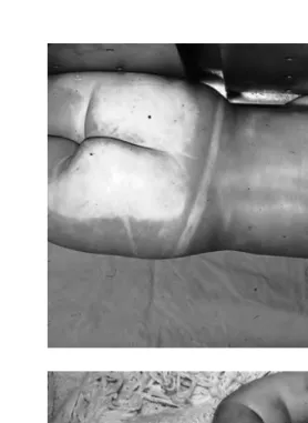
Dependent areas resting against a firm surface will appear pale in contrast to the surrounding livor mortis due to compression of the vessels in this area, which prevents the accumulation of blood. Thus, areas sup-porting the weight of the body, for example, the shoulder blades, buttocks,
and calves in individuals lying on their backs, show no livor mortis, but appear as pale or blanched areas (Figure 2.2B, C). Tight clothing, for example, a brassiere, corset, or belt, which compresses soft tissues, collapsing the vessels, also produces pale areas.
Livor mortis usually, but not invariably, has a cherry-red to pinkish color in deaths due to carbon monoxide. This is due to carboxyhemoglobin. Iden-tical coloration may be caused by exposure of a body to cold temperatures, and in deaths due to cyanide.Localized areas of bright red livor mortis are Figure 2.1 Contact wound of right temple with .357 Magnum. The deceased lived 1 hr and 34 min with no life support systems.
also seen adjacent to chest tubes. In all three of the aforementioned entities, the coloration is caused by predominance of oxygenated hemoglobin.
[image:43.666.83.362.71.453.2]Livor mortis is usually evident within 30 min to 2 h after death. In indi-viduals dying a slow lingering death with terminal cardiac failure, livor mortis may actually appear antemortem. Livor mortis develops gradually, usually reaching its maximum coloration at 8–12 h. At about this time, it is said to become “fixed.” Prior to becoming fixed, livor mortis will shift as the body is moved. Thus, if an individual dies lying on his back, livor mortis develops posteriorly, i.e., on the back. If one turns the body on its face, blood will Figure 2.2 (continued) (B) Blanched areas of buttocks and shoulders due to compression of vessels by weight of body. (C) Infant with pale face from lying face down in crib.
B
drain to the anterior surface of the body, now the dependent aspect. Livor mortis becomes “fixed” when shifting or drainage of blood no longer occurs, or when blood leaks out of the vessels into the surrounding soft tissue due to hemolysis and breakdown of the vessels. Fixation can occur before 8–12 h if decomposition is accelerated, or at 24–36 h if delayed by cool tempera-tures. Thus, the statement that livor mortis becomes fixed at 8–12 h is really just a vague generalization. That livor mortis is not fixed can be demonstrated by applying pressure to a dependent discolored area and noting the subse-quent blanching at the point of pressure.
Although livor mortis may be confused with bruising, bruising is rarely confused with livor mortis. Application of pressure to an area of bruising will not cause blanching. An incision into an area of contusion or bruising shows diffuse hemorrhage into the soft tissue. In contrast, an incision into an area of livor mortis reveals the blood to be confined to vessels, without blood in the soft tissue.
Livor mortis also occurs internally, with settling of the blood in the depen-dent aspects of an organ. This is most obvious in the lungs.
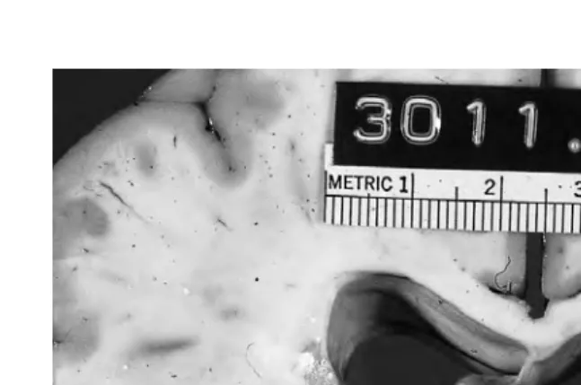
As the blood accumulates in the dependent areas, the pressure of the settling blood can rupture small vessels, with development of petechiae (minute hemorrhages or Tardieu spots) and purpura (patches of purplish discoloration) (Figure 2.3). This usually takes 18–24 h and often indicates that decomposition is fast approaching. This phenomenon is more common in asphyxial or slow deaths. Unfortunately, as time passes, it cannot always be determined with certainty whether the purpura produced are ante- or postmortem. Presence of petechiae and purpura only in dependent areas suggests a postmortem origin. In limbs hanging over the side of a bed or the legs and forearms of an individual who is hanging, Tardieu spots may develop even more rapidly, appearing as early as 2–4 h after death.
diffuse scalp hemorrhage. Rarely, postmortem leakage of blood into the soft tissue and muscle of the anterior aspect of the neck may also occur in drownings. This “bleeding” is minimal.