University of Warwick institutional repository:
http://go.warwick.ac.uk/wrap
A Thesis Submitted for the Degree of PhD at the University of Warwick
http://go.warwick.ac.uk/wrap/73513
This thesis is made available online and is protected by original copyright.
Please scroll down to view the document itself.
The Expression and Function of the
Adenovirus Type 5 E4 region
Ian Dix
Bsc. (Hons.) (Warwick)
A thesis presented for
the degree of PhD in the
Universtiy of Warwick
Department of Biological Sciences
.
.• I."
PAGE'
.
NUMBERING
.
\
AS ORIGINAL
. .~ .
,
Table of contents
Table of contents
List of figures
List of tables
Acknowledgements Declaration Abbreviations Summary 1 lX Xlll XlV xv XVI xx
Chapter 1. Introduction
1.1. Adenoviruses- a general introduction
1.1.1. The adenovirus family
1.1.2. The structure of adenoviruses
1.1.3. Adenovirus replication
1.1.4. Adenovirus gene function
1.1.4.1. The Ela region
1.1.4.2. The Elb region
1.1.4.3. The E2 region
1.1.4.4. The E3 region
1.1.4.5. The E4 region
1.1.4.6. Late transcription units
1.1.4.7. VARNA
1.2. The regulation of gene expression in an adenovirus infection
1.2.1. Introduction
1.2.2. Transcription initiation
1.2.3. mRNA polyadenylation
1.2.4. RNA splicing
1.2.4.1. Introduction
1.2.4.2. Mechanism 1: viral splicing factors
1.2.4.3. Mechanism 2: cis-acting sequences
1.2.4.4. Mechanism 3: general splicing factors
1.2.4.5. Summary
1.2.5. mRNA export
1.2.6. Translation
1.2.6.1. Introduction
23
24
26
26
27
27
28
29
29
30
30
31
35
37
40
44
44
47
47
4850
57
57
60
60
63
1.2.6.2. VAl RNA
1.2.6.3. L4-100K
1.2.6.4. The MLTU tripartite leader
1.3. Elb-55K and cytoplasmic viral mRNA accumulation
1.3.1. Introduction
1.3.2. RNA transport models
1.3.3. The Elb-55K protein - subcellular localisation
1.3.4. The elucidation ofElb-55K
function
1.3.5.
Amodel for Elb-55K function
1.4. The E4 region
1.4.1. E4 expression
1.4.2. The functions of the E4 proteins
1.4.2.1. Introduction
1.4.2.2. ORFI and ORF2
1.4.2.3. ORF3 and ORF6
1.4.2.4. ORF3/4
1.4.2.5. ORF4
1.4.2.6. ORF5
1.4.2.7. ORF617
Chapter 2. Materials and Methods
2.1. Materials
2.1.1. Common buffers and solutions
2.1.2. Oligonucleotide primers
2.2. Suppliers
2.3. Methods
2.3.1. Nucleic acid manipulations
2.3. 1.1. Ethanol precipitation of nucleic acids
2.3.1.2. Nucleic acid quantification
2.3.1.3. Phenol/chloroform extraction
2.3.1.4. Restriction endonuclease digestion
2.3.1.5. End-fill repair of5' overhangs
2.3.1.6. T4 DNA polymerase removal of3' overhangs
2.3.1. 7. Dephosphorylation
2.3.1.8. Agarose gel electrophoresis
2.3.1.9. Purification of dsDNA fragments from agarose gels
2.3.1.10. Ligation ofdsDNA
2.3.1.11. Polymerase chain reaction (PCR)
2.3.1.12. Site-directed mutagenesis using PCR
2.3.1.13. DNA sequencing
2.3.2. Bacteriological techniques
2.3.2.1. The growth and maintenance of bacteria required for
64 65 65 65 66 68 68 68 68 68 69 69 69 70 70 70 71 72 72 74 76
routine cloning 76
2.3.2.2. The transformation of bacteria with plasmid DNA 76
2.3.2.3. Small scale plasmid DNA purification from bacteria (Mini-Prep) 78
2.3.2.4. Large scale plasmid DNA preparation from bacteria (Maxi-Prep) 78
2.3.3. M13 Phage techniques 79
79 2.3.3.1. Transformation procedure
2.3.3.2. Culturing M13 clones 80
2.3.3.3. Preparation ofRF DNA from M13 cultures 80
2.3.3.4. Preparation of single stranded DNA from M13 plaques 80
2.3.4. Tissue culture and virus techniques 81
2.3.4.1. Maintenance and passage of cell lines 81
2.3.4.2. Maintenance and passage of non-transformed cells 81
2.3.4.3. Freezing cell stocks 82
2.3.4.4. Recovery of cell stocks 82
2.3.4.5. Calcium phosphate transfection for the construction of
adenovirus mutants
2.3.4.6. Virus infection of cell monolayers
2.3.4.7. Rapid preparation of adenovirus DNA
2.3.4.8. Large scale preparation of adenovirus DNA
2.3.4.9. Preparation of virus stocks
2.3.4.10. Preparation of purified adenovirus virions
2.3.4.11. Titration of adenovirus by plaque assay
2.3.5. Analysis of viral RNA expression
2.3.5.1. Preparation of cytoplasmic and nuclear RNA
2.3.5.2. Preparation of antisense RNA probes
2.3.5.3. Ribonuclease protection analysis
2.3.5.4. RNA polyacrylamide gel electrophoresis (PAGE)
2.3.6. Analysis of proteins
2.3.6.1. SDS polyacrylamide gel electrophoresis (SDS-PAGE)
2.3.6.2. Coomassie-blue staining ofSDS-polyacrylamide gels
2.3.6.3. Monochromatic silver staining
2.3.6.4. Preparation ofSDS-polyacrylamide gels for autoradiography
2.3.6.5. Bradford assay
2.3.7. Bacterial protein expression
2.3.7.1. Protein expression and purification using the GST system
2.3.7.2. Inclusion body purification ofpGEX fusion proteins
2.3.7.3. Protein expression and purificationusing the histidine tag
~~ ~
2.3.8. Production of mono-specific polyclonal antibodies
93
2.3.9. Immunochemicaltechniques for the detection of proteins
94
2.3.9.1. Westernblotanalysis
94
2.3.9.2. Immunoprecipitation of proteins using polyclonal antibodies
95
2.3.9.3. Enzyme linked immunoadsorbent assay (ELISA)
96
2.3.10. Analysisof viral protein expression
97
2.3.10. 1. 35S-methioninelabelling of proteins in tissue culture
97
2.3.10.2. Quantification of 35S-methionine incorporation into protein
97
2.3.10.3. Subcellularfractionation using hypotonic buffer
98
2.3.10.4. Sucrose gradient analysis
98
2.3.11. In vitro
protein expression
99
2.3.11.1 In vitro
protein expression
99
2.3.11.2. Quantitation of protein produced using rabbit reticulocyte
~~e
~
2.3.12. Autoradiography
99
2.3.13. Densitometry
100
Chapter 3. The Sequence ofORFl
and 2 of Ad5 early region 4
101
3.1. Introduction
102
3.2. Cloning of the AdS DNA to be sequenced
102
3.3. Results
103
3.4. Discussion
103
3.5. Computer prediction and homology studies ofORFI and ORF2
107
3.5. 1. Introduction
107
3.5.2. ORFI structure predictions
107
3.5.3. ORFI homology searches
112
3.5.5. ORF2 homology searches
116
3.5.6. Conclusion
118
Chapter 4. The expression of the Ad5 E4 mRNAs
119
4.1. Introduction
120
4.2. Methods
122
4.2.1. Cells and viruses
122
4.2.2. The antisense RNA probes
124
4.3. Results
126
4.3.1. The temporal classes ofE4 mRNA
126
4.3.2. The D3 to A3 intron is excised late in the infection
129
4.3.3. Relative abundance of the E4 mRNAs
129
4.3.4. Late E4 mRNAs are dependent on DNA replication for accumulation
132
4.3.5. Dependence oflate E4 mRNA expression on the Elb-55K protein
132
4.3.6. E4 mRNA Nfulliength mRNA is dependent on E4 ORF6-34K for
optimal cytoplasmic accumulation
136
4.3.7. E4 mRNA Nfulliength mRNA dependence on Elb-55K is at a
post-processing level
4.4. Discussion
4.4.1. Classes ofE4 mRNA
4.4.2. The expression of the E4 ORF products
4.4.3. Regulation of the expression ofmRNA A and/or full length mRNA
Chapter 5. The construction and analysis of a virus containing a
mutated E4
Dl
donor site
5.1. Introduction
5.2. Experimental strategy
5.3. Construction of the Dl and ORF6 mutant virus
5.4. AnalysisofmRNA Alfulllength mRNA expression from the vDl virus
136
139
139
142
144
146
Chapter
6.
The generation of a polyclonal antisera to E4ORFI
1576.1. Introduction 158
6.2. Viruses and cells used 158
6.3. Expression of the ORFI protein using the pGEX vector 159
6.4. The generation of an anti-ORFI antiserum using the
GST/ORFI
168inclusion body preparation
6.5. The expression ofORFl using the XPRESS expression system 170
6.6. The expression ofORFl using the QIAexpress expression system 174
6.7. Attempts to detect ORFI in infected cells 176
6.8. Discussion 185
Chapter
7.
The E4ORF2
protein7.1. Introduction
7.2. Viruses and cells used
7.3. Expression of the ORF2 protein using the pGEX vector
7.4. The generation of an anti-ORF2 antiserum using the
GST/ORF2
fusionprotein 197
189
190
190
190
7.5. Expression ofE4 ORF2 during a lytic infection 200
7.5.1. The ORF2 protein exists in infected cells 200
7.5.2. The time course of expression of the ORF2 protein 200
7.5.3. Subcellular localisation of the ORF2 protein 204
7.5.4. Interactions of the ORF2 protein with other infected cell components 206
7.6. Discussion 209
Chapter
8.
The function of theORFI
andORF2
proteins 2138.1. Introduction 214
8.2. The growth of adenovirus in non-transformed cells 215
8.4. E4 ORFI and ORF2 mutant virions
8.5. Conclusion
220
222
Chapter 9. General discussion
9.1. Summary and interpretation of the results
9.2. Future work
223
224
227
List of figures
Fig. I. I. Schematic representation of the adenovirus virion 4
Fig. 1.2. Transcription and translation map of adenovirus type 2 9
Fig. 1.3. The Ela region 11
Fig. 1.4. The predicted AdS Elb products 13
Fig.
1.5.
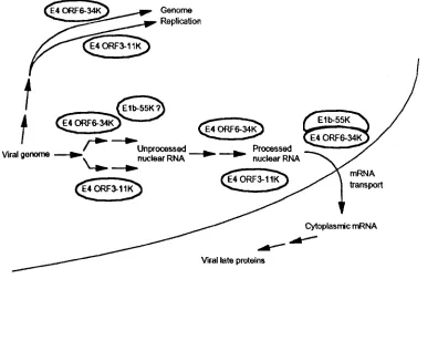
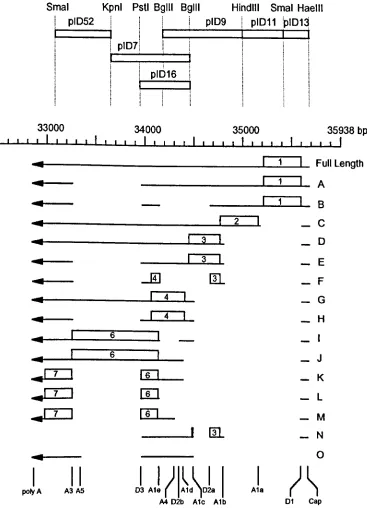
The observed mRNA species from the E4 region with encoded ORFs 46Fig. 1.6. The functions of the E4 ORF3-11K, E4 ORF6-34K and the
Elb-SSK proteins in the lytic viral cycle 58
Fig.2.1. Diagram depicting site directed mutagenesis using PCR 73
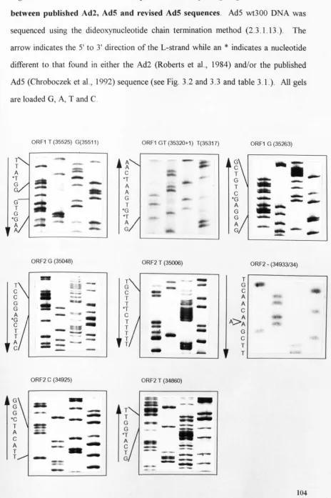
Fig.3.1. ORFI and ORF2 sequence data depicting the regions of variation
between the published Ad2, AdS and revised AdS sequences 104
Fig.3.2. Comparison of the revised sequence of AdS E4 ORFI with AdS
(Chroboczek et al., 1992) and Ad2 (Roberts et al., 1984) 105
Fig.3.3. Comparison of the revised sequence of AdS E4 ORF2 with AdS
(Chroboczek et al., 1992) and Ad2 (Roberts et al., 1984) 105
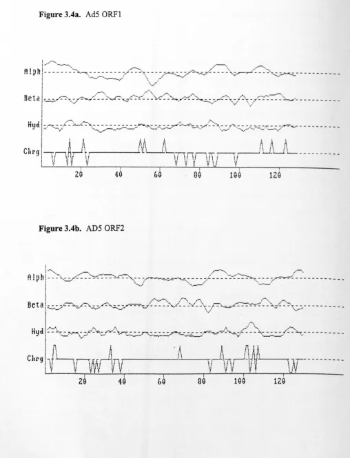
Fig.3.4. The structure of AdS ORF1 and ORF2 108
Fig.3.S. Alignment of the predicted AdS, Ad9, Ad12 and Ad34 E4 ORF1
protein sequences 110
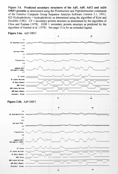
Fig.3.6. Predicted secondary structures of the AdS, Ad9, Ad12 and Ad34 E4
ORFI proteins 111
Fig.3.7. Sequence alignment over 119 amino acids of AdS ORFI and the
Sacchromyces cerevisiae
dUTPase 113Fig.3.8. Alignment of the predicted AdS, Ad9, Adl2 and Ad40 E4 ORF2
protein sequences 115
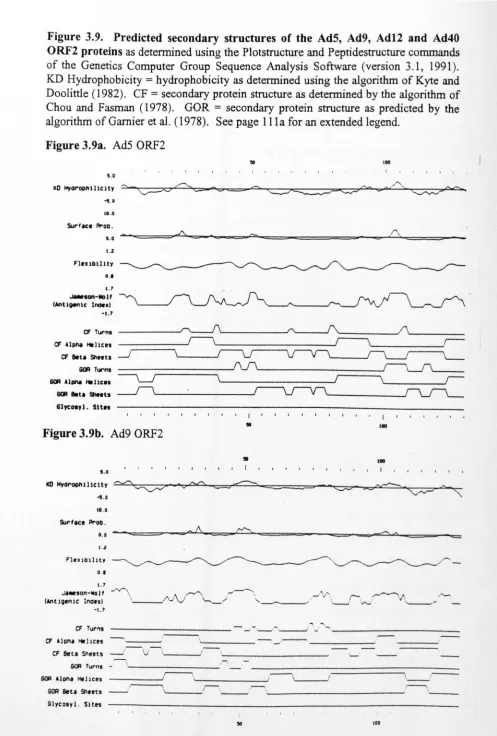
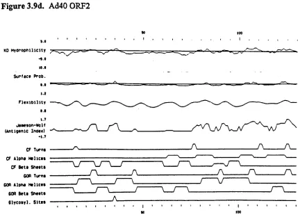
Fig.3.9. Predicted secondary structures of the AdS, Ad9, Ad12 and Ad40 E4
ORF2 proteins 117
Fig.4.1. E4 transcription map showing the location of the RNA probes 125
Fig.4.2. Analysis of cytoplasmic levels ofE1a, p-actin and E4 mRNAs during
Fig.4.3. Analysis of cytoplasmic levels ofE4 mRNAs in HeLa cells infected
by wild-type d1309 or Elb-55K mutant d1338
Fig.4.4. Analysis ofE4 mRNAs expression in the presence and
absence of an inhibitor of DNA replication
Fig.4.5. Analysis of cytoplasmic levels ofEla and E4 mRNAs during infection
of He La cells by wild-type d1309 or Elb-55K mutant virus dl338 134 130
133
Fig. 4.6. Analysis of cytoplasmic levels ofE4 mRNAs in HeLa cells infected
by wild-type d1309, E4 ORF6 mutant d1355, or Elb-55K1E4 ORF6
mutant dl367
Fig.4.7. Analysis of cytoplasmic and nuclear levels ofE4 mRNAs during
infection of He La cells by wild-type dl309 or Elb-55K mutant
virus dl3 38 138
137
Fig.4.8. The E4 donor splice sites as compared to the eukaryotic consensus
sequence of Mount (1982)
Fig. 5.1. The strategy for the detection of the mRNA A derived from the
D 1 and E4 ORF6 mutated virus vD 1
Fig. 5.2. The sequence of the Dl site of dl309 at 35542 bp, the
HindIII
site in vDl at 35543 bp and the mutated KpnI site in the ORF6
reading frame of vD 1
151
141
149
Fig. 5.3. Restriction digest analysis of the Dl mutant virus vDl and dl309 153
Fig.5.4. RNase protection analysis ofd1309, d135S and vDl mRNA Nfull
length mRNA using a probe made from pIDSO
155
Fig.6.l. The pGEX-2T expression vector 160
Fig.6.2. PCR amplification of the ORFI cDNA using primers IDC and IDD 160
Fig.6.3. The construction of the pGEX-2T/ORFl cDNA expression
vector pID22 162
Fig.6.4. Protein expression from the pGEX constructs 164
Fig.6.5. The expression of the GST/ORFIN and GST/ORFIC fusion proteins 166
of the first polyclonal antiserum
167
Fig.6.7. Titration of the
anti-GST/ORFI
activity in the serum samples
by ELISA
169
Fig.6.8. Western blot analysisof the
GST/ORFI
polyclonal antisera
169
Fig.6.9. The pTrcHis vector
171
Fig.6.10. The expression of the HisiORFI fusion at 37°C from pID35
173
Fig.6.11. SOS-PAGE analysisof samples of the 0.5 ml fractions from the
nickel resin column used in the purification of the
His/ORFI
fusion expressed from pID35
173
Fig. 6.12. The pQE-30 and pREP4 plasmids
175
Fig. 6.13. The expression of the
His/ORF
1 fusion at 37°C from pID38
177
Fig.6.14. SOS-PAGE analysisof samples of the 0.5 ml fractions from the
nickel resin column used in the purification of the
His/ORFI
fusion expressed from pID38
177
Fig.6.15. Titration of the anti-HisiORFI activity
in
serum samples by ELISA
179
Fig. 6.16. Western blot analysis employing sequential anti-His/ORFI antisera
to detect 400 ng of purified recombinant ORF 1
179
Fig.6.17. Detection ofORFI in infected cells
181
Fig. 6.18. Immunoprecipitation of an
in vitrotranscribed/translated ORFI
product
182
Fig. 6.19. Western blot analysis employingthe anti-HiS/orfl antisera
184
Fig.7.1. PCR amplificationof the ORF2 cDNA using primers IDE and IDF
192
Fig.7.2. The expression of the
GST/ORF2
fusion protein from pID22 at 30°C
and 37°C
193
Fig.7.3. SOS-PAGE analysis of samples of the 0.5 ml fractions from the
glutathione-sepharose column used for the purification of the
GST/ORF2
fusion expressed from pID19
193
Fig. 7.4. SDS-PAGE analysisof 10
ugof glutathione sepharose-bound
Fig.7.S. Purification ofORF2 protein released from the
GST/ORF2
fusion bythrombin digestion 196
Fig.7.6. Analysis of the
GST/ORF
antisera 198Fig.7.7. Western blot analysis employing sequential anti-GST/ORF2 antisera
to detect 40 ng of purified recombinant ORF2 199
Fig.7.8. Detection of ORF2 in infected cells 201
Fig. 7.9. Steady state levels of the ORF2 protein during the course of an
infection 202
Fig. 7.10. Synthesis of the ORF2 protein during the course of an infection 203
Fig.7.11. The subcellular localisation of the ORF2 protein within the
infected cell
Fig.7.12. Determination of the size of the ORF2 protein by sucrose
gradient analysis
Fig. 7.13. Co-immunoprecipitation analysis to detect ORF2 protein-protein
interactions
Fig.7.14. Immunoprecipitation analysis of the ORF2 protein under reducing
and non-reducing conditions
Fig. 8.1. Analysis of the protein production ofwt300, dll-3, d1327, d13S1 and
dl3S2 during the course of an infection 219
205
207
208
210
List of tables
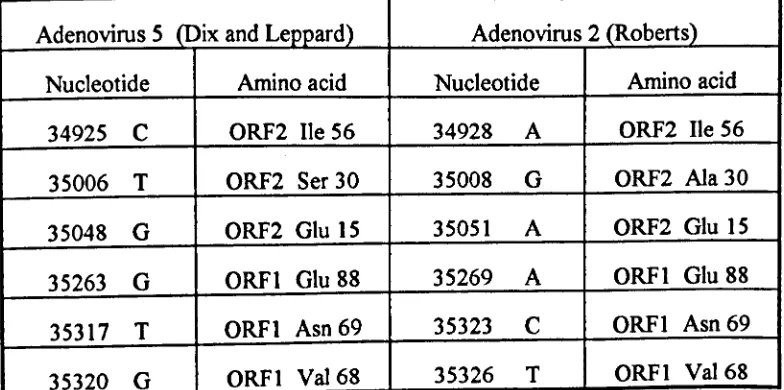
Table 3.1. The DNA sequence variations found in AdS (Chroboczek
et al.,
1992), Ad5 (Dix and Leppard, 1992) and Ad2 (Roberts
et al.,
1984) E4 ORFI and ORF2 106
Table 3.2. Amino acid changes due to the nucleotide differences between Ad2
and AdS ORFI and ORF2 106
Table S.1. Classification of E4
mRNAs
131Table 8.1. The titres ofwt300 and dll-3 virus, 4 days after infection of HeLa,
MRCS, WI-38, HVV-EC-C and HISM cells 216
Table 8.2. The titres ofd1327, d13S1, and d13S2, 4 days after infection of He La
Acknowledgements
First, I would like to thank the members of both the Adenovirus and Rotavirus groups
at the University of Warwick for all their assistance during the course of this study. I
am particularly grateful to Cally Caravokyri and Sue Thomas for their advice, help and
support over the last three years. I would also like to thank Carol Hill for her
assistance in all the animal work contained within this study. I especially want to thank
Keith Leppard, my supervisor, who has given me encouragement and invaluable
guidance during this period, to whom I will always be indebted.
I also would like to thank my parents for their love and support over the last 25 years
and for always encouraging me to 'get an education'. However special thanks must go
to Amanda Young, my wife to be, for putting up with me over the last three years. I
particularly thank her for her love, understanding and support, especially when
experiments 'crashed and burned'! Finally, I thank the Lord Jesus Christ for the
strength and hope that He alone can give.
This work was supported by a grant from the Science and Engineering Research
Declaration
All the work presented in this thesis was performed by the author in the Biological
Sciences Department of the University of Warwick, under the supervision of Dr. KN.
Leppard and with the assistance of the University of Warwick technical staff in the
handling of animals and the synthesis of oligonucleotides. None of this material has
been previously submitted for examination at another institution. However the studies
reported
in
chapter 4 derive from preliminary work performed by the author as part ofan undergraduate honours project and submitted in accordance with the requirement of
the BSc degree, 1991, University of Warwick. None of the data generated in this
preliminary work is presented here.
During the course of this study two publications have been made by the author. The
first was in 1992 and contained data from chapter 3:
Dix, I., &Leppard, KN. (1992). Open reading frames 1 and 2 of adenovirus region E4 are conserved between human serotypes 2 and 5.
J.
Gen. Virol. 73,2975-2976.The second was in 1993 and contained data from chapter 4:
Abbreviations
General abbreviations
35S-met
A260
Ab
35 S-methionine
absorbance at 260
nm
antibody
Ad(5) (2) (9) (12) (34) (40) adenovirus serotype (5) (2) (9) (12) (34) (40)
Ag antigen
Amp ampicillin
ATP adenosine 5'-triphosphate
bp
base pairBSA bovine serum albumin
Ci cune
cpm counts per minute
CTP cytidine 5'-triphosphate
CS newborn calf serum
Da Dalton
dATP deoxyadenosine triphosphate
dCTP deoxycytidine triphosphate
dGTP deoxyguanosine triphosphate
ddATP dideoxyadenosine triphosphate
ddCTP dideoxycytidine triphosphate
ddGTP dideoxyguanosine triphosphate
ddNTP dideoxynucleoside triphosphate
ddTTP dideoxythyrnidine triphosphate
DMEM Dulbecco's modified Eagle medium
DMSO dimethyl sulphoxide
DNA deoxyribonucleic acid
DNase deoxyribonuclease
DOC
deoxycholate
ds
double-stranded (DNA or RNA)
dTTP
deoxythymidine triphosphate
DTT
dithiothreitol
dUMP
deoxyuridine monophosphate
dUTP
deoxyuridine triphosphate
dUTPase
deoxyuridine triphosphate nucleotidohydrolase
EDTA
ethylenediaminetetraacetic acid (disodium salt)
ELISA
enzyme-linked immunosorbent assay
FCS
fetal calf serum
GST
glutathione S-transferase
GTP
guanosine 5
1-triphosphate
hnRNP
heterogeneous nuclear ribonucleoprotein
Ig
immunoglobulin
IPTG
isopropyl-l-thio-B. D-galactosidase
Kan
kanamycin
KLH
keyhole limpet haemacyanin
MCS
multiple cloning site
MLP
Adenovirus major late promoter
MLTU
Adenovirus major late transcription unit
MOl
multiplicity of infection
ut
nucleotides
NP40
Nonidet P40
oligo
oligonucleotide
ORF
open reading frame
PAGE
polyacrylamide gel electrophoresis
PCR
polymerase chain reaction
PEG
polyethylene glycol
PMSF poly(A) PPi
RBS
RF RNA phenylmethylsulphonyl fluoride polyadenylate pyrophosphateribosome binding site
replicative form
ribonucleic acid (prefixes: m
=
messenger,r
=
ribosomal, sn=
small nuclear, t=
transfer) ribonucleaserevolutions per minute
Svedberg unit
sodium dodecyl sulphate
single stranded (DNA or RNA)
trichloroacetic acid N,N,N',N'-tetramethyl-ethylenediamine Tris(hydroxymethyl)aminomethane uridine S'-triphosphate untranslated region ultraviolet light 5-bromo-4-chloro-3-indolyl-J3-D-galactoside RNase rpm S SDS ss
TeA
TEMED Tris UTP UTRuv
XgalVirus genome abbreviations
In adenovirus, genes and proteins are named according to the early region (E 1
a,
El b,E2, E3, E4) major late region (Ll, L2, L3 , L4, LS), or minor late region (IVa2, IX) of
the genome from which they are expressed (Fig. 1.2.). This is normally followed by
either the open reading frame number (e.g. ORFl), or the molecular weight of the of
the protein (e.g. Elb-SSK), or the number of amino acids in the protein (e.g.
sequence of Chroboczek et
al.,
1992 (Gen Bank ace. no. M73260) amended by DixSummary
Human adenovirus type S is a dsDNA virus which replicates in the nucleus of the
infected cell, exploiting a number of host cell mechanisms. This close association with
the eukaryotic cell has made adenovirus the target of numerous studies attempting to
understand how cellular systems function. This study focuses on the E4 transcription
unit, which has the potential to encode at least 7 distinct polypeptides from reading
frames accessed by differential splicing of a single primary transcript. In this study, the
pattern of expression of these mRNAs during lytic infection was examined, and two
distinct temporal classes were defined; early and late. It had been previously shown
that adenovirus mRNAs produced late in the infection depended on a virus-coded
RNA transport regulator, EIb-S5K, for optimal cytoplasmic accumulation. However,
only one of the E4 late class mRNAs was dependent on this EI b protein for
cytoplasmic accumulation leading to the hypothesis that for an E4 late mRNA to be
dependent on EIb, it had to retain intact splice sites or intronic sequences. To examine
this hypothesis, a virus was built lacking an important splice site of the E4 region to
see if, by removing this splice site, the mRNA could leave the nucleus in the absence of
the El b complex. The results of initial experiments reported here supported this
hypothesis.
Two of the E4 open reading frames (ORFI and ORF2) identified in Ad2 were
disrupted in the published AdS E4 sequence, but these differences were subsequently
found to be sequencing artefacts. The presence of these two proteins in the infected
cell had never been previously demonstrated so polyclonal antisera were generated
against bacterially expressed ORFI and ORF2. The ORF2 antiserum allowed the
identification of the ORF2 protein in the cytoplasm of infected cells, from early stages
of the infection. No associations of ORF2 with other infected cell components were
detected. In contrast, the ORFI antiserum only reached a low titre and no ORFI
protein was detected in infected cells. Now that ORF2 has been found in the infected
Chapter 1.
1.t. Adenoviruses - A General Introduction
1.1.1. The adenovirus family
Adenoviruses were first isolated from tonsil and adenoid biopsies from children by
Rowe et al. (1953) Since then adenoviruses have been shown to be the aetiological
agents of a wide variety of diseases in birds and mammals including a number of acute
febrile respiratory illnesses, epidemic keratoconjunctivitis, acute haemorrhagic cystitis
and gastro-enteritis (reviewed in Horwitz, 1990b). Some adenoviruses have also been
associated with the induction of malignant tumours in rodents although there is no
evidence of these viruses having oncogenic potential in humans.
Adenoviruses are of the family Adenoviridae which is divided into two genera;
Mastadenoviridae and Aviadenoviridae. The Mastadenoviridae genus contains
human, simian, bovine, equine, porcine, ovine and canine viruses. The human
adenoviruses have been classified into six subgroups (A-F) by a number of serological
and biochemical techniques such as haemagglutination patterns of rat and rhesus
monkey red blood cells, oncogenic potential in rodents, DNA base composition and
homology, and the ability to transform cells in tissue culture. The antigenic
determinants important in the classification of adenoviruses are found on the hexon,
penton and fibre proteins, for example all human adenoviruses share a cross-reacting
group antigen (a) on the hexon capsomer while the type-specific antigens (s and y) are found on the hexon and fibre proteins respectively (reviewed
in
Horwitz, 1990a).This study focuses on a human subgroup C virus, adenovirus serotype S (AdS).
Subgroup C viruses are characterised by their partial agglutination of rat erythrocytes,
their inability to induce tumours in rodents, their ability to transform cells in tissue
culture and the high G+C content (57-59 %) of their genomes. Of the subgroup C viruses isolated, serotypes 2 and S have been studied in greatest detail. The genomes
of these viruses have been completely sequenced, Ad2 in 1984 by Roberts et
al.
andAdS in 1992 by Chroboczek et
al.,
amended by Dix and Leppard (1992). Sequenceorganisation. The differences between the sequences of these two viruses are
concentrated in regions encoding hexon, fibre and some of the E3 proteins (Kinloch
et
al.,
1984~ Cladaras and Wold, 1985~ Chroboczek and Jacrot, 1987), probably due toselective pressure exerted by the host immune system during infection as the hexon and
fibre proteins contain major antigenic determinants. As these two serotypes are so
closely related, data derived from one or other of these viruses is routinely taken to
apply to both serotypes; in the introduction to this study no distinction between the
two serotypes is usually made.
1.1.2. Structure of adenoviruses
(Fig.1.1.)
The structure of the adenovirus virion has been well characterised and is reviewed by
Horwitz (1990a). Adenovirus virions are non-enveloped, regular icosahedrons of
about 65 to 80 nm in diameter which comprise a double stranded DNA molecule of
about 36,000 bp (Ad5 is 35938 bp) surrounded by a protein shell (capsid) made of at
least 11 distinct structural proteins (figure 1.1). The capsid comprises 240 hexon
capsomeres (3 x 120 kd polypeptide II molecules) and 12 penton capsomeres (5 x 85
kd polypeptide III molecules noncovalentIy attached to 3 x 62 kd polypeptide IV
molecules; the fibre structure). The penton capsomeres are located at each of the 12
vertices of the capsid with the fibre structure projecting out from the virion. Each of
these penton capsomeres is surrounded by five hexon capsomeres (the peripentonal
hexons) associated with polypeptide lIla (66kd). Also associated with the hexon
capsomeres are 3 further polypeptides; VI (24 kd), VIII (13 kd) and IX (12 kd), which
are probably involved in stabilising the capsid structure. Within the capsid shell of the
virion is the core structure which contains the DNA genome covalently attached to the
55 kd terminal protein (TP) at its 5' ends and polypeptides V (48.5 kd), VII (18.5 kd)
and Jl (4 kd). Polypeptides V and VII are basic proteins which form irregular
nucleosome-like structures along the viral genome, each spanning approximately 200
Figure 1.1. Schematic representation of the adenovirus virion (reproduced from
Horowitz, 1990a). The f..l protein and the probable virion protein, L3-23K protease,
are not featured as their location within the virion is not known. The thread-like strand
within the virion shown below is representative of the virion genome.
Il- Hexon III - Penton base lIla - Penton associated
protein TP - terminal protein V - Core protein IV -Fiber
VI - Hexon associated protein VII - Core protein VITI - Hexon associated
protein IX - Hexon associated
1.1.3. Adenovirus replication (reviewed by Horwitz, 1990a)
The adenovirus lytic cycle in cultured cells results in the production of about 10,000
virions per infected cell during the course of one virus life cycle (ca. 36 hours). The
lytic life cycle of adenovirus has been divided, by convention, into two phases, early
and late, separated by the onset of viral DNA replication, with distinct patterns of gene
expression in each phase. This division of the virus life cycle is not rigid, early mRNAs
are also made after vDNA replication has begun and some late mRNAs can been found
prior to vDNA replication.
Adenoviruses enter the host cell by receptor-mediated endocytosis, attaching to the
host cell receptor via the C terminus of the virion fiber protein (Devaux
et al., 1987).
The nature of the host cell receptor is, at present, vague although initial studies have
indicated that the virus binds to a 50 kd protein identical to the receptor used by the
coxsackie B3 virus (Lonberg-Holm
et aI.,
1976; Hennache and Boulanger, 1977). Thepenton cap somers at the base of the fibre protein then interact with cell surface
av
integrins via an RGD peptide sequence within the penton protein (Bai
et al., 1993;
Wickham
et al.,
1993). This secondary interaction is required for efficient virusinternalisation, allowing the formation of endocytic vesicles or receptosomes. Within
the receptosome the pH drops, which is thought to alter the structure of the virion
capsid, rupturing the endocytic vesicle, releasing the virion into the cytoplasm of the
cell (Seth
et al.,
1985). It has been postulated that the penton protein plays animportant role in the rupture of the receptosome, possibly through the interaction with
cellular integrins (Seth
et al.,
1984). The penton capsomere is believed to be lostduring this process (reviewed by Nemerow
et al., 1994).
The virion is then transported through the cytoplasm to the nucleus of the infected cell
in a process which probably involves the interaction of the hexon capsomeres with the
microtubules of the host cell. The viral DNA and the core proteins (V, VII, TP and J.1)
then enter the nucleus through the nuclear pores leaving the remainder of the virion
The viral genome then undergoes early transcription. The virus appears to rely almost
exclusively upon the host cell machinery for transcription and RNA processing with
transcripts being spliced, capped and polyadenylated in a manner similar to host
transcripts (Ziff, 1980~ Darnell, 1982~ Tooze, 1982~ Sharp, 1984). Transcription from
the six early transcription units (E1a, Elb, E2, E3, E4 and L1) results in the synthesis
of a variety of mRNAs which encode proteins required for subsequent events in the
lytic cycle, such as transcriptional activation, viral DNA replication and mRNA
metabolism. Early transcription from these transcription units results in the production
of only low levels of viral mRNA, compared to the level of viral mRNAs late in the
infection.
The late phase of the infection is marked by the onset of DNA replication, about 10-12
hours post infection. Viral DNA replication has been extensively studied, the current
model incorporating both type I (duplex template) and type II (single stranded
template) replication to produce progeny genomes. Replication initiates at either end
of the parental duplex molecule, displacing one of the parental strands to produce a
semi-conservatively replicated genome and a single stranded molecule (type I
replication). The inverted terminal repeats of the genome allow the single-stranded
molecule to form a panhandle structure where the double-stranded region resembles
the end of a duplex genome. This is then replicated by a type II replication mechanism
to produce a second daughter genome (reviewed by Stillman, 1989).
In vivo
andin
vitro
studies have demonstrated that only three viral proteins are directly involved inDNA replication; the 140 kd viral DNA polymerase, the 72 kd ssDNA binding protein,
and the 80K precursor to the terminal protein (reviewed by Stillman, 1989). Recently
it has been demonstrated that the ORF3 and ORF6 proteins of the E4 region are
implicated in the regulation of vDNA replication (Weiden and Ginsberg, 1994, see
section l.4.2.3.). Three host proteins are also important in viral DNA replication;
nuclear factors I and III, transcription factors which bind adjacent to the core origin of
replication; and nuclear factor II, a type I topoisomerase, (reviewed by Stillman ,
Following the onset ofviraI DNA replication the nuclear structure is reorganised with
both DNA replication and late transcription occurring in association with the nuclear
matrix in discrete viraI inclusion bodies (Moyne
et al.,
1978~ Zhongheet al.,
1986~Walton
et al.,
1989~ Moen et aI., 1990~ Jimenez-Garcia and Spector, 1993).Transcription occurring in these inclusion bodies results in the production of late
mRNAs, primarily encoded by the major late transcription unit (ML TU). These late
mRNAs, which mainly encode the structural proteins of the virion, can account for up
to 20-40% of the total cellular mRNAs (Flint, 1986). As the structural proteins are
produced, the progeny virions are assembled in the nucleus of the infected cell; initially
the penton and hexon polypeptides assemble into capsomeres in the cytoplasm and
then form a 600S capsid intermediate within the nucleus. At this stage the viral DNA
enters the capsid along with the core proteins, by virtue of packaging signals located
between 290 and 390 bp from the left end of the genome, at an opening at one of the
vertices. The last stage in virion assembly is the processing of some of the proteins
(pVI, pVII, pVIII, pTP) within the virion, which are assembled in precursor form, by
the viral protease (L3-23 kd) to produce the mature virion. This entire process is
aided by virus-encoded scaffolding proteins which are necessary during virion
formation but are absent from the virion (e.g. L4-100 kd and IVa2-50 kd). The
mechanism of virion release from the cell has not yet been clearly elucidated (reviewed
by Horwitz et aI., 1990a).
Cellular macromolecular synthesis undergoes major changes during the late phase of an
adenovirus infection, with both cellular DNA replication and protein synthesis being
inhibited. The gradual cessation of cellular DNA replication is probably a secondary
effect of the inhibition of cellular protein synthesis. The block in cellular protein
synthesis has been shown to be at the level of translation and not at the level ofmRNA
transcription or mRNA transport into the cytoplasm as cytoplasmic levels of cellular
1.1.4. Adenovirus gene function
Gene expression from the 36 kbp adenovirus genome is a very complicated process,
with transcription of most of both strands of the double stranded genome as can be
seen in Fig. 1.2. (reviewed by Flint
et al.,
1986 and Horwitz, 1990a). The regulationof this expression is discussed in section 1.2 ..
1.1.4.1. The Ela region
The El a transcription unit is encoded on the r strand of the genome. Five mRNAs are
derived from this region by the differential splicing of the primary transcript: two
predominant early mRNAs (I2S and l3S); a predominant late mRNA (9S); and two
minor late mRNAs (lOS and lIS) (Fig. IJa.). The two major products from this
region are encoded by the 12S and l3S mRNAs; the 243R and 289R proteins, which
are identical apart from 46 internal residues. Both these proteins contain discrete
regions conserved among the different adenovirus serotypes, the CRI and CR2
regions, while only 289R contains a third conserved region, CR3. Both proteins also
contain two auxiliary regions (ARI and AR2), shown to be important in functional
studies in addition to CRI-3, and a C terminal nuclear localisation signal (Fig. 1.3b).
The 243R and 289R proteins of Ela are involved in the induction of cellular DNA
synthesis which is related to their role in transformation of non-permissive cells. The
mechanism of action of these proteins in cellular transformation is not fully understood
but it is known that they form complexes, via the CRI and CR2 regions, with cellular
proteins involved in cell cycle control, including the tumour repressor protein p l OSRb
and other related proteins (reviewed by Dyson and Harlow, 1992).
The CR3 domain of 289R is involved in the transactivation of Ela-dependent
promoters. The E I a protein activates transcription by a number of different
mechanisms (reviewed by Akusjarvi, 1993): it regulates certain transcription factors
(e.g. E2F, E4F and TFIIIC) by phosphorylation (Roemer
et al.,
1988; Bagchiet al.,
1989; Raychaudhuri
et al.,
1989); it can bind upstream activating sequence.
N ~ N So,.,
VI UI,.,
~ o..
;, OIl ~ Q N..,~
00_~
~~ I I OIl .. :: :: Q Q'"
'"
e, Q. VI VI 0-'-·W
}~
toIII~}~
+++T
CD0.-t
(>I'0:-j
I I I
~.
0:-", N CD ~:-o~ CDN "O~ N' 0.-", , N ~ 01'
0:-ttll
•• I' u Ol'0:-1••••••• ,•••••••..•• I_
•••••••••••••• "'N _ _ • VI
01 ~ :n z ~
..
"0 ., o•
'"
..
x (11VI UlN ~I Q ~~i
g)~
< •..,
ts
::r_
..
_>< Q N'" (>I ~ "0 -1 Z o..
•
~f}ij.. ·
::J-r
o ::J o I7' (.N <:;. (>I -.
"'0"; 0;;;J
~
II '<
~_
a;O=u.~
~";~:>t~
,I
NFigure 1.2. Transcription and translation map of adenovirus type 2 (reproduced
from Horwitz, 1990a). Early mRNAs are represented by thin lines, late mRNAs (and
early mRNAs which are transcribed late, e.g. L1 transcripts) by bold lines,
polyadenylation sites by arrow heads, nonstructural gene products by their molecular
mass, and virion components by Roman numerals. The three components of the major
late tripartite leader are designated 1, 2 and 3. Some minor mRNA species and
proximity with the core promoter proteins (Liu and Green, 1990~ Chatton
et al.,
1993)~ Ela can also interact directly with the TBP (TATA Box binding protein)
activating transcription (Horikoshi
et al.,
1991~ Leeet al., 1991).
The CRI and CR2 domains of 243R and 289R can also transactivate transcription via
their interaction with tumour suppressor proteins, such as p l OSRb, as this interaction
can cause the dissociation of transcription factors from these regulatory, inhibitor
proteins (Bandara and La Thangue, 1991~ Chellappan
et al.,
1991). A well studiedexample of this phenomenon is the E2F-pl0SRb interaction, where E2F has no
transactivating function when bound to plOSRb but in the presence of the El a protein
it is released in an active form, as El a interacts with plOSRb displacing E2F.
The multifunctional E I a proteins may therefore regulate one promoter in a number of
different ways, e.g. the E2 early promoter. The E2 promoter consists of an activating
transcription factor (ATF) binding site and two inverted E2F sites. It is activated by
El a by three distinct mechanisms: ATF can bind to the upstream ATF binding site and
transactivate the promoter via Ela which associates with ATF; Ela can dissociate E2F
from pIOSRb_E2F complexes where E2F is inactive and it can also phosphorylate E2F,
so increasing its DNA binding activity and so its ability to transactivate the E2
promoter (Bagchi
et al.,
1989~ Bandara and La Thangue, 1991~ Chellappanet al.,
1991~
Chattonetal., 1993).
Evidence is mounting that Ela-243R can also inhibit the activation of some promoters,
a function shown to be a property of the N terminus of the protein (Berk, 1986). The
Ela-243R protein, which lacks the transactivation domain CRJ, is believed to repress
transcription by interacting with a TFIID protein from the core of the transcription
machinery and so reducing expression from certain promoters (Ielsma
et al.,
1989~Stein
et al.,
1990~ Bautistaet al.,
1991).1.1.4.2. The Elb region
The EI b transcription unit is also on the r strand of the genome and encodes proteins
A
98
CAP Donor
1st 12S Acceptor Donor
138 2nd Poly(A) site Donor Acceptor
9S
128
138
10S
B
CR1 CR2 CR3 AR1 AR2 NL8
289R (138)
Figure 1.3. The Eta region. Panel A: The Ela mRNAs
(from Harper and Manley,1991). The open boxes represent exon regions while the lines represent intron regions.
Indicated above the mRNAs are the transcriptional start sites, splice sites and
polyadenylation sites. Indicated to the left of the mRNAs are the popular names given
to the mRNAs.
Panel
B:The multifunctional domains of the major Ela proteins,
289R and 243R.
289R and 243R are the products of the 12 and 13S mRNAsrespectively. CRI, CR2 and CR3 are regions conserved in Eia proteins of all the
adenovirus serotypes. ARI and AR2 are auxiliary regions shown to be functionally
derived from this region (not including the pIX mRNA) which potentially encode 7
distinct proteins (perricaudet
et al.,
1979; Boset al.,
1981; Andersonet al., 1984;
Virtanen and Petterson, 1985; Lewis and Anderson, 1987; Takayesu
et al.,
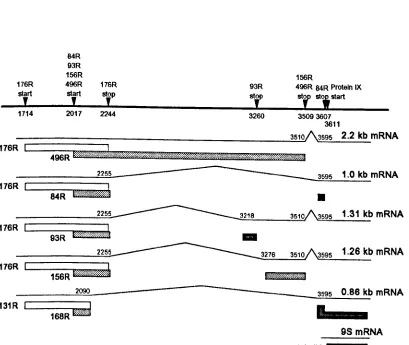
1994, seeFig. l.4.). A 2.2kb mRNA is predominant early in the infection and is translated to
produce the major E1b proteins 19K (176R) and 55K (496R), the E1b-55K protein
being translated from a reading frame overlapping that of the 19K protein, initiating at
a site within the 19K reading frame. Late in the infection a 1.0 kb mRNA accumulates,
due to differential splicing, encoding the 19K protein and an 84 residue protein (84R)
which comprises the N- and C- termini of the 55K protein. Also produced at late
times are three less abundant mRNAs of 0.86 kb, l.26 and l.31 kb producing various
products related to the major El b products.
The 19K protein is involved in a number of different aspects of the viral infection
including the protection of viral DNA, and in non-permissive cells, cellular
transformation (Chinnadurai, 1983; Subramanian
et al.,
1984; Stillman, 1986; Whiteand Stillman, 1987). The 19K protein also protects against programmed cell death
(apoptosis), induced as a result of the disruption of cell growth control pathways by
Ela (White
et al.,
1991; 1992; Raoet al.,
1992; Debbas and White, 1993), andcytolysis induced by the tumour necrosis factor-a (White
et al.,
1992). The 19Kprotein may also have the ability to transactivate early viral genes (Yoshida
et al.,
1987), although no transcritpional activity was found by Telling
et al. (1994).
The 55K protein is also a multifunctional protein being involved in cellular
transformation (Barker and Berk, 1987; McLorie
et al.,
1991) and in the cytoplasmicaccumulation of late mRNAs, host cell shutoff and late protein synthesis during lytic
infection (Babiss and Ginsberg, 1984; Babiss
et al.,
1985; Pilderet al.,
1986a,b;Williams
et al.,
1986). The 55K protein derives some or all of its transforming activityfrom its interaction with the tumour suppressor protein p53 (Samow
et al.,
1982; Kaoet al.,
1990), inhibiting p53-mediated transcriptional activation (Yew and Berk, 1992).Malette
et al.
(1983) demonstrated that the 55K protein is phosphorylated at threeFigure 1.4. The predicted Ad5 Elb products
(reproduced from Takayesu et al.,1994). The Elb
mRNAs
are depicted as lines, with the protein products indicated byboxes. The 3 translation reading frames are shown by different shading. Also included
are the relevant nuc1eotides for translation start and stop sites and for splice start and
donors.
176R
start
,
84R 93R 156R
496R 176R
start
,
stop,
2017 2244
93R stop
,
156R
496R 84RProtein IX
str st; start
1714 3260 35093607
3611
~======:;-
23~5101\3595 2.2 kb mRNA176R 496R
bmuwu6w$M_%W§%~~Mi$#i%i1l
;-;:=====~22~55~ __ --- 3595 1.0 kbmRNA
176R L...---S4-R---'U""'W""d""''''''.'''''''
•
2255 3218 3510 3595 1.31 kb mRNA
176R L...---93-R--"t""'M"""i""b"",,;,,,,,,d
-2255 3276 3510 3595 1.26 kb mRNA
176R L...--1!155f6iRR~M.,,*mMm@a.
________ ~2~090~ --- ~3~59~5~0~.8~6~kbmRNA
131 R
L---...Fiiicl"_,,j
168R
•
98 mRNA
phosphorylation is important in regulating 55K function, especially in transformation
(Teodoro
et al.,
1994). Although the El b-55K protein is multifunctional, the viral
replication and transforming functions are not divided into discrete functional
domains, as in the El A proteins, although the two functions can be separated by
mutational analysis (Yew
et al.,
1990). The function of the Elb-5SK protein in viral
replication is dealt with in detail in section 1.3. The functions of the minor El b
proteins has not yet been established.
As mentioned, both the Ela and the Elb proteins are involved in the immortalisation
and transformation of primary rodent cells infected with adenovirus, however an
extensive review of the literature reporting these functions is outside the scope of this
thesis. In addition to the above references the subject is reviewed by Horwitz
et al.
(1990a) and Boulanger and Blair (1991).
1.1.4.3. The E2 region
The E2a region encodes a nuclear phosphoprotein with an apparent molecular weight
of 72 kDa (DBP) which is essential in viral DNA replication, binding non-specifically
to single-stranded and double-stranded DNA changing its structure (reviewed by Hay
and Russell, 1989; Stillman 1989; Horwitz
et al.,
1990a).
It has also been
demonstrated to be involved in the regulation of transcription (Nevins and Winkler,
1980; Johnston
et al.,
1985), mRNA stability (Babich and Nevins, 1981), mRNA
processing (see section 1.2.4.) and in virus assembly (Nicolas
et al., 1983).
The E2b region encodes two phosphoproteins; the 80 kDa pre-terminal protein (PTP)
and the 140 kDa DNA polymerase.
The DNA polymerase catalyses the covalent
attachment of dCMP to a serine residue of the pTP protein in the presence of specific
DNA residues at the origin of replication. This acts as a novel priming mechanism for
DNA replication (reviewed by Hay and Russell, 1989; Stillman, 1989). Later in the
infection the protein is cleaved by the L3-23K viral protease to give the 55K form
matrix so allowing efficient transcription and possibly DNA replication (Shaack
et al.,
1990; Fredman and Engler, 1993), it protects the DNA ends from exonuclease activity
(Dunsworth-Browne
et al.,
1980),it
prevents the attachment of DNA termini bindingproteins which inhibit DNA replication (DeVries
et al.,
1989) and it ensures thecorrect initiation site for DNA replication (reviewed by Hay and Russell, 1989;
Stillman, 1989).
The viral DNA polymerase is involved in the initiation and elongation of the nascent
DNA strand and is probably regulated by phosphorylation (reviewed by Hay and
Russell, 1989; Stillman, 1989).
1.1.4.4. The E3 region
The E3 region is not required for viral replication in tissue culture, being involved in
the virus' ability to circumvent immune surveillance in the natural host (reviewed by
Wold and Gooding, 1991). The AdS E3 region expresses approximately 9
alternatively spliced and polyadenylated mRNAs coding for at least seven proteins;
gp19K, 14.7K, 14.SK, 12.SK, 11.6K, 10.4K, and 6.7K. The 14.7K, 14.SK and the
10.4K proteins prevent tumour necrosis factor (TNF)-mediated cytolysis of the
infected cell. The 10.4K and 14.SK proteins also form a complex which
down-regulates the cell surface epidermal growth factor receptor levels although it is unclear
whether this function is linked to the proteins' ability to prevent TNF-mediated
cytolysis. Recently the 10.4K and 14.SK proteins have been associated with the down
regulation of the expression ofEla gene products, at the level of translation (Zhang
et
al.,
1991, 1994). This reduction in Ela gene expression is believed to cause areduction in cytolysis by adenovirus-specific T cells (Zhang
et a!.,
1991). The gp19Kbinds to the major histocompatibility complex (MBC) class I antigens and retains them
in the endoplasmic reticulum so preventing their transport to the cell surface. This
failure of virus-infected cells to present MHC class I antigens, and consequently virus
of the host immune system failing to recognise and lyse virus-infected cells. No
functions have yet been ascribed to the 12.5K, 1l.6K or the 6.7K proteins.
l.l.4.5. E4 region
The E4 region encodes potentially seven different polypeptides; E4-0RF1, ORF2,
ORF3,
ORF3/4,
ORF4, ORF6, and ORF6/7. The expression and functions of theseproteins will be discussed in detail in section 1.4.
l.l.4.6. Late transcription units
Late in the infection the major late promoter (MLP) is strongly activated to produce 5
families of late transcripts (LI-L5) by alternative splicing and polyadenylation. Every
RNA from the ML TU contains an identical 5' non-coding region of about 200
nucleotides called the tripartite leader which is believed to be important in the
translation of MLTU transcripts (see section l.2.6.). Most of the late proteins are
encoded by these ML TU transcripts although the minor late polypeptides IX and IVa2
are encoded elsewhere on the genome. The expression of the late products is
restricted to the period after viral DNA replication with the exception of L 1 mRNA
which is also expressed at low levels during the early phase. In addition to structural
proteins, the ML TU encodes a number of late, nonstructural proteins such as
'scaffolding' proteins, involved in virion assembly (e.g. LI-55K, L4-100K), and other
nonstructural proteins (e.g. L4-33K).
Three other late-specific transcription units are also active late in the infection, IVa2,
IX and E2-L. The IVa2 and IX transcription units are often referred to as the
intermediate transcription units as they are transcribed before late ML TU transcription,
at the end of the early phase. The IVa2 region encodes a 50K scaffolding protein
while region IX encodes a 12K virion protein. The activation of the E2-L promoter at
late times results in an increase in the levels of the viral proteins required for DNA
l.l.4.7. VARNA
Adenoviruses also encode two distinct virus-associated (VA) RNAs; VA RNAI and
VA RNAn, each about 160 nucleotides long, transcribed by RNA pol III (Price and
Penman, 1972; Weinmann
et al.,
1974; Mathews, 1975; Pettersson and Philipson,1975). These RNAs are initially synthesised early in the infection but production
increases at late times and has been shown to be necessary for efficient viral mRNA
translation (see section l.2.6.).
1.2.
The regulation of gene expression in an adenovirus infection1.2.1
IntroductionAdenovirus genes are expressed in a highly regulated fashion during the course of a
lytic infection. This regulation of gene expression results in a clearly defined temporal
expression pattern of the gene products so that they are produced at the required times
and at the required levels to ensure the efficient replication of the virus. This highly
regulated pattern of gene expression has been the subject of intense research, in an
attempt to elucidate the mechanisms by which the virus controls gene expression.
Studies have shown that adenovirus gene expression is regulated both at the level of
transcription and at post-transcriptional levels. Details of these studies are summarised
below.
1.2.2.
Transcription initiation (reviewed by Akusjarvi, 1993)Adenovirus genes are transcribed by two host cell, DNA-dependent RNA polymerases;
RNA polymerase II which transcribes both strands of the virus producing over 99% of
the viral mRNAs, and RNA polymerase III which transcribes the short, noncoding
RNAs known as VA RNA (sections 1.1.4.7. and l.2.6.2.). Transcription from the
adenovirus genome occurs from five early (Ela, Elb, E2, E3 and E4) and four late
(MLTU, IX, IVa2 and E2-L) promoters. These genes can be subdivided further into
IX) genes according to when transcription initiates in the virus lytic cycle.
This
temporal regulation is not fully understood but a number of viral proteins have been
demonstrated to be involved in the formation and the activity of transcription
complexes within adenovirus infected cells, so regulating gene expression. Firstly,
Ela transactivates Ela, Elb, E2, E3, E4, VA RNA and a limited number of cellular
promoters such as
hsp70, c-fos, c-jun
and proliferating cell nuclear antigen (peNA).
This transactivation has been shown to be due to a number of indirect mechanisms
(reviewed in section 1.1.4.1.). Secondly, both the Elb-19K and the Elb-55K proteins
also have the potential to regulate transcription during an adenovirus infection.
However, mutational analysis suggests that these proteins have no significant effects
on viral promoters during lytic infection (see sections 1.1.4.2. and 1.3), but their
involvement in promoter transactivation is probably limited to cellular transformation.
Thirdly, the multifunctional E2-DBP protein has a specific repressive effect on the E4
promoter, although the mechanism by which this repression occurs is unclear.
Fourthly, two E4 proteins have been shown to be possibly important in transcriptional
regulation, the ORF617protein and the ORF4 protein. The ORF617 protein can bind
to the host transcription factor E2F and induce the co-operative binding of this factor
to the two inverted E2F binding sites in the E2 early promoter so inducing E2
transcription (see section 1.4.2.7.).
The importance of the E4-0RF4 protein in
transcription is not yet clear; initial experiments have shown it is involved in the
phosphorylation of a number of transcription factors, so regulating their activity (see
section 1.4.2.5.).
1.2.3. mRNA polyadenylation
The formation of a 3' end is an essential part in the biogenesis of a mature mRNA
molecule.
In higher eukaryotes (and adenovirus) the 3' end is formed by
endonucleolytic cleavage of a pre-mRNA molecule followed by the addition of 200 to
250 adenylate residues to the new 3' OH terminus (reviewed by Proudfoot, 1991;
within the adenovirus genome there are a number of 'complex' transcription units
which produce multiple mRNAs from a single promoter by utilising alternative splice
sites and/or polyadenylation sites (Flint
et al.,
1986; Leffet al.,
1986). The utilisationof alternative polyadenylation signals can lead to the production of different gene
products and in the case of the adenovirus MLTU, can be temporally regulated.
The MLTU produces five 3' co-terminal families of mRNA, Ll through to L5, each
defined by a unique polyadenylation site (reviewed by Nevins and Chen-Kiang 1981;
Horwitz 1990a). By the temporally regulated utilisation of these sites the virus
regulates the production of the structural and nonstructural proteins encoded by this
region. Early in the infection Ll through to L3 RNAs are transcribed but the
utilisation of the L 1 site is three times that of the L3 site. This difference is even
higher in the cytoplasmic mRNA population indicating many of the L3 polyadenylation
events non-productive, resulting in mRNAs which fail to be exported from the nucleus.
After DNA replication all 5 polyadenylation sites are utilised with L3 RNA out
numbering the Ll RNA by 3:1 (Nevins and Darnell, 1978; Chow
et al.,
1979; Shawand Ziff, 1980; Akusjarvi and Persson, 1981; Nevins and Wilson, 1981). This change
in polyadenylation allows the virus to produce nonstructural proteins at early times and
structural proteins later in the infection from the same transcription unit.
Recently, Larsson
et al.
(1992) proposed that there is an intermediate step between theearly and late patterns of ML TU polyadenylation, with mRNAs from regions L 1 and
L4 being selectively over expressed as compared to the L2, L3, and L5 mRNAs. They
proposed that for the late pattern of polyadenylation to occur both viral DNA
replication and late protein synthesis are required.
Although a number of the sequences involved in poly(A) site selection have been
identified, along with some of the protein factors responsible for the recognition and
processing of poly(A) sites, the selection and regulation of alternative poly(A) sites is
not understood. Work carried out on the MLTU has suggested the involvement of the
RNA sequence of the poly(A) sites (DeZazzo
et al.,
1991; Prescott andenhance or depress the use of a specific poly(A} site and the relative levels of general
factors involved in poly(A} site usage (late in the infection the high levels of ML TU
precursor RNA may sequester the factors involved in the formation ofthe 3' termini of
mRNAs,
so reducing their relative levels in the nucleus (Mannet al.,
1993}).The other adenovirus transcription unit which contains more than one polyadenylation
site is the E3 unit. This region contains multiple splice sites and two polyadenylation
sites, E3A and E3B, allowing the regulation of gene expression from this region by
alternative splicing and polyadenylation (reviewed by Wold and Gooding, 1991; Brady
eta!',1992).
1.2.4. RNA splicing
1.2.4.1. Introduction
Eukaryotic pre-mRNA splicing is one of the steps in the nuclear processing of
precursor RNA (pre-mRNA) into mature, translatable mRNA, common to almost all
eukaryotic transcripts. RNA splicing occurs within large, nuclear ribonucleoprotein
complexes, known as spliceosomes, where RNA sequences (introns) are precisely
excised from the pre-mRNA and the remaining RNA sequences (exons) are accurately
ligated together. The mechanism by which cells accomplish RNA splicing has been
studied intensively but is still not fully understood (reviewed by Smith
et al., 1989;
Green, 1991).
All but one of the adenovirus primary transcripts (the exception is IX) are spliced, with
transcripts often being differentially spliced to produce more than one mRNA from a
given transcription unit. This alternative pre-mRNA splicing is not unique to
adenovirus, being found m a number of cellular transcription units (e.g.
n-tropomyosin, calcitoninlCGRP, immunoglobulin 1Clight chain). In adenovirus, primarytranscripts can be differentially spliced by the use of either alternative 5' donor sites
with the same 3' acceptor site (e.g. Ela), or the same 5' donor site but alternative 3'
There are some notable differences between cellular and viral exon-intron
arrangements: first, adenoviral transcripts have relatively few introns, the maximum
discovered is six (the fibre mRNAs) and these introns are relatively short (probably
because the genetic material has to be kept as short as possible if it is to be compressed
into the viral capsid) while cellular genes commonly have numerous large introns;
second, the viral introns tend to occur in the 5' or 3' untranslated regions (UTRs) of the
transcripts while cellular genes generally have introns disrupting the actual coding
regions, although a number of cellular genes have now been found where differential
splicing only alters the UTRs, e.g. colony stimulating factor-1 RNA (Ladner
et al.,
1987). The difference in the positioning of the introns is probably due to a subtle
difference between the function of viral intronslexons and those intronslexons found in
cellular genes. Eukaryotic cells differentially splice transcripts
in
order to: (a) localiseproteins to alternative cell compartments (e.g. immunoglobulin Il, neural cell adhesion
molecules, decay-acceleration factor, all of which are generated as either
membrane-bound or secreted forms (Alt
et al.,
1980; Caraset al.,
1988; Goweret al.,
1988»);(b)
modulate the function of a protein either by deleting a region (e.g. the Drosophila
transposase involved in the germ line-dependent transposition of P elements (Laski
et
al.,
1986)) or exchanging a segment(s) of a protein for an alternative sequence toproduce a isoform of the protein (e.g. troponin-T, pyruvate kinase (Medford
et al.,
1984»; (c) produce proteins with completely different functions from the same
transcript (e.g. calcitonin and the calcitonin gene-related peptide (Amara
et al.,
1982»;(d) regulate the efficiency of mRNA stability, transport and translation (e.g. CSF-l
(Ladner
et al.,
1987». In comparison, the majority of alternative splicing found inadenovirus gene expression is involved in the production of several completely distinct
viral proteins from a single promoter (one exception is the El a transcription unit where
transcripts are differentially spliced to produce the two protein isoforms 243R and
289R).
Alternative splicing of most eukaryotic gene transcripts is regulated either in a
in an adenovirus lytic infection is regulated in a temporal manner, with the early
splicing pattern within a given transcription unit often being different from the pattern
late in the infection. Studies of the biochemistry of temporal splicing regulation in
adenovirus infections have drawn various conclusions as to the mechanisms for
splicing regulation (1.2.4.2. to 1.2.4.4.). The mechanisms identified can be arranged
into a number of groups but it must be noted that these are not mutually exclusive but
are probably highly interlinked.
1.2.4.2. Mechanism 1: Viral splicing factors
Recent work by Nordqvist
et al.
(1994) has demonstrated that adenovirus producesfunctional analogues of some splicing factors so regulating the temporal splicing of
ML TU transcripts. All the ML TU mRNAs contain the tripartite leader sequence at
their 5' end consisting of exons 1,2 and 3. The splicing of this tripartite leader is
temporally regulated with an extra exon being included at early times to give a leader
of 1-2-i-3. Ohman
et al.
(1993) and Nordqvistet al.
(1994) demonstrated that thisshift was due, in part at least, to two E4 proteins: ORF3-11K and the ORF6-34K.
These two E4 proteins had different effects on the splicing of the tripartite leader
sequences, with the E4 ORF3-11K protein facilitating 'i' leader exon inclusion while
the E4 ORF6-34K protein facilitated 'i' leader exon exclusion (Nordqvist
et al., 1994).
They suggest that the ORF3-11K and ORF6-34K have antagonistic functions in the
modulation of RNA splicing comparable to that of the
ASF/SF2
and hnRNP Alldistalsplicing factor (DSF) proteins respectively (Ge and Manley, 1990~ Harper and Manley,
1991~ Krainer
et al.,
1991~ Mayeda and Krainer, 1992). It was also proposed thatthese E4 proteins may actually be involved in splice site selection (Ohman
et al., 1993;
Nordqvist
et al.,
1994). However the homology between the E4 proteins and theASF/SF2
andhnRNP
A2IDSF proteins is poor, as are the homologies for ORF3-11Kand ORF6-34K with all the other known splicing factors, although the ORF6-34K
protein does have a weak homology to an RNA binding domain at its C-terminus