Original Article
Expression of IL-17, IL-17R, and MMP-13 mRNA in
spinal tuberculosis and its correlation with lesions
Xuegang Liang, Qingfeng Wang, Zhen Wang, Lijun Cai, Cheng Li
Department of Spine and Orthopedics, Ningxia People’s Hospital, Yinchuan, Ningxia, China
Received December 12, 2018; Accepted January 11, 2019; Epub December 15, 2019; Published December 30, 2019
Abstract: Objective: Expression of IL-17, IL-17R, and MMP-13 in spinal tuberculosis lesions and its mutual correla-tion in different types of lesions was evaluated. Method: Retrospective analysis was performed on 40 patients with ST that underwent surgery from January 2014 to December 2017, including 22 patients with proliferative lesions and 18 patients with caseous necrotic lesions. Twenty subjects with normal cancellous bones served as the control group. Moreover, mRNA expression of IL-17, IL-17R, and MMP-13 in lesions was detected by qRT-PCR. The relation-ship was analyzed using Pearson’s and Spearman’s correlation. Results: Expression of IL-17, IL-17R, and MMP-13 was higher in the necrotic lesion group than in the proliferative lesion and control groups (P<0.05). Expression of IL-17, IL-17R, and MMP-13 was higher in the proliferative lesion group than the control group (P<0.05). For patients with hyperplasia and patients with caseous necrosis, a positive correlation was found between IL-17 and IL-17R, between IL-17 and MMP-13, and between IL-17R and MMP-13 expression. IL-17, IL-17R, and MMP-13 were higher in the necrotic lesion group than in the proliferative lesion group. Indicators gradually increased with the severity of the disease. IL-17, IL-17R, and MMP-13 in the lesions of proliferative lesion group were positively correlated with each other (P<0.05). In the necrotic lesion group, these indicators were also positively correlated (P<0.05). Conclusion: IL-17, IL-17R, and 13 are highly expressed in lesions of patients with ST. Expression of IL-17, IL-17R, and MMP-13 is mutually correlated.
Keywords: IL-17, IL-17R, MMP-13, spinal tuberculosis
Introduction
Tuberculosis is the most infectious chronic dis-ease in humans, worldwide. Mycobacterium tuberculosis is the causative agent of tubercu-losis. One survey revealed that occurrence of tuberculosis is mainly in Asia. The two countries with the highest incidence rates are found in East Asia [1]. China is among the 22 countries with a high burden of tuberculosis, ranking sec-ond only to India [2]. In China, about one-third of the total population is infected with tubercu-losis. More than 1.4 million new cases are reported, with 130,000 people dying of the dis-ease every year. The number of deaths has increased year by year. Treatment of tuberculo-sis is, therefore, a great challenge [3].
Spinal tuberculosis (ST) is the most common form of bone and joint tuberculosis. More than 50% of all-body osteoarticular tuberculosis is of the ST type [4], more common in young people. Based on differences in the pathological
condi-tions, ST may be divided into proliferative and caseous necrosis types. As clinical manifesta-tions vary between patients, those with mild lesions may suffer from a lack of medication and patients with severe disease may have
severe inflammatory reactions, along with vary -ing degrees of bone destruction and granula-tion tissue formagranula-tion [5]. Disease progression is more rapid in patients with caseous necrosis, often accompanied with the formation of dead bones and cold abscesses. Patients with mul-tiple vertebral body involvement and vertebral deformities may present with paraplegia and other symptoms, thereby severely affecting quality of life [6].
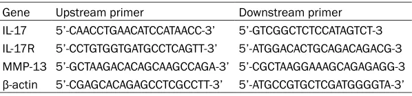
infec-Table 1. Primer sequences
Gene Upstream primer Downstream primer
IL-17 5’-CAACCTGAACATCCATAACC-3’ 5’-GTCGGCTCTCCATAGTCT-3 IL-17R 5’-CCTGTGGTGATGCCTCAGTT-3’ 5’-ATGGACACTGCAGACAGACG-3 MMP-13 5’-GCTAAGACACAGCAAGCCAGA-3’ 5’-CGCTAAGGAAAGCAGAGAGG-3
β-actin 5’-CGAGCACAGAGCCTCGCCTT-3’ 5’-ATGCCGTGCTCGATGGGGTA-3’
tion, showing differential expression in serum and thoracic edema in patients with tubercu-lous pleurisy [8]. Matrix metalloproteinase-13 (MMP-13), also known as collagenase-3, is a member of the matrix metalloproteinase family. It exhibits a strong type II collagen degradation ability [9]. Studies have shown that high expres-sion of MMP-13 is conducive for the destruc-tion and degradadestruc-tion of the extracellular matrix in tuberculosis and is closely related to the spread, metastasis, and prognosis of tubercu-losis [10]. However, few reports have been pub-lished regarding IL-17, IL-17R, and MMP-13 expression in ST disease.
Therefore, this study explored changes in IL-17, IL-17R and MMP-13 expression in ST lesions, examining mutual correlations in different types of lesions.
Materials and methods
In this study, 40 patients with ST that under-went surgical treatment, from January 2014 to December 2017, were retrospectively analyzed. Patients with ST were diagnosed according to clinical manifestations, pathological tions, acid-fast staining, and imaging examina-tions. Of these, 22 patients with proliferative lesions were included. There were 12 male and 10 female patients, with an age range of 20-65 years. In addition, 18 patients with necrotic lesions, including 8 males and 10 females, aged 19-70 years, were included. Another 20 subjects with normal cancellous bones served as the control group, including 10 males and 10 females, between 21 and 69 years of age. The study was approved by the Medical Ethics Committee of Ningxia People’s Hospital. All family members and patients provided informed consent.
Inclusion and exclusion criteria for patients with tuberculosis
Inclusion criteria: Age > 18 years old, no other hereditary diseases, no autism, no memory im-
ease, malignant tumors, and history of cancer and rheumatoid immune system disease.
Main reagents and instruments
RNA reversing transcription and real-time poly-merase chain reaction (PCR) kits were pur-chased from TransGen Biotech, China. TRIzol Reagent was obtained from Invitrogen, USA,
while the ABI 7500 PCR amplification instru -ment was procured from ABI, USA. Primers were designed and synthesized by Shanghai Biotech Co., Ltd. (Table 1).
Quantitative reverse-transcription polymerase chain reaction (qRT-PCR) analysis
During the operation, lesion tissues were ex- cised from patients (5 mm × 5 mm) and stored in liquid nitrogen for 1 hour. Total RNA was extracted using TRIzol. Ehe extracted RNA was detected by agarose gel electrophoresis and ultraviolet spectrophotometer for purity, con-centration, and integrity. Total RNA was reverse transcribed using a reverse transcription kit. Recombinant cDNA was collected and stored for use, in strict accordance with manufacturer instructions. PCR reaction system included the
following reagents: 10 μL of 2 × TransStart® Top Green qPCR SuperMix, 0.6 μL of each of
the upstream and downstream primers, and
2.0 μL of cDNA supplemented to 20 μL of
nuclease-free water. ABI 7500 was used for
amplification. PCR reaction conditions were as
follows: Pre-denaturation at 95°C for 10 min-utes and 40 cycles of 94°C for 30 seconds, 55°C for 30 seconds, and 72°C for 60 onds. This was followed by 55°C for 30 sec-onds and 72°C for 60 secsec-onds. In this study,
β-actin was used as an internal reference. Data were analyzed using 2-Δct method. The experi -ment was performed three times.
Statistical analysis
SPSS 20.0 software package (Guangzhou Bomai) was used to perform statistical analysis
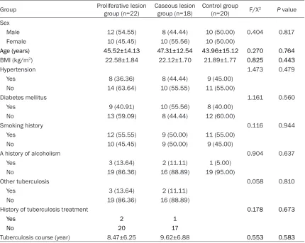
[image:2.612.91.390.83.153.2]dis-Table 2. Comparison of baseline clinical data among the three groups of patients
Group Proliferative lesion group (n=22) Caseous lesion group (n=18) Control group (n=20) F/X2 P value
Sex
Male 12 (54.55) 8 (44.44) 10 (50.00) 0.404 0.817
Female 10 (45.45) 10 (55.56) 10 (50.00)
Age (years) 45.52±14.13 47.31±12.54 43.96±15.12 0.270 0.764
BMI (kg/m2) 22.58±1.84 22.12±1.70 21.89±1.77 0.825 0.443
Hypertension 1.473 0.479
Yes 8 (36.36) 8 (44.44) 9 (45.00)
No 14 (63.64) 10 (55.55) 11 (55.00)
Diabetes mellitus 1.161 0.560
Yes 9 (40.91) 10 (55.56) 8 (40.00)
No 13 (59.09) 8 (44.44) 12 (60.00)
Smoking history 0.116 0.944
Yes 12 (55.55) 9 (50.00) 11 (55.00)
No 10 (45.45) 9 (50.00) 9 (45.00)
A history of alcoholism 0.904 0.637
Yes 3 (13.64) 2 (11.11) 1 (5.00)
No 19 (86.36) 16 (88.89) 19 (95.00)
Other tuberculosis 0.058 0.810
Yes 3 (13.64) 2 (11.11)
No 19 (86.36) 16 (88.89)
History of tuberculosis treatment 0.178 0.673
Yes 2 1
No 20 17
Tuberculosis course (year) 8.47±6.25 9.62±6.88 0.553 0.583
on collected data. Data were plotted using GraphPad Prism 7 (Shanghai Beka), wherein count data usage rates (%) were expressed. Chi-squared test was also used. Measurement data are expressed as mean ± standard devia-tion (means ± SD). Three groups were tested using one-way ANOVA, indicated by F values. LSD was used for pairwise comparisons. Analysis of variance was used for comparisons
between groups. Correlation coefficients of
IL-17, IL-17R, and MMP-13 were calculated by Pearson’s correlation analysis. Correlations between IL-17, IL-17R, MMP-13, and lesion types were investigated by Spearman’s correla-tion. Statistical differences are indicated by P<0.05.
Results
Baseline characteristics
Comparison of the clinical data of the three groups of patients revealed no statistical differ-ences between proliferative lesions, caseous necrotic lesions, and control groups in gender,
age, body mass index (BMI), hypertension, dia-betes, smoking history, history of tuberculosis, and alcohol abuse history (P > 0.05) (Table 2).
Expression of IL-17, IL-17R, and MMP-13 in tissues from three groups of patients
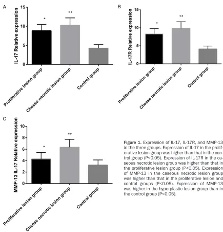
Relative expression levels of IL-17, IL-17R, and MMP-13 were detected in the tissues from pro-liferative lesions, caseous necrosis, and con-trol groups of patients. IL-17, IL-17R, and
MMP-13 expression was significantly higher in the
necrotic lesion group than in the proliferative lesion and control groups (P<0.05). Expression of IL-17, IL-17R, and MMP-13 was higher in the proliferative lesion group than in the control group (P<0.05) (Figure 1 and Table 3).
Correlation between IL-17, IL-17R, and MMP-13 expression in patients with proliferative le-sions and caseous necrotic lele-sions
[image:3.612.93.523.84.427.2]Table 3. Expression of IL-17, IL-17, IL-17, and MMP-13 in lesions of the three groups of patients
Group Proliferative lesion group (n=22) Caseous lesion group (n=18) group (n=20) F-numberControl P value IL-17 8.84±1.65* 10.28±1.92*,** 4.25±0.95 80.379 <0.001
IL-17R 8.35±1.44* 9.84±1.86*,** 4.11±0.83 84.794 <0.001
MMP-13 4.27±1.15* 6.34±1.38*,** 3.24±0.89 35.520 <0.001
Note: *P<0.05 compared with the control group; **P<0.05 when proliferative lesion group
compared with caseous lesion group.
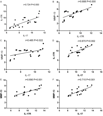
group than in the proliferative lesion group. In- dicators gradually increased with the severity of the disease (P<0.05). Relative expression levels of IL-17, IL-17R, and MMP-13 in patients with proliferative lesions showed a positive
cor-A chronic skeletal joint disease, ST is a type of secondary tissue injury disease caused by M. tuberculosis in patients with tuberculosis via blood circulation into the spine [11]. Statistics have shown that ST is mainly a type of vertebral Figure 1. Expression of IL-17, IL-17R, and MMP-13 in the three groups. Expression of IL-17 in the prolif-erative lesion group was higher than that in the con-trol group (P<0.05). Expression of IL-17R in the ca-seous necrotic lesion group was higher than that in the proliferative lesion group (P<0.05). Expression of MMP-13 in the caseous necrotic lesion group was higher than that in the proliferative lesion and control groups (P<0.05). Expression of MMP-13 was higher in the hyperplastic lesion group than in the control group (P<0.05).
relation (P<0.05). Furth- ermore, a positive corre-lation was observed be- tween relative expres-sion levels of IL-17, IL- 17R, and MMP-13 and degree of patient condi-tions (P<0.05) (Figure 2, Tables 4, 5).
[image:4.612.90.397.565.629.2]Figure 2. Correlation between IL-17, IL-17R, and MMP-13 expression levels in patients with proliferative lesions and caseous necrotic lesions. A. Proliferative lesions (r=-0.724). B. Hyperplastic lesion group shows a positive correla-tion between expression of IL-17R and MMP-13 (r=-0.668). C. Proliferative type group shows a positive correlacorrela-tion between expression of IL-17 and MMP-13 (r=0.485 and P=0.022). D. Caseous necrotic type group shows a positive correlation between expression of IL-17 and IL-17R in the lesion tissues (r=0.670 and P=0.002). E. Proliferative type group shows a positive correlation between expression of IL-17R and MMP-13 (r=0.690 and P=0.001). F. Caseous necrotic type group shows a positive correlation between expression of IL-17 and MMP-13 (r=0.715 and P=0.001).
tuberculosis, with a large number of cases are reported in adolescents [12]. ST may be misdi-agnosed in clinic, causing patients to miss the best treatment opportunity. Patients with severe disease may also have spinal cord
inju-ries and paraplegia. Like other types tuberculo-sis, ST also has three pathological forms of exu-dation, hyperplasia, and necrosis. The exuda-tive type of lesions mainly manifests as serous
Table 4. Correlation analysis of IL-17, IL-17, and MMP-13 between the two groups
Group ρ P
Proliferative lesion group VS Cheese necrotic lesion group (IL-17) 0.499 0.035 Proliferative lesion group VS Cheese necrotic lesion group (IL-17R) 0.496 0.036 Proliferative lesion group VS Cheese necrotic lesion group (MMP-13) 0.472 0.048
manifestations of clinically exudative ST pa- tients, thus it is rarely diagnosed and treated [13].
Interleukin, a type of cytokine that regulates cell growth and differentiation, is secreted by leukocytes. The main role of this cytokine is to mediate T-cell and B-cell activation and prolif-eration, regulate immune cell differentiation
and activation, and fight against inflammatory
reactions [14]. IL-17 is a class of ILs that is not well-studied. Recent studies have shown
that IL-17 secretes pro-inflammatory cytokines
through CD4+ memory T lymphocytes and monocytes [15]. IL-17 and its receptors are widely expressed in the tissues. It mainly
func-tions by binding to IL-17R, promoting inflamma -tory response and recruiting central granulo-cytes. It has been shown that IL-17 upregulates MMP and IL-6 expression during cell activation and stimulates chondrocytes to produce nitric oxide and nitric oxide synthase [16]. High con-centrations of oxidative nitrogen stimulate the activity of MMP and promote the degradation of chondrocytes.
Matrix metalloproteinases are a class of calci-um-dependent zinc-containing endopeptidas-es. Studies have shown that MMPs play a key role in tissue remodeling and are involved in various pathological processes, including angio- genesis, tissue repair, and arthritis [17]. An important member of the MMP family, MMP-13
has a specific cleavage effects on helicase
enzymes. The ability of MMP-13 to degrade type II collagen is 5-10 times higher than that of MMP-1, the highest among all MMPs [18]. Studies have shown that high expression of MMP-13 may promote the degradation of dam-aged outer matrix of tuberculosis-infected
tis-sue and is associated with tuberculosis metas-tasis [19]. In the study by González-Avila G et al. [17], M. tuberculosis was thought to promote
expression of MMP-13 in lung fibroblasts and
participate in the formation and decomposition of various collagens. However, very little is known about these two factors in ST. Whether IL-17, IL-17R, and MMP-13 play any roles in the development of ST remains unclear. Therefore, this study explored the relationship between these factors and the extent of lesions by eval-uating expression levels of IL-17, IL-17R, and MMP-13 in the lesions of patients with ST, aim-ing to provide clinical reference.
The current study detected expression levels of IL-17 and IL-17R in patients with proliferative lesions and necrotic lesions. Expression of MMP-13 was higher in these groups than in the control group. Expression of IL-17, IL-17R, and MMP-13 in the lesions of patients with caseous necrosis was higher than in the lesions of patients with proliferative lesions. This obser-vation indicates that IL-17, IL-17R, and MMP-13 are differentially expressed in ST and that expression levels vary in different pathological lesion tissues. These biomarkers may be poten-tially useful for ST pathological typing. It has been shown that IL-17 expression was higher in patients with tuberculosis potential and active infections than in normal subjects [20]. It was found that IL-17 mediates immune-protection from M. tuberculosis (MtbHN 878) by IL-17 receptor signaling in non-hematopoietic cells [21]. These effects are mainly induced by the chemokine CXCL-13. CXCL-13 is an important B lymphocyte chemokine. Its expression may be related to the pathogenesis of various
diseas-es, such as autoimmune and inflammatory dis -Table 5. Correlation analysis of IL-17, IL-17R, and MMP-13 between the groups
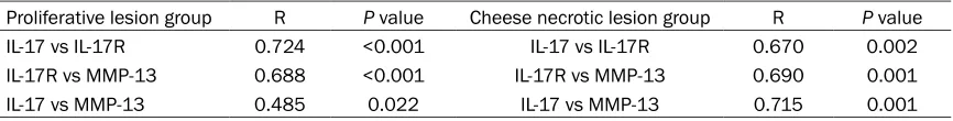
Proliferative lesion group R P value Cheese necrotic lesion group R P value
IL-17 vs IL-17R 0.724 <0.001 IL-17 vs IL-17R 0.670 0.002
IL-17R vs MMP-13 0.688 <0.001 IL-17R vs MMP-13 0.690 0.001
[image:6.612.90.523.172.227.2]eases. Ong [22] showed that high expression of MMP-13 in patients with tuberculosis was closely related to clinical and radiological mark-ers of lung tissue destruction.
The current study examined expression of IL-17, IL-17R, and MMP-13 in patients with different
pathological types of ST, finding that IL-17,
IL-17R, and MMP-13 are involved in the devel-opment of ST. At the end of the study, the cor-relation between IL-17, IL-17R, and MMP-13 expression was analyzed. Results showed a positive correlation between each indicator, suggesting the presence of a regulatory rela-tionship between IL-17, IL-17R, and MMP-13. In the study by Koenders M, IL-17 was shown to enhance expression of MMP-13 in
chondro-cytes and synovial fibroblasts, thereby promot -ing a reduction in cartilage proteoglycans and collagen [23]. Another study showed that IL-17 regulates the activity of MMP-13 [24]. Therefore, it was speculated that, due to long-term infec-tion in patients, Th17 cells are activated to secrete IL-17 and IL-17 binds to its receptor (IL-17R, etc.). This promotes the secretion of
various inflammatory factors and MMP-13 pro -tein. Secretion is involved in the development of the disease. However, more in vitro experi-ments and animal experiexperi-ments should be
con-ducted to verify the specific mechanisms.
Present results and previous studies indicate that differential expression of IL-17, IL-17R, and MMP-13 in ST lesions may serve as a potential observation index.
The present study had a few limitations, how-ever. First, the sample size was small and whether the results may have caused errors is unclear. Second, this study failed to follow-up with patients to observe whether differential expression of each index had an impact on patient survival. Present experiments have not been thoroughly studied. How to regulate the relationship between IL-17, IL-17R, and MMP-13 expression has not been proven. Moreover, the study did not determine protein levels and did not observe the relationship between the above factors and clinical symptoms. These should be examined in the future.
In summary, IL-17, IL-17R, and MMP-13 are highly expressed in lesions of patients with ST. These markers showed differential expression in lesions of patients with different degrees of disease. Expression levels of IL-17, IL-17R, and
MMP-13 are correlated, serving as potential observation indicators of ST.
Disclosure of conflict of interest None.
Address correspondence to: Cheng Li, Department of Spine and Orthopedics, Ningxia People’s Ho- spital, No.301 North Zhengyuan Street, Jinfeng District, Yinchuan 750011, Ningxia, China. Tel: +86-18709506610; E-mail: [email protected]
References
[1] Treerat P, Prince O, Cruz-Lagunas A, Munoz-Torrico M, Salazar-Lezama MA, Selman M, Fal-lert-Junecko B, Reinhardt TA, Alcorn JF, Kaush-al D, Zuniga J, Rangel-Moreno J, Kolls JK and Khader SA. Novel role for IL-22 in protection during chronic Mycobacterium tuberculosis HN878 infection. Mucosal Immunol 2017; 10: 1069-1081.
[2] Chen C, Zhu T, Wang Z, Peng H, Kong W, Zhou Y, Shao Y, Zhu L and Lu W. High latent TB infec-tion rate and associated risk factors in the Eastern China of low TB incidence. PLoS One 2015; 10: e0141511.
[3] Lin CH, Lin CJ, Kuo YW, Wang JY, Hsu CL, Chen JM, Cheng WC and Lee LN. Tuberculosis mor-tality: patient characteristics and causes. BMC Infect Dis 2014; 14: 5.
[4] Bhat S, Kangoo K, Zahoor A and Nazir A. Con-comitant pyogenic spondylodiscitis with large psoas abcess in known case of tuberculosis spine; presenting as refractory tuberculosis. J Spine 2015; 4: 2.
[5] Liu P, Jiang H, Li S, Lin Z and Jiang J. Determi-nation of anti-tuberculosis drug concentration and distribution from sustained release micro-spheres in the vertebrae of a spinal tuberculo-sis rabbit model. J Orthop Res 2017; 35: 200-208.
[6] Tang L, Liu S, Bao YC, Gao RX, Han CF, Sun XC, Zhang WL and Feng SQ. Study on the
relation-ship between vitamin D deficiency and suscep -tibility to spinal tuberculosis. Int J Surg 2017; 44: 99-103.
[7] Coffelt SB, Kersten K, Doornebal CW, Weiden J, Vrijland K, Hau CS, Verstegen NJM, Ciampri-cotti M, Hawinkels LJAC, Jonkers J and de Viss-er KE. IL-17-producing gammadelta T cells and neutrophils conspire to promote breast cancer metastasis. Nature 2015; 522: 345-348. [8] Basile JI, Kviatcovsky D, Romero MM, Balboa
L, Monteserin J, Ritacco V, Lopez B, Sabio y Garcia C, Garcia A, Vescovo M, Montaner PG, Palmero D, Del Carmen Sasiain M and de la Barrera S. Mycobacterium tuberculosis multi-drug-resistant strain M induces IL-17+
and TGF-beta-dependent mechanism in pa-tients with MDR-TB tuberculosis. Clin Exp Im-munol 2017; 187: 160-173.
[9] Gerber A, Sonnet J, Goldklang M, Stearns K, Arteaga-Solis E and D’Armiento J. A Novel Role of Matrix Metalloproteinase-13 (MMP-13) in
Aging and Lung Inflammation. In: editors. A69.
NOVEL SIGNALING CASCADES IN LUNG INJU-RY, INFLAMMATION, AND REPAIR. American Thoracic Society; 2018. p. A2243-A2243. [10] Al Shammari B, Shiomi T, Tezera L, Bielecka
MK, Workman V, Sathyamoorthy T, Mauri F, Jayasinghe SN, Robertson BD, D’Armiento J, Friedland JS and Elkington PT. The extracellu-lar matrix regulates granuloma necrosis in tu-berculosis. J Infect Dis 2015; 212: 463-473. [11] Shen X, Liu H, Wang G, Pang X, Luo C, Zeng H,
Xu Z, Liu X and Wang X. The role of single-stage posterior debridement, interbody fusion with titanium mesh cages and short-segment in-strumentation in thoracic and lumbar spinal tuberculosis. J Neurosurg Sci 2017; 61: 473-480.
[12] Yu WY, Lou C, Liu FJ and He DW. Clinical effi -cacy of one stage posterior debridement joint
graft fixation for lumbar vertebral fractures in
spinal tuberculosis patients with compression. Eur Rev Med Pharmacol Sci 2016; 20: 3161-3167.
[13] Tang Y, Yin L, Tang S, Zhang H and Lan J. Ap-plication of molecular, microbiological, and im-munological tests for the diagnosis of bone and joint tuberculosis. J Clin Lab Anal 2018; 32.
[14] Zhou Y, Wu J, Wang W and Sun M. Association between interleukin family gene polymor-phisms and recurrent aphthous stomatitis risk. Genes Immun 2019; 20: 90-101.
[15] Tonaco MM, Moreira JD, Nunes FFC, Loures CMG, Souza LR, Martins JM, Silva HR, Porto AHR, Toledo VPCP, Miranda SS and Guimarães
TMPD. Evaluation of profile and functionality of
memory T cells in pulmonary tuberculosis. Immunol Lett 2017; 192: 52-60.
[16] Eruslanov EB, Lyadova IV, Kondratieva TK, Majorov KB, Scheglov IV, Orlova MO and Apt AS. Neutrophil responses to Mycobacterium tuberculosis infection in genetically suscepti-ble and resistant mice. Infect Immun 2005; 73: 1744-1753.
[17] González-Avila G, Sandoval C, Herrera MT, Ruiz V, Sommer B, Sada E, Ramos C and Sarabia
MC. Mycobacterium tuberculosis effects on fi -broblast collagen metabolism. Respiration 2009; 77: 195-202.
[18] Stearns K, Goldklang M and D’Armiento JM. A Novel Role For Alpha-1 Antitrypsin In Protecting The Lung From Destruction By Mmp-13. In: editors. A73. BEYOND CIGARETTE SMOKE IN COPD. American Thoracic Society; 2017. p. A2430-A2430.
[19] Di G, Chao F and Huirong Z. The effect of mini-mally invasive operation on the serum interleu-kins and matrix metalloproteinases of old pa-tients with spinal tuberculosis and periopera-tive nursing. Biomedical Research 2017; 28. [20] Cowan J, Pandey S, Filion LG, Angel JB, Kumar
A and Cameron DW. Comparison of interferon-gamma-, interleukin (IL)-17- and IL-22-express-ing CD4 T cells, IL-22-expressIL-22-express-ing granulocytes
and proinflammatory cytokines during latent
and active tuberculosis infection. Clin Exp Im-munol 2012; 167: 317-329.
[21] Gopal R, Monin L, Slight S, Uche U, Blanchard E, Fallert Junecko BA, Ramos-Payan R, Stall-ings CL, Reinhart TA, Kolls JK, Kaushal D, Nag-arajan U, Rangel-Moreno J and Khader SA. Unexpected role for IL-17 in protective immu-nity against hypervirulent Mycobacterium tu-berculosis HN878 infection. PLoS Pathog 2014; 10: e1004099.
[22] Ong CW, Elkington PT and Friedland JS. Tuber-culosis, pulmonary cavitation, and matrix me-talloproteinases. Am J Respir Crit Care Med 2014; 190: 9-18.
[23] Koenders MI, Kolls JK, Oppers-Walgreen B, van den Bersselaar L, Joosten LA, Schurr JR, Schwarzenberger P, van den Berg WB and
Lub-berts E. Interleukin-17 receptor deficiency re -sults in impaired synovial expression of inter-leukin-1 and matrix metalloproteinases 3, 9, and 13 and prevents cartilage destruction dur-ing chronic reactivated streptococcal cell wall-induced arthritis. Arthritis Rheum 2005; 52: 3239-3247.