organic papers
Acta Cryst.(2006). E62, o1155–o1156 doi:10.1107/S1600536806006179 Jinget al. C
7H8N2O2
o1155
Acta Crystallographica Section E Structure Reports
Online
ISSN 1600-5368
2-Methyl-6-nitroaniline
Zuo-Liang Jing,* Qiao-Zhen Zhang, Jia Jia and Ming Yu
College of Sciences, Tianjin University of Science and Technology, Tianjin 300222, People’s Republic of China
Correspondence e-mail: [email protected]
Key indicators
Single-crystal X-ray study T= 294 K
Mean(C–C) = 0.002 A˚ Rfactor = 0.037 wRfactor = 0.107
Data-to-parameter ratio = 11.9
For details of how these key indicators were automatically derived from the article, see http://journals.iucr.org/e.
Received 6 February 2006 Accepted 20 February 2006
#2006 International Union of Crystallography
All rights reserved
In the crystal structure of the title compound, C7H8N2O2, the
molecules are linked through N—H O and N—H N hydrogen bonds, forming an extended supramolecule, which contributes to the stability of the structure in the solid state. There are two molecules in the asymmetric unit.
Comment
The syntheses of new intentionally designed crystal structures are part of a major strand of modern chemistry (Belloniet al., 2005; Tynanet al., 2005). Metal complexes of Schiff bases have attracted much attention because they can be utilized as model compounds for the active centres in various enzymes and proteins (Kahwa et al., 1986; Santoset al., 2001). One of the aims of crystal engineering is to establish control over the preparation of crystalline solid materials so that their subse-quent architecture and properties are predictable. In order to investigate a crystal structure with strong intermolecular bonding that might provide useful information in the field of crystal engineering, we report here the synthesis and the molecular and crystal structures of the title compound (I).
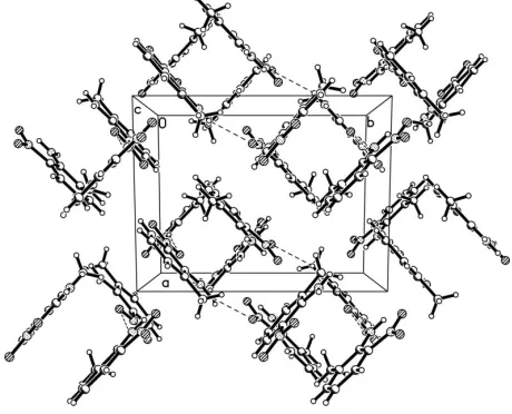
A view of the two molecules of the asymmetric unit of (I) is shown in Fig. 1, and selected bond lengths and angles are reported in Table 1. They are all within the normal ranges for such values. The molecules are linked through N—H O and N—H N hydrogen bonds (Table 2), forming a supra-molecule which leads to a stable crystal structure. Fig. 2 shows a portion of this extensively hydrogen-bonded supramolecular assembly.
Experimental
obtained by slow evaporation of anN,N-dimethylformamide solution of (I).
Crystal data
C7H8N2O2
Mr= 152.15
Monoclinic,P21=c a= 8.9844 (16) A˚
b= 11.359 (2) A˚
c= 14.785 (3) A˚
= 103.814 (2)
V= 1465.1 (5) A˚3
Z= 8
Dx= 1.380 Mg m
3 MoKradiation Cell parameters from 2333
reflections
= 2.9–23.4
= 0.10 mm1
T= 294 (2) K Block, yellow 0.220.160.14 mm
Data collection
Bruker SMART CCD area-detector diffractometer
’and!scans
Absorption correction: multi-scan (SADABS; Sheldrick, 1996)
Tmin= 0.968,Tmax= 0.986 7772 measured reflections
2589 independent reflections 1931 reflections withI> 2(I)
Rint= 0.014
max= 25.0
h=9!10
k=13!13
l=15!17
Refinement
Refinement onF2
R[F2> 2(F2)] = 0.037
wR(F2) = 0.107
S= 1.04 2589 reflections 217 parameters
H atoms treated by a mixture of independent and constrained refinement
w= 1/[2(F
o2) + (0.0597P)2 + 0.1509P]
whereP= (Fo2+ 2Fc2)/3 (/)max< 0.001
max= 0.11 e A˚
3
min=0.17 e A˚
[image:2.610.310.565.74.193.2]3
Table 1
Selected geometric parameters (A˚ ,). O1—N1 1.2316 (17) O2—N1 1.2368 (17) O3—N3 1.2208 (19) O4—N3 1.2393 (19) N1—C4 1.4312 (18) N2—C3 1.346 (2)
N2—H2A 0.92 (2) N2—H2B 0.84 (2) N3—C13 1.434 (2) N4—C14 1.349 (2) N4—H4A 1.02 (3) N4—H4B 0.902 (19) O1—N1—O2 120.81 (13)
O1—N1—C4 119.33 (13) O2—N1—C4 119.86 (12) C3—N2—H2A 116.4 (13) C3—N2—H2B 121.4 (14) H2A—N2—H2B 122.0 (19)
O3—N3—O4 120.65 (15) O3—N3—C13 119.87 (16) O4—N3—C13 119.47 (15) N2—C3—C4 123.94 (14) N2—C3—C2 119.31 (14)
Table 2
Hydrogen-bond geometry (A˚ ,).
D—H A D—H H A D A D—H A
N4—H4B O1 0.902 (19) 2.341 (19) 3.161 (2) 151.2 (16) N4—H4A O4 1.02 (3) 1.84 (2) 2.616 (2) 130 (2) N2—H2B O4i
0.84 (2) 2.38 (2) 3.151 (2) 152.6 (18) N2—H2A N1 0.92 (2) 2.55 (2) 2.915 (2) 103.8 (14) N2—H2A O2 0.92 (2) 1.90 (2) 2.607 (2) 131.4 (17)
Symmetry code: (i)xþ1;y1 2;zþ
3 2.
All C-bound H atoms were positioned geometrically and refined using the riding-model approximation, with C–H = 0.93 A˚ and U (H) = 1.2U (C). H atoms attached to N and O atoms were
Data collection:SMART(Bruker, 1999); cell refinement:SAINT (Bruker, 1999); data reduction:SAINT; program(s) used to solve structure:SHELXS97(Sheldrick, 1997); program(s) used to refine structure: SHELXL97 (Sheldrick, 1997); molecular graphics: SHELXTL (Bruker, 1997); software used to prepare material for publication:SHELXTL.
References
Belloni, M., Kariuki, B. M., Manickam, M., Wilkie, J. & Preece, J. A. (2005).
Cryst. Growth Des.5, 1443–1449.
Bruker (1997).SHELXTL. Version 5.10 for Windows NT. Bruker AXS Inc., Madison, Wisconsin, USA.
Bruker (1999).SMART(Version 5.0) andSAINT(Version 4.0) for Windows NT. Bruker AXS Inc., Madison, Wisconsin, USA.
Kahwa, I. A., Selbin, J., Hsieh, T. C.-Y. & Laine, R. A. (1986).Inorg. Chim. Acta,118, 179–185.
Rabjohn, N. (1963).Org. Synth. Coll. IV [M], pp. 42–44.
Santos, M. L. P., Bagatin, I. A., Pereira, E. M. & Ferreira, A. M. D. C. (2001).J. Chem. Soc. Dalton Trans.pp. 838–844.
Sheldrick, G. M. (1996).SADABS. University of Go¨ttingen, Germany. Sheldrick, G. M. (1997). SHELXS97 and SHELXL97. University of
Go¨ttingen, Germany.
Tynan, E., Jensen, P., Lees, A. C., Moubaraki, B., Murray, K. S. & Kruger, P. E.
Figure 1
The asymmetric unit of the title compound, (I), with 30% probability displacement ellipsoids.
Figure 2
[image:2.610.324.554.246.432.2]supporting information
sup-1 Acta Cryst. (2006). E62, o1155–o1156
supporting information
Acta Cryst. (2006). E62, o1155–o1156 [https://doi.org/10.1107/S1600536806006179]
2-Methyl-6-nitroaniline
Zuo-Liang Jing, Qiao-Zhen Zhang, Jia Jia and Ming Yu
2-Methyl-6-nitroaniline
Crystal data
C7H8N2O2 Mr = 152.15
Monoclinic, P21/c Hall symbol: -P 2ybc a = 8.9844 (16) Å b = 11.359 (2) Å c = 14.785 (3) Å β = 103.814 (2)° V = 1465.1 (5) Å3 Z = 8
F(000) = 640 Dx = 1.380 Mg m−3
Mo Kα radiation, λ = 0.71073 Å Cell parameters from 2333 reflections θ = 2.9–23.4°
µ = 0.10 mm−1 T = 294 K Block, yellow
0.22 × 0.16 × 0.14 mm
Data collection
Bruker SMART CCD area-detector diffractometer
Radiation source: fine-focus sealed tube Graphite monochromator
φ and ω scans
Absorption correction: multi-scan (SADABS; Sheldrick, 1996) Tmin = 0.968, Tmax = 0.986
7772 measured reflections 2589 independent reflections 1931 reflections with I > 2σ(I) Rint = 0.014
θmax = 25.0°, θmin = 2.3° h = −9→10
k = −13→13 l = −15→17
Refinement
Refinement on F2 Least-squares matrix: full R[F2 > 2σ(F2)] = 0.037 wR(F2) = 0.107 S = 1.04 2589 reflections 217 parameters 0 restraints
Primary atom site location: structure-invariant direct methods
Secondary atom site location: difference Fourier map
Hydrogen site location: inferred from neighbouring sites
H atoms treated by a mixture of independent and constrained refinement
w = 1/[σ2(F
o2) + (0.0597P)2 + 0.1509P] where P = (Fo2 + 2Fc2)/3
(Δ/σ)max < 0.001 Δρmax = 0.11 e Å−3 Δρmin = −0.17 e Å−3
Special details
Geometry. All e.s.d.'s (except the e.s.d. in the dihedral angle between two l.s. planes) are estimated using the full
Refinement. Refinement of F2 against ALL reflections. The weighted R-factor wR and goodness of fit S are based on F2, conventional R-factors R are based on F, with F set to zero for negative F2. The threshold expression of F2 > σ(F2) is used only for calculating R-factors(gt) etc. and is not relevant to the choice of reflections for refinement. R-factors based on F2 are statistically about twice as large as those based on F, and R- factors based on ALL data will be even larger.
Fractional atomic coordinates and isotropic or equivalent isotropic displacement parameters (Å2)
x y z Uiso*/Ueq
O1 0.28792 (14) 0.54144 (11) 0.41093 (8) 0.0733 (4)
O2 0.37257 (15) 0.56793 (12) 0.55800 (8) 0.0800 (4)
O3 0.8289 (2) 0.99462 (14) 0.60405 (10) 0.1025 (5)
O4 0.64899 (17) 0.87510 (15) 0.61259 (9) 0.0954 (5)
N1 0.28697 (15) 0.51650 (11) 0.49186 (9) 0.0551 (3)
N2 0.25384 (19) 0.45531 (14) 0.67751 (10) 0.0657 (4)
H2A 0.325 (2) 0.5098 (17) 0.6686 (14) 0.088 (6)*
H2B 0.246 (2) 0.4356 (18) 0.7308 (16) 0.093 (7)*
N3 0.72369 (18) 0.93022 (13) 0.56607 (10) 0.0694 (4)
N4 0.48473 (19) 0.77306 (16) 0.46317 (12) 0.0755 (5)
H4A 0.515 (3) 0.776 (2) 0.5342 (18) 0.127 (8)*
H4B 0.425 (2) 0.7185 (16) 0.4276 (13) 0.076 (6)*
C1 0.0331 (2) 0.29133 (16) 0.70392 (11) 0.0688 (5)
H1A −0.0453 0.2325 0.6986 0.103*
H1B 0.0024 0.3614 0.7309 0.103*
H1C 0.1270 0.2623 0.7429 0.103*
C2 0.05660 (17) 0.31901 (12) 0.60917 (10) 0.0502 (4)
C3 0.16933 (16) 0.40320 (12) 0.60046 (9) 0.0453 (3)
C4 0.18314 (15) 0.42848 (12) 0.50911 (9) 0.0452 (3)
C5 0.09387 (18) 0.37194 (13) 0.43156 (10) 0.0550 (4)
H5 0.1064 0.3897 0.3724 0.066*
C6 −0.0115 (2) 0.29084 (14) 0.44216 (11) 0.0639 (4)
H6 −0.0713 0.2524 0.3906 0.077*
C7 −0.02885 (19) 0.26593 (13) 0.53135 (12) 0.0607 (4)
H7 −0.1019 0.2107 0.5380 0.073*
C8 0.4243 (2) 0.75291 (17) 0.26788 (13) 0.0776 (5)
H8A 0.4214 0.7584 0.2027 0.116*
H8B 0.3266 0.7756 0.2780 0.116*
H8C 0.4467 0.6733 0.2885 0.116*
C9 0.54645 (16) 0.83340 (13) 0.32171 (10) 0.0514 (4)
C10 0.63353 (17) 0.89972 (14) 0.27732 (11) 0.0579 (4)
H10 0.6151 0.8953 0.2128 0.069*
C11 0.74836 (19) 0.97342 (14) 0.32481 (12) 0.0628 (4)
H11 0.8065 1.0170 0.2925 0.075*
C12 0.77552 (19) 0.98172 (13) 0.41864 (12) 0.0602 (4)
H12 0.8529 1.0308 0.4511 0.072*
C13 0.68729 (17) 0.91653 (13) 0.46679 (10) 0.0514 (4)
supporting information
sup-3 Acta Cryst. (2006). E62, o1155–o1156
Atomic displacement parameters (Å2)
U11 U22 U33 U12 U13 U23
O1 0.0855 (9) 0.0889 (9) 0.0522 (7) −0.0110 (7) 0.0299 (6) 0.0102 (6)
O2 0.0809 (8) 0.0968 (9) 0.0590 (7) −0.0363 (7) 0.0103 (6) 0.0030 (7)
O3 0.1262 (13) 0.1014 (11) 0.0636 (9) −0.0324 (10) −0.0095 (8) −0.0131 (7)
O4 0.1006 (10) 0.1383 (13) 0.0502 (7) −0.0117 (9) 0.0235 (7) 0.0033 (8)
N1 0.0574 (8) 0.0626 (8) 0.0474 (8) −0.0022 (6) 0.0170 (6) 0.0054 (6)
N2 0.0795 (10) 0.0767 (10) 0.0375 (8) −0.0159 (8) 0.0075 (7) 0.0047 (7)
N3 0.0793 (10) 0.0752 (10) 0.0502 (8) 0.0042 (8) 0.0088 (8) 0.0003 (7)
N4 0.0739 (10) 0.0901 (11) 0.0644 (10) −0.0252 (9) 0.0205 (8) 0.0114 (9)
C1 0.0792 (12) 0.0713 (11) 0.0634 (11) −0.0009 (9) 0.0320 (9) 0.0128 (8)
C2 0.0582 (9) 0.0451 (8) 0.0507 (9) 0.0070 (7) 0.0198 (7) 0.0060 (6)
C3 0.0492 (8) 0.0463 (8) 0.0403 (8) 0.0067 (6) 0.0103 (6) 0.0035 (6)
C4 0.0500 (8) 0.0451 (8) 0.0423 (8) 0.0038 (6) 0.0143 (6) 0.0025 (6)
C5 0.0693 (10) 0.0546 (9) 0.0421 (8) 0.0038 (8) 0.0152 (7) −0.0036 (7)
C6 0.0767 (11) 0.0580 (9) 0.0542 (10) −0.0079 (8) 0.0101 (8) −0.0128 (8)
C7 0.0666 (10) 0.0488 (8) 0.0691 (11) −0.0077 (7) 0.0208 (9) −0.0040 (8)
C8 0.0716 (11) 0.0872 (13) 0.0674 (11) −0.0170 (10) 0.0034 (9) −0.0056 (10)
C9 0.0489 (8) 0.0542 (8) 0.0490 (9) 0.0005 (7) 0.0075 (7) 0.0030 (7)
C10 0.0616 (10) 0.0669 (10) 0.0453 (8) 0.0037 (8) 0.0130 (7) 0.0099 (7)
C11 0.0663 (10) 0.0626 (10) 0.0623 (10) −0.0068 (8) 0.0205 (8) 0.0158 (8)
C12 0.0610 (10) 0.0501 (9) 0.0665 (11) −0.0092 (7) 0.0092 (8) 0.0034 (7)
C13 0.0573 (9) 0.0509 (8) 0.0442 (8) 0.0034 (7) 0.0086 (7) 0.0020 (6)
C14 0.0491 (8) 0.0517 (8) 0.0492 (9) 0.0026 (7) 0.0138 (7) 0.0087 (6)
Geometric parameters (Å, º)
O1—N1 1.2316 (17) C4—C5 1.390 (2)
O2—N1 1.2368 (17) C5—C6 1.357 (2)
O3—N3 1.2208 (19) C5—H5 0.9300
O4—N3 1.2393 (19) C6—C7 1.393 (2)
N1—C4 1.4312 (18) C6—H6 0.9300
N2—C3 1.346 (2) C7—H7 0.9300
N2—H2A 0.92 (2) C8—C9 1.502 (2)
N2—H2B 0.84 (2) C8—H8A 0.9600
N3—C13 1.434 (2) C8—H8B 0.9600
N4—C14 1.349 (2) C8—H8C 0.9600
N4—H4A 1.02 (3) C9—C10 1.362 (2)
N4—H4B 0.902 (19) C9—C14 1.420 (2)
C1—C2 1.499 (2) C10—C11 1.383 (2)
C1—H1A 0.9600 C10—H10 0.9300
C1—H1B 0.9600 C11—C12 1.353 (2)
C1—H1C 0.9600 C11—H11 0.9300
C2—C7 1.362 (2) C12—C13 1.397 (2)
C2—C3 1.421 (2) C12—H12 0.9300
O1—N1—O2 120.81 (13) C5—C6—C7 119.08 (15)
O1—N1—C4 119.33 (13) C5—C6—H6 120.5
O2—N1—C4 119.86 (12) C7—C6—H6 120.5
C3—N2—H2A 116.4 (13) C2—C7—C6 122.81 (15)
C3—N2—H2B 121.4 (14) C2—C7—H7 118.6
H2A—N2—H2B 122.0 (19) C6—C7—H7 118.6
O3—N3—O4 120.65 (15) C9—C8—H8A 109.5
O3—N3—C13 119.87 (16) C9—C8—H8B 109.5
O4—N3—C13 119.47 (15) H8A—C8—H8B 109.5
C14—N4—H4A 115.3 (14) C9—C8—H8C 109.5
C14—N4—H4B 116.0 (12) H8A—C8—H8C 109.5
H4A—N4—H4B 126.7 (18) H8B—C8—H8C 109.5
C2—C1—H1A 109.5 C10—C9—C14 119.86 (14)
C2—C1—H1B 109.5 C10—C9—C8 120.83 (15)
H1A—C1—H1B 109.5 C14—C9—C8 119.31 (14)
C2—C1—H1C 109.5 C9—C10—C11 122.33 (15)
H1A—C1—H1C 109.5 C9—C10—H10 118.8
H1B—C1—H1C 109.5 C11—C10—H10 118.8
C7—C2—C3 119.44 (13) C12—C11—C10 119.52 (15)
C7—C2—C1 121.31 (14) C12—C11—H11 120.2
C3—C2—C1 119.24 (14) C10—C11—H11 120.2
N2—C3—C4 123.94 (14) C11—C12—C13 119.99 (15)
N2—C3—C2 119.31 (14) C11—C12—H12 120.0
C4—C3—C2 116.74 (13) C13—C12—H12 120.0
C5—C4—C3 121.93 (13) C12—C13—C14 121.50 (14)
C5—C4—N1 116.63 (12) C12—C13—N3 116.46 (14)
C3—C4—N1 121.42 (13) C14—C13—N3 122.03 (14)
C6—C5—C4 119.98 (14) N4—C14—C13 124.00 (15)
C6—C5—H5 120.0 N4—C14—C9 119.23 (15)
C4—C5—H5 120.0 C13—C14—C9 116.77 (13)
C7—C2—C3—N2 −179.85 (14) C14—C9—C10—C11 0.5 (2)
C1—C2—C3—N2 0.8 (2) C8—C9—C10—C11 −179.06 (16)
C7—C2—C3—C4 1.3 (2) C9—C10—C11—C12 −0.7 (3)
C1—C2—C3—C4 −178.07 (13) C10—C11—C12—C13 −0.3 (3)
N2—C3—C4—C5 179.65 (14) C11—C12—C13—C14 1.4 (2)
C2—C3—C4—C5 −1.6 (2) C11—C12—C13—N3 179.88 (15)
N2—C3—C4—N1 −2.3 (2) O3—N3—C13—C12 −1.3 (2)
C2—C3—C4—N1 176.52 (12) O4—N3—C13—C12 179.67 (15)
O1—N1—C4—C5 3.1 (2) O3—N3—C13—C14 177.17 (16)
O2—N1—C4—C5 −178.11 (14) O4—N3—C13—C14 −1.9 (2)
O1—N1—C4—C3 −175.05 (13) C12—C13—C14—N4 178.90 (16)
O2—N1—C4—C3 3.7 (2) N3—C13—C14—N4 0.5 (2)
C3—C4—C5—C6 0.8 (2) C12—C13—C14—C9 −1.5 (2)
N1—C4—C5—C6 −177.38 (14) N3—C13—C14—C9 −179.88 (13)
C4—C5—C6—C7 0.3 (2) C10—C9—C14—N4 −179.85 (15)
C3—C2—C7—C6 −0.3 (2) C8—C9—C14—N4 −0.3 (2)
supporting information
sup-5 Acta Cryst. (2006). E62, o1155–o1156
C5—C6—C7—C2 −0.5 (3) C8—C9—C14—C13 −179.87 (14)
Hydrogen-bond geometry (Å, º)
D—H···A D—H H···A D···A D—H···A
N4—H4B···O1 0.902 (19) 2.341 (19) 3.161 (2) 151.2 (16)
N4—H4A···O4 1.02 (3) 1.84 (2) 2.616 (2) 130 (2)
N2—H2B···O4i 0.84 (2) 2.38 (2) 3.151 (2) 152.6 (18)
N2—H2A···N1 0.92 (2) 2.55 (2) 2.915 (2) 103.8 (14)
N2—H2A···O2 0.92 (2) 1.90 (2) 2.607 (2) 131.4 (17)