Light and scanning electron microscopy of the
developing lingual papillae in the green iguana,
Iguana iguana
P. Cizek
1, L. Krejcirova
2, I. Kocianova
1, F. Tichy
11Faculty of Veterinary Medicine, University of Veterinary and Pharmaceutical Sciences, Brno,
Czech Republic
2Faculty of Medicine, Masaryk University, Brno, Czech Republic
ABSTRACT: Reptiles have recently become a popular group of pet animals. A relatively large number of studies
on the morphology of the oral cavity and method of feeding in adult individuals have been published. Nevertheless, embryological descriptions of reptile body parts or structures are rare. In this study, we describe the morphology of the developing tongue, in particular its dorsal surface, in pre-hatched green iguanas. Microscopic examination of the oral cavity of early embryos revealed that the tongue was divided into three different areas: apex, corpus and radix. The dorsal lingual surface was smooth and covered by nonkeratinised stratified squamous epithelium with slight prominences in some cases. In the underlying mesenchyme of the tongue, striated muscular tissue was formed. The epithelium thickness was reduced during formation of the lingual papillae and in later stages remained simple cuboidal. No developing taste buds could be recognised in the lingual epithelium.
Keywords: green iguana; tongue; development; morphology
Supported by the Internal Grant Agency of the University of Veterinary and Pharmaceutical Sciences, Brno (Grant No. 166/2008/FVL IGA VFU).
The tongue is a typical organ of tetrapodes and is closely related to a terrestrial lifestyle. Fish have an elevation of mucosa in the floor of the oral cav-ity but this structure does not contain a voluntary controlled musculature like the tongue of land ani-mals. The majority of adult amphibians, as well as all known reptiles, birds and mammals have a tongue (Helff, 1929). The lingual epithelium of am-phibians is not keratinised, whereas in reptiles it is keratinized to varying degrees. Reptiles live in a variety of habitats, from the seashore to regions of high temperature and high or low air humidity. Keratinisation of the epithelium is considered to be a key feature of the evolution of amniotes. The different degrees of epithelial keratinisation appear to be secondary and reflect the environmental con-ditions of different species (Iwasaki, 2002).
Basic attributes of tongue development in rep-tiles (as in other amniotes) correspond to those in
amphibians. A rounded swelling, named the tuber-culum impar, initially originates between the man-dibular and hyoid arch in all these species (Butschli, 1924). The second swelling, named the copula, is located between the hyoid arch and the following first branchial arch. The tuberculum impar is ac-companied by a pair of lateral swellings, which arise from the inner surface of the mandibular arch and meet at the mid line (Torrey, 1979). The site of their junction persists post-embryonically in the form of the median sulcus of the tongue.
The tongue of the rough tailed rock agama (Laudakia stellio) is not forked. The lingual apex does not form any papillae and is covered by kerat-inised stratified epithelium. The dorsal surface of the tongue extends in the form of fungiform pa-pillae which are covered by cells with only a small amount of keratin. The lingual body contains cy-lindriform papillae with serous and mucous glands in the basal portion (Koca et al., 2007).
Lingual papillae are also formed in turtles. As in other species, there is a considerable correlation between adaptation to the environment and the appearance of lingual papillae (Winokur, 1988). Turtles which live permanently or even partly in water have tongues covered by stratified epithe-lium. Short papillae can be formed in this case, as in Clemmys japonica (Iwasaki et al., 1992) and
Geoclemys reevesii (Iwasaki, 1992). These turtles fall between the purely aquatic and purely terres-trial species. A well-developed complex of lingual papillae is found in Testudo hermanni which is typically terrestrial (Weisgram et al., 1989). On the other hand, the seawater olive ridley turtle (Lepidochelys olivacea) does not possess any lin-gual papillae. Only transversely orientated folds are present on the lingual body and root in this species. The epithelium which covers the tongue in this tur-tle species is stratified squamous with a thick layer of desquamating cells (Iwasaki et al., 1996).
The dorsal lingual surface of the apex linguae in the iguana Oplurus cuvieri is covered by flattened columnar papillae. There are no papillae present on the lingual body which facilitates transport of food to the pharynx. Conical papillae are formed later-ally on the lingual radix and present a rough area to assist swallowing. Epithelial cells on the surface of the papillae contain microvilli and invaginations. There are numerous secretory granules in the cy-toplasm of these epithelial cells (Delheusy et al.,
removed after 135 days of incubation. One embryo from each interval was collected and examined. The embryos were immediately fixed in neutral formal-dehyde (1.33 mol/l). During the fixation process, the heads were cut off. Due to ongoing ossification of the skull, almost all the embryos were submerged in a solution of 5.5% EDTA in 4% formaldehyde (for three weeks in the youngest decalcified embryo up to a maximum of nine months in the oldest embryo). Samples were dehydrated in a graded ethanol series (ethanol concentration in each subsequent bath was increased by 0.1 mmol/l), acetone and three baths of xylene. At the end of dehydration process, sam-ples were infiltrated with hot paraffin and embed-ded in paraffin wax. Three to four µm thin sections were cut sagitally in a routine manner. The sections were dried, stained with haematoxylin and eosin, mounted and examined and photographed under an Olympus BX 51 light microscope.
The same embryonic material was used for scan-ning electron microscopy. Samples of specimens from 87, 95, 103, 111, 127, and 135 days of incuba-tion were dehydrated in a graded alcohol series (0.6, 0.7, 0.96, and 1.0 mmol/l) and transferred to absolute acetone. Finally the samples were dried at the criti-cal point and coated with gold. These samples were examined and photographed under a Tescan VEGA TS 5136 XM scanning electron microscope.
Morphological staging followed the criteria used by Wise et al. (2009) for the leopard gecko (Eublepharis macularius).
RESULTS
ros-trally, whereas the caudal regions are covered by simple epithelium. In addition, the number of layers decreases laterally so that the tallest epithelium covers the dorsal area of the lingual apex. There are no mucosal prominences present in the early stages so the dorsal lingual surface remains
[image:3.595.138.463.85.323.2]com-pletely smooth. However, the developing muscula-ture in the underlying connective tissue becomes more prominent, and at the 83 day stage of intra ovo development (stage 38), the muscle fibres lie in horizontal, vertical and longitudinal directions. No lingual glands have yet been formed.
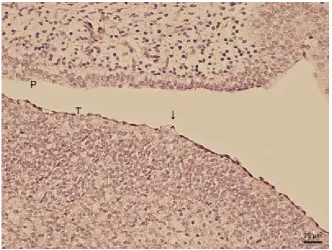
Figure 1. Sagittal section of the oral cavity and associated structures. Nasal cavity (NC), dental lamina (DL), lingual apex (LA), lingual body (LC), lingual radix (LR), upper lip (UL), lower lip (LL)
[image:3.595.134.467.474.724.2]During development, the lingual apex, corpus and radix can be distinguished (79 days of intra ovo de-velopment, stage 37; Figure 1). The apex and radix are bifurcated. The tongue is completely covered by stratified squamous epithelium, which appears lower on the ventral lingual surface. The uppermost epithelial cells form sessile prominences in some developmental stages. These structures are found
not only on the dorsal lingual surface (79 and 83 days of intra ovo development, stage 37 – Figure 2, and 38) but also on the ventral surface of the tongue (87 days of intra ovo development, stage 39).
[image:4.595.148.468.87.326.2]The lingual corpus bulges dorsally during sub-sequent development. The lingual apex becomes elongated. At day 95 of intra ovo development (stage 40), prominences of different shapes and
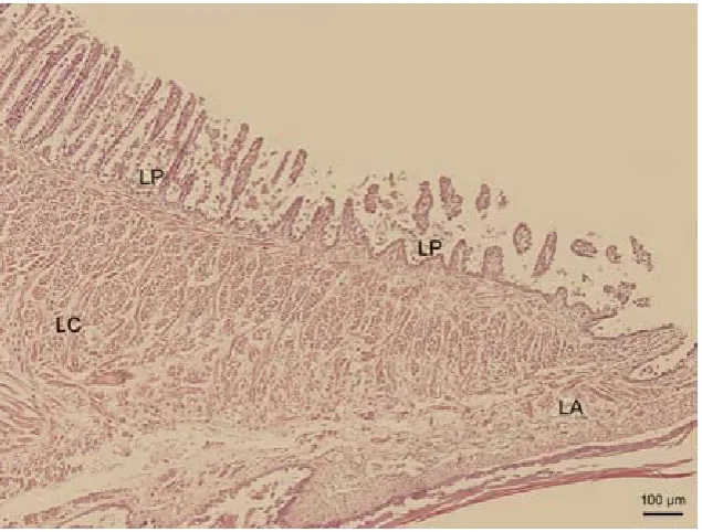
Figure 3. Sagittal section of the tongue (T) in the area of the lingual body. Lingual papillae on the dorsal lingual sur-face (LP)
[image:4.595.146.464.495.736.2]sizes develop on the lingual body. The lamina pro-pria forms numerous finger-like processes in the re-maining areas of the body of the tongue so that the base of the epithelium in these areas is not straight. Since the stratified epithelium flattens the exten-sions of the lamina propria, only the prominences previously mentioned can be observed. Two more structures develop during this period: a thicken-ing of the lthicken-ingual apex which is covered by lower keratinised squamous epithelium and the hyaline cartilage plate in the lingual musculature.
Development in the last weeks before hatching is irregular. The most differentiated embryonic stages from all the data processed are those of the 111 and 119 days of intra ovo development (end of stage 42 and end of stage 42). The dorsal lingual surface ex-tends in the form of papillae which resemble me-chanical lingual papillae in mammals. They appear as slender mucosal processes, the length of which decreases caudally. The free papillary endings proxi-mally on the lingual body tend to bend caudally, therefore papillae on the lingual radix and distally on the lingual body are similar to conical papillae (Figure 3), and papillae found proximally on the lin-gual body resemble filiform papillae (Figure 4) in mammals. Some papillae on the lingual apex bifur-cate. Compared to these two clearly differentiated stages, the mucosa of the dorsal lingual surface in younger stages (95 and 103 days of intra ovo de-velopment, stage 40 and 41; Figure 5) constitutes tortuous mucosal strips, whereas in older embryonic categories (127 and 135 days of intra ovo develop-ment, start of stage 42 – Figure 6, and start of stage 42) these strips develop into less differentiated
papil-lae. These papillae appear as rounded prominences with a wide base.
The dorsal lingual surface is covered by nonk-eratinised stratified squamous epithelium in the early stages, the tongue of which is more or less smooth. The flat surface cells have a mosaic ar-rangement (Figure 7) and contain numerous bizarre formations (microvilli) and droplets of secreta. The height of epithelium decreases with the formation of lingual papillae. In these cases, the epithelium is simple cuboidal. The apical surface of the epithelial cells is dome-shaped and these cells also have a mosaic arrangement (Figure 8). The only area of the tongue where the epithelium remains stratified is at the thickening of the lingual apex. Moreover, keratinisation of this structure is more prominent during the ongoing embryonic development.
Several mucous secreting glandular tubules are present in the connective tissue stroma of the ventral lingual surface at day 127 of intra ovo development (start of stage 42). The epithelial cells differentiate later and form more layers by day 135 of intra ovo development (start of stage 42). These glandular tubules open on the ventral lingual surface between the papillary processes of the mucosa.
No developing taste buds were found in the lin-gual epithelium of the investigated green iguana embryonic material.
DISCUSSION
In the presented study we focused on the second half of embryogenesis, i.e., 63rd to 135th day of
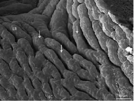
incu-Figure 5. Dorsal lingual surface in the area of the lingual body
[image:5.595.65.293.85.256.2] [image:5.595.306.533.86.255.2]bation under our conditions. We assume that matu-ration and differentiation of the tongue in this period is insignificant and that differences between individ-ual embryonic stages are small. Since temperature and air humidity are essential factors in the devel-opment of the oviparous green iguana, fluctuations in these values are reflected as a discontinuity in ontogenesis. In other words, variable external con-ditions are probably responsible for the accelerated development in some earlier stages in comparison to later stages prior to hatching. Moreover, these exter-nal conditions also affected the relatively extended development of embryos in our egg yield, which ex-ceeded almost twice the breeder’s standard.
The tongue is only mildly differentiated in the early stages at the beginning of our observation. The surface of the lingual mucosa is smooth. Developing lingual papillae, which were described in adults of the green iguana (Abbate et al., 2008), were not present. The lingual epithelium consti-tutes sessile formations in some later stages but these structures do not correlate with the further differentiation of the lingual papillae as we suggest. These epithelial prominences are found not only on the dorsal lingual surface (at 79 or 83 days of intra ovo development, stage 37 and 38) but also on the ventral surface (at 87 days of intra ovo de-velopment, stage 39). There are no lingual papillae formed ventrally on the tongue of adult green igua-nas (Cizek, 2004). It seems that these formations originate as a result of uneven differentiation of the lingual epithelium.
Lingual papillae, regarded as connective tissue prominences covered by epithelium, were
[image:6.595.306.534.83.253.2]ob-served at 111 days of development (end of stage 42). The appearance of the lingual papillae is in fact connected with their function in food intake. On the other hand, the epithelium covering the lingual papillae which contains secretory cells, with droplets of secreta on the surface, as well as glands at the cores of the papillae, hint at the possibility that lingual papillae have more than a purely mechanical function in the future ani-mal. It appears that they play an important role in food prehension and also in its digestion (Cizek, 2004). Thickening of the lingual apex, which is formed together with lingual papillae, represents a protective adaptation that allows safe contact of the tongue with structures in the environment. Glandular complexes of the tongue follow simi-lar developmental dynamics. Their differentiation accelerates shortly before hatching. Our findings can be compared only with results from different reptile species (Rabinowitz and Tandler, 1991). We obviously assume that there is a lower occur-rence of glands on the dorsal lingual surface. We believe that the different diet and subsequently different specialisation of the lingual glands in certain areas of the dorsal lingual surface makes, in our case, the glandular apparatus of the tongue species-specific. The form of the ingested food can substitute for an insufficient number of lingual glands resulting in a reduced amount of saliva. It is also possible that a considerable expansion of the glands on the dorsal lingual surface takes place postnatally. However, we did not find an expansion of glandular parenchyma in our previous studies (Cizek, 2004).
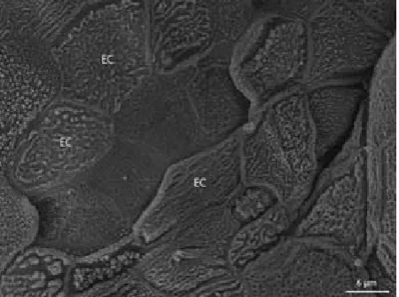
[image:6.595.64.289.85.252.2]Figure 7. Dorsal lingual surface, detail of Figure 6. Epi-thelial cells (EC) of the lingual papilla with numerous formations (microvilli)
Acknowledgement
The authors are grateful to Brno Zoo for its gen-erous supply of embryonic material.
REfERENCES
Abbate F, Latella G, Montalbano G, Guerrera MC, Le-vanti MB, Ciriaco E (2008): Scanning electron micro-scopical study of the lingual epithelium of Green iguana (Iguana iguana). Anatomia Histologia Embry-ologia 37, 314–316.
Butschli O (ed.) (1924): Ernaehrungsorgane. In: Vorle-sungen über vergleichende Anatomie. Verlag von Ju-lius Springer, Berlin. 263–281.
Cizek P (2004): Morphological differences of the lingual mucosa in some mammals and reptiles. [Diploma The-sis.] University of Veterinary and Pharmaceutical Sci-ences, Brno, 30–31.
Delheusy V, Toubeau G, Bels VL (1994): Tongue struc-ture and function in Oplurus cuvieri (Reptilia: Igua-nidae). Anatomical Record 238, 263–276.
Helff OM (1929): Studies on amphibian metamorphosis IV. Growth and differentiation of anuran tongue during metamorphosis. Physiological Zoology 2, 334–341. Iwasaki S (1990): Fine structure of the dorsal lingual
epi-thelium of the lizard, Gekko japonicus (Lacertilia, Gek-konidae). American Journal of Anatomy 187, 12–20. Iwasaki S (1992): Fine structure of the dorsal lingual
epithelium of the tongue of the freshwater turtle, Geoclemys reevesii (Chelonia, Emydinae). Journal of Morphology 211, 125–135.
Iwasaki S (2002): Evolution of the structure and function of the vertebrate tongue. Review. Journal of Anatomy 201, 1–13.
Iwasaki S, Asami T, Asami Y, Kobayashi K (1992): Fine structure of the dorsal epithelium of the tongue of the Japanese terrapin, Clemmys japonica (Cheloia, Emydi-nae). Archives of Histology and Cytology 55, 295–305. Iwasaki S, Wanichanon C, Asami T (1996): Histological
and ultrastructural study of the lingual epithelium of the juvenile Pacific ridley turtle, Lepidochelys olivacea (Chelonia, Cheloniidae). Annals of Anatomy 178, 243–250.
Koca YB, Oguz EO, Osanc E (2007): Morphology, and mus-cle- and papilla-volume ratios, of the tongue of Laudakia stellio (Agamidae, Squamata): a histological and stere-ological study. Zostere-ological Science 24, 899–905. Rabinowitz T, Tandler B (1991): Ultrastructure of lingual
salivary glands in the American chameleon: Anolis carolinensis. Anatomical Record 229, 489–494. Torrey TW (1979): The alimentary canal and its
deri-vates. In Morphogenesis of the Vertebrates. John Wi-ley & Sons, inc. New York, London. 314–324. Weisgram J, Ditrich H, Splechtna H (1989): Comparative
functional anatomical study of the oral cavity in two turtle species (in Czech). Plzensky Lekarsky Sbornik 59, 117–122.
Winokur RM (1988): The buccopharyngeal mucosa of the turtles (testudines). Journal of Morphology 196, 33–52.
Wise PAD, Vickaryous MK, Russel AP (2009): An em-bryonic staging table for in ovo development of Eublepharis macularius, the Leopard gecko. Anatom-ical Record 292, 1198–1212.
Received: 2011–06–28 Accepted after corrections: 2011–12–04
Corresponding Author:
MVDr. Petr Cizek, Faculty of Veterinary Medicine, University of Veterinary and Pharmaceutical Sciences, Palackeho 1–3, 612 42 Brno, Czech Republic