Original Article
Clinicopathological and immunohistochemical
features of primary central nervous system
germ cell tumors: a 24-years experience
Yuping Gao1, Jiyao Jiang2, Qiang Liu3
1Shanghai Key Laboratory for Assisted Reproduction and Reproductive Genetics, Center for Reproductive
Medi-cine, Ren Ji Hospital, School of MediMedi-cine, Shanghai Jiao Tong University, 845 Lingshan Road, Shanghai 200135, China; 2Department of Neurosurgery, Ren Ji Hospital, School of Medicine, Shanghai Jiao Tong University, 160
Pujian Road, Shanghai 200127, China; 3Department of Pathology, Ren Ji Hospital, School of Medicine, Shanghai
Jiao Tong University, 160 Pujian Road, Shanghai 200127, China
Rceived August 12, 2014; Accepted September 13, 2014; Epub September 15, 2014; Published October 1, 2014
Abstract: Primary central nervous system (CNS) germ cell tumors (GCTs) are a rare heterogeneous group of lesions, which the clinicopathological features have a marked degree of heterogeneity comparing with that of gonadal GCTs.
Accurately diagnosing CNS GCTs might be extremely difficult and requires immunohistochemical verification. This
study was to investigate the biological feature of CNS GCTs and diagnostic value of immunohistochemical markers OCT3/4, C-kit, PLAP, and CD30 in CNS GCTs. A retrospective study was performed on 34 patients with CNS germ cell tumorsbetween 1990 and 2014. 34 CNS GCTs account for 9.2% of all primary CNS neoplasms. The sellar region (35.3%) and pineal gland (17.6%) were the most common sites of intracranial GCTs. Hydrocephalus (82.4%) and diplopia (46.9%) were the two most common clinical presentations. The most common histological subtypes were germinoma (67.6%). PLAP, c-kit, OCT3/4 were highly expressed in gernimomas. CD30 and CK AE1/3 stainings were
positive in embryonal carcinoma. Yolk sac tumor component showed positive staining for AFP and CK AE1/3. β-HCG
staining was positive in choriocarcinoma and STGC. Patients with mature teratomas and germinomas had a better prognosis (a 5-year survival rate) than those with embryonal carcinoma and choriocarcinoma (a 5-year survival rates
were 0). Our finding suggest that the incidences of primary CNS GCTs are higher in South China than in the West, but
mixed GCTs are uncommon in our study. The judicious use of a panel of selected markers is helpful in diagnosing and predicting the prognosis for CNS GCTs.
Keywords: Central nervous system, germ cell tumors, C-kit, PLAP, OCT3/4, CD30
Introduction
Primary central nervous system (CNS) germ cell tumors (GCTs) are a rare heterogeneous group of lesions in the CNS. CNS GCTs occur predomi-nantly in children and adolescents with approxi-mately 90% of the cases before the age of 20 years. The peak incidence of CNS-GCT is between 10 and 20 years of age. CNS GCTs are more common in male. Histologically, these tumors are very similar to those in gonadal and other extragonadal sites. CNS GCTs most fre-quently locate in the midline sites of body (pine -al, suprasella region, thalamus, basal ganglia, et al). Pathological classifications of CNS GCTs by the WHO contain germinoma, teratoma, yolk sac tumor, embryonal carcinoma,
choriocarci-can be divided into two broad groups: germino-mas and non-germinoamtous germ cell tumors (NGGCT) [1-4].
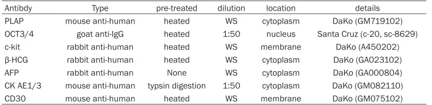
Table 1. Summary of immunohistochemical antibodies
Antibdy Type pre-treated dilution location details
PLAP mouse anti-human heated WS cytoplasm DaKo (GM719102)
OCT3/4 goat anti-IgG heated 1:50 nucleus Santa Cruz (c-20, sc-8629)
c-kit rabbit anti-human heated WS membrane DaKo (A450202)
β-HCG rabbit anti-human heated WS cytoplasm DaKo (GA023102)
AFP rabbit anti-human None WS cytoplasm DaKo (GA000804)
CK AE1/3 mouse anti-human typsin digestion 1:50 cytoplasm DaKo (GM082110)
CD30 mouse anti-human heated WS membrane DaKo (GM075102)
WS: working solution; goat anti-mouse, rabbit anti-goat, goat anti-rabbit.
from CNS GCT are often very limited in size as special lesion sites. Accurately diagnosing CNS GCTs might be extremely difficult and requires immunohistochemical verification. This study was to investigate the biological feature of CNS GCTs, and diagnostic value of immunohisto-chemical markers OCT3/4, C-kit, PLAP, and CD30 in CNS GCTs. In addition, we assessed treatment outcomes and prognostic factors.
Materials and methods
Patient data
Between 1990 and 2014, 25 male and 9 female Chinese patients (male: female ratio 2.8:1), with median age of 16.9 years (ranging from 7 to 36 years) diagnosed with primary CNS GCTs from Ren Ji Hospital, School of Medicine, Shanghai Jiaotong University. Diagno- ses are based on the combination of clinical symptoms, tumor imaging characteristics, ser- um tumor markers, as well as cytological and histological confirmation. None of the patients had a previous history of gonadal GCTs or non-CNS extragonadal GCTs. The study was approved by the ethics committee of Ren Ji Hospital, School of Medicine, Shanghai Jiaotong University.
Follow-up study
Thirty-four patients were followed up by tele-phone or mail and only 29 patients had com-pleted follow-up results. Median follow-up peri-od was 6.5 years, ranging from 4 months to 17 years.
Immunohistochemistry and staining evaluation
Surgical specimens were fixed in 10% formalin and embedded in paraffin. 4 μm sections were cut and stained with hematoxylin and eosin.
Additional 4 μm sections were deparaffinized with xylene and rehydrated in a graded series of ethanol. The deparaffinised sections were then incubated with 3% H2O2 to inhibit the endogenous peroxidase, followed by micro-wave-treated or trypsin digestion for antigen retrieval before incubation with different prima-ry antibodies, using a two-step polymer method (EnVisionTM). The sections were incubated in a humid chamber at 4°C overnight after adding primary antibodies and the Table 1 showed the details of primary antibodies used in our study. Subsequently, second antibodies were added after PBS rinse. The sections were incubated at room temperature for 30 minutes, and then colored with DAB for 15 minutes, and finally light counterstained with hematoxylin. As posi-tive control staining we used placental tissue, testicular neoplasm and Gastrointestinal stro-mal tumor, respectively. Negative controls were performed using blocking serum in place of pri-mary antibody. Immunohistochemical expres-sion was graded using a semi-quantitative scoring system based on the proportion of posi-tive cells over total cells (percent positivity) ranging from 0 to 100% where 0% was negative expression, <50% was weak expression, ≥50% was strong expression [7, 8]. This study proto-col was approved by the Human Ethics Committee of Renji Hospital, School of Medi- cine, Shanghai Jiao Tong University.
Statistics analysis
Results
Clinical characteristics of the patients
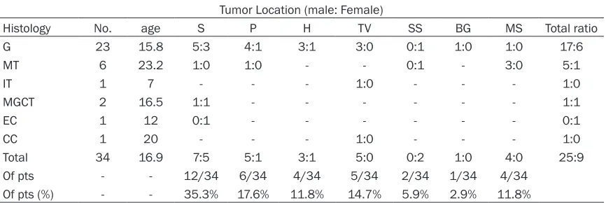
Thirty-four CNS GCTs accounted for 9.2% of all primary CNS neoplasms and 37.7% of extrago-nadal GCTs, respectively. The duration of symp-toms before diagnosis ranged from 5 days to 5 years, with a mean time of 10 months. 7 patients (20.6%) were younger than 10 years, 9 patients (26.5%) were older than 20 years, and 18 patients (52.9%) were between 10 and 20 years. Affected sites included intracranial (88.2%) and intraspinal canal (11.8%). The sel-lar region and pineal gland were the most com-mon sites for intracranial GCTs, accounting for 35.3% and 17.6% respectively. The hypothala-mus, third ventricle and suprasella were also common tumor locations. Other locations included: basal ganglia and medulla spinalis (Table 2). There is an overall male predomi-nance in CNS GCTs except suprasella (n = 2, both female). Clinical presentations of CNS GCTs were dependent on the location and size of the tumor in the CNS. Symptoms varied at diagnosis including endocrine abnormalities, headache, visual disturbances and signs of increased intracranial pressure. 10 patients displayed endocrine abnormalities, including precocious puberty (n = 6), growth retardation (n = 2, one with Cushing syndrome), low libido (n = 1) and amenorrhoea (n = 1).
Elevations of serum beta-human chorionicgo-nadotropin (β-HCG) have been detected in one germinoma patient with syncytiotrophoblastic (1,3900 mIU/ml, normal < 25 mIU/ml) and one
choriocarcinoma patient (2,5980 mIU/ml, nor-mal < 25 mIU/ml), respectively. Both the serum β-HCG levels dropped dramatically after the operation. Elevation of serum alpha fetoprotein (AFP) level was found in one mixed GCT patient (G+YST+EC). Additionally, 7 patients with pure germinoma had normal AFP, β-HCG, TT3, TT4, TSH, ACTH, LH, FSH and PRL levels.
Histological diagnosis
The sizes of tumor varied from 2 cm × 1.8 cm × 1.5 cm to 5 cm × 4 cm × 4 cm. 34 patients had undergone surgical resection. 23 patients were histologically diagnosed for germinoma (67.6%), one for germinoma with STGCs, 7 for teratoma (20.6%, 1 immature teratoma and 6 mature teratoma), 2 for mixed malignant GCTs (5.9%, 1 G+YST+EC, 1 G+EC), one for embryonal carci-noma (2.9%) and choriocarcicarci-noma (2.9%) respectively.
Macroscopic examination: The macroscopic appearance of the germinoma was pale grey solid nodule with less hemorrhage, necrosis and cystic change. 17 of 34 CNS GCTs cases had well defined tumor borders with well cir -cumscribed, and the other 17 had poorly defined tumor borders with infiltrative growth pattern. Two mixed GCTs, one embryonal carci-noma, and one choriocarcinoma also had poor-ly defined borders with hemorrhage, necrosis.
[image:3.612.91.529.85.230.2]Microscopic examination: The histological fea-tures of 23 germinoma were similar to semino-ma of testis and dysgerminosemino-ma of ovarian. The
Table 2. Distribution of locations in different histological subtype of 34 CNS GCTs Tumor Location (male: Female)
Histology No. age S P H TV SS BG MS Total ratio
G 23 15.8 5:3 4:1 3:1 3:0 0:1 1:0 1:0 17:6
MT 6 23.2 1:0 1:0 - - 0:1 - 3:0 5:1
IT 1 7 - - - 1:0 - - - 1:0
MGCT 2 16.5 1:1 - - - 1:1
EC 1 12 0:1 - - - 0:1
CC 1 20 - - - 1:0 - - - 1:0
Total 34 16.9 7:5 5:1 3:1 5:0 0:2 1:0 4:0 25:9
Of pts - - 12/34 6/34 4/34 5/34 2/34 1/34 4/34
Of pts (%) - - 35.3% 17.6% 11.8% 14.7% 5.9% 2.9% 11.8%
tumor cell of germinoma in CNS was round or polygonal with abundant pale to clear cyto-plasm and moderate sized polygonal nuclei and prominent nucleoli. The architecture was a sheet or nest pattern associated lymphocytic infiltrates along fibrovascular septae (Figure 1A). One of the 23 germinoma cases revealed a germinoma containing syncytiotrophoblastic giant cells (STGCs). 6 mature teratomas had tis-sue originating from three fully differentiated tissue elements of ectoderm, mesoderm, and endoderm. One immature teratoma contained incompletely tissue from embryonic mesen-chyme-like stroma. 2 mixed GCT contained ger-minoma, yolk sac tumor and embryonal carci-noma component (G+YST+EC), and germicarci-noma and embryonal carcinoma (G+EC), respectively.
One embryonal carcinoma was composed of large cells with indistinct cell borders, promi-nent nucleoli, mitoses forming irregular gland-like architecture. One choriocarcinoma was composed of cytotrophoblasts and syncytiotro-phoblasts with severe hemorrhage, necrosis and scattered syncytiotrophoblastic giant cells.
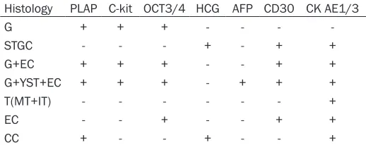
Immunohistochemical results (Table 3)
[image:4.612.91.523.70.428.2]Placental alkaline phosphatase (PLAP) was mainly located in the cell membrane and cyto-plasm. It was detected in 19 of 23 germinomas (82.6%), 2 mixed GCTs, one embryonal carci-noma and one choriocarcicarci-noma. Strong posi-tive staining was found in 14/23 germinomas, weak positive in 5/23, negative in 4/23. None Figure 1.A: Histological features of CNS germinoma: A tumor cells with abundant clear cytoplasm, sheet growth
pattern, lymphocytic infiltrates along fibrovascular septae. (HE×200). B: OCT3/4 immunolabeling of tumor cells in
CNS germinoma (×200). C: CNS Mixed GCT showing embryonal carcinoma component with CD30 immonolabelling (×200). D: Immunohischemical features of germinoma with syncytiotrophoblastic giant cells. Immunostaining for
of 7 patients with teratoma (mature and imma-ture) had a positive expression of PLAP. YST component of mixed GCT showed negative expression of PLAP. An uncertain immuno-hischemical evaluation was performed in 2 cases with weak positive expression because of insufficient tumor samples and obscuration by crushing artifact. C-kit was detected in all germinomas (23/23) and 2 mixed GCTs. Strong positive staining was found in 18/23 germino-mas, and weak positive in 5/23. None of 7 patients with teratoma (mature and immature), one patient with embryonal carcinoma, and one patient with choriocarcinoma had a posi-tive expression. C-kit was posiposi-tive in YST com-ponent in mixed GCT, but negative in embryo-nal component. STGC was also negative. OCT3/4 was detected in all germinomas (23/ 23), germinoma component and embryonal carcinoma component in 2 mixed GCTs. Strong positive staining was detected in 19/23 germi-nomas, and weak positive in 4/23. None of 7 patients with teratoma (mature and immature), YST component, and STGCs had a positive expression (Figure 1B).
In addition, CD30 and CK AE1/3 staining were positive in embryonal carcinoma component of mixed GCT and embryonal carcinoma (Figure 1C). AFP and CK AE1/3 staining were positive in YST. β-HCG and CK AE1/3 staining were both positive in STGC. β-HCG staining was positive in choriocarcinoma (Figure 1D).
Treatment outcomes
Total excision and subtotal excision were per-formed in 15 and 34 patients, respectively. All
[image:5.612.90.349.98.201.2]patients except 6 patients with mature teratomas received subse-quent adjuvant radiotherapy and chemotherapy after surgical resec-tion. 2 patients with germinomas recurrence 4/8 months after primi-tive surgical excision received tumor excision again, and neither local recurrence nor distal metastasis was observed according to a followed up of 3 and 6 years, respectively. One patient with mature teratoma showed local relapse 4 years after surgical treatment. One death case of germi-noma was reported two weeks after operation for serious intranoperative
Table 3. Expression of immunohischemical markers in 34 CNS GCTs
Histology PLAP C-kit OCT3/4 HCG AFP CD30 CK AE1/3
G + + + - - -
-STGC - - - + - + +
G+EC + + + - - + +
G+YST+EC + + + - + + +
T(MT+IT) - - - +
EC - - + - - + +
CC + - - + - - +
G: germinoma; MT: mature teratoma; IT: immature teratoma; MGCTs: mixed germ cell tumors; EC: embryonal carcinoma; YST: yolk sac tumor; CC: cho-riocarcinoma; STGCs: syncytiotrophoblastic giant cells.
bleeding. One patient with mixed GCT (G+YST) had recurrence and died 4 years after opera-tion. One patient with embryonal carcinoma had recurrence 4 month after tumor resection and died 10 months for repeated surgical resection. One death case of choriocarcinoma was reported 6 months after operation. One mixed GCT (G+EC) recurrence 2 months after primitive surgical excision received tumor exci-sion again, and neither local recurrence nor dis-tal metastasis was observed according to a fol-lowed up of 5 years. Neither local recurrence nor distal metastasis was observed in the rest 23 patients with germinoma, mature teratomas and immature teratomas, according to follow-up records.
The overall 5-year survival rate of the patients with CNS GCTs was 86.2%. Teratomas and ger-minomas had good prognosis with a 5-year sur-vival rates of 100% and 94.4%, respectively. The overall 3 and 5-year survival rates of the patients with malignant NG-GCTs were 60% and 40%. The prognosis of the patients with germinoma was significantly better than that of the patients with malignant NGGCT (P < 0.05). The overall 5-year survival rates of the patients with or without recurrence were 66.7% and 95.7%, but there was no significantly diffe-rence.
Discussions
West [4, 6]. Our data confirmed the incidence of CNS GCT is high in China. In this study, CNS GCTs account for 9.2% of all primary CNS tumors, with a high rate in the prepubertal chil-dren (73.5%), and especially in the second ten years of the life (52.9%). There is an overall male predominance in CNS GCTs except 2 cases of suprasella. 2 patients in suprasella region tumors were female, suggesting a female predominance in this site. This result was in accordance with Lee et al who reported that the suprasella region was the only anatom-ical site demonstrating a female predominance in South Korea [3].
Clinical features of CNS GCTs are similar to that of other tumors, such as gliomas, pituitary ade-noma, or pineoblastomas, whose symptoms were largely caused by tumor effect on sur-rounding structures. Clinical presentations of CNS GCTs frequently include signs of increased intracranial pressure, visual changes, and endocrine abnormalities depending on the tumor location, size of lesion, and patient’s age. In our study, the sellar region (35.3%) and pine-al gland (17.6%) were the most common sites of intracranial GCTs. Patients with tumors located in pineal region usually present with progres-sive hydrocephalus and signs of increased intracranial pressure, which result from com-pression and obstructive of the aqueduct of midbrain. Patients with GCTs in sellar and suprasellar most commonly present with visual symptoms including failure of upper gaze and obtundation related to compression of the optic chiasm, or present with hypothalamic/pituitary axis dysfunction such as delayed sexual devel-opment, hypopituitarism, precocious puberty, and diabetes insipidus [3, 6, 9]. In this study, 6 patients with precocious puberty were all male, and the histological types were pure germino-na, G+STGCs, and choriocarcinoma, respec-tively, with the locations of sella regions, pineal region, hypothalamic and medulla spinal. Though the cause of precocious puberty in patients with CNS GCTs has not been com-pletely clarified, we found some pure germino -mas can produce β-HCG at a high level, which may be responsible for precocious puberty. This speculation was also supported by previ-ous studies that germinoma with β-HCG secret -ing syncytiotrophoblasts and choriocarcinomas seems to be more likely to present with preco-cious puberty than any other type of GCTs [10-12].
CNS GCTs are composed of several different tumor types showing varying degrees of malig-nancy. However, researchers found various his-tological types of GCTs may share a common origin of primordial germ cells (PGCs) at differ-ent developmdiffer-ent stages. For example, yolk sac tumor and choriocarcinoma were thought to originate from PGCs at extraembryonic differ-entiation, while germinoma were thought to originate from undifferentiated PGCs, and tera-toma from PCGs at embryonal differentiation [2, 13, 14]. In this study, 67.6% of GCTs were germinomas, followed by mature and immature teratomas (20.6%), mixed GCTs (5.9%), embryo-nal carcinoma (2.9%), and choriocarcinoma (2.9%). The results were rather similar to a report based on 389 published cases by Jennings et al, in which 65% of GCTs were ger-minomas, 18% were teratomas, 5% were embryonal carcinomas, 7% were endodermal sinus tumors, and 5% were choriocarcinomas [4]. However, the present study reveals a mark-edly lower proportion of mixed GCTs (2.9%) compared with 27.4% and 13.8% that reported in the studies performed in Korea and Taiwan, respectively [3, 5]. In our study, germinomas with STGCs constituted 4.3% of geminoma, which is similar with a reported proportion of 5.2% by Matsutani et al [6]. It should be paid more attention that scattered STGCs in the ger-minoma cannot be misdiagnosed as choriocar-cinoma [11, 12].
and those without it [16-18]. Matsutani report-ed that 5-years, 10-year, 15-year and 20-year survival rates for pure germinomas were 95.4%, 92.7%, 87.9% and 80.6%, respectively. The 1-year survival rates for embryonal carcinomas, yolk sac tumors and choriocarcinomas was 80%, 33.3% and 0, respectively. The 5-year survival rates for mixed GCTs and immature and malignant teratomas were 57.1% and 70.7%, respectively [6]. A previous study report-ed that germinomas with STGCs usually has a poor prognosis largely for high incidence of recurrence [12]. In our study, one case of germi-noma with STGCs has a good prognosis without occurrence and metastasis 6 years after operation.
Compared with other extragonadal sites GCTs, CNS GCTs are usually diagnosed based on his-tological assessments, neuroimaging charac-teristics, and tumor markers. However, the neu-roimaging characteristics of all types of CNS GCTs are similar, and the tumor markers are usually nonspecific. The diagnosis of CNS GCTs usually requires a tumor biopsy; however it is sometimes difficult to perform complete resec -tion of the tumor due to its special sites in CNS. Accurately diagnosing CNS GCTs can be very difficult, since tumor specimens from these lesions are often so limited in size that it is dif-ficult to obtain them. Immunohistochemical staining is often necessary for the diagnosis of CNS GCTs. This may be the reason why more patients were diagnosed during 2001~2014 than during 1990~2000 (8 vs. 26 cases) in our series.
PLAP, a characteristic marker of primordial germ cells, is detected in 82.6% of germino-mas. However, the PLAP marker has its own shortcomings of heavy staining background due to surface membrane and diffuse cytoplas-mic staining. It was very difficult to interpret in small biopsies where tumor cells were obscured by crushing artifact and focal necrosis in 2 cases. C-kit and OCT3/4 are also characteristic sensitive markers of germinomas with mem-brane staining and nuclear staining, respective-ly, and the staining sensitivities are higher than PLAP (100% positive staining). CK AE1/3 stain-ing is usually negative or focal weak positive in germinomas, but positive in embryonal carcino-mas, yolk sac tumors, teratomas and chorio-cacinomas. STGCs show strong immunoposi-tive reactions for CK AE1/3, human placental
lactogen and β-HCG [7, 8, 10, 13]. Embryonal carcinomas and embryonal carcinoma compo-nent show positive stainings for CK AE1/3, EMA, OCT3/4 and CD30. CD30 staining is neg-ative in germinoma, yolk sac tumors and cho-riocacinomas [19, 20]. PLAP, OCT3/4, C-kit and CD30 used in combination are useful for distin-guishing germinoma from embryonal carcino-ma. Yolk sac tumors are positive for AFP, CK AE1/3 and EMA. Immunoreactivity for AFP of yolk sac tumors is characteristic and valuable in distinguishing these tumors from embryonal carcinomas [20, 21]. Choriocarcinomas show immunopositivity for β-HCG, HPL, and CK AE1/3 [21, 22].
In summary, the incidences of primary CNS GCTs are higher in South China than that in West countries. The mixed GCTs are uncom -mon in our study and the incidences are very lower than that in other countries. Mature tera-tomas and germinomas have a better progno-sis. The prognosis of the patients with germi-noma was significantly better than that of the patients with malignant NGGCT. An accurate diagnosis of CNS GCTs is critical for patient management. Immunohistochemistry has play- ed a major role in accurately diagnosing and distinguishing the different histological types. The judicious use of a panel of selected mark-ers is helpful in diagnosing and predicting the prognosis for CNS GCTs.
Acknowledgements
This work was partly supported by the funding of Science and Technology Commission of Shanghai Municipality (NO. 134119a9502). 12DZ2260600.
Disclosure of conflict of interest
None.
Address correspondence to: Dr. Yuping Gao, Shang- hai Key Laboratory for Assisted Reproduction and Reproductive Genetics, Center for Reproductive Medicine, Ren Ji Hospital, School of Medicine, Shanghai Jiao Tong University, 845 Lingshan Road, Shanghai 200135, China. E-mail: ginagao53@ yahoo.com
References
update on treatment. Med Oncol 2013; 30: 496-515.
[2] Packer RJ, Cohen BH, Cooney K. Intracranial germ cell tumors. Oncologist 2000; 5: 312-320.
[3] Lee D, Suh YL. Histologically confirmed intra -cranial germ cell tumors; an analysis of 62 pa-tients in a single institute. Virchows Arch 2010; 457: 347-357.
[4] Jennings MT, Gelman R, Hochberg F. Intracra-nial germ-cell tumors: natural history and pathogenesis. J Neurosurg 1985; 63: 155-167.
[5] Liang SY, Yang TF, Chen YW, Liang ML, Chen HH, Chang KP, Shan IK, Chen YS, Wong TT. Neuropsychological functions and quality of
life in survived patients with intracranial germ cell tumors after treatment. Neuro Oncol 2013; 15: 1543-1551.
[6] Matsutani M, Sano K, Takakura K, Fujimaki T, Nakamura O, Funata N, Seto T. Primary intra-cranial germ cell tumors: a clinical analysis of
153 histologically verified cases. J Neurosurg
1997; 86: 446-455.
[7] Cheng L, Sung MT, Cossu-Rocca P, Jones TD, MacLennan GT, De Jong J, Lopez-Beltran A, Montironi R, Looijenga LH. OCT4: biological functions and clinical applications as a marker of germ cell neoplasia. J Pathol 2007; 211: 1-9.
[8] Takeshima H, Kuratsu J. A review of soluble c-kit (s-c-kit) as a novel tumor marker and possible molecular target for the treatment of CNS ger-minoma. Surg Neurol 2003; 60: 321-4; discus-sion 324-5.
[9] Kaur H, Singh D, Peereboom DM. Primary cen-tral nervous system germ cell tumors. Curr Treat Options Oncol 2003; 4: 491-498. [10] Allen J, Chacko J, Donahue B, Dhall G, Kre-
tschmar C, Jakacki R, Holmes E, Pollack I. Di-agnostic sensitivity of serum and lumbar CSF bHCG in newly diagnosed CNS germinoma. Pe-diatr Blood Cancer 2012; 59: 1180-1182. [11] Ogino H, Shibamoto Y, Akanaka T, Suzuki K,
Ishihara S ,Yamada T, Sugie C, Nomoto Y, Mimura M. CNS germinoma with elevated se-rum human chorionic gonadotropin level: clini-cal characteristics and treatment outcome. Int J Radiat Oncol Biol Phys 2005; 62: 803-808. [12] Shibamoto Y, Takahashi M, Sasai K. Prognosis
of intracranial germinoma with syncytiopho-blastic giant cells treated by radiation therapy. Int J Radiat Oncol Biol Phys 1997; 37: 505-510.
[13] Hoei-Hansen CE, Sehested A, Juhler M, Lau YF, Skakkebaek NE, Laursen H, Rajpert-de Meyts E. New evidence for the origin of intracranial germ cell tumours from primordial germ cells: expression of pluripotency and cell differentia-tion markers. J Pathol 2006; 209: 25-33. [14] Sano K. Pathogenesis of intracranial germ cell
tumors reconsidered. J Neurosurg 1999; 90: 258-264.
[15] Brandes AA, Pasetto LM, Monfardini S. The treatment of cranial germ cell tumours. Cancer Treat Rev 2000; 26: 233-242.
[16] Ogawa K, Toita T, Nakamura K, Uno T, Onishi H, Itami J, Shikama N, Saeki N, Yoshii Y, Muraya-ma S. Treatment and prognosis of patients with intracranial nongerminomatous malig-nant germ cell tumors: a multiinstitutional ret-rospective analysis of 41 patients. Cancer 2003; 98: 369-376.
[17] Kawabata Y, Takahashi JA, Arakawa Y, Shira-hata M, Hashimoto N. Long term outcomes in patients with intracranial germinomas: a sin-gle institution experience of irradiation with or without chemotherapy. J Neurooncol 2008; 88: 161-167.
[18] Kanamori M, Kumabe T, Saito R, Yamashita Y, Sonada Y, Ariga H, Takai Y, Tominaga T. Optimal treatment strategy for intracranial germ cell tumors: a single institution analysis. J Neuro-surg Pediatr 2009; 4: 506-514.
[19] Ulbright TM. Germ cell tumors of the gonads: a selective review emphasizing problems in dif-ferential diagnosis, newly appreciated, and controversial issues. Mod Pathol 2005; 18 Suppl 2: S61-79.
[20] Kim A, Ji L, Balmaceda C, Diez B, Kellie SJ, Dunkel IJ, Gardner SL, Sposto R, Finlay JL. The prognostic value of tumor markers in newly di-agnosed patients with primary central nervous system germ cell tumors. Pediatr Blood Cancer 2008; 51: 768-773.
[21] Sugiyama K, Arita K, Tominaga A, Hanaya R, Taniguchi E, Okamura T, Itoh Y, Yamasaki F, Kurisu K. Morphologic features of human cho-rionic gonadotropin- or alpha-fetoprotein-pro-ducing germ cell tumors of the central nervous system: histological heterogeneity and surgical meaning. Brain Tumor Pathol 2001; 18: 115-122.
[22] Wildi-Runge S, Crevier L, Carret AS, Robitaille