COMPARATIVE EVALUATION OF THE MICROLEAKAGE OF METAL CROWNS CEMENTED WITH
TWO DIFFERENT LUTING AGENTS
*Dr. Siddhant Bumb, Dr.
Prosthodontics and Implantology,
ARTICLE INFO ABSTRACT
Microleakage
cements irrespective of their contents show some amount of dissolution when they come in contact with oral environment. Marginal opening between cemented crowns and the
related to cement dissolution. This in vitro study investigated the effect of two different luting agent namely resin cement and resin modified glass ionomer cement on microleakage under metal crown. Twenty intact human premolars w
prepared in a standardized manner for receiving full metal crowns. Crowns were made following a standard laboratory technique and cemented on their respective abutments. The specimens were subjected
cut in the buccolingual direction. The microleakage in the area of tooth as linear penetration of methylene blue and was determined wi microleakage scores using a five
type and degree of microleakage. The smallest degree of microleakage was observed in specimens luted with resin cement, follo
formation and microleakage has been clearly established but the exact amount that becomes clinically significant remains undefined.
Copyright©2017, Dr. Siddhant Bumb et al. This is an open
use, distribution, and reproduction in any medium, provided the original work is properly cited.
INTRODUCTION
In restorative dentistry, dental cements have been used since ages. The choice of cement depends on specific clinical circumstances that is basically decided by the properties of various luting agents (Osborne et al., 1978; Swartz
Jacobs and Windeler, 1991). Microleakage is the seepage or penetration of oral fluids, bacteria, molecules or ions between tooth surface and restoration or cement layer.
of microleakage along the interface has been related to hypersensitivity, recurrent caries, pulpal problems and tooth discoloration. Tooth discoloration is the most common reason why replacement of restoration is required.
determination can be undertaken using various
vitro techniques. They are namely chemical agents measurement by neutron activation analysis, bacterial activity,
scanning electron microscope, air pressure,
radioisotope, ionization, autoradiography, decay, staining
reversible radioactive adsorption and thermal
application. Freshly, a new technique has been introduced to validate microleakage for adhesive using silver methenamine.
*Corresponding author: Dr. Siddhant Bumb,
Prosthodontics and Implantology, Dr, D.Y.Patil Dental College and Hospital, Pune, Mahatashtra, India.
ISSN: 0975-833X
Article History:
Received 18th January, 2017
Received in revised form
15th February, 2017
Accepted 27th March, 2017
Published online 30th April,2017
Key words:
Marginal, Microleakge, Resin Cement, Stereomicroscope.
Citation: Dr. Siddhant Bumb, Dr. Dilip Kakade, Dr.
crowns cemented with two different Luting agents : In vitro study
RESEARCH ARTICLE
COMPARATIVE EVALUATION OF THE MICROLEAKAGE OF METAL CROWNS CEMENTED WITH
TWO DIFFERENT LUTING AGENTS :
IN
VITRO
STUDY
Dilip Kakade, Dr. Amit Jagtap and Dr. Riddhi Kulkarni
Prosthodontics and Implantology, Dr, D.Y.Patil Dental College and Hospital, Pune,
ABSTRACT
Microleakage and marginal opening are related to most of the fixed restoration failures. The luting cements irrespective of their contents show some amount of dissolution when they come in contact with oral environment. Marginal opening between cemented crowns and the
related to cement dissolution. This in vitro study investigated the effect of two different luting agent namely resin cement and resin modified glass ionomer cement on microleakage under metal crown. Twenty intact human premolars were randomized to two groups of ten teeth each. They were prepared in a standardized manner for receiving full metal crowns. Crowns were made following a standard laboratory technique and cemented on their respective abutments. The specimens were subjected to thermocycling for 500 cycle, placed in methylene blue solutions for 24hrs and vertically cut in the buccolingual direction. The microleakage in the area of tooth
as linear penetration of methylene blue and was determined wi
microleakage scores using a five-point scale. A significant association was found between a cement type and degree of microleakage. The smallest degree of microleakage was observed in specimens luted with resin cement, followed by modified resin glass-ionomer cement. The importance of gap formation and microleakage has been clearly established but the exact amount that becomes clinically significant remains undefined.
is an open access article distributed under the Creative Commons Attribution License, which use, distribution, and reproduction in any medium, provided the original work is properly cited.
In restorative dentistry, dental cements have been used since ages. The choice of cement depends on specific clinical circumstances that is basically decided by the properties of Swartz et al., 1971; . Microleakage is the seepage or penetration of oral fluids, bacteria, molecules or ions between tooth surface and restoration or cement layer.The occurrence along the interface has been related to hypersensitivity, recurrent caries, pulpal problems and tooth discoloration. Tooth discoloration is the most common reason The microleakage various in vivo and in vitro techniques. They are namely chemical agents measurement by neutron activation analysis, bacterial activity,
air pressure, markers,
radioisotope, ionization, autoradiography, decay, staining and
nd thermal cycles
application. Freshly, a new technique has been introduced to validate microleakage for adhesive using silver methenamine.
Dr, D.Y.Patil Dental College and Hospital,
Out of these various techniques the use of dye penetration is most commonly used and a swift technique to estimate microleakage in complete crown
This study was performed for comparative evaluation of microleakage by dye penetration method under full metal crown luted with two different luting agents. (Resin modified glass ionomer cement and Resin cement).
MATERIALS AND METHODS
Twenty maxillary premolars without caries were collected for the study. The criteria for selection was premolar having intact clinical crowns that were obtained either by periodontal compromised condition or for orthodontic reasons. A maxillary extracted premolar was mounted in an
acrylic resin block, with dimension of 30 mm in height
in width and 30mm in breadth. While mounting the tooth over acrylic resin block, it was placed in such a way that 2 mm portion of tooth beyond the cemto enamel junction
uncovered by the resin block. For achieving parallelism between acrylic block and long axis of the tooth, a surveyor was used. Ideal tooth preparation was carried out for full veneer metal crown using micromotor
International Journal of Current Research Vol. 9, Issue, 04, pp.49308-49312, April, 2017
INTERNATIONAL
OF CURRENT RESEARCH
Dr. Siddhant Bumb, Dr. Dilip Kakade, Dr. Amit Jagtap and Dr. Riddhi Kulkarni, 2017. “Comparative evaluation of the Microleakage of metal
In vitro study”, International Journal of Current Research, 9, (04),
COMPARATIVE EVALUATION OF THE MICROLEAKAGE OF METAL CROWNS CEMENTED WITH
STUDY
Riddhi Kulkarni
ntal College and Hospital, Pune, Mahatashtra, India
and marginal opening are related to most of the fixed restoration failures. The luting cements irrespective of their contents show some amount of dissolution when they come in contact with oral environment. Marginal opening between cemented crowns and their tooth margins mainly related to cement dissolution. This in vitro study investigated the effect of two different luting agent namely resin cement and resin modified glass ionomer cement on microleakage under metal crown. ere randomized to two groups of ten teeth each. They were prepared in a standardized manner for receiving full metal crowns. Crowns were made following a standard laboratory technique and cemented on their respective abutments. The specimens were to thermocycling for 500 cycle, placed in methylene blue solutions for 24hrs and vertically cut in the buccolingual direction. The microleakage in the area of tooth-cement interface was defined as linear penetration of methylene blue and was determined with a stereomicroscope to assign point scale. A significant association was found between a cement type and degree of microleakage. The smallest degree of microleakage was observed in specimens ionomer cement. The importance of gap formation and microleakage has been clearly established but the exact amount that becomes clinically
ribution License, which permits unrestricted
Out of these various techniques the use of dye penetration is most commonly used and a swift technique to estimate microleakage in complete crown (Pessutti Nunes et al., 2005). udy was performed for comparative evaluation of microleakage by dye penetration method under full metal crown luted with two different luting agents. (Resin modified glass ionomer cement and Resin cement).
MATERIALS AND METHODS
without caries were collected for the study. The criteria for selection was premolar having intact clinical crowns that were obtained either by periodontal compromised condition or for orthodontic reasons. A maxillary extracted premolar was mounted in an autopolymerizing ck, with dimension of 30 mm in height, 30 mm in width and 30mm in breadth. While mounting the tooth over acrylic resin block, it was placed in such a way that 2 mm yond the cemto enamel junction remains uncovered by the resin block. For achieving parallelism between acrylic block and long axis of the tooth, a surveyor was used. Ideal tooth preparation was carried out for full-veneer metal crown using micromotor attached to a dental
INTERNATIONAL JOURNAL OF CURRENT RESEARCH
surveyor. This surveyor consists of a clamp, to which high speed handpiece can be attached. This clamp maintained the handpiece in a constant relation with the surveyor. Its horizontal position could be change by adjustable horizontal arm of surveyor (Bio-art Fresadora 1000N). Tooth preparation was carried out using Sofu crown & bridge prepration Kit (SHOFU INC) diamond bur . Impression of the prepared tooth was taken in perforated custom made autopolymerizing acrylic resin special tray with a handle was fabricated for making an impression of a prepared tooth. A uniform space of 5-6 mm was provided for the polyvinylsiloxane impression material. A one step putty/light body (Extreme Lite, Medicept UK LTD) wash impression technique was utilized for impression making. Impressions were then poured in type IV gypsum product according to the manufacturer's instruction after 24hrs. Fabrication of metal crown was done in two layers of die spacer were coated over each stone die keeping it short by 0.5 mm from the finishing line . Die lubricant was then applied over which a wax pattern was fabricated using an Inlay casting wax (BEGO, USA). The wax margins were carefully checked for accurate fit. Wax gauged was used to check the uniform thickness. When the electric furnace was at room temperature, a ring was placed in the center. The temperature was gradually raised at the rate of 8°C per minute upto 427°C and then for the second cycle, rate of 14°C per minute upto 950°C was used. After finishing of casting the metal crowns were retrieved from the investment material followed by sand blasting with 50mm Al2O3 particles under 40 psi pressures from approximately 5cm space. The intaglio surface of each castingwere studied under a magnifying lens and all discernible nodules were eliminated using a round carbide bur with a high -speed hand piece. Final finishing and polishing was carried out using the metal finishing kit.
Twenty metal crowns were randomly allocated in two groups. Each group included ten crowns. Two different cementing media were used for the two groups as follows:
Group 1: Resin Modified Glass Ionomer Cement (RelyX Luting 2, 3M ESPE) n=10 (Fig.3)
Group 2: Resin cement (RelyX U200, 3M ESPE) n=10
(Fig.4)
Thermocycling Procedure-Prior to thermocycling procedure all samples were stored for in distilled water for twenty four hours at room temperature. The thermocycling procedure was carried out in a thermocycling unit (LG & MAHAVIR Make Chamber). Each thermal cycle composed of submerging the samples alternatively in water bath maintained at 50 and 550C. 500 cycles which is equivalent to six month were performed in each water bath with 30 seconds dwell time and 5 seconds of transition time. The specimens were placed in distilled water for 1 week at room temperature after thermocycling procedure. The specimens were then stained by 5% aquous methylene blue solution for 24 hours. Each sample was sectioned in the buccolingual direction by using a slow-speed diamond saw with water-cooling provision in two parts i.e. PART I & PART II. Each part now had one flat surface. Microleakage was then evaluated over each surface at two positions.
PART I-- Position a - the tooth- cement interface on buccal side Position b - the tooth- cement interface on lingual side PART II -- Position c - the tooth- cement interface on lingual side- Position d- the tooth- cement interface on buccal side
Stereomicroscope (Wuzhou new found instrument co-Ltd, CHINA) was used to assess the microleakage around tooth-cement interface. It is a linear penetration of methylene blue stain which begins from the restorative crown margins (For each tooth four readings were taken). Tjan et al advocated a criteria for the qualitative assessment of microleakage.
0- No microleakage.
1- Microleakage less than 1/3rd the axial wall length.
2- Microleakage more than 1/3rd but less than 2/3rd the axial wall length.
3- Microleakage all along the axial wall length. 4- Microleakage on the occlusal surface.
RESULTS
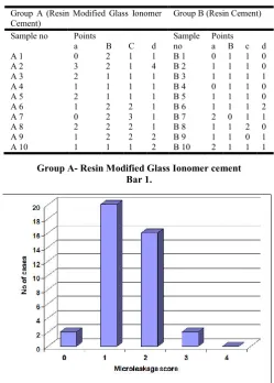
[image:2.595.311.555.54.182.2]Microleakage scoring of Resin Modified Glass Ionomer Cement (Group A), Resin Cement (Group B) based on 40 points for each group.
Table 1- Microleakage Score
Group A (Resin Modified Glass Ionomer Cement)
Group B (Resin Cement)
Sample no Points Sample
no
Points
a B C d a B c d
A 1 0 2 1 1 B 1 0 1 1 0
A 2 3 2 1 4 B 2 1 1 1 0
A 3 2 1 1 1 B 3 1 1 1 1
A 4 1 1 1 1 B 4 0 1 1 0
A 5 2 1 1 1 B 5 1 1 1 0
A 6 1 2 2 1 B 6 1 1 1 2
A 7 0 2 3 1 B 7 2 0 1 1
A 8 2 2 2 1 B 8 1 1 2 0
A 9 1 2 2 2 B 9 1 1 0 1
A 10 1 1 1 2 B 10 2 1 1 1
Group A- Resin Modified Glass Ionomer cement Bar 1.
[image:2.595.306.556.442.791.2]Group A
Microleakage score No of cases Percentage
0 2 5
1 20 50
1 16 40
3 2 5
4 0 0
Total 40 100
Group B- Resin Cement
[image:3.595.42.285.70.383.2]Bar 2
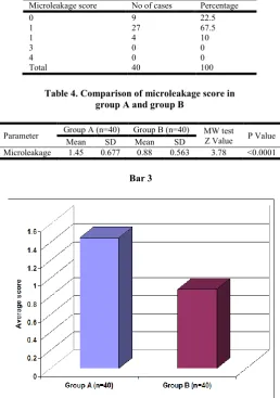
Table 3. Distribution of score according to microleakage in Group B
Microleakage score No of cases Percentage
0 9 22.5
1 27 67.5
1 4 10
3 0 0
4 0 0
Total 40 100
Table 4. Comparison of microleakage score in group A and group B
Parameter Group A (n=40) Group B (n=40) MW test
Z Value P Value
Mean SD Mean SD
Microleakage 1.45 0.677 0.88 0.563 3.78 <0.0001
Bar 3
DISCUSSION
The disadvantage of this method is that the marker particle has a smaller diameter as compared to the bacterial toxin or the bacteria themselves. Thus it was believed that the results procured by the dye penetration method were not clinically significant. Therefore, application of liposaccharides was recommended. However, it was observed that application of dye or liposaccharides categorized with radioisotopes were equally efficacious for determination of microleakage and level of dye penetration at the dentin-cement interface (Pessutti Nunes et al., 2005).Shane N. White, B Dent, Sue Ingles, and Victor Kipnis (White et al., 1994) - presented many reasons for the capacity of the cement to reduce microleakage was considered less favourable as compared to that of in vivo studies. The most important reasons were the difference in size of marker particles and size of the particles. The dentinal fluid in vital teeth may contrast molecular penetration and the buildup of proteins in the marginal opening may improve the seal. Therefore, it may be accomplished that if the cement material resists marker penetration in invitro studies, its response is anticipated to be better in vivo studies. He pointed out that the results of microleakage in an in-vitro study depend on a factors such as biological dentine properties, applied experimental model (teeth storage, teeth preparation, thermocycling, occlusal loading) and cement properties (Mash
et al., 1991). All the factors in this study were relatively alike except for cement type. It can also be established that properties of the luting agent have played a conclusive role in marginal opening formation. In the present study, the degree of microleakage for different cements were evaluated that were based on the results obtained by the analysis of 10 crowns cemented with the same type of cement. Each sample evaluated at the 4 points. Therefore the degree of microleakage for each cement was measured at total 40 points. The results of investigation of microleakage was obtained by measuring depth of linear dye penetration and is presented in Table 2, 3 & 4. It explains that the maximum linear dye penetration was found with resin modified glass ionomer cement and minimum with resin cement. The mean microleakage score of group A and Group B are 1.45 and 0.88.
Vesna Medic, Kosovka Obradovic, Slobodan Doic, Renata Petrov (Medic et al., 2010) explored the effect of various cements (polycarboxylate, resin cement, glass-ionomer and zinc-phosphate) on microleakage in various ceramic crown systems (Empress 2, metal ceramic crown with a porcelain margin, metal ceramic crown, and In Ceram all-ceramic crowns) secured on extracted human teeth. The study elicited that the crown type does not influence microleakage but it allows comparison of the degrees of microleakage pertaining to a particular cement type. Results were found that least degree of microleakage was noticed in specimens luted with resin cement (X = 1.73), followed by glass-ionomer cement (X=2.45). So the authors concluded that various dental cements exhibited different sealing abilities. The use of resin cement resulted in the percentage of 0 microleakage scores. Due to this unique characteristic, resin cement is to be recommended in everyday clinical practice.
G. A. Crim, M. L. Swartz, and R. W. Phillips (1985 & 1987) showed that even if low number of thermal cycles are used, it can cause failure and microcracks of the weak links at the
restoration interface. They proposed that prolonged
[image:3.595.33.292.425.793.2]produce microleakage. Anthony H.L, D. Dent, James (Tjan et al., 1991; Anthony Tjan et al., 1992) et al assessed teeth which were restored with composites. No significant differences were found in dye penetration between 100 and 1500 cycles (Gavelis
et al., 1981). Moreover, high linear of coefficient of thermal expansion is seen in acrylic resins as compared to tooth structure and filled composites. When the acrylic resin is used in single crowns, its thickness near the margin is 1 to 1.5 mm. Hence, the crown gets subjected to dimensional changes caused by temperature variations. Chung-Ming Hung, Saul Weiner, Dastane, and K. Vaidyanathan (Hung et al., 1993) described the effect of thermocycling and occlusal force on microleakage. Different crowns made of different material under different mechanical loading tend to show different properties. When stress is applied in the region of crown margin, it instigates tensile and shear strength in the cement layer which cannot be resisted. This may be the cause for weakening of the bond between the cement and dentine surface proving an onset for microcracks and microleakage. In this study, the assessment of the influence of different cement types on crown microleakage which was measured at 4 points showed a statistically significant difference in microleakage values between different cement types at all the observed points (Table 2, 3 & 4). Statistically significant lowest values were apparent in resin cement and higher values were observed with resin modified glass-ionomer cement. The results of the study provide useful information and helps the clinicians in choosing the best cementing material. The clinician must be well versed with all properties of the available cements as well as with their manipulation methods. Practical approval includes use of luting agents depending on clinical situation, type of indicated fixed restoration and finally the cost-effectiveness of luting agent. The significance of gap formation and microleakage has been clearly confirmed but the exact amount that becomes clinically relevant remains unspecified. The choice of luting agent depends upon appropriate criteria for any specific clinical situation.
REFERENCES
Anthony Tjan A, Dunn J.R and Grant B.E. 1992. Sealing ability of cast gold complete crown restoration. J. Prosthet Dent, 67;224.
Berg J H, David E MS. Pettey, Max O. Hutchins, 1988. Microleakage of three luting agents used with stainless steel crowns. Pediatric Dentistry: September, Volume 10. Bhandari S, Aras M, and Chitre V. 2012. An In Vitro
Evaluation of the Microleakage under Complete Metal Crowns Using Three Adhesive Luting Cements. J Indian Prosthodont Soc., Jun; 12(2): 65–71.
Cardoso M, Torres M.F, Moraes Rego M, and Santiago L.C. 2008. Influence of application site of provisional cement on the marginal adaptation of provisional crowns; J Appl Oral Sci., Jun; 16(3): 214–218.
Castro A, MS Robert F. Feigal. 2002. Microleakage of a new improved glass ionomer restorative material in primary and permanent teeth. Pediatric Dentistry – 24:1,
Crim G.A, and Garcia-Godoy F. 1987. Microleakage. The effect of storage and cycling Duration. J Prosthet Dent, 57(5):574-6.
Crim G.A, Swartz M.L. and Phillips R.W. 1985. Comparison of four thermocycling. J Prosthet Dent, 53(1):50-3.
Diaz-Arnold A.M, Vargas M.A, and Debra R.H. 1999. Current status of luting agents for fixed prosthodontics. J Prosthet Dent, 81:135-41.
Fusayama T, Ide K, Kurosu A and Hosoda H. 1963. The thickness between cast restoration and preparation. J Prosthet Dent, 13(2):354-64.
Gavelis J.R, Morency J.D, Riley E.D, Sozio R.B. 1981. The effect of various finish line preparation on marginal seal. J Prosthet Dent, 45(2):138-42.
Harish V,Mohamed Ali S.A.,Jagadesan N,Ifthikar
M,4Senthil S, Basak D, Huda F,and Priyanka. 2014. Evaluation of Internal and Marginal Fit of Two Metal
Ceramic System – In Vitro Study. Journal of Clinical and
Diagnostic Research, Dec; 8(12).
Holmes JR, Bayne SC, Holland GA, Sulik WD. 1989. Considerations in measurement of marginal fit. J Prosthet Dent., Oct;62(4):405-8.
Hung CM, Weiner S, Dastane A, and Vaidyanathan T.K. 1993. Effects of thermocycling and occlusal force on the margins of provisional acrylic resin crowns. J Prosthet Dent, 69:573-7.
Jacobs M.S. and Windeler A.S. 1991. An investigation of dental luting cement solubility as a function of the marginal gap. J Prosthet Dent, 65:436-42.
Lindquist TJ. and Connolly J. 2001. In vitro microleakage of luting cements and crown foundation material. J Prosthet Dent., Mar;85(3):292-8.
Mash L.K, Beninger C.K, Bullard J.T and Staffanou R.S. 1991. Leakage of various types of luting agents. J. Prosthet Dent., 66:763-6.
Medic V, Obradovic K, Doic S, Petrovi R. 2010. In Vitro Evaluation of Microleakage of Various Types of Dental Cements. Srp Arh Celok Lek, 138(3):143-9
Medić V, Obradović-Djuričić K, Dodić S, Petrović R. 2010. In Vitro Evaluation of Microleakage of Various Types of Dental Cements; Srp Arh Celok Lek., Mar-Apr;138(3-4):143-149.
Orlato P.H, Accacio L do, Ricardo M de, Fernando De M, Fernando L. 2008. Correlation between margin fit and microleakage in complete crowns cemented with three luting agents. J Appl Oral Sci., 16(1):64-9
Osborne L.W, Swartz L.M, Goodacre C.J, Phillips R.W, Gale E.N. 1978. A method for assessing the clinical solubility and disintegration of luting cements. J Prosthet Dent, 40: 413-7.
Pessutti Nunes M.C, Franco E.B and Pereira J.C. 2005. Marginal microleakage: critical analysis of methodology. Salusvita, Bauru, ;3:487-502.
Piwowarczyk A, Lauer HC, Sorensen JA. 2005. Microleakage of various cementing agents for full cast crowns. Dent Mater. May;21(5):445-53.
Sener I, Turker S.B, Valandro L.F, Ozcan M. 2014. Marginal gap, cement thickness, and microleakage of 2 zirconia crown systems luted with glass ionomer and MDP-based cements. General Dentistry,62(2):67-70 · March.
Shah R. and Shah D. 2012. An evaluation of microleakage under crowns cemented with different luting agents. Journal of Advanced Oral Research, Vol 3; Issue 3: Sept – Dec.
Shiflett K, Shane N. White, B Dent Sc. 1997. Microleakage of cements for stainless steel crowns. American Academy of Pediatric Dentistry . 19:4.
Swartz M.L, Sears C and Phillips R.W. 1971. Solubility of cement as related to time of exposure in water. J Prosthet Dent, 3:294-7.
Tsitrou E.A, Northeast S.E, Noort R. 2007. Evaluation of the marginal fit of three margin designs of resin composite
crowns using CAD/CAM. Journal of Dentistry, 35(1): 68–
73.
Wang CJ, Millstein PL, Nathanson D. 1992. Effects of cement, cement space, marginal design, seating aid materials, and seating force on crown cementation. J Prosthet Dent., Jun;67(6):786-90.
White S.N, Ingles S, and Kipnis V. 1994. In vivo microleakage of luting cements for cast crowns. JProsthetDent, 71:333-8.
Wu, JC., Lai LC, Sheets CG, Earthman J, and Newcomb R. 2011. A comparison of the marginal adaptation of cathode-arc vapor-deposited titanium and cast base metal copings. J Prosthet Dent, Jun; 105(6): 403–409.
Yüksel E. and Zaimoğlu, A. 2011. Influence of marginal fit and cement types on microleakage of all-ceramic crown systems. Braz Oral Res., May-Jun;25(3):261-6 2.