DIFFUSE AXONAL INJURY IMAGING (DAII) AND FUTURE PROSPECTIVE: A COMPARATIVE STUDY
WITH RECENT ADVANCES APPLICATION IN SYSTEMATIC REVIEW OF LITERATURE
1,*
Dr. MayadharBarik,
2Dr
5
Dr. Ghanshyam Kumawat,
1
Department of Nuclear Medicine,
2
Post Graduate Patna Medical College Bihar,
3Department of Radiology
4
Senior Resident Orthopaedics Patna Medical
5Department of Radiology
7
Ex-HOD Orthopaedics Patna Medical
ARTICLE INFO ABSTRACT
Background:
extensive lesions in white matter tracts occurs over a widespread area. DAI is one of the most common and devastating types of traumatic brain injury (TBI).
Material and Met
resources were retrospectively we reviewed to identify patients underwent both CT and MRI examinations of the head and patients were found with diagnostic images were available for DAI and de-identified images reported by ealier world literature.
hemorrhage, diffuse axonal injury (DAI), and skull fracture also reviewed here in systematically.
Results:
with over 97% of patients wit
often remain significantly impaired. So DAI can occur in every degree of severity from very mild or moderate to very severe. MRI more frequently reported intracranial findings of CT scanning. statistically significant difference observed between CT and MRI in the detection of any intracranial injury.
Conclusion:
of the underlying pathophysiological change
diagnostic and prognostic accuracy of MRI. But we should be ignored CT findings for DAI for decission making.
Copyright ©2016,Dr. MayadharBarik et al. This is an open access article distributed
unrestricted use, distribution, and reproduction in any medium, provided the original work is properly cited.
INTRODUCTION
Diffuse axonal injury (DAI) is a brain injury in which damage in the form of extensive lesions in white matter tracts occurs over a widespread area. DAI is one of the most common and devastating types of traumatic brain injury (TBI)
2004). DAI having major cause of unconsciousness and persistent vegetative state after head trauma
Corresponding author: Dr. MayadharBarik, Department of Nuclear Medicine, AIIMS, NewDelhi
ISSN: 0975-833X
Available online at http://www.journal
Article History:
Received 18th December, 2015 Received in revised form 20th January, 2016
Accepted 05th February, 2016
Published online 31st March,2016
Key words:
Diffuse axonal Injury, Traumatic Brain Injury, X-Ray,
CT, MRI.
Citation: Dr. MayadharBarik, Dr. Arun kumar, Dr. Priyanka Rathore, Dr. Anjit Kuma
Dr. R. R. Kanaugia, 2016. “Diffuse axonal injury imaging (daii) and future prospective: a comparative study with recent advances application in systemat
review of literature”, International Journal of Current Research,
RESEARCH ARTICLE
DIFFUSE AXONAL INJURY IMAGING (DAII) AND FUTURE PROSPECTIVE: A COMPARATIVE STUDY
WITH RECENT ADVANCES APPLICATION IN SYSTEMATIC REVIEW OF LITERATURE
Dr. Arun kumar,
3Dr. Priyanka Rathore,
4Dr
Dr. Ghanshyam Kumawat,
6Dr. Vikash Kumar and
7Dr. R. R
Department of Nuclear Medicine, AIIMS, New Delhi-29 India
Post Graduate Patna Medical College Bihar, India
of Radiology, Patna medical College, Patna Bihar,
Senior Resident Orthopaedics Patna Medical College, Bihar, India
of Radiology, Patna medical college, Patna Bihar,
6
IIT Patna, Bihar, India
HOD Orthopaedics Patna Medical College, Bihar, India
ABSTRACT
Background: Diffuse axonal injury (DAI) is the brain injury in which damage in the form of an extensive lesions in white matter tracts occurs over a widespread area. DAI is one of the most common and devastating types of traumatic brain injury (TBI).
Material and Methods: We reviewed from from different data set like pubmed/medline and google resources were retrospectively we reviewed to identify patients underwent both CT and MRI examinations of the head and patients were found with diagnostic images were available for DAI and
identified images reported by ealier world literature. Presence
hemorrhage, diffuse axonal injury (DAI), and skull fracture also reviewed here in systematically.
Results: It occurs in about half of all cases of severe head trauma and outcome is frequently coma, with over 97% of patients with severe DAI never regaining consciousness.
often remain significantly impaired. So DAI can occur in every degree of severity from very mild or moderate to very severe. MRI more frequently reported intracranial findings of CT scanning. statistically significant difference observed between CT and MRI in the detection of any intracranial
Conclusion: The multimodal MRI approach in patients with DAI results differentiated representation of the underlying pathophysiological changes of the injured nerve tracts.
diagnostic and prognostic accuracy of MRI. But we should be ignored CT findings for DAI for decission making.
is an open access article distributed under the Creative Commons Attribution License, which use, distribution, and reproduction in any medium, provided the original work is properly cited.
Diffuse axonal injury (DAI) is a brain injury in which damage white matter tracts occurs over a widespread area. DAI is one of the most common and
(TBI) (Iwata et al.,
DAI having major cause of unconsciousness and
Department of Nuclear Medicine, AIIMS, NewDelhi-29 India.
(Wasserman and Koenigsberg,
DAI can occur in every degree of severity from very mild or moderate to very severety (Vik
2000). Unlikely, brain trauma occured due to direct impact and deformation. The brain, DAI is the result of traumatic shearing forces that occur when the head is rapidly accelerated or decelerated, occur in auto accidents, falls,
assaults (Sivák et al., 2005; Wolf
results from rotational forces or severe deceleration and McKenna, 2001; Shepherd
Available online at http://www.journalcra.com
International Journal of Current Research
Vol. 8, Issue, 03, pp. 28440-28445, March, 2016
Dr. MayadharBarik, Dr. Arun kumar, Dr. Priyanka Rathore, Dr. Anjit Kumar Dr. Ghanshyam Kumawat, Dr. Vikash Kumar and
Diffuse axonal injury imaging (daii) and future prospective: a comparative study with recent advances application in systemat
Journal of Current Research, 8, (03), 28440-28445.
z
DIFFUSE AXONAL INJURY IMAGING (DAII) AND FUTURE PROSPECTIVE: A COMPARATIVE STUDY
WITH RECENT ADVANCES APPLICATION IN SYSTEMATIC REVIEW OF LITERATURE
Dr. Anjit Kumar
R. Kanaugia
India
Patna Bihar, India
India
India
India
Diffuse axonal injury (DAI) is the brain injury in which damage in the form of an extensive lesions in white matter tracts occurs over a widespread area. DAI is one of the most
We reviewed from from different data set like pubmed/medline and google resources were retrospectively we reviewed to identify patients underwent both CT and MRI examinations of the head and patients were found with diagnostic images were available for DAI and Presence of any injury, intracranial hemorrhage, diffuse axonal injury (DAI), and skull fracture also reviewed here in systematically.
It occurs in about half of all cases of severe head trauma and outcome is frequently coma, h severe DAI never regaining consciousness. Those who do wake up often remain significantly impaired. So DAI can occur in every degree of severity from very mild or moderate to very severe. MRI more frequently reported intracranial findings of CT scanning. No statistically significant difference observed between CT and MRI in the detection of any intracranial
The multimodal MRI approach in patients with DAI results differentiated representation s of the injured nerve tracts. It helps to improve the diagnostic and prognostic accuracy of MRI. But we should be ignored CT findings for DAI for
ribution License, which permits
, 2007; Vinas and Pilitsis, 2006) DAI can occur in every degree of severity from very mild or
Vik et al., 2006; Smith and Meaney,
Unlikely, brain trauma occured due to direct impact The brain, DAI is the result of traumatic shearing forces that occur when the head is rapidly accelerated occur in auto accidents, falls, addiction and
Wolf et al., 2001). It was usually
results from rotational forces or severe deceleration (Sanders Shepherd, 2004).
INTERNATIONAL JOURNAL OF CURRENT RESEARCH
Dr. Ghanshyam Kumawat, Dr. Vikash Kumar and
Vehicle accidents most frequent cause of DAI the result of child abuse (Hardman and Manoukian, 2002
shaken baby syndrome (SBS) (Smith and Greenwald
The major cause of damage in DAI is the disruption of axons in the neural processes. It allows one neuron to communicate
with another Tracts of axons (Vik et al., 2006
white due to myelination, are referred to as white matter. Acceleration due causes shearing injury are damage inflicted as tissue slides over other tissue. When the brain is accelerated by parts of differing densities. Distances from the axis of rotation slide over each other stretching axons.
junctions between areas of different density at junctions between white and grey matter, Two thirds of DAI lesions occur in areas point at (grey and white matter meet
MATERIALS AND METHODS
We reviewed from from different data set like pubmed/medline and google resources were retrospectively we reviewed to identify patients underwent both CT and MRI examinations of the head and patients were found with diagnostic images were available for DAI and de-identified images reported by ealier world literature. Presence of any injury, intracranial hemorrhage, diffuse axonal injury (DAI), and skull. THe important studies published from 2000 to 2016 on the topic of head trauma, even mild head injury, neurocognitive deficits, medical imaging has assumed preeminence for detecting abnormalities associated with TBI. Advanced MRI modalities such as DTI and MRS have an important role in the diagnosis of lesions for TBI patients were taken into account of this review.
RESULTS
It occurs in about half of all cases of severe head trauma and outcome is frequently coma, with over 97% of patients with severe DAI never regaining consciousness.
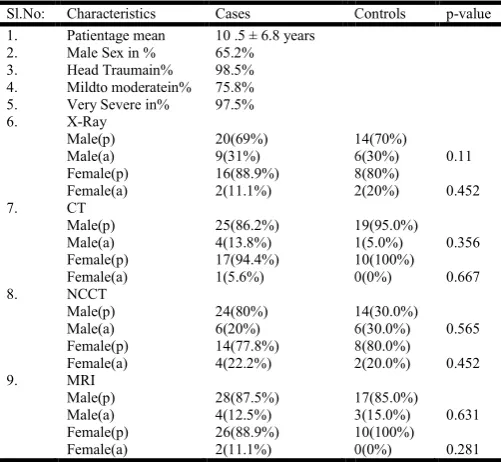
Table 1. (Comparison of various imaging modalities in DAI
Sl.No: Characteristics Cases Controls
1. Patientage mean 10 .5 ± 6.8 years
2. Male Sex in % 65.2%
3. Head Traumain% 98.5%
4. Mildto moderatein% 75.8%
5. Very Severe in% 97.5%
6. X-Ray
Male(p) Male(a) Female(p) Female(a) 20(69%) 9(31%) 16(88.9%) 2(11.1%) 14(70%) 6(30%) 8(80%) 2(20%)
7. CT
Male(p) Male(a) Female(p) Female(a) 25(86.2%) 4(13.8%) 17(94.4%) 1(5.6%) 19(95.0%) 1(5.0%) 10(100%) 0(0%)
8. NCCT
Male(p) Male(a) Female(p) Female(a) 24(80%) 6(20%) 14(77.8%) 4(22.2%) 14(30.0%) 6(30.0%) 8(80.0%) 2(20.0%)
9. MRI
Male(p) Male(a) Female(p) Female(a) 28(87.5%) 4(12.5%) 26(88.9%) 2(11.1%) 17(85.0%) 3(15.0%) 10(100%) 0(0%)
N.B. P:present,a:absent and p-value lesser then.0005 considered as significant.
28441 Dr. MayadharBarik et al. Diffuse axonal injury imaging (daii) and future prospective: a comparative study with recent advances application
Vehicle accidents most frequent cause of DAI the result of 2002). It is like as in Smith and Greenwald, 2003). The major cause of damage in DAI is the disruption of axons in the neural processes. It allows one neuron to communicate 2006). This appeared white due to myelination, are referred to as white matter.
eleration due causes shearing injury are damage inflicted as tissue slides over other tissue. When the brain is accelerated by parts of differing densities. Distances from the axis of rotation slide over each other stretching axons. That traverse junctions between areas of different density at junctions Two thirds of DAI lesions grey and white matter meet).
We reviewed from from different data set like pubmed/medline and google resources were retrospectively we reviewed to identify patients underwent both CT and MRI examinations of the head and patients were found with diagnostic images were identified images reported by ealier Presence of any injury, intracranial hemorrhage, diffuse axonal injury (DAI), and skull. THe important studies published from 2000 to 2016 on the topic of neurocognitive deficits, nce for detecting abnormalities associated with TBI. Advanced MRI modalities such as DTI and MRS have an important role in the diagnosis of lesions for TBI patients were taken into account of this
occurs in about half of all cases of severe head trauma and outcome is frequently coma, with over 97% of patients with
Comparison of various imaging modalities in DAI)
Controls p-value
14(70%) 6(30%) 8(80%) 2(20%) 0.11 0.452 19(95.0%) 1(5.0%) 10(100%) 0(0%) 0.356 0.667 14(30.0%) 6(30.0%) 8(80.0%) 2(20.0%) 0.565 0.452 17(85.0%) 3(15.0%) 10(100%) 0(0%) 0.631 0.281
value lesser then.0005 considered as significant.
Those who do wake up often remain significantly impaired. So DAI can occur in every degree of severity from very mild or moderate to very severe. MRI more frequently reported intracranial findings of CT scanning.
significant difference observed between CT and MRI in the detection of any intracranial injur
patients cohort varies 10 .5 ± 6.8 years, and 65.2% were male. The mean Injury Severity Score
mean Glasgow Coma Scale score
statistically significant difference between CT and MRI observed majority of the papers till date (attached table no1 and interesting Figures (1 to 9).
[image:2.595.370.499.248.400.2]Interesting Images of X-Ray, Ct, N
Fig shows X-ray head diffuse axonal injury (DAI)
Fig. 1. X ray DAI control
[image:2.595.37.288.521.752.2]Fig. 2. X ray DAI cases
Fig. 3. X ray DAI cases
use axonal injury imaging (daii) and future prospective: a comparative study with recent advances application in systematic review of literature
Those who do wake up often remain significantly impaired. So DAI can occur in every degree of severity from very mild or moderate to very severe. MRI more frequently reported CT scanning. No statistically significant difference observed between CT and MRI in the detection of any intracranial injury. The mean age of the cohort varies 10 .5 ± 6.8 years, and 65.2% were male. The mean Injury Severity Score (MESS) varies 15.7 ± 8.2.The mean Glasgow Coma Scale score (GCSS) was 10 ± 5.8. No statistically significant difference between CT and MRI observed majority of the papers till date (attached table no1
(1 to 9).
Ray, Ct, Ncct, Mri Of Dai
ray head diffuse axonal injury (DAI)
Fig. 1. X ray DAI control
X ray DAI cases
Fig. 3. X ray DAI cases
Fig shows CT head diffuse axonal injury (DAI)
Fig. 4. Shows CT (DAI
Fig. 5. Case CT (DAI
Fig. 6. Control CT (DAI)
DISCUSSION
Older approach and comparison
MRI as sensitive as CT scanning in the detection of THI, DAI, and intracranial hemorrhage.
28442 International Journal of Current Research,
Fig shows CT head diffuse axonal injury (DAI)
MRI as sensitive as CT scanning in the detection of THI, DAI,
Fig shows NCCT head diffuse axonal injury (DAI) comparing with MRI
Fig. 7. Shows
Fig. 8. Shows MRI (DAI)
Fig. 9. ShowsT1 weighted MRI (DAI)
We still missed out skull fractures among patients. MRI may be a useful alternative to CT scanning in select stable patients with mild THI&DAI neuroimaging made clinical decision to
(Roguski et al., 2015).
International Journal of Current Research, Vol. 08, Issue, 03, pp. 28440-28445, March,
Fig shows NCCT head diffuse axonal injury (DAI)
Shows NCCT (DAI)
hows MRI (DAI)
Fig. 9. ShowsT1 weighted MRI (DAI)
missed out skull fractures among patients. MRI may be a useful alternative to CT scanning in select stable patients with mild THI&DAI neuroimaging made clinical decision to easier
Traumatic brain injury (TBI) result in immediate long-lasting coma. So far attention has been given to predict this outcome. From the initial examination because these predictions guide future treatment and interactions with the patient's family. DAI in these cases have ascribed the coma to widespread damage located in the deep white matter that disconnects the hemispheres from the ascending arousal system (AAS). Brainstem lesions are also present in the AAS interrupt at the brainstem level. This review examined that autopsy and imaging literature that assesses the presence, extent, and predictive value of lesions in both sites in DAI. An evidence suggests that diffuse injury to the deep white matter is not the usual cause of immediate long-lasting posttraumatic coma (LLPTC). Brainstem lesions in the rostral pons or midbrain are always cause not only if the lesions but also bilateral.
Recovery is possible if critical brainstem inputs to the AAS are spared. In precise localization of the latter is subject to ongoing investigation with advanced imaging techniques using magnets of very high magnetic gradients were very Limited availability. As of this equipment plus the need to verify the findings continue to require meticulous autopsy examination (Rosenblum, 2015). Diffuse axonal injury (DAI) plays a major role after traumatic brain injury (TBI). Its imaging is based on computed tomography (CT) or magnetic resonance imaging (MRI). DAI in a histological diagnosis, histopathological findings on survival after TBI are very rare reported that global macrostructural damage commonly associated with traumatic axonal injury (TAI). It contributes to structural disconnection of anatomically distributed regions that underlie ToM. This study suggests that SWI may be a valuable imaging biomarker to predict outcome and recovery of social cognition after
pediatric TBI (Ryan et al., 2015).
The SWI technique is extremely sensitive to blood breakdown to products and appear as small signal voids at three locations, at the gray-white interface, in the corpus callosum and in the brain stem. Functional MRI comprises a group of constantly developing techniques that have a great potential in optimal evaluation of the white matter in patients after craniocerebral trauma. These imaging techniques allowed the visualization of changes associated with shear injuries, such as functional impairment of axons and decreased blood flow including an abnormal metabolic activity of the brain parts affected.
The multimodal MRI approach in patients with DAI resulted in a more detailed and differentiated representation of the underlying path physiological changes of the injured nerve tracts and helps to improve the diagnostic and prognostic accuracy of MRI. If DAI is suspected multimodal MRI should be performed as soon as possible after craniocerebral injury (Mallouhi, 2014). This prospective approach improve in moderate-severe TBI patients were to investigate volume change in cortical gray matter (GM). Moreover, hippocampus, lenticular nucleus, lobar white matter (WM), brainstem and ventricles be analyzed using through design and repeated MRI. So in the early phase (1-30 days) and 4 and 12 months postinjury and to assess changes in GM apparent diffusion coefficient (ADC) in normal appearing tissues be varified throughly.
Recent advances
In addition to the cortex, hippocampus and brainstem. The impact of Glasgow Coma Scale (GCS) score at admission, duration of post-traumatic amnesia (PTA), and diffusion axonal injury (DAI) grade on brain volumes and ADC values overt the time was assessed and determined if MRI-derived brain volumes. Higher ADC values were detected in the cortex in individuals with severe TBI, DAI and PTA > 2 weeks, from 3 months. There were no associations between ADC values and brain volumes, ADC values did not predict outcome in better
way (Brezova et al., 2014). Neuroimaging is commonly used
for the assessment of children with traumatic brain injury and has greatly advanced how children are acutely evaluated emphasis has been given on how advanced magnetic resonance imaging (MRI) methods can detect subtler injuries. It could be
related to the structural underpinnings of the
neuropsychological and behavioral alterations frequently occured. We examined several methods used for the assessment of pediatric brain injury (PBI). Susceptibility-weighted imaging is a sensitive 3-dimensional high-resolution technique in detecting hemorrhagic lesions associated with diffuse axonal injury (DAI). Magnetic resonance spectroscopy acquires all metabolite information. This serves as a proxy for neuronal (and glial, lipid, etc). Structural integrity and provides sensitive assessment of neurochemical alterations in Diffusion-weighted imaging (DWI) is useful for the early detection of ischemic and shearing injury. Diffusion tensor imaging (DTI) allows better structural evaluation of white matter tracts. These methods more sensitive than conventional imaging in demonstrating subtle injury that underlies a clinically child's symptoms. It is an increasing desire to develop computational methods to fuse imaging data to provide a more integrated analysis of the extent to which components of the neurovascular unit are affected in the future of traumatic brain injury (TBI) neuroimaging research is promising and will lead to novel approaches to predict and improve the better
outcomes (Ashwal et al., 2014).
Future prospective
Our view provides a summary of some of the important studies published from 2000 to 2016 on the topic of MRI findings in head trauma. With the growing realization that even mild head injury can lead to neurocognitive deficits, medical imaging has assumed preeminence for detecting abnormalities associated with TBI. Advanced MRI modalities such as DTI and MRS have an important role in the diagnosis of lesions for TBI
patients care fully (Moen et al., 2014). Neuroradiologists
endanger diagnostics and lead to false treatment decisions and medico-legal problems and faced trouble. Standardized quantitative imaging analysis programs and advances in MRI technology should be utilized to improve radiological TBI
diagnosis (Laalo et al., 2014). We summarized in this paper
adds significantly to the creation of a fundamental knowledge for the improvement of bicycle helmets as well as other head protective measures to be taken care. We also described investigations and experimental results are of crucial importance in DAI cases. We also add DAI information for forensic research angel to neural recovery of an injured cingulum following brain injury.
In head trauma had consistently reduced axonal spike amplitude. The susceptibility of an axon to trauma could be modulated by the function of an ATP-dependent sodium-potassium pump suggest a mechanism by concussive mTBI could lead to the immediate impairment of signal propagation through the axon and the emerging dysfunctional neuronal
information exchange (Seo et al., 2014).
Conclusion
DAI result in immediate long-lasting coma. So, attention has been given to predicting this better outcome from the initial examination.These predictions guide future treatment and interactions with the patient's family. Reports of diffuse axonal injury (DAI) in these cases have ascribed the coma to widespread damage in the deep white matter that disconnects the hemispheres. It from the ascending arousal system (AAS), brainstem lesions re present in such cases.AAS may be interrupted at the brainstem level in patients with DAI. Therefore, diagnosis might have more severe cerebral injury. Hence, the identification process, one should pay attention to the possible missed diagnosis and misdiagnosis, and meanwhile avoid relying on those evidences provided only by CT and MRI only. Particularly, this review examines autopsy and imaging literature that assesses the presence, extent, and predictive value of lesions in both sites as well. Anevidence suggests that diffuse axonal injury (DAI) to the deep white matter is not the usual. Because of this immediate long-lasting posttraumatic coma. Even brainstem lesions in the rostral pons or midbrain are almost always the cause.
The lesions are bilateral and recovery is possible if critical brainstem inputs to the AAS are spared. So, the precise localization of the latter is subject to ongoing investigation required with advanced imaging techniques. We were using magnets of very high magnetic gradients and Limited availability of this equipment plus. This need to verify the findings continue to require meticulous autopsy examination (MAE).Multimodal MRI approach (MMA) in patients with DAI results in a more detailed. We differentiated representation of the underlying path physiological changes of the injured nerve tracts and helps to improve the diagnostic and prognostic accuracy of MRI. This study helps to new and innovative protocol development in future progress of diagnostic and prognostic value.
Acknowledgement: We thanks to our patients and their
parents and relatives.
Conflict of Interest: NILL
REFERENCES
Ashwal, S., Tong, K.A., Ghosh, N., Bartnik-Olson, B. and Holshouser, B.A. 2014. Application of advanced neuroimaging modalities in pediatric traumatic brain
injury. J Child Neurol., Dec;29(12):1704-17.
Brezova, V., Moen, K.G., Skandsen, T., Vik, A., Brewer, J.B., Salvesen, O. and Håberg, A.K. 2014. Prospective longitudinal MRI study of brain volumes and diffusion
changes during the first year after moderate to severe traumatic brain injury.Neuroimage Clin. Mar 28;5:128-40. Hardman, J.M. and Manoukian, A. 2002. "Pathology of head
trauma". Neuroimaging Clinics of North America 12 (2):
175–187.
Iwata, A., Stys, P.K., Wolf, J.A., Chen, X.H., Taylor, A.G., Meaney D.F. and Smith D.H. 2004. Traumatic axonal injury induces proteolytic cleavage of the voltage-gated sodium channels modulated by tetrodotoxin and protease
inhibitors. The Journal of Neuroscience, 24 (19): 4605—
4613.
Laalo, J.P., Kurki, T.J. and Tenovuo, O.S. 2014. Interpretation of magnetic resonance imaging in the chronic phase of traumatic brain injury: what is missed in the original reports?Brain Inj.28(1):66-70.
Mallouhi, A. 2014 .Craniocerebral trauma: magnetic resonance
imaging of diffuse axonal injury. Radiologe.
Sep;54(9):907-15.
Moen, K.T., Jørgensen, L., Olsen, A., Håberg, A., Skandsen, T., Vik, A., Brubakk, A.M. and Evensen, K.A. 2014. High-level mobility in chronic traumatic brain injury and its relationship with clinical variables and magnetic
resonance imaging findings in the acute phase. Arch Phys
Med Rehabil. Oct;95(10):1838-45.
Roguski, M., Morel, B., Sweeney, M., Talan, J., Rideout, L., Riesenburger, R., Madan, N., Hwang, S. Magnetic resonance imaging as an alternative to computed tomography in select patients with traumatic brain injury: a
retrospective comparison. J Neurosurg Pediatr. 2015
May;15(5):529-34.
Rosenblum, W.I. 2015. Immediate, irreversible, posttraumatic coma: a review indicating that bilateral brainstem injury rather than widespread hemispheric damage is essential for
its production. J Neuropathol Exp Neurol.
Mar;74(3):198-202.
Ryan, N.P., Catroppa, C., Cooper, J.M., Beare, R., Ditchfield, M., Coleman, L., Silk, T., Crossley, L., Rogers, K., Beauchamp, M.H., Yeates, K.O. and Anderson, V.A. 2015. Relationships between acute imaging biomarkers and theory of mind impairment in post-acute pediatric traumatic brain injury: A prospective analysis using susceptibility
weighted imaging (SWI). Neuropsychologia. Jan;66:32-8.
Sanders, M.J. and McKenna, K. 2001. Mosby’s Paramedic Textbook, 2nd revised Ed. Chapter 22, "Head and Facial Trauma." Mosby.
Seo, J.P., Jang, S.H. Monea, A.G., Van der Perre, G., Baeck, K., Delye, H., Verschueren, P., Forausebergher, E., Van Lierde, C., Verpoest, I., Vander Sloten, J., Goffin, J. and Depreitere, B. 2014. The relation between mechanical impact parameters and most frequent bicycle related head
injuries. J Mech Behav Biomed Mater., May;33:3-15.
Shepherd, S. 2004. Head Trauma. Emedicine.com. Retrieved on 2008-01-17.
Sivák, Š., Kurča, E., Jančovič, D., Petriščák, Š. and Kučera, P. 2005. "An outline of the current concepts of mild brain injury with emphasis on the adult population" (PDF). Časopis Lėkařů Českých 144 (7): 445–50.
Smith, D. and Greenwald, B. 2003.Management and staging of traumatic brain injury. Emedicine.com. Retrieved through web archive on 17 January 2008.
Smith, D.H. and Meaney D.F. 2000. Axonal damage in
traumatic brain injury. The Neuroscientist. 6 (6): 483—495.
Vik, A., Kvistad, K.A., Skandsen, T. and Ingebritsen, T. 2006. Diffus aksonal skade ved hodetraume. Tiddskr. Nor. Lægeforen. 126: 2940-44.
Vik, A., Kvistad, K.A., Skandsen, T., Ingebrigtsen, T. 2006. "Diffuse axonal injury in traumatic brain injury". Tidsskrift for den Norske Laegeforening (in Norwegian) 126 (22): 2940–2944. PMID 17117192.
Vinas, F.C. and Pilitsis, J. 2006. Penetrating head trauma.
Emedicine.com. Retrieved on 2008-01-14.
Wasserman, J. and Koenigsberg, R.A. 2007. Diffuse axonal
injury. Emedicine.com. Retrieved on 2008-01-26.
Wolf, J.A., Stys, P.K., Lusardi, T., Meaney, D. and Smith, D.H. 2001. Traumatic axonal injury induces calcium influx modulated by tetrodotoxin-sensitive sodium channels.
Journal of Neuroscience. 21 (6): 1923–1930