Raspberry Pi Implementation of Skull Stripping in
MR Images using fast and Robust Technique
Mr. Mahesh Kalyankar1, Mr. Sameer Nagtilak2
1, 2
Department of Electronica Engineering, Kit’s College of engineering, Kolhapur
Abstract: In brain related abnormalities tumor detection Magnetic Resonance Imaging (MRI) is an extremely valuable tool. Due to error prone and time consuming manual segmentation, various methods have been developed without human intervention such as automatically removal of extra-cerebral tissues. Implementation simplicity and processing speed along with high accuracy in automatic extra cerebral tissue removal is very important factor to be considered. This paper focuses on the segmentation methods based on detection in sharp, local changes in intensity, skull stripping using edge following technique. The implementation on raspberry pi and its performance evaluation.
Keywords: Edge growing, skull stripping, opencv, raspberry pi.
I. INTRODUCTION
Accurate prediction due to better visualization has become possible due to computer aided medical diagnosis with various image processing techniques., MRI (magnetic resonance image) is an effective imaging tool amongst the various tools available. Due to its high spatial resolution it has added simplicity to study the anatomical structure. T1-weighted, T2-weighted, and PD-weighted are some of conventional MR sequences. T1-weighted are better suited for showing anatomical structures because it has highest signal on fat tissue. In liquid for detecting lesions, T2-weighted has highest signal and hence suited. By differentiating the contrast of the brain tissues [3], PD-weighted is suited for characterizing lesions. Axial, coronal and sagittal planes of brain can be seen in MRI images. Detailed pictures in MRI scans can produce effective scenery of organs, soft tissues, bone and other internal body structure [4,5]. In medical imaging tasks image segmentation is a common need for the delineation of anatomical structures and other regions of interests. Extracting the brain from an MR Image is the main focus of this work. Accurately segmentation of brain from non-brain tissue is the main requirement for cortical surface reconstruction, pre-surgical planning, brain morphometry. However, there is requirement of robust solution which will be able to remove skull region from brain MR images. The skull stripping problem by region growing segmentation method is the main approach considered here. The simplicity and robustness of the former are main advantages of our method. For detection of brain abnormalities such as brain tumors Magnetic Resonance Imaging (MRI) is an extremely valuable tool. Diagnostic confusion may be produced due to artefacts and undesired tissues along with undesired processing. Thus, one of the important steps in the processing of brain images is skull stripping where the brain tissue is completely segmented from the skull. For reducing unwanted information from the MR images it is done for the purpose of clearing away non-brain backgrounds. The region-based approaches are based on similarity of regional image data. Some of the more widely used approaches are thresholding, clustering, region growing, and splitting and merging. However, as segmentation methods fail to extract the correct boundaries of objects in noisy images it is still a challenging problem of performance evaluation of image segmentation results.
II. RELATEDWORK
and histogram analysis for skull stripping was presented by Galdameset al [7]. A method based on histogram analysis was proposed by Balanet al. [8] Proposed and compared the segmentation accuracy between their proposed method and two widely used techniques, namely BSE and BET. Proposed method outperforms these methods based on this factor. Pratibha [9] compared two methods of skull stripping (BET, BSE) over two datasets containing T1, T2. They showed that BSE is superior to BET in segmentation of brain images. To eliminate non brain tissues from brain MR images skull stripping is a preliminary step, required for many clinical applications and analysis. Depending on factors such as accuracy and speed the best suitable method can be selected. For the sake of improvement in speed and accuracy of prognostic and diagnostic procedures in medical applications automated skull stripping is considered as most helpful strategy. Based on performances using commonly available datasets, various automated skull stripping methods can be comparatively studied and analyzed. Based on merits and limitations of individual method of automated skull stripping suitable method can be chosen.
III.PROPOSEDWORK
To develop fast and robust edge detection technique for MR images to locate object boundaries with following sub objectives:
A. To develop the algorithm for detecting boundaries of an object in the BR images
B. To develop the hardware platform using raspberry pi for proposed algorithm
C. To analyze the performance of the proposed algorithm.
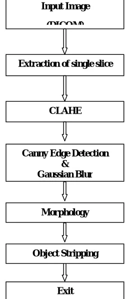
The proposed system block diagram is shown in figure 1. The various stages have been considered as below.
1) Pre-processing: To preprocess image is taken as input from DICOM image with better details of objects. The image is then denoise using Gaussian filter and contrast limited adaptive histogram equalization. The canny edge detection method can be used to differentiate the ROI object from image.
2) Edge Detection: The canny edge detection method can be used to differentiate the ROI object from image.
3) Morphological Operation: Morphological operations like erosion and dilation can be used to remove unwanted objects from the image.
[image:2.612.252.377.417.714.2]4) Stripping: The stripping leads to extraction of ROI object from the MR image.
Figure 1: Block Diagram of proposed work
Input Image
(DICOM)
Extraction of single slice
CLAHE
Canny Edge Detection &
Gaussian Blur
Morphology
Object Stripping
IV.RESULTSANDANALYSIS
[image:3.612.49.487.340.711.2]We have implemented the proposed work in Raspberry pi along with opencv as image processing tool and evaluated the performance in terms of accuracy of segmentation.
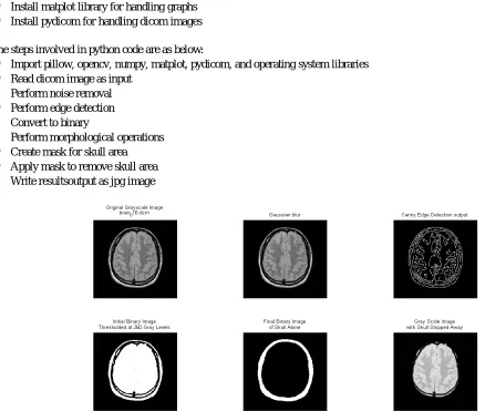
Figure 2, 3 shows the results on brain MR image for various stages output processed in MATLAB and raspberry pi respectively. The input image is taken and Gaussian blur filter is used to remove Gaussian noise in the image. As a first step in skull region detection canny edge detection is done. The result in figure can be seen with optimum value of threshold for canny edge detection. The morphological operations are performed on edge detected image to obtain exact skull area. After morphological operations exact skull area mask is obtained which then can be used to mask the skull are. The mask obtained is applied on Gaussian noise removed image which shows perfect skull stripped away and only brain region is remaining.
Raspberry pi is installed with raspbian operating system which is debian linux. On this system to perform image processing we have installed open computer vision. Using open computer vision image processing can be done such as noise removal, binary conversion, edge detection.
The steps followed for implementation on raspberry pi are as follows:
1) Install raspbian operating system
2) Install python 3 for programming
3) Install open computer vision system
4) Install numpy package for image handling in matrix format
5) Install Pillow package for image Input output handling
6) Install scikit learn for handling histogramsof image
7) Install matplot library for handling graphs
8) Install pydicom for handling dicom images
The steps involved in python code are as below:
a) Import pillow, opencv, numpy, matplot, pydicom, and operating system libraries
b) Read dicom image as input
c) Perform noise removal
d) Perform edge detection
e) Convert to binary
f) Perform morphological operations
g) Create mask for skull area
h) Apply mask to remove skull area
[image:3.612.120.486.494.708.2]i) Write resultsoutput as jpg image
Figure 3: Results of various stages in skull stripping away on raspberry pi
A. Performance Evaluation
The performance of proposed technique is done by experimentation various images. The pixel actually coming under skull area and not coming under skull area and after getting mask is considered as factor of performance evaluation. Based on this true positive, true negative, false positive, false negative are calculated. Results are assessed via comparison with reference skull maps, using metrics P and R based on true positives, false negatives and false positives.
P represents the detection precision, which is given by the ratio between true positives TP and the total region of skull, which is the sum of TP and false negatives FN. It shows the percentage of true skull correctly detected.
P=(TP/TP+FN)
R is given by the ratio between true positives TP of equation and the total region of pixels classified as skull. This measure tells how many of the classified skulls are true skulls.
R=(TP/TP+FP)
A third factor, called F-measure, corresponds to a combination of P and R. The three measures correspond to pixel-wise analysis of the predicted skull map and the respective ground truth. The higher they are, the better the results.
F=(2P.R/P+R)
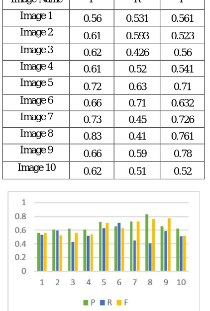
TABLE 1
Performance evaluation of each image for P,R, F calculations for existing method [3]
Image Name P R F
Image 1 0.56 0.531 0.561
Image 2 0.61 0.593 0.523
Image 3 0.62 0.426 0.56
Image 4 0.61 0.52 0.541
Image 5 0.72 0.63 0.71
Image 6 0.66 0.71 0.632
Image 7 0.73 0.45 0.726
Image 8 0.83 0.41 0.761
Image 9 0.66 0.59 0.78
Image 10 0.62 0.51 0.52
[image:4.612.202.413.403.716.2]TABLE2
Performance evaluation of each image for P,R, F calculations for proposed method Image
Name
P R F
Image 1 0.81 0.63 0.56
Image 2 0.83 0.62 0.52
Image 3 0.86 0.63 0.51
Image 4 0.88 0.62 0.53
Image 5 0.78 0.78 0.52
Image 6 0.71 0.79 0.42
Image 7 0.79 0.62 0.63
Image 8 0.81 0.631 0.81
Image 9 0.82 0.63 0.71
Image 10 0.83 0.61 0.42
Figure 4: Performance evaluation for proposed method
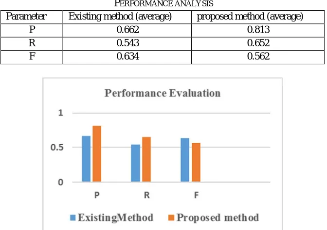
TABLE 3
PERFORMANCE ANALYSIS
Parameter Existing method (average) proposed method (average)
P 0.662 0.813
R 0.543 0.652
F 0.634 0.562
[image:5.612.170.441.96.428.2] [image:5.612.139.475.463.699.2]V. CONCLUSION
We have designed a new edge following technique based boundary detection and applied it to object segmentation for skull stripping in brain MRI images. Our edge following technique incorporates a vector image model and the edge map information. The proposed technique shows outstanding results in terms of skull stripping away. The implementation on raspberry pi is less complex and cost of implementation is considerably low.
REFERENCES
[1] Ségonne F1, Dale AM, Busa E, Glessner M, Salat D, Hahn HK, Fischl B. , “A hybrid approach to the skull stripping problem in MRI”, Neuroimage. 2004 Jul;22(3):1060-75.
[2] Forkert, Nils Daniel Säring, Dennis Fiehler, Jens Illies, Till Färber, Matthias Möller, Dietmar Handels, Heinz, “Fully Automatic Skull-Stripping in 3D Time-of-Flight MRA Image Sequences”, VCBM, 2008
[3] K. Somasundaram and P. Kalavathi, "A hybrid method for automatic skull stripping of magnetic resonance images (MRI) of human head scans," 2010 Second International conference on Computing, Communication and Networking Technologies, Karur, 2010, pp. 1-5.
[4] N. Theera-Umpon and P. D. Gader, “System level training of neural networks for counting white blood cells,” IEEE Trans. Syst., Man, Cybern. C, App. Rev., vol. 32, no. 1, pp. 48–53, Feb. 2002.
[5] N. Theera-Umpon and S. Dhompongsa, “Morphological granulometric features of nucleus in automatic bone marrow white blood cell classification,” IEEE Trans. Inf. Technol. Biomed., vol. 11, no. 3, pp. 353–359,May 2007.
[6] J. Carballido-Gamio, S. J. Belongie, and S. Majumdar, “Normalized cuts in 3-D for spinal MRI segmentation,” IEEE Trans. Med. Imag., vol. 23, no. 1, pp. 36– 44, Jan. 2004.
[7] H. Greenspan, A. Ruf, and J. Goldberger, “Constrained Gaussian mixture model framework for automatic segmentation ofMR brain images,” IEEE Trans. Med. Imag., vol. 25, no. 9, pp. 1233–1245, Sep. 2006.
[8] J.-D. Lee, H.-R. Su, P. E. Cheng, M. Liou, J. Aston, A. C. Tsai, and C.-Y. Chen, “MR image segmentation using a power transformation approach,” IEEE Trans. Med. Imag., vol. 28, no. 6, pp. 894–905, Jun. 2009.
[9] P. Jiantao, J. K. Leader, B. Zheng, F. Knollmann, C. Fuhrman, F. C. Sciurba, and D. Gur, “A computational geometry approach to automatedpulmonary fissure segmentation in CT examinations,” IEEE Trans. Med Imag., vol. 28, no. 5, pp. 710–719, May 2009.
[10] I. Isgum, M. Staring, A. Rutten, M. Prokop, M. A. Viergever, and B. van Ginneken, “Multi-Atlas-based segmentation with local decision fusion—Application to cardiac and aortic segmentation in CT scans,” IEEE Trans. Med. Imag., vol. 28, no. 7, pp. 1000–1010, Jul. 2009.