Immunological Variations in Epileptic Children
Sanjeev Kumar1*, Vinod Kumar1, D. C. Jain2, R. Mittal3
1Department of Physics, Medical Physics Research Laboratory, D.A.V. (P.G.) College, Muzaffarnagar, India 2Department of Neurology, Safdarganj Hospital, New Delhi, India
3Department of Mathematics, Shri. K.K. Jain College, Khatauli, India
Email: *[email protected], *[email protected], [email protected], [email protected]
Received September 30,2012; revised November 3, 2012; accepted November 12, 2012
ABSTRACT
Epilepsy is one of the most frequent neurological problems. Despite of the advances and improvements in treatment of seizure disorders, immunological alterations such as immunoglobulins and complements have been measured. The lev- els of IgG in epileptic patients are found to be higher than controls. The levels of IgA, IgM, C3 and C4 were found to be
lower in all the controls. Student’s t-test were also applied. Multiple and regression analysis, have been also carried out. A trend RM AG RG MA RA GM &rGM A rM GA rM AG has also set up in the present study. An alteration in the immune mechanism of epileptic patients is required. Ketogenic diet may be given and the balance of trace elements like Na, K, Zn, Fe, Ca, Mg and Cu may be maintained to alter the immune mechanism of the epileptic patients. Green leafy vegeta- bles may be given to the patients to control the seizure. Immunity is related with the food we eat. By adjusting the im- munity with proper diet the severity of epileptic attack or any disease may be reduced.
Keywords: Epilepsy; Immunoglobulins; Central Nervous System; Complements
1. Introduction
Humoral immunity is manifested by the production of antibodies. The antibodies are special chemical substanc- es that react against foreign substances. Antibodies are called as immunoglobulins. Immunoglobulins are serum proteins, which possess antibody activity and which are classified according to the antigens and stimulate their production such as IgA, IgG, IgM, IgD and IgE. In order
for antibody to have a cytotoxic effect, an extra sub- stance is required called complement and much has yet to be discovered about its nature and precise functions. The presence of antigens in the body stimulates certain types of defense cells to produce antibodies. Humoral immune response depends on a group of small lymphocytes called B-lymphocytes (B-cells). B-cells originate in bone mar- row and travel directly into lymphoid tissues. Cell medi- ated immunity is directly expressed by certain type of defense against the antigens. These cells are also lym- phocytes, but with a slightly different maturation having the influence of the thymus gland. These are called T- lymphocytes in contrast to B-lymphocytes, which pro- duce humoral immunity.
T-cells also originate in bone marrow but unlike B- cells, they do not travel directly into lymphoid tissues, but first enter the thymus where they undergo a condi-
tioning process, hence the name thyms dependent lym- phocytes. After leaving the thymus thus migrate to spe- cial regions known as thyms-dependent areas, namely the paracortical area of lymph.
It was thought that the central nervous system (CNS) is inaccessible to the immune system in human beings. The concept of immune privilage originated from four historical findings. Ehrlich [1] reported some of the main findings regarding parenterally administration of aniline dyes, which stained almost all body tissues except CNS. Due to this magnificant observation concept of blood brain barrier (BBB) physiologically separated central ner- vous tissue from the systemic circulation and systemic immune response.
Murphy and Sturm [2] have studied mouse sarcoma tissue, which was found not to be rejected implantation into rat brain. It implies that the CNS was exempt from the immunological processes responsible for graft rejec- tion. Sabin [3] studied the absence in brain of direct lym- phatic drainage and by implication of circulating lym- phocytes-depriving the CNS of an apparently essential requirement for immune response generation, which pro- vide an argument in favour of immune isolation. Ediden [4] has reported findings regarding normal nervous tissue, which was not to express major histocompatibility com- plex (MHC) antigens. It exempts the brain from partici- pation in cell-medicated immune reactions.
Immunocompetence of CNS was under some objec- tions. Adverse immune reactions were recognized and reported in the literature. A rabies virus vaccine pricipi- tated with a small number of patients and a neuropara- lytic illness was found to constitute an autoimmune hy- per sensitivity reaction; and an acute disseminated CNS inflammatory disorder precipitated by inoculation with nervous tissue. These findings and favourable outcome of infectious encephalitides and the brisk cerebrospinal (CSF) Pleocytosi, which accompanies brain infection, in- dicate that immune responses can and must be generated within the brain.
The major role of immune system in the CNS is to protect the host against infection and perhaps also neo- plasia. It has been seen that three steps in the protection are first identification of the invaliding microbe or tumor clearance and finally memory for the pathogen so that the immune system may equipped to defend against repeated infections.
CNS and immune system should not be separated but rather as elements of a vast commutation system in which they may exchange information both via an atomic connections and though the hormones and mediators re- leased by hypothalamopituitary axis [5-8]. It is well es- tablished that any excessive or uncontrolled immune re- action may be expected to interfere with the brain’s phy- siology. We may say that any neurologic dysfunction carries the risk of disturbing the delicate equilibrium of the immune network conversely it is true.
This statement is helpful to provide the rationale for the therapeutic concept of immunomodulation of certain immune disorders [7]. Abnormalities of the immune sys-tem are indeed increasing number of neuropathies as well as in some psychiatric syndroms [6,7].
Pharmocologic manipulations of immune pathologies such as rheumatoid arthritis systemic lupus erythmatius etc., by means of neurohormones have been routinely performed.
The process of inflammations increases vascular per- meability and allows anti body, complement and other proteins to pass out the circulation and enter the extra- vaseular space. It may also induce inflammatory cells, including lymphocytes, to cross the vascular endothelium and accumulate in the tissues. Total net effect is to de- ploy all the resources of the immune system at the site of injury. Cells antibody and complement leave the blood and go into action where the demand is high. It may be in the affected tissue outside the vessel wall.
The effect is to abrogate in CNS, if only temporarily, its isolation from the immune processes of the body. The barrier, which excludes plasma proteins from the brain breaks down, allowing antibody to enter the extra vascu- lar space. The amount of protein in CSF increases and with it the level of immunoglobulin may also change.
Immuno competent cells enter the CNS. CNS now be- comes capable to generating an immune response [9]. Antibody, which synthesized locally, adds to that leaking from vessels and increased the level of immunoglobulin. This is a complete reversal of the normal stage and the antibody levels very low. Lymphoid cells may be exclud- ed. Inflammation has three main consequences. It allows lymphocytes and antibody forming cells to enter the CNS and immunoglobulin to be synthesized in the brain and spinal cord.
Lymphocytes accumulate in the CNS in so many in- flammatory diseases. They enter from the blood stream, which passed out of the capillary wall between the endo- thelial cells through altered tight junctions by a process of emperopolesis. Emperopolesis is a process, which de- scribe the behaviour of lymphocytes in tissue culture. They appear to move about inside the cytoplasm of the sebbile cells.
Electron micrograph of the brain shows then travering the endothelial cells of the cerebral capillary. They are enveloped by the plasma membrane and completely sur- rounded by the host cell, only to extrude on the other side.
Lymphocytes and macrophages entering the CNS ac- cumulate in the Virchow-Rabin space. A sharply demar- cated perivascular cuff off inflammatory cells. They may also extend out into the neurophil infiltrating the paren- chymal tissue. Similar peri vascular accumulation can be seen in menings large numbers of lymphocytes, which may enter the subarchnoid space.
The permeability of the cerebral vessels to protein may increase as a result of inflammation. The antibody enters the extravascular space. The vascular permeability changes are often transitory and disappear, but the cells continue to enter from the circulation. In the later stages of the disease the vessels may be impermeable to protein al- though surrounded by densely packed cuff off mono- nuclear cells. Oldstone et al. [10] have studied immu-
nofluorescence technique and reported that albumin is a small molecule and convenient as a marker, but larger protein such as fibrinogen leak from vessel as well. It may be deposited as fibrin in and around the cerebral vessel. Immunoglobulin and complement C3 also pass
out of the vessel and diffuse into the brain parenchyma. A vascular leak directly may be seen in human dis- eases. It changes the composition of CSF. The inflamma- tion may increase the total protein level. This level is found normaly (200 - 400) mg/l and raise in many neu- rological diseases. CSF protein level is not a measure of the efficiency of the blood brainbarrier (BBB).
sized within the CNS and boost up the CSF protein level without vascular leak. Albumin is a good marker, which drives from plasma. It is synthesized in the liver and measured by radial immuno diffusion technique. It is found with high concentration in CSF.
It has been reported that in many inflammatory dis- eases of the nervous system, lymphocytes enter the brain and cord. Both T and B cells may appear in CSF in large amount. It is seen that the percenatage of T cell is in- creased in many specific conditions compared with that in the blood and the percentage of B cells is reduced re-ciprocaly.
In acute and chronic infections B-lymphocytes may differentiate into plasma cells. This transformation may be observed in CSF, where all the cells of the lymphoid series may be found, ranging from small, medium and large lymphocytes and immunoblasts to nature plasma cells.
In many neurological diseases the immunoglobulin level of CSF is found to be higher. This feature is often disproportionate to the rise in total protein and it is due to antibody synthesis in the nervous system. It is very well known that the percentage of immunoglobulin in CSF may be raised in many diseases.
It has been found that the proportion between the con- centration of a protein in the serum and the CSF remains the same, irrespective of the serum level. Serum IgG and
the albumin ratios remain constant and their quotient is also constant as well This has been designated the
. Index IgG CSF CSF Index Serum Serum IgG Alb IgG IgG Alb
(1)
From the equilibrium position reached between CSF and plasma we can calculate the proportion of immu- noglobulin entering from the circulation.
The amount of IgG entering from plasma as exudates
or transudate cannot be calculated directly. We can get some useful informations from levels of IgG and albumin
in the CSF and plasma.
Serum Synthesis CSF Serum CSF Serum Serum 369 230 0.43 5 IgG IgG IgG Alb Alb IgG Alb (2) excudate Serum Serum CSF Serum 0.43 230 IgG Alb IgG Alb Alb (3)
where IgGCSF is equal to the levels of IgG measured
in CSF Seru is equal to the levels of meas- ured in Serum m
IgG IgG
CSF Alb
Alb
is equal to the levels of albumin measured in CSF Se is equal to the levels of albu- min measured in serum rum
We assume that the plasma contribution is made up of two separate elements the normal transudate across the blood brain barrier, supplemented by an exudate or in- flammatory leak at the stage of breaking of BBB. we have a situation like
Synthesis CSF
transudate exudate
IgG IgG
IgG IgG
(4)
The amount of in the exudate is calculated from the albumin levels in CSF and plasma. It is assumed that these two proteins cross the barrier in a fixed quotient directly related to serum concentration:
IgG
Serum
Serum IgG
Alb (5)
and inversely to molecular weight
69000 4.3
150000 (6)
Albumin in the exudates is estimated by subtracting the amount in the transudate
Serum
230
Alb
(7)
From the total, allowing the in the exudate to be calculated as follows:
IgG excudate Serum Serum CSF Serum 0.43 230 IgG Alb IgG Alb Alb (8)
1.1. Tolerance and Autoimmunity in the Nervous System
It was believed that only foreign proteins are considered true antigens. Ehrlich [1] had introduced horror auto-toxicus. Due to this fact, it is possible for body, which may generate antibodies against itself. Autoimmunity suggests that an appropriate immune response is directed against a normal tissue component and leads directly to disease in the absence of persisting infection. Microbes are responsible for autoimmunity. If autoimmunity per- sists during the disease it is inappropriate to term the dis- ease autoimmune.
The distinction between autoimmune response trigger- ed by an infection and inflammation directed against a persistent microbe has been studied by Miller et al. [11].
tides of proteolipid proteins and myelin basis protein.
1.2. Mechanism of Auto Immunity
Auto immunity may be initiated in the following ways: 1) A self-antigen may be modified and appear as for-eign.
2) Ignorant clones may be educated. Microbes may cross-react with self antigens to which the immune sys- tem is ignorant. Epitope is at low concentration perhaps. A cross-reacting microbes present in heavy numbers than the original value of antigen and is able to activate and prime T-cells. Once so primed, the original antigen is sufficient to perpetuate the inflammation.
3) Removal of suppression of auto reactive processes. Microbes might cross-react with idiotope and so disrupt the anti-idiotypic network in favour of immunity. The normal regulatory mechanisms of the immune response should restore self tolerance after the initiation of auto- immunity. The maintenance of autoimmune disease must require either multiple rounds of autoimmunity to differ- ent self-antigens or a single autoimmune response that is perpeturated by defective regulation. A different form of altered immune regulation is an abnormal cytokine re- sponse.
Majority of autoimmune processes are driven by T- celluler processes. Important exceptions are the anti ace- tyl choline receptor antibody of mysthenia gravis and antibodies against epithelial adhesion molecules in the bullous skin disease. In the CNS, the pathogenicity of autoantibodies, such as those associated with the para- neoplastic syndrome or stiff man syndrome is not clearly well documented.
1.3. Antigen Specific Tolerance to Unknown Auto Antigens
It has been seen in many autoimmune disease of the nervous system, driving auto antigen is not known. A treatment is required that makes no assumptions about the provoking antigen and induces antigen -specific tol- erance. A short pulse of antigen nonspecific therapy set up a sequence of events leading to the perpeturation of antigen, specific tolerance. This strategy has been used to justify the use of humanized monoclonal antibody. The earlier work on complement deficiency of specific com- plement components is responsible for a couple of dis- eases. It is very important to remember that this is similar to the disorders, which occur with selective deficiencies of the immune system [12].
There are two types of deficiencies of complements: hereditary and acquired.
Acquired deficiencies persist over a long periods and also become causative factor for certain disease. The complement system consists of a series of proteins, there
are only a handfull of proteins in the complement system, floating freely in the blood. Complements are created in a person’s liver and are activated by the work with anti- bodies. Complement cause lysing or brusting of cells and signal to phagocytes that a cell needs to be removed.
Complement are sequentially reacting proteins on ac- tivation these proteins mediate a number of biological reactions significant to host defense against bacteria, viruses and other injurious stimuli. Antigen-antibody com- plexes, bacterial and plant polysacharides and microbial and tissue enzymes initiate the activation. Biological activities mediated by the activated complements pro- teins or by their fragments include increased capillary permeability, chemotaxis of leukocytes, enhanced phago- cytosis, retention of leukocytes at the site of tissue injury and cytolysis. The most thoroughly studied biological complement system has 18 proteins. These are in higher concentration in plasma. The basic role of complement system is mediation of host defense against microbial infection. This goal is full-filled during activation of complement by the elaboration of peptides, larger protein fragments and multimolecular complexes that opsonize and lyse the activating target; induce chemotactic, secre- tory and metabolic response of leukocytes and alter vas- cular permeability. These activities constitute an inflam- matory response. Fearon, D.T. [13] has given informa- tion regarding complement system and he suggested that this complement system is the most complex of the sev- eral protein activation system in blood, but this complex- ity can be reduced by considering the system to be com- prised of there functional division: two pathways for ac- tivation the classical and alternative pathways and a com- mon effector sequence to which the activating pathways are directed and from which are derived many of the biological activities of complement. Molecular weight that of complement C3 is 185,000 while that of comple-
ment C4 is 180,000. Serum concentration of complement C3 and C4 are 1500 μg/ml and 400 μg/ml.
Some of the important phenomena have been studied by Valanakis, J.E. [14] occurring during activation of the complement sequence and these are related to
1) Acute inflammation.
2) Cell killing namely, which has the following pro-cedure:
a) Increased vascular permeability; b) Chemotaxis of leukocytes; c) Enhanced phagocytosis; d) Membrane damage.
in plasma levels. Thus, an ongoing immunologic event, which is activating the complement system in vivo is
likely to generate a decrease in plasma levels of these proteins. Conversely, the abatement of the complement activating stimulus may be paralleled by a reform of the complement levels toward normal has been reported by Stein, J.H. [15].
The main mediator mechanism of humoral immunity is the complement system. It is an important and essen- tial mechainsm for the destruction both of foreign organ- isms and immunocomplexes in the presence or absence of specific antibodies. It is a double edged sword and may also destroy host tissue. Fishman R.A. [16] reported that CNS is immunologically unusual tissue do to the presence of BBB. It may exclude many components of the immune sustem like antibodies and complement sys- tem. These studies imply a suppressive environment for immune reactivity in the CNS. Monocytes or macro- phases are the important and major extrathepatic sources of complement components. Microglia or astrocytes have been suspected to be the cells which produce comple-ment proteins in brain such as C3.
Milica, T.C. et al. [17] have been studied the role of
complement system. This system does not support in chronic and neurodegenerative disorders. There are two separate complement pathways active by distinctly def- erent classes of activators, specific; identification of com- plement factors in CSF promises to be diagnostic not only of complement activation but also of the mode of activation. Epilepsy is a common neurological problem, which has occupied clinicians for many centuries. It has been noticed that there is strong evidences for polygenic predisposition and in some of cases genetic factors caus- ing epilepsy. Toxins, which is environmental factor plays an important role in some cases of epilepsy. Immuno- logical mechanisms have also been implicated as aetiol- ogy of epilepsy
Some clinical observations show that the aberrations of immunologic system may be associated with untreated epilepsy and pharmacologically treated epileptic patients. The nervous system and the immune system posses a number of interesting similarities. Both of these use well- defined, specialized cell types that communicate through a variety of intercellular mechanisms and develop a spe- cific response to external stimuli. These two demonstrate memory and adapt to historical experience. Dysfunction in the brain can lead to seizures. It can lead to autoimmu- nity [18] in the immune system.
We may see many examples of the direct interactions of the immune system and the brain in patients with epi- lepsy. There is an increased incidence of epilepsy after stroke, trauma, or infection. These environmental condi- tions may release brain antigens, stimulate cytokine re- lease, and result in immune activation. Some of the pro- teins with epilepsy well have a transient worsening of
their symptoms with inter current illness. Such worsen- ing does not necessarily imply a CNS infection, but ra- ther an example of the brain’s response to active immune surveillance. Epilepsy is primarily a paroxysmal disorder of brain function. The brain has a special relationship with the immune system. An understanding of this rela- tionship is essential to realize some of the immunological problems meet in epileptic patients.
Bouma, P.A. [19] suggested that IgA deficiency is
found in most of the epileptic patients and it goes upto 25%. Golamali, Y.P. [20] studied immunoglobulins in idiopathic generalized tonic-clonic epilepsy and found that the changes in serum level of auto antibodies in pa- tients were found to be very high. The above-mentioned abnormalities are associated with both seizure disorders per se and also anti epileptic drugs (AEDS). Baziel, G.M.V.E. et al. [21] have reviewed and studied immu-
noglobulin levels in epilepsy. They have reported that comulative meta-analysis of the earlier studied is not possible due to lack of controlled studies. Savory, J, et al.
[22] have studied cerebrospinal fluid levels of IgG, IgA,
and IgM in neurological disorders such multiple sclerosis,
subacute sclerosing, panencephalities bacterial meningi- tis, viral meningitis, Guillian-Barre syndrome and some miscellaneous disorders. They have reported finding such as elevation of IgG levels in multiple sclerosis goes upto
eighty eight percent. Masi, M., et al. [23] have studied
immuno suppression by Phenytoin and reported the findings as serum. Immunoglobulin levels were observed in mg/dl unit. The level of IgM
95 0
mg/dl and IgG,
1180 0 mg dl
. Their findings reinforce the suspicion that phenytoin may cause severe impairment of both hu- moral and cellular immunity. Bassanini et al. [24] haveexamined serum immunoglobulins in some epileptic pa- tients and compared with controls with the aim to explore whether the previously reported alterations of Ig concen-
trations in epileptic patients are due to anticonsultant the- rapy or due to epilepsy itself. Walker [25] proposed im- munological approach for future work on epilepsy; no subsequent explanation for epileptogensis in immuno- logical terms has been reported. Some of the earlier inve- stigators [26] have proposed that the alterations in elec- trical discharges, which comprise epilepsy could be the result of an autoimmune response directed against trans- mitter receptor sites at synapses. Most of the familiar types of epilepsy have been attributed to be associated with tissue damage. Amman and Hong [27] have found that low serum IgA concentration is frequently associated
with low IgA levels in secretions. Slavin et al. [28] con-
reduced in some patients on long term oral medications with hydantion and also many other patients were having immunologic deficiency. There is no well defined con- stituent defect in cellular immune function in all patients with IgA deficiency. Aman and Hong [31] suggested that
the maintenance of cellular immunity be taken as one of the criteria for diagnosis for the disease.
Yabuki and Nakaya [32] observed that a significant number of patients, had slightly low serum IgM level
among epileptic patients on oral antiepileptic drugs. Conversely, there were also many patients, who had high
IgG levels above the normal range. Phenytoin was the
only drug with which these patients of abnormal serum immunoglobulin levels were commonly mediated. No such immune abnormalities were observed by them in some patients not treated with oral antiepileptic drugs. Allansmith et al. [33] reported that the serum concentra-
tion of immunoglobulins changes insignificantly within individuals. Anderson and Moseklide [34] exhibited that immunological abnormalities do not develop in all pa- tients receiving long term anti convulsant therapy and it is possible that genetic predisposition is of importance in the development of Ig deficiency and auto immunity in
drug-treated epileptics. Matsuoka et al. [35] demon-
strated that the cellular events involved in IgA deficiency
in epileptic patients were heterogeneous but were similar to those involved in primary IgA deficiency. This might
point out that some of the patients studied were not in- duced by anti convulsants. The reason of this heterogen- ity with regard to defects in terminal differentiation of
IgA-B cell is not known. Probable factors are kinds of
drugs used, the type of epilepsy and genetic factor [36] involved in the development of the disease. The com- parative study of drug-induced IgA deficiency and selec-
tive IgA deficiency can throw light on the reason why the
defective Ig class is IgA, not IgG or IgM in the epilepsy.
Dashora, U.K. et al. [37] have reported some abnor-
malities in the immunoglobulins of the epileptic patients. In the untreated epileptic patients, the main serum values of IgA, IgM, and IgG have been found to be lower than
the controls. However the observed differences have not been found to be statistically significant. Individual va- riations have been noticed. Aarli, J.A. et al. [38] have
studied immunological aspects in epilepsy and reported their finding as epileptogenic activity can be provoked in animals by topical application on the cerebral cortex of antiserum to brain tissue. Some of the epileptic patients may have IgA deficiency. This deficiency may be caused
by antiepileptic drugs or may be associated with the con- dition of epileptic patients. A few epileptic patients de- velop autoimmune disorders when given antiepileptic drugs. Aarli, J.A. [39] has also worked on the same pro- blem of immunological aspects in epilepsy and found
that these aspects of epilepsy are not confined to the de- pressive effect of some antiepileptic drugs upon the im- mune system. They also comprise factors relevant to the pathogensis of some forms of epilepsy as well as variety of clinical manifestations met in some epileptic patients. Al-Hakeim, H.K. [40] studied serum cortisol immuno- globulins & some complements among depressed pa- tients and found a slight positive correlation was observ- ed between cartisol verses IgG in depressed patients and
it was absent in healthy controls. Mirdha, B.R. [41] stud- ied status of toxoplasma gondii infection in the etiology of epilepsy. It has been reported that seropositivity was higher in children compared to adults. The detection of antibodies to toxoplasma in sera from patients suffering from recurrent unprovoked seizures. Riddoch, D. et al.
[42] have measured immunoglobulins in CSF in different neurological disorders. They have reported as the levels of IgG were found to be higher in multiple sclerosis. IgG
level may be a predictor as good indicator in multiple sclerosis Callenbach, P.Mc. et al. [43] have measured
immunoglobulins in children with epilepsy. They have reported that valproic acid used as a monotherapy for patients. The levels go down to normal values. Humoral immunity has already altered in children with epileptic seizures. Ashrafi, M. etal., [44] have studied the effect
of antiepileptic drugs on serum immunoglobulin levels in children. They have reported that the changes in serum immunoglobulin concentrations in patients treated with anti convulsant medicines deserve consideration, because of their frequency of attacks and possible clinical reper- cussions. The changes in immunoglobulin level can be due to the consumption of antiepileptic drugs. IgA serum
concentration with carbamazepine has a decreasing trend. Bostantjopoulou. S., et al. [45] have studied
immu-nological parameters in patients with epilepsy. They have reported their findings as levels of IgG, IgM and Cu were
decreased. The levels of IgG and IgM were found to be
higher and the levels of Cu decreased after administra-tion of Carbamazepine and Valproate respectively. The findings indicate that there is defective immune mecha-nism in epilepsy it should be modified accordingly.
De Ponti, F., et al. [46] have studied immunological
adverse effects of anti epileptic drugs. It has been well established that certain adverse reactions to antiepileptics may have an immunotoxological origin e.g. lymphade- nophathy, pseudolymphoma and systemic lupus erythe- omatosus (SLE). They have also reported that the im- munotoxic potential of anticonvulsants appears very low and the monitoring of Ig levels not required in patient
with known immune defects. Basta-Kaim, A. et al. [47]
Phenytion, Carbamazepine both attenuate humoral and cellular response. Phenytion, Carbamazepine and Val- proate show an immunosuppressive trend and inhibit protein synthesis in lymphocytes decrease IgA and in-
duce changes in IgG and IgM plasma levels. Studies on
chornic administration of traditional and new antiepilep- tic drugs on immune system activity are clearly restricted and warranted. Ranua, J. et al. [48] have also reported
their findings on IgA, IgG and IgM, concentrations with
epileptics patients. They have reported that no difference in serum IgG and IgM concentrations were found be-
tween patients and healthy controls. The low value of serum IgA levels were reported on Phenytoin medica-
tions. This medication may have characteristics of immu- nological effects in patients of epilepsy. Baziel, G.M.V.E.
et al. [20] have studied immunoglobulin treatment in epi-
lepsy with a detailed review work. During the immuno- logical treatment mean clinical seizure were reduced and mean EEG improvements seen. Bardana, E.J. et al. [49]
have studied effects of Phenytoin on man’s immunity with reference to immunoglobulin and complement and some other factors related to antinuclear antibody. They have reported that the serum IgA concentration was high.
A decrease of IgA level was also observed with the
Phenytoin treatment. Minor decrease in serum IgG and IgM were reported. Serum IgD and complement were
unaffected. Antinuclear antibodies were also observed as same as before the Phenytion treatment. Vezzani, A. et al.
[50] studied innate immunity and inflammation in tem- poral lobe epilepsy with new emphasis of complement activation. They have given idea of complement and it is a double edge sword with the capacity to harm as well as heal. The general role for complement in neurodegenera- tive processes comes from the evidence of chronic com- plement activation and synthesis in different neuropa-thological conditions. There is a prominent activation of the complement cascade during the epileptogensis phase in human also. Aronica, E. et al. [51] have studied com-
plement activation in experimental and human temporal lobe epilepsy with the findings of persistence of com- plement activation could contribute to a sustained inflam- matory response. It could be helpful in destabilizing neu- ronal networks involvement.
Price P. et al. [52] have studied cerebrospinal fluid
(CSF) complement proteins in neurological disease. They have shown that the CSF levels of C3 and C4 were similar
to controls. The levels of IgG were very high. The roles
of local production and consumption of complement protein and damage to the blood brain barrier were also taken into consideration for the data analysis. Basaran, N.
et al. [53] have studied humoral and cellular immune
parameter in untreated and Phenytoin or Carbamazepine treated epileptic patients. They have reported their find- ings as the levels of IgM and C3 were significantly higher
in untreated epileptics rather than healthy controls. IgM
levels were lower in Carbamazepine treated epileptic patients. The levels of IgA and IgG were lower than
healthy controls with the application of Phenytoin treat- ment. Oettinger, B., et al. [54] have studied antiepileptic
drug levels and side effects in man. They have suc- ceceded in reducing the frequency of attack by Pheno- barbital. Side effects of the drug were completly disap- peared. Gilhus, N.E. et al. [55] have studied respiratory
disease and nasal immunoglobulin concentrations in the patient of epilepsy treated with Phenytoin. They have reported negative findings such as no difference found with IgA deficiency in serum and nasal secretion. IgG
and IgM levels in nasal secretion do not show any direct
relationship to the frequency of respiratory symptoms. The main aim of research work is to throw light on some of the aspects of epilepsy with special reference to im- munology. Immunological parameters play an important role in understanding the causative mechanisms for this typical brain syndrome.
We wish to determine the levels IgA, IgG, IgM, C3 and C4 in epileptic children. Students “t” test will also be ap-
plied for the statistical point of view. Multiple correlation coefficient analysis will also be carried out.
2. Materials and Method
2.1. Selection and Exclusion of Patients
We have selected epileptic children whose aged group has a range of zero to twelve years. The patients were on standard medicines. We did not select above this age group. Blood samples of epileptic patients were collected from the Department of Neurology, Safdarjang Hospital, New Delhi 110016 after the approval of ethical commit- tee of the hospital. 10 milliliters freshly drawn blood from each patient was collected in clean and dry test tube without any anti-coagulant. The test tube was kept for 45 minutes at room temperature (22˚C ± 2˚C) for the forma- tion of clot. Sera of different patients were separated by centrifugation at 1500 r.p.m. upto 15 minutes and were collected in screw capped test tubes.
The immunological parameters (IgA, IgG, IgM, C3 & C4) were quantitated by using singles radial
immunodif-fusion method of Mancini et al. [56] using commercially
available antibody-agar plates. The plates were standard-ized with purified immunoglobulins. The purpose of this study is to measure serum levels of immunoglobulins (IgG, IgA & IgM) and complements (C3 & C4). These mea-
surements aid in the clinical diagnosis, assessment of dis- ease activity, response to treatment, and follow-up in pa- tients with various clinical conditions. Measurements of immunoglobulin A (IgA) and immunoglobulin M (IgM)
surements of IgG aids in the diagnosis of autoimmune
diseases, sarcoidosis, chronic liver disease, chronic and recurrent infections, lymphoid malignancies, multiple myeloma and severe and variable immunodeficiencies.
2.2. Statistical Analysis
We have used a regression analysis regarding the validity of our data. A multiple correlation coefficients analysis has also been applied to the study Regression analysis with different equations has also applied to calculate multiple correlation coefficient.
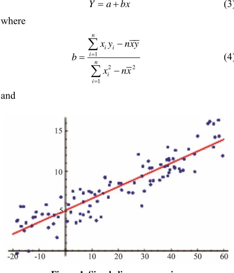
1) Regression analysis is used to find equations that fit data. Once we have the equation, we can use the statisti- cal model to make predictions. One type of regression analysis is linear analysis. When a correlation coefficient shows that data is likely to be able to predict future out- comes and a scatter graph of the data appears to form a straight line, statisticians may use linear regression to find a predictive function. The equation for a line is
ymx b (1) We can take data to calculate linear regression, and we can find the regression equation as
y a bx (2) is a way to describe a relationship between two variables through an equation of a straight line, called line of best fit, that most closely models this relationship (Figure 1).
Linear Regression Formula can be used to derive the equation for the line of best fit:
Y a bx (3) where
1
2 2
1 n
i i i
n i i
x y nxy
b
x nx
(4) [image:8.595.56.286.461.730.2]and
Figure 1. Simple linear regression.
a y bx (5) 2) The Simple Multiple Correlation Coefficient (R)is
a measure of the strength of the association between the independent (explanatory) variables and the one depen- dent (prediction) variable. Interpretation of R can be very
well explained with the strength of the association: The strength of the association is measured by the sample Multiple Correlation Coefficient, R. R can be any value
from 0 to +1. If it is closer R and is equal to one, the lin-
ear association will be stronger. If Rto zero, then there is
no linear association between the dependent variable and the independent variables. Unlike the simple correlation coefficient, r, which tells both the strength and direction
of the association, Rtells only the strength of the associa-tion. Ris never a negative value.
This can be seen from the formula below, since the square root of this value indicates the positive root.
1 2 1 2 1 2
1 2
2 2
2
2
1
yx yx yx yx x x
x x
r r r r r
R
r
3. Results
Experimental findings along with work carried out by re- searchers on different modes of analysis and diseases are tabulated in Tables 1-3. Statistical analysis like regres-
sion analysis and multiple correlations were also tabulat- ed in Tables 4-7.
We have shown our dataon the basis of experimental procedure of Radial immuno diffusion technique of Man- cini, G. et al. [56]. Data reveal that the levels of C3 do
not have any deflection from the normal healthy controls. The levels of C4 were lower about 20% and levels of IgA
and IgM were found to be lower with the normal data.
The higher values of IgG show that the patients may get
some infection, which may be a causative factor in this disease. Recently nervous system disorders have been shown to be associated with autoantibodies. It is well re- cognized that the epileptic children producing one auto- antibody have an increased likelihood of having other au- toantibodies. It is possible that the epilepsy represent the first manifestation of the syndrome itself. The antibodies themselves may be implicated directly triggering the epi- lepsy.
4. Discussion
Table 1. Values of C3, C4, IgA, IgG and IgM measured in epileptic patients and normal healthy controls are presented.
S.No. Types of Samples C3 gm/l C4 gm/l IgG gm/l IgM gm/l IgA gm/l
1 E 1.31 0.43 22.80 1.59 4.17 2 E 1.92 0.44 15.00 0.57 1.06 3 E 1.48 0.17 15.20 0.83 1.70 4 E 1.83 0.28 17.90 1.23 0.56 5 E 1.07 0.09 27.00 1.57 3.55 6 E 1.19 0.13 21.80 1.38 3.84 7 E 1.65 0.38 13.30 1.82 1.16 8 E 1.80 0.20 23.30 1.49 0.92 9 E 1.95 0.40 15.10 1.82 1.47
10 E 1.46 0.46 14.60 1.72 3.53
11 E 1.76 0.44 13.40 1.43 1.76
12 E 1.37 0.15 28.40 1.43 1.38
13 E 1.58 0.16 15.30 1.36 2.63
14 E 1.42 0.13 15.40 1.32 1.09
15 E 1.31 0.18 17.20 1.23 1.38
16 E 1.48 0.26 15.30 1.39 2.74
17 N 1.72 0.45 17.70 1.17 1.96
18 N 1.58 0.34 16.10 1.33 2.51
19 N 1.31 0.23 14.60 0.69 2.88
20 N 1.64 0.33 17.50 5.47 3.72
21 N 1.75 0.16 22.00 3.42 3.29
22 N 1.63 0.21 17.30 4.12 2.56
23 N 1.38 0.39 16.70 2.42 1.88
24 N 1.73 0.26 12.00 0.80 3.11
[image:9.595.55.542.101.550.2]25 N 1.53 0.17 21.30 2.43 3.62
Table. 2 Experimental findings along with earlier work carried out by researchers.
S.No. Immuno-logical Parameter Types of Samples Mean ± S.D Unit Disease/Control Reference
1 C3 Serum 133.8 29.7 mg dl Control Al-Hakeim. H.K.
et al. [40]
2 C3 Serum 171.3 81.2 mg dl Depressed Do
3 C4 Serum 26.8 7.9 mg dl Control Do
4 C4 Serum 5.6 21.7 mg dl Depressed Do
5 IgA Serum 218.9 127.6 mg dl Control Do
6 IgA Serum 253.3 188.7 mg dl Depressed Do
7 IgG Serum 1128.4 413.7 mg dl Control Do
Continued
9 IgM Serum 176.4 92.3 mg dl Control Do
10 IgM Serum 158.5 83.4 mg dl Depressed Do
11 C3 Serum 0.80 0.10 mg dl Control Olsson, R. et al. [57]
12 C3 Serum 0.85 0.16 g l Epilepsy Do
13 C4 Serum 86 26 g l Control Do
14 C4 Serum 85 33 g l Epilepsy Do
15 IgG Serum 11 2.5 g l Control Do
16 IgG Serum 9.5 1.1 g l Epilepsy Do
17 IgA Serum 1.7 0.6 g l Control Do
18 IgA Serum 1.5 1.1 g l Epilepsy Do
19 IgM Serum 1.5 0.4 g l Control Do
20 IgM Serum 1.6 0.8 g l Epilepsy Do
21 IgG CSF 0.019 0.005 g l Control Milica, T.C. et al. [17]
22 IgG Serum 9.8 2.7 g l Control Do
23 C3 CSF 0.0020 0.004 g l Control Do
24 C3 Serum 1.13 0.21 g l Control Do
25 C4 CSF 0.0007 0.0002 g l Control Do
26 C4 Serum 0.25 0.08 g l Control Do
27 IgG CSF 0.386 0.658 g l Hemorrhages into CNS Accuta Do
28 IgG Serum 14.4 1.8 g l Do Do
29 C3 CSF 0.015 0.016 g l Do Do
30 C3 Serum 1.15 0.15 g l Do Do
31 C4 CSF 0.0049 0.0044 g l Do Do
32 C4 Serum 0.29 0.06 g l Do Do
33 IgG CSF 0.060 0.027 g l Ischemic Cerebrovascular Accident Do
34 IgG Serum 13.0 2.3 g l Do Do
35 C3 CSF 0.0045 0.0016 g l Do Do
36 C3 Serum 1.17 0.18 g l Do Do
37 C4 CSF 0.0021 0.009 g l Do Do
38 C4 Serum 0.30 0.08 g l Do Do
39 C3 CSF 0.0014 0.0003 g l Meningism Do
40 C3 Serum 1.14 0.14 g l Do Do
41 C4 CSF 0.004 0.003 g l Do Do
Continued
43 IgG CSF 0.012 0.005 g l Do Do
44 IgG Serum 12.8 1.9 g l Do Do
45 C3 CSF 0.0039 0.0015 g l
Meningitis Serosa + Aspetic
Meningitis Do
46 C3 Serum 1.31 0.20 g l Do Do
47 C4 CSF 0.0012 0.0005 g l Do Do
48 C4 Serum 0.32 0.0008 g l Do Do
49 IgG CSF 0.034 0.018 g l Do Do
50 IgG Serum 13.2 3.4 g l Do Do
51 C3 CSF 0.0048 0.0020 g l Ig Synthesis Proven Do
52 C3 Serum 1.35 0.32 g l Do Do
53 C4 CSF 0.0020 0.0008 g l Do Do
54 C4 Serum 0.31 0.09 g l Do Do
55 IgG CSF 0.048 0.031 g l Do Do
56 IgG Serum 12.2 2.9 g l Do Do
57 C3 CSF 0.0054 0.0030 g l Encephalitis + Meningo Encephalitis Do
58 C3 Serum 1.07 0.30 g l Do Do
59 C4 CSF 0.0019 0.0010 g l Do Do
60 C4 Serum 0.22 0.07 g l Do Do
61 IgG CSF 0.049 0.025 g l Do Do
62 IgG Serum 11.2 3.2 g l Do Do
63 C3 CSF 0.0120 0.0064 g l Guillain-Barre Syndrone Acute Do
64 C3 Serum 1.25 0.27 g l Do Do
65 C4 CSF 0.0022 0.0013 g l Do Do
66 C4 Serum 0.26 0.07 g l Do Do
67 IgG CSF 0.141 0.082 g l Do Do
68 IgG Serum 11.0 2.8 g l Do Do
69 C3 CSF 0.0080 0.0046 g l Guillain-Barre Syndrome Chronic Course Do
70 C3 Serum 1.10 0.20 g l Do Do
71 C4 CSF 0.0022 0.0013 g l Do Do
72 C4 Serum 0.26 0.07 g l Do Do
73 IgG CSF 0.100 0.64 g l Do Do
74 IgG Serum 12.8 3.0 g l Do Do
Continued
76 C3 Serum 1.00 0.23 g l Do Do
77 C4 CSF 6.0012 0.0005 g l Do Do
78 C4 Serum 0.23 0.07 g l Do Do
79 IgG CSF 0.070 0.030 g l Do Do
80 IgG Serum 10.6 1.5 g l Do Do
81 C3 CSF 0.0033 0.0009 g l Complement Activation Do
82 C3 Serum 0.93 0.20 g l Do Do
83 C4 CSF 0.0017 0.0004 g l Do Do
84 C4 Serum 0.21 0.05 g l Do Do
85 IgG CSF 0.064 0.033 g l Do Do
86 IgG Serum 11.1 2.0 g l Do Do
87 C3 CSF 0.0032 0.0014 g l Suspected MS with Progressive Course
88 C3 Serum 0.89 0.21 g l Do Do
89 C4 CSF 0.0013 0.0005 g l Do Do
90 C4 Serum 0.18 0.07 g l Do Do
91 IgG CSF 0.032 0.015 g l Do Do
92 IgG Serum 11.7 2.3 g l Do Do
93 C3 CSF 0.0039 0.0027 g l Compressive Accuta Radiculopathies Do
94 C3 Serum 1.08 0.20 g l Do Do
95 C4 CSF 0.0016 0.0005 g l Do Do
96 C4 Serum 0.23 0.05 g l Do Do
97 IgG CSF 0.049 0.031 g l Do Do
98 IgG Serum 12.0 2.2 g l Do Do
99 C3 CSF 0.0045 0.0018 g l Complecated Sequele Do
100 C3 Serum 1.06 0.25 g l Do Do
101 C4 CSF 0.0020 0.0006 g l Do Do
102 C4 Serum 0.22 0.06 g l Do Do
103 IgG CSF 0.044 0.016 g l Do Do
104 IgG Serum 11.6 2.0 g l Do Do
105 C3 CSF 0.0041 0.0015 g l Spinal Cord Neoplastic Processes Do
106 C3 Serum 1.05 0.18 g l Do Do
107 C4 CSF 0.0015 0.0005 g l Do Do
108 C4 Serum 0.19 0.05 g l Do Do
Continued
110 IgG Serum 12.6 3.0 g l Do Do
111 C3 CSF 0.0033 0.0015 g l Motor Neuron Disease Do
112 C3 Serum 1.26 0.20 g l Do Do
113 C4 CSF 0.0015 0.0005 g l Do Do
114 C4 Serum 0.26 0.10 g l Do Do
115 IgG CSF 0.030 0.017 g l Do Do
116 IgG Serum 11.4 2.6 g l Do Do
117 C3 CSF 0.0780 0.1155 g l CNS Tumors Do
118 C3 Serum 1.08 0.22 g l Do Do
119 C4 CSF 0.0220 0.0286 g l Do Do
120 C4 Serum 0.25 0.07 g l Do Do
121 IgG CSF 0.970 1.110 g l Do Do
122 IgG Serum 11.5 4.3 g l Do Do
123 IgG CSF 7.4 0 g 100 Epilepsy Riddoch, D. et al.,[42]
124 IgG CSF 9.0 0 ml 100 Motor Neurone Disease Do
125 IgA Serum 2.26 1.15 IU ml Control Gholamali, Y.P. et al. [20]
126 IgA Serum 2.23 1.05 IU ml Tonic-Clonic Epilepsy Do
127 IgG Serum 12.87 6.3 IU ml Control Do
128 IgG Serum 12.77 6.4 IU ml Tonic-Clonic Epilepsy Do
129 IgM Serum 2.13 1.72 IU ml Control Do
130 IgM Serum 2.23 1.82 IU ml Tonic-Clonic Epilepsy Do
131 IgA Serum 248 2 mg 100ml C Slavin, et al., [28]
132 IgA Serum 196 2 mg 100ml E Do
133 IgG Serum 950 2 mg 100ml C Do
134 IgG Serum 1206 2 mg 100ml E Do
135 IgM Serum 94 2 mg 100ml C Do
136 IgM Serum 142 2 mg 100ml E Do
137 IgM Serum 153 11.5 mg 100ml C Do
138 IgM Serum 64.1 5.4 mg 100ml E. (Grade I) Moustafa, S. et al.[58]
139 IgM Serum 55.1 5.9 mg 100ml E. (Grade II) Do
140 IgM Serum 35.1 5.8 mg 100ml E. (Grade III) Do
141 IgG Serum 945 107 mg 100ml C Do
142 IgG Serum 795.6 90 mg 100ml E. (Grade I) Do
Continued
144 IgG Serum 403.4 10.5 mg 100ml E. (Grade III) Do
145 IgA Serum 154 69 mg dl C Kumar, S. [59] Thesis
146 IgA Serum 320 21 mg dl GME Do
147 IgG Serum 1169 351 mg dl C Do
148 IgG Serum 2774 161 mg dl GME Do
149 IgM Serum 188 62 mg dl C Do
150 IgM Serum 280 24 mg dl GME Do
151 C3 Serum 93 7 mg dl GME Do
152 C3 Serum 108 24 mg dl C Do
153 C4 Serum 30 4 mg dl C Do
154 C4 Serum 37 4 mg dl GME Do
155 IgA Serum 161 10 mg dl E Anderson, P. & Moseklide, L. [34]
156 IgG Serum 1217 359 mg dl E Do
157 IgM Serum 157 72 mg dl E Do
158 IgA Serum 142 66 mg dl Migraine Moore et al., [60]
159 IgG Serum 1344 448 mg dl Do Do
160 IgM Serum 141 80 mg dl Do Do
161 IgA Serum 143 66 mg dl Headache free Do
162 IgG Serum 1394 530 mg dl Do Do
163 IgM Serum 144 75 mg dl Do Do
164 IgA Serum 160 63 mg dl Prodromal Migraine Do
165 IgG Serum 1456 400 mg dl Do Do
166 IgM Serum 133 80 mg dl Do Do
167 IgA Serum 133 70 mg dl Non-Prodromal (Migrance) Do
168 IgG Serum 1250 433 mg dl Do Do
169 IgM Serum 144 74 mg dl Do Do
170 IgA Serum 144 66 mg dl Prodroml (Headachetree) Do
171 IgG Serum 1518 530 mg dl Do Do
172 IgM Serum 150 64 mg dl Do Do
173 IgA Serum 142 69 mg dl Non-Prodromal
(Head Ache Free) Do
174 IgG Serum 1258 473 mg dl Do Do
175 IgM Serum 131 69 mg dl Do Do
176 IgA Serum 212 18 mg dl Migraneous System Lord and Duckworth [61]
Continued
178 IgM Serum 161 11 mg dl Do Do
179 C3 Serum 106 9 mg dl Late Hecdacae Do
180 C4 Serum 101 11 mg dl Do Do
181 C3 Serum 81 5 mg dl Earlyheadache Do
182 C4 Serum 71 5 mg dl Do Do
183 C3 Serum 20 9 mg dl Migraine Do
184 C4 Serum 16 6 mg dl Do Do
185 C3 Do 142 38 mg dl Prodromal Headache Free Moore, et al., [60]
186 C4 Do 28 10 mg dl Do Do
187 C3 Do 167 10 mg dl Non-Prodromal (Migraine) Do
188 C4 Do 27 7 mg dl Do Do
189 C3 Do 176 64 mg dl (Headache Free) Non Prodromal Do
190 C4 Do 27 8 mg dl Do Do
191 C3 Do 172 65 mg dl Prodromal (Migraine) Do
192 C4 Do 24 6 mg dl Do Do
193 C3 Do 165 56 mg dl Controls Do
194 C4 Do 28 8 mg dl C Do
195 IgG Serum 18.18 4.71 mg l E Present work
196 IgG Serum 17.24 2.89 mg l C Present work
197 IgM Serum 1.38 0.31 mg l E Present work
198 IgM Serum 2.42 1.54 mg l C Present work
199 IgA Serum 2.05 1.13 mg l E Present work
200 IgA Serum 2.83 0.62 mg l C Present work
201 C3 Serum 1.53 0.25 mg l E Present work
202 C3 Serum 1.58 0.14 mg l C Present work
203 C4 Serum 0.26 0.13 mg l E Present work
204 C4 Serum 0.28 0.09 mg l C Present work
most infectious agents, the same barrier may stop viral clearance. Immune responses in the CNS during infec- tion are recruited from the systemic circulation in a rela- tively selective and specific fashion. Cells and antibodies found in the CNS during infections differ from these that follow non specific rupture in the BBB such as it occurs after a traumatic injury of any type injury may be one of the causes of epileptic attack. In traumatic lesion the tran- sudate of serum contains antibodies and cells of all types enter, but with a predominance of monocytes, which dif-
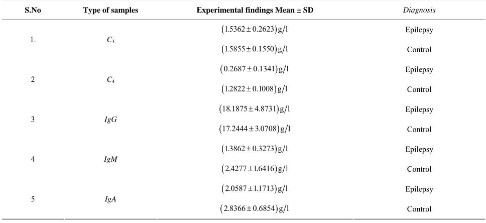
Table 3. Mean levels and standard deviation of C3, C4, IgG, IgM, IgA in epileptic patient and normal healthy control.
S.No Type of samples Experimental findings Mean ± SD Diagnosis
1.5362 0.2623 g l Epilepsy
1. C3
1.5855 0.1550 g l Control
0.2687 0.1341 g l Epilepsy
2 C4
1.2822 0.1008 g l Control
18.1875 4.8731 g l Epilepsy
3 IgG
17.2444 3.0708 g l Control
1.3862 0.3273 g l Epilepsy
4 IgM
2.4277 1.6416 g l Control
2.0587 1.1713 g l Epilepsy
5 IgA
[image:16.595.55.539.103.325.2]2.8366 0.6854 g l Control
Table 4. Regression and correlation coefficient studies on C3, C4, IgG, IgM and IgA in normal blood samples.
S.No Correlation Co. Regression Co. Regression Equation Correlation Coff. Partial Multiple Co.
C3 C C3 4–0.0256
3 4 3 4 0.0394 –0.0167 C C C C b b
C3 0.0394C41.5966 -
C4 C C4 3–0.0256 C4 0.0167C30.3087 -
IgG IgG IgM 0.4814 0.9006
0.2574 GM MG b b 0.9006 15.0577 0.2574 2.0109 IgG IgM IgM IgG
rGM A 0.4201 RG MA 0.2386
IgM IgM IgA 0.4140 1.0214
0.1678 MA AM b b 0.1678 2.4291 1.0214 0.4698 IgA IgM IgM IgA
rAG M 0.0943 RA GM 0.1788
IgA IgG IgA 0.2746 1.2673
0.0595 GA AG b b 1.2673 13.6493 0.0595 1.8102 IgG IgA IgA IgG
[image:16.595.56.539.560.732.2] rMA G 0.3344 RM AG 0.3177
Table 5. Regression and correlation coefficient studies on C3, C4, IgG, IgM and IgA in epileptic blood samples.
S.No Correlation Co. Regression Co. Regression Equation Coefficient of
Partial co-relation Multiple Co.
C3 C C3 40.5566 bC C341.0887 C3 1.0887 C41.2436 -
C4 C C4 30.5566 bC C430.2845 C4 0.2845 C30.1684 -
IgG IgG IgM 0.1404 2.0907
0.0094 G M M G b b 2.0907 15.2891 0.0094 1.2146 IgG IgM IgM IgG
rGM A 0.0609 RG MA 0.0778
IgM IgM IgA 0.3103 0.0867
1. 1104 M A A M b b 0.0867 1.2077 1.1104 0.5193 IgM IgA IgA IgM
rAG M 0.2436 RA GM 0.1499
IgA IgG IgA 0.2728 1.1351
0.0655 G A A G b b 1.1351 15.8504 0.0655 0.8658 IgG IgA IgA IgG
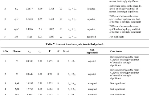
Table 6. Student t test analysis, two tailed paired.
S.No Element ttest ttheo P df Result Null hypothesis Conclusion
1 C3 0.5572 0.69 0.583 23 texpttheo rejected
Difference between the mean C3
levels of epilepsy and that of normal is strongly significant
2 C4 0.2617 0.69 0.796 23 texpttheo rejected
Difference between the mean C4
levels of epilepsy and that of normal is strongly significant
3 IgG 0.5224 0.69 0.606 23 texpttheo rejected
Difference between the mean
IgG levels of epilepsy and that of normal is strongly significant
4 IgM 2.4906 2.5 0.02 23 texpttheo rejected
Difference between the mean
IgM levels of epilepsy and that
[image:17.595.55.545.141.452.2]of normal is strongly significant 5 IgA 1.823 1.71 0.081 23 texpttheo accepted Not significant
Table 7. Student t test analysis, two tailed paired.
S.No Element texp ttheo P df Result
Null
hypothesis Conclusion
1 C3 0.0588 0.71 0.955 8 texpttheo rejected
Difference between the mean
C3 levels of epilepsy and that
of normal is strongly significant
2 C4 0.0649 0.71 0.95 8 texpttheo rejected
Difference between the mean
C4 levels of epilepsy and that
of normal is strongly significant
3 IgG 1.0262 0.71 0.335 8 texpttheo accepted Not significant
4 IgM 1.9763 1.86 0.084 8 texpttheo accepted Not significant
5 IgA 1.355 0.71 0.212 8 texpttheo accepted Not significant
searchers have proposed the hypothesis of immunologi-cal mechanism for the involvement of pathogenesis in epileptic attacks. Many of the patient of epilepsy have immune deficient state.
It has been seen that some of the epileptic patients de-velop auto immune disorders on antiepileptic drugs me- dication. Many of epileptic patients exhibit different autoantibodies without any clinically manifest autoim- mune disorder. The immunological aspects of epilepsy are not confined to the depressive effect of some of the antiepileptic drugs (AEDs) upon the immune system. They also comprise factors relevant to the pathogenesis of some form of epilepsy as well as variety of clinical manifestations met in some patients with epilepsy. The immunologic reactions can be involved in the pathoge- nesis of some of the epileptic patients is not unexpected. Local immune reaction can give rise to focal cerebral le- sions. Focal lesions may develop epileptic attack. It has been established this anti neuronal antibodies lead to epi- leptic attacks. Immune complexes are trapped in small vessels giving rise to attack of epilepsy. Anti phosphol- ipids antibodies lead to small vascular lesion.
If the antiepileptic effect is due to a direct action upon the brain, the immunoglobulins have to cross the BBB. Many research studies indicate that the BBB has been broken down locally during generalised cerebral seizures. It has been established that the increased expression of proinflammatory molecule has been demonstrated in the brain of epileptic patients after surgery. Inflammatory reactions occur in epilepsy of different types and do not invoke an inflammatory pathophysiology such as tempo- ral lobe epilepsy. Brain inflammation may be a common factor contributing of predisposing to the occurrence of seizures and cell death in different type of epilepsy. We would like to add here that a reversible induction of a selective IgA deficiency might occur in some patients
receiving antiepileptic drugs such as Phenytoin. Humoral immunity may alter in patients after the first attack of seizure.
established that content of the food have some trace ele- ments. The trace elements play a role in human immunity. If the level of these elements goes beyond the limit of normalcy even death may occur. On the other hand if the levels are lower side of the normal range something un- natural can happen. A relation between immunoglobulin, complement and trace element be consider in the future preview of the study.
Granata, T. et al. [62] have given their views on the
pathogenic role of immunity in epilepsy. They have ob- served the efficacy of immune-modulating treatment. On the basis of clinical and experimental findings they also reported that innate and adaptive immunity may be in- volved in epilepsy. Epilepsy may be present as a symp- tom of different neurological perturbations. Aetiological explanation can not be identified directly. Some evidenc- es show that autoimmune mechanisms might behave a role in epilepsy. The evidence for immunological mecha- nisms in epilepsy can be examined as, childhood epilepsy syndrome, epilepsy associated with other immunologicly mediatd disease and unselected groups of patients with epilepsy. Autoimmunity has also suspected to involve in the pathology of certain types of epilepsies. Antibodis can be epileptogenic. We are able to say that the level of
IgG in epileptic patients are measured as
18.1875 4.8737 mg l
and
17.2444 3.0708 mg l
in normals respectively. The IgG levels are higher in the
present finding of epileptic patients, while IgA, IgM, C3 & C4 levels are lower than normal values. On the basis of
statistical analysis in multipole correlation a trend has been found in epileptic cases i.e.
A GM M AG G MA
R R R
A partial correlation coefficients analysis shows a trend, that is
MA G AG M GM A
r r r
In the present work an attempt is made to relate the circulatory level of IgG, IgA and IgM and complement C3 & C4 among the subjects undergoing an epileptic at-
tack and comparing them with that of normal individuals.
5. Conclusions
It has been seen that the conventional antiepileptic treat- ment have many limitations in the treatment of epileptic cases. Although there has been great advance in the management of the epileptic patients by the application of newly invented developed antiepileptic drugs and sur- gical techniques. Some of the cases still remain in an interactable position. Immunoglobulin, steroids and ke- togeinc diet may be tried and better results may be seen in the treating cases. Immunoglobulin treatment shows benefits in some autoimmune related epilepsy. This treatment has its own limitations in long term efficiency.
Steroids show significant improvement in many epileptic syndromes. Ketogenic diet has become one of the most reliable treatment for epileptic children. Ketoegenic diet is difficult to maintain because of low palatability. It shows a very high antiepileptic efficacy. These are some of the suggestive directions to neutralize the effect of the epileptic seizure and try to control. Humoral immunity is altered in children early after the first attack of epilepsy. This may be a consequence of an exogenous event, such as occurrence of infection and related to an interaction of the CNS. The alterations in the immunity of the epileptic patients may be done by the adjusting the level of these in the blood of epileptic patients after the supplementa- tion of proper diet, which is rich in all necessary trace elements. The immunotoxic potential of anticonvulsant drugs appear to be very low. The immunological moni- toring is required in all the patients. This monitoring is also required in those patients who are at stage of im- mune defect.
Our results show that the levels of IgA, IgM, C3 and C4
are lower than controls and the levels of IgG are higher
in all the epileptic cases. Statistical analysis shows a trend i.e.
A GM M AG G MA
R R R
The contributions of all these immunoglobulins are very strong i.e. M AG has a value 0.0996. If we have a higher value of IgG the other values of IgM and IgA will
be adjusted according to the application of multiple cor- relation coefficient analysis. We would like to add here that it is quite possible to have a genetic predisposition to develop IgA deficiency is completely unrelated to the
genetic factors involved the pathogenesis of epilepsy.
IgA deficiency may appear in many types if epilepsy and
irrespective of a family background. Some of the studies showed that the deficiency of IgA occurs in patients with
generalized cerebral seizure and some time in partial epilepsies.
R
Aarli, J. A. [39] had reported some of the findings on allergy in epilepsy. A small percentage of patients treated with antiepileptic drugs develop transient exanthema. Exfoliative dermatitis or erythema multiforme exsudati-vum may develop in some cases. Various immunological parameters like IgA. IgM, IgA, C3 and C4 complement
were determined in almost one hundred cases and heal- thy controls were taken about twenty. Values of these parameters were very low in compression with healthy controls except IgG. The values of IgG levels are higher
than controls.