ANTICANCEROUS ACTIVITY OF ANTHOCYANIN ANALYZED USING DIFFERENT CELL LINES
FROM THE ONION PEEL (
M. Geetha, M.
P.G. Department of Biotechnology, Dr. Mahalingam Centre for Research and Development, N.G.M. College,
ARTICLE INFO ABSTRACT
Anthocyanins and flavonoids are polyphenolic compounds and capable of inhibiting the growth of human cancer cells. It is mainly responsible for cyanic colors ranging from salmon
and violet to dark blue of most flowers, fruits, leaves and stems. Quercetin, a novel flavonoid, was present in the onion peel (
anthocyanin on human epithelial cells 2,5-diphenyltetrazolium bromide (MTT) assay.
culture plates in different concentrations of red onion and big onion peel extracts of
determine their anticancer effects using the MTT assay. Anthocyanin extracted from big onion peel showed approximately 92% inhibition on HEp
anthocyanin extracted from red onion peel, big onion peel showe
anthocyanin extracted from red onion peel and big onion peel was tested against MCF
Red onion peel showed higher activity around 78% inhibitions than big onion peel. So we can assume that, the anthocyanin compounds
cells. So the anthocyanins extracted from easily available onion peel would be a valuable source for antiproliferative activity in food industry.
INTRODUCTION
Cancer is the second leading cause of death in the United States and in many other nations in the world. The prognosis for a patient with metastatic carcinoma of the lung, colon, breast, or prostate remains a concern and accounts for more than half of all cancer deaths. (Aziz et al.
Chemoprevention or chemotherapy via nontoxic agents could be one approach for decreasing the incidence of these cancers. Many naturally occurring agents have shown chemopreventive and chemotherapeutic (anticancer) potential in a variety of bioassay systems and animal models. (Middleton
An effective and acceptable chemopreventive or anticancer agent should have certain properties: (i) notoxic effects in normal and healthy cells, (ii) high efficacy against multiple cancers, (iii) capability of oral consumption, (iv) known mechanism of action, (v) low cost, and (vi) acceptance by the human population (Skibola et al., 2000, Formica
Flavonoids and anthocyanins have been known as plant pigments for over a century and belong to a vast group of phenolic compound that are widely distributed in all foods of plant origin. In the normal North American diet, flavonoid glycosides are unavoidably consumed daily, with an estimated total consumption of 1 g/d (Formica et al., 1995
be much higher if dietary supplements are also consumed. As an example, dietary supplements of quercetin have been
*Corresponding author: [email protected]
ISSN: 0975-833X
International Journal of Current Research Vol.
Article History:
Received 12th
January, 2012 Received in revised form
19th February, 2012
Accepted 08th
March, 2012
Published online 30th April, 2012
Key words:
Allium cepa;
Red, Big varieties; Anthocyanin;
HEp-2 and MCF-7 cancer cell line; 3-(4,5-dimethylthiazol-2-yl)-2,5-Diphenyltetrazolium bromide; Cytotoxicity.
RESEARCH ARTICLE
ANTICANCEROUS ACTIVITY OF ANTHOCYANIN ANALYZED USING DIFFERENT CELL LINES
FROM THE ONION PEEL (
ALLIUM CEPA
) EXTRACTION
Geetha, M. Saravanakumar and P. Suganyadevi*
P.G. Department of Biotechnology, Dr. Mahalingam Centre for Research and Development, N.G.M. College, Pollachi, Tamil Nadu, India.
ABSTRACT
Anthocyanins and flavonoids are polyphenolic compounds and capable of inhibiting the growth of human cancer cells. It is mainly responsible for cyanic colors ranging from salmon
and violet to dark blue of most flowers, fruits, leaves and stems. Quercetin, a novel flavonoid, was present in the onion peel (Allium cepa). In the present study, we explored the cytotoxic effects of anthocyanin on human epithelial cells and the Breast cancer cells by 3
diphenyltetrazolium bromide (MTT) assay. The HEp-2 and MCF-7 cells were seeded in 96 culture plates in different concentrations of red onion and big onion peel extracts of
determine their anticancer effects using the MTT assay. Anthocyanin extracted from big onion peel showed approximately 92% inhibition on HEp-2cells at 1000 µg/ml (table 1). When compared with anthocyanin extracted from red onion peel, big onion peel showed a highest inhibition. The anthocyanin extracted from red onion peel and big onion peel was tested against MCF
Red onion peel showed higher activity around 78% inhibitions than big onion peel. So we can assume that, the anthocyanin compounds present in red onion peel are inhibiting the proliferation of cells. So the anthocyanins extracted from easily available onion peel would be a valuable source for antiproliferative activity in food industry.
Copy Right, IJCR, 2012, Academic Journals
Cancer is the second leading cause of death in the United States and in many other nations in the world. The prognosis for a patient with metastatic carcinoma of the lung, colon, breast, or prostate remains a concern and accounts for more
et al., 2003)
Chemoprevention or chemotherapy via nontoxic agents could be one approach for decreasing the incidence of these cancers. Many naturally occurring agents have shown chemopreventive and chemotherapeutic (anticancer) potential in a variety of systems and animal models. (Middleton et al.,1994). An effective and acceptable chemopreventive or anticancer agent should have certain properties: (i) notoxic effects in normal and healthy cells, (ii) high efficacy against multiple ity of oral consumption, (iv) known mechanism of action, (v) low cost, and (vi) acceptance by the 2000, Formica et al., 1995). Flavonoids and anthocyanins have been known as plant a vast group of phenolic compound that are widely distributed in all foods of plant origin. In the normal North American diet, flavonoid glycosides are unavoidably consumed daily, with an estimated 1995), which could be much higher if dietary supplements are also consumed. As an example, dietary supplements of quercetin have been
suggested to contain doses which are up to 20 times higher than those which would be obtained in a typical vegetarian diet (Skibola et al., 2000). Recent work is beginning to highlight the potential health-beneficial properties of flavonoids, known to be powerful antioxidants. The clinical trials indicate that flavonoids have important effects on cancer chemoprevention and therapy (Pannala
Flavonoids may interfere in several of the steps that lead to the development of malignant tumors, including protecting DNA from oxidative damage, inhibiting carcinogen activation, and activating carcinogen detoxifying systems (Kerry
Galati et al., 2000). Anthocyanins are considered as potential replacements for synthetic colors because of their bright attractive hue and water solubility that allows their incorporation into aqueous food systems; they may also possess health benefits (Nayak et al., 2009). Anthocyanins are reported to have some therapeutic benefits including vasoprotective and anti-inflammatory properties (Kallithraka
et al., 1995), anti-cancer and chemoprotective properties as
well as antineoplastic properties (Ferguson P, Kurowsk 2004). There is a rising demand for natural sources of food colorants with nutraceutical benefits and alternative sources of natural anthocyanins are becoming increasingly important. Our objective was to determine the antiproliferative effects anthocyanin extracted from onion peel (red and big varieties
of Allium cepa) against the different cell lines.
ternational Journal of Current Research Vol. 4, Issue, 04, pp.008-012, April, 2012
INTERNATIONAL
OF CURRENT RESEARCH
ANTICANCEROUS ACTIVITY OF ANTHOCYANIN ANALYZED USING DIFFERENT CELL LINES
P.G. Department of Biotechnology, Dr. Mahalingam Centre for Research and Development, N.G.M. College,
Anthocyanins and flavonoids are polyphenolic compounds and capable of inhibiting the growth of human cancer cells. It is mainly responsible for cyanic colors ranging from salmon pink through red and violet to dark blue of most flowers, fruits, leaves and stems. Quercetin, a novel flavonoid, was ). In the present study, we explored the cytotoxic effects of and the Breast cancer cells by
3-(4,5-dimethylthiazol-2-yl)-7 cells were seeded in 96-well culture plates in different concentrations of red onion and big onion peel extracts of Allium cepa to determine their anticancer effects using the MTT assay. Anthocyanin extracted from big onion peel 2cells at 1000 µg/ml (table 1). When compared with d a highest inhibition. The anthocyanin extracted from red onion peel and big onion peel was tested against MCF-7 cell lines. Red onion peel showed higher activity around 78% inhibitions than big onion peel. So we can present in red onion peel are inhibiting the proliferation of cells. So the anthocyanins extracted from easily available onion peel would be a valuable source for
, Academic Journals. All rights reserved.
are up to 20 times higher than those which would be obtained in a typical vegetarian Recent work is beginning to beneficial properties of flavonoids, known to be powerful antioxidants. The human linical trials indicate that flavonoids have important effects on cancer chemoprevention and therapy (Pannala et al., 1998). Flavonoids may interfere in several of the steps that lead to the development of malignant tumors, including protecting DNA idative damage, inhibiting carcinogen activation, and activating carcinogen detoxifying systems (Kerry et al., 1999, ., 2000). Anthocyanins are considered as potential replacements for synthetic colors because of their bright water solubility that allows their incorporation into aqueous food systems; they may also ., 2009). Anthocyanins are reported to have some therapeutic benefits including inflammatory properties (Kallithraka cancer and chemoprotective properties as well as antineoplastic properties (Ferguson P, Kurowska et al., 2004). There is a rising demand for natural sources of food colorants with nutraceutical benefits and alternative sources of natural anthocyanins are becoming increasingly important. Our objective was to determine the antiproliferative effects of anthocyanin extracted from onion peel (red and big varieties
) against the different cell lines.
MATERIALS AND METHODS
Materials required in mem
Monolayer culture bottle of HEp-2 cell lines.
5ml, 10ml serological pipette
Minimal essential media (MEM) with 10%, 2% foetal calf serum
TPVG (Trypsin PBS versene glucose)
Discarding jar, inverted microscope, desiccators
Gloves, spirit, cotton, label pad, marker pen.
Materials required in cytotoxicity assay
1. Monolayer culture in log phase 2. Drug extracts (different concentrations) 3. MEM without FCS
4. 0.45μ filter
5. 5ml sterile storage vial
6. Tissue paper, spirit, cotton, marker pen and gloves 7. Micropipette and tips
Materials required in MTT assay
1. MTT (3-(4,5-dimethyl thiazol-2yl)-2,5-diphenyl tetrazolium bromide)stock solution 5mg/ml
2. DMSO-dimethyl sulfoxide
3. Micropipette and 200μl of sterile tips 4. Spectrophotometer with 1ml cuvette holder.
Minimal essential media preparation
Media is defined as a complex source of nutritional supplementation vital for the growth proliferation and maintenance of cells in vitro. The MEM dissolved in the pre sterilized Millipore distilled water and mixed well, closed and sterilized at 15lbs 121ºc for 15mins. Allow ingredients in the quantity, depending on the concentration of foetal calf serum (2% or 10%) mix well by shaking. Take care avoid spills pass CO2 using sterile pipette, Shake the bottle, check Ph and adjust to 7.2 to 7.4. The MEM bottles are kept for 2 days at 37ºc and checked for sterility, PH drop and floating particles they are then transferred to the refrigerator.
MEDIA PREPARATION: INGREDIENTS
10% GROWTH MEDIA
2%GROWT H MEDIA
MAINTANCE MEDIA WITHOUT FCS
MEM 857ml 937ml 957ml
Penicillin and
streptomycin
1ml 1ml 1ml
Phenol red 1ml 1ml 1ml
Amphotericin B 1ml 1ml 1ml
3% L-glutamine 10ml 10ml 10ml
Foetal calf serum 100ml 20ml nil
7.5%NaHCo3 30ml 30ml 30ml
Total volume 1000ml 1000ml 1000ml
PREPARATION OF INGREDIENT
1. Penicillin and streptomycin: (concentration 100IU of penicillin and 100 μg of streptomycin)
Dissolve both antibiotics in sterile Millipore distilled water, so as to give a final concentration 100 IU of penicillin and 100μg of streptomycin/ml. Mix well and distribute in 1ml aliquots. Store at -20º C Check sterility.
2. Fungi zone (amphotericin B): (conc.: 20μg/ml)
Dissolve in sterile Millipore distilled water so as to give a final concentration of 20μg/ml and distribute in 1ml aliquots in vials. Store at -20ºc. Check sterility.
3. L.glutamine: 3%
Weigh 3g of l-glutamine accurately and dissolve in 100ml sterile Millipore distilled water and mix well. Filter through Millipore membrane filter 0.22μ and distribute in 5ml aliquots in vials. Store at -20ºc. Check sterility.
4. 7.5% sodium-bi-carbonate
Weigh requisite quantity of sodium-bi-carbonate (to give 7.5% solution) accurately and dissolve in 100ml of sterile Millipore distilled water. Filter through what man filter paper No.1, distribute into bottles and at 121ºc, 15lbs, 15mins. Cool and store at +4ºc.
4. Foetal calf serum
Bring FCS at room temperature. Inactivated at 56º C in water bath for ½ hour and cool at room temperature. If floating particles are seen filter through Seitz filter. Distribute in 100ml, 50ml, and 20ml quantities in sterile bottles. Store at -20ºc.
Trypsin, PBS,versene, glucose solution: (TPVG)
2% trypsin: 100ml
Weigh 2g of trypsin accurately; dissolve in 100 ml sterile Millipore distilled water with magnetic stirrer for ½ hour. Filter through membrane filter. Store at -20ºC.
0.2%EDTA (versene)
Weigh 200mg of EDTA accurately. Dissolve in 100 ml of sterile Millipore distilled water. Autoclave at 121º C 15 lbs/15mins.
10%glucose -100ml
Weigh 1g of glucose accurately. Dissolve in 100 ml of sterile Millipore distilled water and filter through what man filter paper and autoclave at 15lbs/15mins.
TPVG-100ml
PBS - 840ml 2%trypsin -50ml 0.2%EDTA -100ml 10%glucose -5ml
Penicillin & streptomycin -5ml
Mix all ingredients and adjust the pH to 7.4 with 0.1 N HCl or 0.1 N NaOH. Distribute in 100 ml aliquots. Store at -20ºc.
METHOLDOLOGY
Maintenance of cell line
Preservation of cells in repository. Revival of cells from repository
Subculturing and maintenance of cell line
Bring the medium and TPVG to room temperature for thawing.
Observe the tissue culture bottles for growth, cell degeneration, pH and turbidity by seeing in inverted microscope.
If the cells become 80% confluent it goes for sub culturing process
Wipe the mouth of the bottle with cotton soaked in spirit to remove the adhering particles.
Discard the growth medium in a discarding jar keep distance between the jar and the flask.
Then add 4 – 5 ml of MEM without FCS and gently rinsed with tilting. The dead cells and excess FCS are washed out and then discard the medium.
TPVG was added over the cells. And incubate at 37º C for 5 minutes for disaggregation. The cells become individual and it’s present as suspension.
Add 5ml of 10% MEM with FCS by using serological pipette.
Gently give passaging by using serological pipette. If any clumbs is present then repeat the process.
After passaging split the cells into 1:2, 1:3 ratio for cytotoxicity studies for plating method
“Seeding of cells”
After homogenize take one ml of suspension and pour in to 24 well plates. In each well add 1ml of the suspension and kept in a desiccators in 5% CO2 atmosphere. After 2 days incubation observe the cells in inverted microscope. If the cells became 80% confluent
Cytotoxicity assay
In order to study the antitumor activity of a new drug, it is important to determine the cytotoxicity concentration of the drug. Cytotoxicity tests define the upper limit of the extract concentration, which is non-toxic to the cell line. The concentration nontoxic to the cells is chosen for antiviral assay. After the addition of the drug, cell death and cell viability was estimated. The result is confirmed by additional metabolic intervention experiment such as MTT assay
Stock drug concentration
0.5ml of drug is dissolved in 4.5 ml of DMSO giving a working concentration of 1mg/ml. the working concentration is prepared fresh and filtered through 0.45 μ filter before each assay.
1. To prepare 5 ml of extract and giving conc. (1mg/ml). 2. 500μl of MEM without FCS was taken in 9 eppendroff tubes. /each samples
3. Then 500μl of the working conc. was added to the first eppendroff tube and mixed well then 500μl of this volume was transferred from first to last tube by serial dilution to obtain the desired concentration of the drug.
4. As a result the volume remains constant but there is a change in concentration.
Sampling
1. 48hr monolayer culture of Hep2cells at a concentration of one lakh /ml /well (10 cells / ml / well) seeded in 24 well titer plates.
2. The plates were microscopically examined for confluent monolayer, turbidity and toxicity if the cells become confluent.
3. The growth medium (MEM) was removed using micropipette. Care was taken so that the tip of the pipette did not touch the cell sheet.
4. The monolayer of cells was washed twice with MEM without FCS to remove the dead cells and excess FCS. 5. To the washed cell sheet, add 1ml of medium (without FCS) containing defined concentration of the drug in respective wells.
6. Each dilution of the drug ranges from 1:1 to 1:64 and they were added to the respective wells of the 24 well titer plates. 7. To the cell control wells add 1ml MEM (w/o) FCS. 8. The plates were incubated at 37ºc in 5% CO2 environment and observed for cytotoxicity using inverted microscope.
MTT ASSAY
MTT assay is called as (3-(4, dimethyl thiazol-2yl)-2, 5-diphenyl tetrazolium bromide. MTT assay was first proposed by Mossman in 1982.
Procedure
After incubation, remove the medium from the wells carefully for MTT assay.
In each well wash with MEM (w/o) FCS for 2 – 3 times. And add 200μl of MTT conc of (5mg/ml).
And incubate for 6-7hrs in 5% CO2 incubator for Cytotoxicity.
After incubation add 1ml of DMSO in each well and mix by pipette and leave for 45sec
If any viable cells present formazan crystals after adding solublizing reagent (DMSO) it shows the purple color formation.
The suspension is transferred in to the cuvette of spectrophotometer and an O.D values is read at 595nm by taking DMSO as a blank.
Graph is plotted by taking concentration of the drug on X axis and relative cell viability on Y axis.
Cell viability (%) = Mean OD/Control OD x 100
RESULTS AND DISCUSSION
Several compounds particularly plant products and dilatory constituent found to exhibits chemo preventive activities both
in vitro and in vivo. Antiproliferative analysis of anthocyanin
extracted from onion peel was tested against HEp-2 and MCF-7 cell lines.
Antiproliferative analysis of anthocyanin against HEp-2cell lines
15.6-1000 µg/ml and the cell viability was counted. Control assay was carried out for sample containing only the approximately volume of blank solution and those showed no effect on cell growth. Anthocyanin extracted from big onion peel showed approximately 92% inhibition on HEp-2cells at 1000 µg/ml (table 1). Shilpa et al., 2000 reported a significant decrease in Melanoma B16 F 10 cell population by the aqueous extract of onion (Allium cepa). Anthocyanin extracted from red onion peel showed approximately 79% inhibition on HEp-2cells at 1000 µg/ml (table 2). When compared with anthocyanin extracted from red onion peel, big onion peel showed a highest inhibition.
Antiproliferative analysis of anthocyanin against MCF-7 cell lines
The anthocyanin extracted from red onion peel and big onion peel was tested against MCF-7 cell lines. The anthocyanin extracted from red onion peel showed higher activity around 78% inhibitions than big onion peel which showed 70% inhibition. From our ealier reports (Geetha et al., 2011) total anthocyanin content are higher in red onion peel (Table) extract compared to big onion peel. These results suggested that the anthocyanin compounds present in red onion peel are inhibiting the proliferation of cells. Chun et al., 2009), demonstrated the antiproliferative activity for Etlingera elatior
on human colorectal carcinoma cells and reported that the phenolics compounds may be responsible for its Antiproliferative activity. Cell growth reduction can be attributed to a decreased proliferation rate or an enhanced cell death by apoptosis or necrosis.
Apoptosis is a programmed cell death, which eliminates redundant or damaged cells. Cancer cells have deregulated proliferation and they are not able to undergo apoptosis naturally. To quantify and further support to the finding that anthocyanin extract from onion peel causes apoptosis on MCF-7 cells. In apoptotic cells the DNA fragments intact within the cell membrane or apoptopic body. Conversely, lysosomal enzymes in necrotic cells digest the cell membrane and cause the release of DNA fragments from the cells. In our results at 1000µg/ml concentration of anthocyanin extracted from big onion on MCF-7 cells showed apoptopic bodies when viewed under phase contrast microscope, where the cells become spherical or round shape when compared with normal MCF-7 cell lines, (Plate 1, 2).
Similarly the anthocyanin extracted from red onion peel showed 78% inhibition on MCF-7 cell line at toxicity (1000µg/ml) the viability is decreased and also the MCF-7 cells become spherical shape (plate) when compared with normal MCF-7 cell lines (untreated). These results suggested that the cell growth reduction may be attributed by apoptosis. Indap et al., (2006) examined the Antiproliferative effect of quercetin both in vitro and in vivo. They showed quercetin could exert Antiproliferative effect against MCF-7 cell line in a dose and time dependent manner was found to arrest the MCF-7 cell growth in G21M phase of all cycle.
CONCLUSION
Red and big onion peels were a very good source of anthocyanins compared to other anthocyanin sources easily
[image:4.612.316.547.103.397.2]available and cheap. From this study the results demonstrated that the amount of anthocyanin extracted from Allium cepa
Table 1: Different concentrations used in sample-A for MTT assay
Sample: A
Serial no Concentrati
on (μg/ml)
Dilutions Absorbance Cell
viability
1 1000 Neat 0.05 8.77
2 500 1:1 0.11 19.29
3 250 1:2 0.14 24.56
4 125 1:4 0.25 43.85
5 62.5 1:8 0.32 56.14
6 31.25 1:16 0.39 68.42
7 15.625 1:32 0.47 82.45
8 Cell control - 0.578.77 100
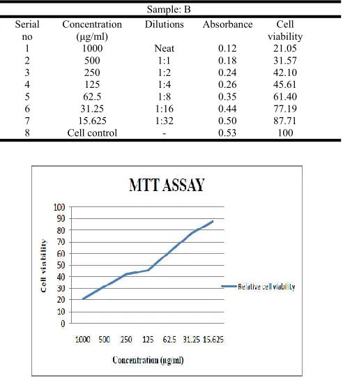
Table 2: Different concentrations used in sample-B for MTT assay
Sample: B Serial
no
Concentration (μg/ml)
Dilutions Absorbance Cell
viability
1 1000 Neat 0.12 21.05
2 500 1:1 0.18 31.57
3 250 1:2 0.24 42.10
4 125 1:4 0.26 45.61
5 62.5 1:8 0.35 61.40
6 31.25 1:16 0.44 77.19
7 15.625 1:32 0.50 87.71
8 Cell control - 0.53 100
[image:4.612.311.557.427.698.2]ACKNOWLEDGEMENT
We gratefully acknowledge, the Head of the department R.Kavitha Krishna, Nallamuthu Gounder Mahalingam College, and Pollachi, Tamil nadu, for permitting this research work, they also thank-ful to all staff members and friends of PG department of Biotechnology for their kind co-operation for complete this successful research work.
REFERENCES
1. Aziz, M. H.; Kumar, R.; Ahmad, N. Cancer chemoprevention by resveratrol: in vitro and in vivo studies and the underlying mechanisms. Int. J. Oncol. 23:17– 28; 2003.
2. Ferguson P, Kurowska E, Freeman DJ, Chambers AF, Koropatnick DJ.(2004).A flavonoid fraction from cranberry extract inhibits proliferation of human tumor cell lines. J Nutr.134,1529–35.
3. Formica, J. V.; Regelson, W. Review of the biology of quercetin and related bioflavonoids. Food Chem. Toxicol. 33:1061–1080; 1995.
4. Galati, G.; Teng, S.; Moridani, M. Y.; Chan, T. S.; O’Brien, P. J.Cancer chemoprevention and apoptosis mechanisms induced by dietary polyphenolics. Drug Metab. Drug Interact. 17:311 – 349; 2000.
5. M. Geetha, P. Ponmozhi, M. Saravanakumar, P. Suganyadevi, Extraction of anthocyanin and analyzing its antioxidant properties from differ-ent onion (Allium cepa) varieties. Int. J. Res. Pharm. Sci. Vol-2, Issue-1, 1-10, 2011.
6. Kallithraka, S., Garcia-Viguera, C., Bridle, P., & Bakker, J. (1995). Survey of solvents for the extraction of grape seed phenolics. Phytochemical Analysis, 6, 265–267. 7. Kerry, N.; Rice-Evans, C. Inhibition of
peroxynitrite-mediated oxidation of dopamine by flavonoid and phenolic antioxidants and their structural relationships. J.
Neurochem. 73:247 – 253; 1999.
8. Middleton, E.; Kandaswami, C. The impact of plant flavonoids on mammalian biology: implications for immunity, inflammation and cancer. In: Harborne J. B., ed., the flavonoids: advances in research since 1986. London: Chapman & Hall; 1994: 619–6522.
9. Nayak, C.A., Rastogi, N.K., and Raghavarao, K.S.M.S. (2009). Bioactive constituents present in Garcinica indica Choisy and its potential food applications. International
journal of food properties, In press.
10. Skibola, C. F.; Smith, M. T. Potential health impacts of excessive flavonoid intake. Free Radic. Biol. Med. 29:375 – 383; 2000.
11. Pannala, A. S.; Razaq, R.; Halliwell, B.; Singh, S.; Rice-Evans,C. Inhibition of peroxynitrite dependent tyrosine nitration by hydroxycinnamates: nitration or electron donation/Free Radic. Biol. Med. 24:594– 606; 1998.