In stark contrast to the human stomach, where the pH is less than 1 (e.g. Ruch and Fulton, 1960), the lumen of caterpillar and larval mosquito midgut can be as high as 12 in some species (Senior-White, 1926; Dadd, 1975; Dow, 1984). The digestive enzymes are adapted to this high pH (Eguchi et al., 1990), which is thought to dissociate tannin–protein complexes, leading to an increased ability to assimilate and utilize dietary protein (Berenbaum, 1980). Whatever its function, the mechanism by which the high pH is generated has defied explanation.
In caterpillar midgut, an electrogenic H+ V-ATPase hyperpolarizes the apical plasma membranes of midgut epithelial cells (for a review, see Wieczorek et al., 1999). This inside-negative potential is thought to energize lumen alkalization via an electrophoretic K+/2H+ antiporter (Wieczorek et al., 1991; Azuma et al., 1995; for reviews, see Lepier et al., 1994; Wieczorek et al., 1999). However, the antiporter hypothesis has been questioned on thermodynamic grounds (Moffett and Cummings, 1994; Klein et al., 1996; Kppers and Bunse, 1996; Clark et al., 1998).
The alkalization mechanism is even less understood in mosquito larval midgut. Like other freshwater organisms, mosquito larvae can live in a Na+-poor environment from which they are able to accumulate Na+ for nerve and muscle function. H+ V-ATPases energize ion uptake in transporting epithelia of many freshwater organisms (for a review, see Harvey et al., 1998). For example, Na+(and Cl−) uptake in frog skin appears to be energized by an apical H+V-ATPase acting in series with the basolateral Na+/K+-ATPase (for a review, see Ehrenfeld and Klein, 1997).
Filippova et al. (Filippova et al., 1998) showed that an H+ V-ATPase is highly expressed in the anterior midgut of Aedes aegypti larvae, and Zhuang et al. (Zhuang et al., 1999) showed that it is localized on the basal membranes in this midgut region. Is it possible that a V-ATPase located on the basal membrane in the anterior midgut of larval mosquitoes could energize the apical membrane and generate the extremely high pH found in the lumen (Senior-White, 1926; Dadd, 1975)? Proton transport from cells to hemolymph via a basally JEB3196
The alkaline environment, pH approximately 11, in the anterior midgut lumen of mosquito larvae is essential for normal nutrition and development. The mechanism of alkalization is, however, unknown. Although evidence from immunohistochemistry, electron microscopy and electrophysiology suggests that a V-ATPase is present in the basal membranes of the epithelial cells, its physiological role in the alkalization process has not been demonstrated. To investigate a possible role of the V-ATPase in lumen alkalization, pH gradients emanating from the hemolymph side of the midgut in semi-intact mosquito larvae were measured using non-invasive, self-referencing, ion-selective microelectrodes (SERIS). Large H+ concentration gradients, with highest concentrations
close to the basal membrane (outward [H+] gradients),
were found in the anterior midgut, whereas much smaller gradients, with concentrations lowest close to this membrane (inward [H+] gradients), were found in the
gastric caeca and posterior midgut. Similar region-specific pH gradients, with consistent anterior-to-posterior profiles, were observed in individuals of two Aedes species,
Aedes aegypti from semi-tropical Florida and Aedes canadensis from north-temperate Massachusetts. The
gradients remained in a steady state for up to 6 h, the maximum duration of the recordings.
Bafilomycin A1 (10−5, 10−7mol l−1) on the hemolymph
side greatly diminished the [H+] gradients in the anterior
midgut but had no effect on the gradients in the gastric caecum and posterior midgut. These physiological data are consistent with the previous findings noted above. Together, they support the hypothesis that a basal, electrogenic H+ V-ATPase energizes luminal alkalization
in the anterior midgut of larval mosquitoes.
Key words: midgut, H+V-ATPase, bafilomycin, liquid ion exchanger
microelectrode, mosquito, Aedes aegypti, Aedes canadensis. Summary
Introduction
IN SITU ANALYSIS OF pH GRADIENTS IN MOSQUITO LARVAE USING
NON-INVASIVE, SELF-REFERENCING, pH-SENSITIVE MICROELECTRODES
DMITRI Y. BOUDKO1,*, LEONID L. MOROZ1, PAUL J. LINSER1, JAMES R. TRIMARCHI2, PETER J. S. SMITH2 ANDWILLIAM R. HARVEY1
1The Whitney Laboratory, University of Florida, 9505 Ocean Shore Boulevard, St Augustine, FL 32086, USA and 2The BioCurrents Research Center, Woods Hole, MA 02543, USA
*e-mail: [email protected]
localized V-ATPase would tend to acidify the hemolymph. The extruded protons must be replaced if the cells are to remain in a pH steady state, and the lumen is a possible source of protons. Movement of protons from lumen to cells would tend to alkalize the lumen. But this movement would require that the apical membrane, as well as the V-ATPase-containing basal membrane, be hyperpolarized by the enzyme. Clark et al. (Clark et al., 2000) found that, indeed, the cytoplasmic sides of both basal and apical cell membranes are negative relative to the outside, especially in the presence of 5-hydroxytryptamine. A similar hyperpolarization of both basal and apical plasma membranes was found to be energized by a V-ATPase that was located in only one membrane sector in adult mosquitoes (Beyenbach et al., 2000).
Taken together, these published results provide strong evidence that a basal H+V-ATPase hyperpolarizes both basal and apical membranes in Aedes aegypti larval anterior midgut, but direct evidence linking these hyperpolarizations to lumen alkalization is lacking. To investigate the role of the V-ATPase in generating [H+] gradients that emanate from specific midgut regions, we used self-referencing ion-selective (SERIS) liquid ion exchanger (LIX) microelectrodes (Smith et al., 1999) to probe the hemolymph-facing surface of a novel, semi-intact preparation of mosquito larvae. Our results support the hypothesis that an H+ V-ATPase energizes the basal membrane, hyperpolarizing both basal and apical membranes and mediating anterior midgut alkalization.
Materials and methods Experimental insects
Aedes aegypti eggs were obtained from the United States Department of Agriculture laboratory at The University of Florida, Gainesville, FL. To obtain larvae, approximately 1000 eggs and 500µl of mosquito food (15 g of bovine liver powder, 10 g of brewer’s yeast in 500 ml of distilled water) were added to 20 ml of 2 % artificial seawater solution (8.4 mmol l−1NaCl, 1.7 mmol l−1 KCl, 0.1 mmol l−1 CaCl
2, 0.46 mmol l−1 MgCl2, 0.51 mmol l−1MgSO
4and 0.04 mmol l−1NaHCO3), which was agitated to ensure wetting. After 3–4 h, insects were transferred into 20.3 cm×25.4 cm Pyrex rearing trays filled with 400 ml of 2 % sea water and supplied with 50 ml of mosquito food every other day. Larvae were grown at 18–20 °C on a 12 h:12 h light:dark cycle. Aedes canadensis larvae were collected from their natural habitat in Cape Cod, MA, USA, by personnel from the local mosquito control district. They were held at 18–20 °C in the water in which they were collected for a day or two prior to use.
Solutions
Dissection and all experiments were performed in an artificial hemolymph-substitute medium (HSM) described previously (Edwards, 1982a; Edwards, 1982b) consisting (in mmol l−1) of: NaCl, 42.5; KCl, 3.0; MgSO
4, 0.6; CaCl2, 5.0; NaHCO3, 5.0; succinic acid, 5.0, malic acid, 5.0; L-proline, 5.0; L-glutamine, 9.1; L-histidine, 8.7; L-arginine, 3.3; dextrose,
10.0; Hepes, 25, pH 7.0 (adjusted with NaOH). Bafilomycin A1 (Fluka) was diluted in HSM from a 2 mmol l−1stock solution in dimethylsulfoxide (DMSO). Control experiments with SERIS-LIX probes and the pH indicator m-Cresol Purple indicate that DMSO at concentrations up to 2 % does not affect [H+] gradients and lumen alkalization in the midgut of mosquito larvae.
Semi-intact preparation of mosquito larvae
Fourth-instar larvae were used in all experiments. Prior to use, the larvae were placed in a 0.5 % solution of m-Cresol Purple (Aldrich; pH 7.0 yellow → pH 10.5 purple) in HSM, which they ingested by filter feeding for approximately 1 h. Only preparations with a bright, deep-purple stain in the anterior midgut, indicating a normal alkalization, were used. Semi-intact larvae were prepared with minimal damage to the tracheae and visceral neuronal plexuses. Briefly, larvae were immobilized after short-term exposure to ice-chilled HSM and attached with fine stainless-steel needles to a Sylgard layer on the bottom of a plastic dish. The intact midgut was exposed via a slit in the cuticle near either the ventral or dorsal midline. Recordings were performed at room temperature (23±1 °C).
SERIS-LIX microelectrode techniques
bathing solution using an agar bridge (3 mol l−1KCl/3 % agar in a 1.5 mm glass capillary). The settings of the probe were: travel distance (trek), 10µm; frequency, 0.3 Hz; mode, proportional square pulse. Near-pole (closest approach to the tissue) pH values were recorded at a distance of 1–10µm from the preparation. Background measurements were taken outside the biologically generated gradients at a distance of 500µm from the surface.
Signal conditioning included analog bandpass-filtering, amplification, digital averaging and estimation of gradient value with a real-time, phase-lock algorithm (Smith et al., 1994; Smith et al., 1999). Resulting data were exported into an Excel 2000 spreadsheet format (Microsoft Corp.) and analyzed with SigmaPlot 5 (SPSS Science, Chicago, IL, USA). The voltage differentials were calculated by subtracting far-pole signals from near-pole signals. Video signals were recorded on a standard video recorder and subsequently transferred to the computer using a Dazzle video-capturing device. Appropriate frames were extracted for representative images.
Values are presented as means ±S.D.
Results
Properties of semi-intact larvae preparation
The internal structures of mosquito larvae (Fig. 1A) are easily observed with a dissecting microscope (Fig. 1B). The midgut of mosquito larvae is a one-cell-thick, largely straight, tubular structure bounded outside with a web of circular and longitudinal muscles. A peritrophic membrane encloses the lumen contents and is separated from the surface of the midgut epithelium by a narrow peritrophic space (Fig. 1A). The size of the gut cells varies greatly. The largest cells, 30µm×150µm
in Aedes aegypti and 30µm×400µm in Aedes canadensis, are found in the anterior part of the gastric caeca. Most of the cells in the midgut are cuboidal type (approximately 50µm in the middle of the anterior and posterior midgut, but smaller in other parts).
The pH indicator m-Cresol Purple produced bright purple labeling in the anterior midgut of intact Aedes aegypti and Aedes canadensis larvae, confirming the well-known high pH (10–11) there (Senior-White, 1926; Dadd, 1975). The labeling extended through the anterior midgut from just behind the gastric caeca to the middle of the second abdominal segment in Aedes canadensis and to the middle of the third abdominal segment in Aedes aegypti. The labeling pattern was unaltered after dissection. In preparations with minimal damage to the respiratory plexus and visceral neural complex, alkalization remained unchanged for more than 6 h, without supplementary oxygenation of the bathing media. Preparations with extensive damage to the tracheal system showed a slow decay of labeling intensity and purple-to-yellow shifts in labeling color (indicating loss of alkalization) within 2–6 h, depending upon the amount of damage. Midgut neutralization, indicated by the pH-sensitive dye, was correlated with decay of external [H+] gradients (N=5) measured by SERIS-LIX microelectrodes. Minimal damage to anterior and middle midgut epithelium induced irreversible loss of midgut alkalization within a few minutes.
Evaluation of the SERIS-LIX technique
Microelectrodes with a tip diameter of 1.5–2.0µm, filled with a 30–50µm column of pH-sensitive liquid ion exchanger, were found to be suitable for recording [H+] gradients in mosquito gut, having the best combination of spatial resolution, noise and
GC AMG CMG PMG Py Ai
1 2 3 4 5 6 7 8
DTT
MT DTT
TP
lc
A
B
1 2 3 4 5 6 7 8
HG
MT Ac
PMG CMG
AMG GC
Oe Ca
pm cm lc ps
Ai Rc
[image:3.612.202.561.481.707.2]Py Ph
Fig. 1. (A) Diagram showing the components of the alimentary canal in a mosquito larva. Ac, anal canal; AMG, anterior midgut; Ai, anterior intestine; Ca, cardia (oesophageal invagination); CMG, central midgut; cm, caecal membrane (dotted line); lc, lumen contents; HG, hindgut; GC, gastric caeca; PMG, posterior midgut; MT, Malpighian tubules; Oe, oesophagus; pm, peritrophic membrane (dotted line); Ph, pharynx; ps, peritrophic space; Py, pylorus; Rc, rectum; 1–8 abdominal segments (nomenclature and abbreviations after Clements, 1992). The gradient shadow in the anterior midgut indicates relative intensities of purple color after feeding larvae with a pH indicator, m-Cresol Purple (yellow→ purple, pH 8.5→10.5). (B) Light micrograph showing a dorsal view of a semi-intact larva of Aedes aegypti. The
sensitivity factors. The microelectrodes exhibited near-Nernstian linear slopes. Initial slopes were 54–59 mV pH unit−1 (23 °C). A small linear decay of the slope value (<0.4 mV pH unit−1h−1) was observed but ignored because most of the recordings were performed shortly after calibration. The natural flicker noise of the probes was less than 5µV (peak-to-peak maximum); therefore, the theoretical detection limit of the SERIS-LIX technique approaches 10−6pH units mm−1.
The sizes of the SERIS-LIX probes relative to gastric caeca and anterior midgut cells are shown in Fig. 2. In theory, the maximum spatial resolution of the probes is limited only by their internal diameter (Ammann, 1986). In practice, several factors such as instrumental noise, efficiency of electrode motion and tissue movement tend to moderate this value. An interesting property of the SERIS assay is its adjustable spatial resolution, which is a function of the distance from the probe to the tissue and of the the microelectrode diameter. Using a probe with a tip ⭐2µm in diameter, it was possible to resolve distances of approximately 2µm, which enabled us to measure [H+] gradients near individual cells of gastric caeca, leak fluxes through gap junctions between the cells and those through puncture holes in the gastric caeca. We could even observe variations in proton gradients emanating from different parts of individual cells (data not shown). However, scanning at
5–10µm from the tissue allowed us to determine an integrated value of [H+] gradients, which reflects the net transepithelial acid–base load in specific regions of mosquito gut.
[H+] gradients in mosquito gut
Region-specific [H+] gradients with similar anterior-to-posterior profiles were observed in Aedes aegypti from semi-tropical Florida and Aedes canadensis from north-temperate Massachusetts (Fig. 3). No significant differences were detected among individuals fed on diets with different [Na+] concentrations. The [H+] gradients remained in a steady state after dissection for up to 6 h, the maximum duration of the recordings.
[image:4.612.46.292.381.701.2]To determine the net acid–base load over the epithelium, the basal surface of the larval midgut was scanned laterally at a distance of 10µm from the tissue. The [H+] gradients were designated as outwardly directed if the SERIS-LIX probe detected the highest H+ activity close to the membrane. Large, outward [H+] gradients were detected in the anterior midgut; they reached an absolute maximum of 0.13± 0.015 pH units mm−1 (N=4) in an area that projects to the anterior part of the first abdominal segment (Fig. 3). Approximately three- to fourfold smaller, inward [H+] gradients were recorded in the middle of the posterior midgut, with a maximum of −0.033±0.008 pH units mm−1, and near the distal surfaces of gastric caecum segments, with a maximum
Fig. 2. Light micrographs showing the relative sizes of pH-sensitive liquid ion exchanger (LIX) microelectrodes and cells in (A) gastric caeca and (B) anterior midgut. Scale bars, 100µm.
1 2 3 4 5
-20
0 0
0.1
20 40 60
80 Aedes aegypti
Aedes canadensis
∆
pH (pH units mm
-1)
∆ψ (µ
V)
1 2 3 4 5
Recording site
Fig. 3. External pH gradients around the midgut in larvae of two mosquito species from different environments and climates: Aedes
aegypti were from the laboratory stock grown on liver- and
yeast-containing artificial diet at 23 °C with 12 h:12 h light:dark cycle; wild-type Aedes canadensis were from their natural habitat near Woods Hole, MA, USA. Numbers on the horizontal scale bar indicate recording sites referred to in the inset diagram, which is similar to that shown in Fig. 1A. Values are means ± S.D. (N=4 individual assays); a paired t-test indicates no significant differences in the [H+] gradients between the two mosquito species (t=0.039; P=0.97). Left ordinate values are actual signals recorded with
[image:4.612.313.562.404.568.2]of −0.038±0.017 pH units mm−1 (Fig. 3). The [H+] gradients decreased exponentially with distance from the tissue and were practically undetectable at 500µm. Near-zero values of [H+] gradients were detected in the central midgut, i.e. in the region between the second and fourth abdominal segments.
Lateral scanning at a distance of 1µm from the tissue in increments of 2 and 5µm revealed a relatively smooth transition of [H+] gradients in the anterior, middle and posterior midgut. In contrast, a patchy [H+] gradient profile was recorded near the tip of gastric caecum segments. In several areas of gastric caeca, we repeatedly recorded a complete inward/ outward reversal of the [H+] gradients (approximately 20µV in absolute value) when the probe was repositioned between two points 2µm apart. Small outward [H+] gradients (0.02–0.01 pH units mm−1) were recorded adjacent to cellular junctions in the epithelium of gastric caeca. A large outward gradient (0.15±0.015 pH units mm−1) was recorded near a pinhole puncture made with a sharp tungsten needle in a gastric caecum (N=3).
Effects of bafilomycin A1on [H+] gradients
The selective inhibitor of H+ V-ATPase, bafilomycin A 1 (Dröse and Altendorf, 1997), applied on the basal side of the mosquito midgut had no effect on [H+] gradients in gastric caecum and posterior midgut but greatly diminished [H+] gradients in anterior midgut (Figs 4, 5). Bafilomycin A1 rapidly eliminated the [H+] gradients in the middle and distal parts of the anterior midgut. These effects were concentration-dependent, requiring approximately 3 min for complete inhibition at 10−5mol l−1 and approximately 7 min at 10−7mol l−1(compare recordings 3 and 4 in Fig. 4). Complete inhibition was never observed in the proximal area of the anterior midgut (Fig. 4, recording 2). No significant changes in [H+] gradients were determined near the gastric caeca and the posterior midgut after a 2 h exposure of the semi-intact midgut of mosquito larvae to 10−5mol l−1bafilomycin A
1(recordings 1 and 5 in Fig. 4). Bafilomycin induced similar changes in midgut pH and [H+] gradient profiles in both Aedes aegypti and Aedes canadensis.
0 20 40 60 80 -20 0 20
0 200 400
-20 0 20 40 60 0 20 40 60 80
0 20 40 60 80
2
3
4
5 1 1
2 3,4 5
Bafilomycin 10-5 mol l-1
0 0.1
0 0.1
0 0.1
0 0.1
0 0.1
*
*
*
*
*
**
**
**
**
**
∆
pH (pH units mm
-1)
∆ψ (µ
V)
10-7 mol l-1 10-5 mol l-1 10-5 mol l-1 10-5 mol l-1
[image:5.612.324.561.69.733.2]Time (s)
Fig. 4. Effects of bafilomycin on the [H+] gradients near the gut of
mosquito larvae. Each part of the figure shows signals recorded with pH-sensitive self-referencing, ion-selective pH-sensitive liquid ion exchanger (SERIS-LIX) probes from different sites of Aedes aegypti midgut (for numbering of sites, see the inset in 1): (1) gastric caeca; (2) anterior midgut a few micrometers posterior to the gastric caeca; (3) anterior midgut 100µm posterior to the gastric caeca, 10−5mol l−1 bafilomycin A1; (4) same position as in 3, 10−7mol l−1bafilomycin;
(5) middle of the posterior midgut. Asterisks indicate background signal recorded 500µm from the midgut tissue. Double asterisks indicate control [H+] gradient data collected 10µm from the tissue.
Discussion
Mosquito larvae have many advantages for physiological studies of epithelial ion transport. Their high transparency and simple internal structure made it possible to prepare semi-intact larvae with minimal damage to peripheral nerves and tracheae, which allowed easy access with microelectrodes to an intact midgut (Figs 1B, 2). An intact preparation of M. sexta anterior midgut was introduced previously (Moffett and Cummings, 1994; Clark et al., 1998). In contrast to isolated epithelia, these semi-intact larval preparations allow direct analysis of the ion-transport components and of the electrical activities of individual epithelial cells under virtually natural conditions. The main disadvantages of the semi-intact larval mosquito preparation are the difficulty in replacing the luminal contents with solutions of known composition and in
measuring transepithelial ion fluxes directly. The main advantages are (i) that the internal side of the epithelium operates under natural conditions and (ii) that the peripheral pathways involved in the neuronal regulation of epithelial ion transport and the oxygen-supplying tracheal system remain virtually intact (Figs 1B, 2).
Our data from SERIS-LIX microelectrodes, combined with data from pH-sensitive dyes (Dadd, 1975; Dadd, 1976), data from electrical measurements (Clark et al., 1998; Clark et al., 2000), data from molecular biology (Filippova et al., 1998) and data from immunohistochemistry and electron microscopy (Zhuang et al., 1999), can be summarized in a model for plasma membrane energization and lumen alkalization in larval mosquito midgut (Fig. 6). Using microelectrodes, Clark et al. (Clark et al., 2000) found that the basal membrane is hyperpolarized and that the transepithelial potential (TEP) is lumen-negative in larval mosquito anterior midgut. This reversed polarity of the TEP between caterpillar and mosquito larval midgut led Clark et al. (2000) to conclude that it ‘appears to be generated by different primary ion-motive forces’ than those in caterpillars. However, Filippova et al. (Filippova et al., 1998) found that V-ATPase subunit B is highly expressed in anterior midgut, and Zhuang et al. (Zhuang et al., 1999) found that the basal membranes are immuno-labeled with a monoclonal antibody to V-ATPase subunit E and that portasomes (V1ATPase particles) stud this membrane in this region. Hyperpolarization of the basal membrane (Clark et al., 2000) is consistent with a basal location of the V-ATPase because these ‘proton pumps’ expel H+ from the cell and, therefore, hyperpolarize the membranes in which they reside (Harvey, 1992).
Anterior midgut
The [H+] gradient profiles in anterior larval mosquito midgut (Fig. 3) are consistent with these earlier findings and strongly support a basal location of the H+V-ATPase. As expected if a basal H+ V-ATPase is extruding H+, the extracellular space adjacent to the basal membrane is acidic, with the H+ concentration decreasing towards the bulk lumen solution (Fig. 3). The extruded H+ could be replaced by H+ from the 0
0.1
1 2 3 4 5
Control Aedes aegypti Bafilomycin 10-5 M
-20 0 20 40 60 80
1 2 3 4 5
∆
pH (pH units mm
-1)
∆ψ (µ
V)
[image:6.612.49.291.74.228.2]Recording site
Fig. 5. Effects of bafilomycin on [H+] gradients around the midgut of
larval Aedes aegypti. Values are mean ± S.D., N>3 for each value. Left ordinate values are actual signals recorded with self-referencing, ion-selective pH-sensitive liquid ion exchanger (SERIS-LIX) probes (⌬ψ, potential difference in µV at 10µm), right ordinate values are calculated ∆pH. Numbers on the horizontal scale bar indicate the recording sites referred to in the inset diagram. Independent t-tests at the 0.05 level: (1) t=−0.61, P=0.56, N=4; (2) P<0.0001, N=5; (3)
P<0.0001, N=5; (4) t=−0.097, P=0.93; N=3; (5) t=−0.70, P=0.51,
N=4.
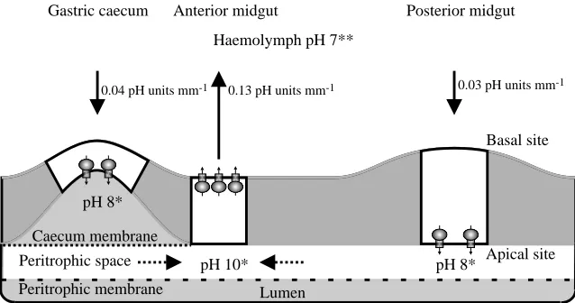
pH 8* pH 8*
Basal site
Apical site Haemolymph pH 7**
Peritrophic space
Peritrophic membrane Caecum membrane
Lumen
Gastric caecum Anterior midgut Posterior midgut
0.13 pH units mm-1
0.04 pH units mm-1 0.03 pH units mm-1
pH 10*
Fig. 6. Summary diagram showing stationary pH values and pH gradients in the midgut of mosquito larvae. The location of V-ATPase molecules is shown by mushroom-shaped symbols. Solid arrows indicate [H+] gradients determined with
self-referencing, ion-selective pH-sensitive liquid ion exchanger (SERIS-LIX) probes in this study. Dashed arrows indicate [H+] gradients in the
[image:6.612.45.364.571.739.2]metabolism. However, if it were replaced by H+ from the lumen in exchange for cell K+, then this addition of the ‘strong’ ion K+ to the lumen and the removal of the ‘weak’ ion H+ would contribute to the high lumen pH (>10; Dadd, 1975; Dadd, 1976) assuming that the gegenion is carbonate, as suggested for lepidopteran midgut (Dow, 1984).
Gastric caecum
The [H+] gradients near the gastric caeca are small, with the extracellular space adjacent to the basal membranes being slightly alkaline. The H+V-ATPase was localized on the apical membrane by immunohistochemistry and by the presence of portasomes (Zhuang et al., 1999). The basal alkalinity is presumably the result of the movement of H+ from the hemolymph to the cells, replacing the H+that is pumped from the cells to the lumen. An electrophoretic K+/nH+antiporter in the apical membrane could account for the luminal pH of 8, estimated with dyes (M. K. Dasher and W. R. Harvey, unpublished observations) and measured with pH-selective glass microelectrodes (D. Y. Boudko and W. R. Harvey, unpublished observations). The cavity of lepidopteran midgut goblet cells has a near-neutral pH (Chao et al., 1991) even though H+ is being transported towards it (Wieczorek et al., 1991; for a review, see Harvey, 1992). In a parallel manner, strong ions in the caecal cavity might be balanced by sulfate ions associated with proteins. If the anions associated with strong cations on the lumen side of the caecal membrane were bicarbonate ions, then the mildly alkaline pH observed there would be explained. Alternatively, highly alkaline fluid might move from the anterior midgut into the gastric caecum (Volkman and Peters, 1989). A Cl−channel in parallel with the apical V-ATPase would allow the secretion of acid into the caecum and neutralize such a fluid.
Posterior midgut
The small, inward [H+] gradients near the basal membrane of the posterior midgut are consistent with the near-neutral pH values observed there. Here, the V-ATPase appears to be localized on the apical membrane and hyperpolarizes it. The transmembrane potential is thought to drive amino acid:K+ symport (cotransport) from lumen to cells. Several amino acid transporters have been characterized in brush-border membrane vesicles from the posterior midgut of caterpillars (for a review, see Wolfersberger, 2000) and two have been cloned (Castagna et al., 1998; Feldman et al., 2000). A similar K+-coupled amino acid symporter has been identified and cloned using representative cDNA from the posterior midgut of Aedes aegypti larvae (M. K. Dasher, A. B. Kohn, R. M. Greenberg and W. R. Harvey, unpublished results).
Bafilomycin
The highly lipophilic V-ATPase inhibitor bafilomycin A1 diminished the [H+] gradients in the anterior midgut when applied to the basal membrane. However, it did not change the [H+] gradients when applied to gastric caeca and posterior midgut (Figs 4, 5). These results were expected because, in the
anterior midgut, the V-ATPase is located in the basal plasma membrane (Zhuang et al., 1999), where the bafilomycin binding site is immediately accessible. In contrast, in posterior midgut and gastric caeca, the V-ATPase is located in the apical plasma membrane, and the binding site is not accessible to inhibitor applied near the basal surface. To reach the enzyme on the apical membrane, the inhibitor would have to elude entrapment by the lipophilic basal membrane and pass through the hydrophobic barrier of the intracellular space.
One might argue that the abolition of pH gradients in the anterior midgut by bafilomycin was not due to an effect on the midgut itself but on an unidentified neuronal regulatory mechanism. Indeed, the anterior midgut is moderately innervated, with a few serotonergic neuronal terminals (Harvey et al., 2000). However, the re-uptake of 5-hydroxytryptamine from synapses by serotonin transporters is thought to be electroneutral (Blakely et al., 1994) and is presumably independent of membrane energization by V-ATPases. Moreover, if bafilomycin were to inhibit V-ATPase-mediated 5-hydroxytryptamine re-uptake, it would tend to increase the neurotransmitter concentration near its target and, therefore, would stimulate alkalization rather than inhibit it. Indeed, application of 5-hydroxytryptamine to the isolated midgut of Aedes aegypti larvae did lead to strong hyperpolarization of a specific population of epithelial cells (Clark et al., 1999).
Alkalization mechanism
Although the results reported here establish the importance of the H+ V-ATPase in larval mosquito anterior midgut alkalization, they do not explain the mechanism of lumen alkalization. The postulate (Küppers and Bunse, 1996) that the V-ATPase is a K+-transporting enzyme with a low affinity for H+ would not account for luminal alkalization in the anterior midgut of the larval mosquito. A basal K+ V-ATPase would move K+towards the blood and would tend to acidify rather than to alkalize the lumen.
epithelial cells, K+gradients emanating from the cells, and the effects of amiloride and other antiporter inhibitors on these properties.
Whatever its mechanism, our results firmly establish a link between the basally located V-ATPase and midgut alkalization in the anterior midgut of mosquito larvae. Specifically, we found (i) that a large [H+] gradient is directed away from the basal membrane in anterior midgut, and (ii) that the gradient is completely abolished by 10−7mol l−1 bafilomycin A
1, a specific inhibitor of the H+V-ATPase.
We thank Michelle K. Dasher for technical assistance with pH-sensitive dyes and for helpful discussion of the manuscript. We thank Katherine Hammar for assistance in the preparation of pH-sensitive LIX microelectrodes and Gabrielle Sakolsky, Staff Entomologist of the Cape Cod Mosquito Control Project, for obtaining Aedes canadensis larvae. This research was supported in part by NIH grants AI22444 (W.R.H.), A145098 (P.J.L.) and P41 RR01395 (P.J.S.S.).
References
Ammann, D. (1986). Ion-Selective Microelectrodes. Principles,
Design and Application. Berlin: Spring-Verlag.
Azuma, M., Harvey, W. R. and Wieczorek, H. (1995). Stoichiometry of K+/H+ antiport helps to explain extracellular
pH 11 in a model epithelium. FEBS Lett. 361, 153–156.
Berenbaum, M. (1980). Adaptive significance of midgut pH in larval Lepidoptera. Am. Nat. 115, 138–146.
Beyenbach, K. W., Pannabecker, T. L. and Nagel, W. (2000). Central role of the apical membrane H+-ATPase in electrogenesis
and epithelial transport in Malpighian tubules. J. Exp. Biol. 203, 1459–1468.
Blakely, R. D., De Felice, L. J. and Hartzell, H. C. (1994). Molecular physiology of norepinephrine and serotonin transporters.
J. Exp. Biol. 196, 263–281.
Breton, S., Hammar, K., Smith, P. J. S. and Brown, D. (1998). Proton secretion in the male reproductive tract: involvement of Cl− independent HCO3− transport. Am. J. Physiol. 275,
1134–1142.
Breton, S., Smith, P. J. S., Lui, B. and Brown, D. (1996). Acidification of the male reproductive tract by a proton-pumping ATPase. Nature Medicine 2, 470–472.
Brown, D., Smith, P. J. S. and Breton, S. (1997). Role of V-ATPase rich cells in acidification of the male reproductive tract. J. Exp. Biol. 200, 257–262.
Castagna, M., Shayakul, C., Trotti, D., Sacchi, V. F., Harvey, W. R. and Hediger, M. A. (1998). Cloning and characterization of a potassium-coupled amino acid transporter. Proc. Natl. Acad. Sci.
USA 95, 5395–5400.
Chao, A. C., Moffett, D. F. and Koch, A. R. (1991). Cytoplasmic pH and goblet cavity pH in the posterior midgut of the tobacco hornworm (Manduca sexta). J. Exp. Biol. 155, 503–514.
Clark, T. M., Koch, A. and Moffet, D. F. (1998). Alkalinization by
Manduca sexta anterior midgut in vitro: requirements and
characteristics. Comp. Biochem. Physiol. A 121, 181–187.
Clark, T. M., Koch, A. and Moffett, D. F. (1999). The anterior and posterior stomach regions of larval Aedes aegypti midgut: regional
specialization of ion transport and stimulation by 5-hydroxytryptamine. J. Exp. Biol. 202, 247–252.
Clark, T. M., Koch, A. and Moffet, D. F. (2000). The electrical properties of the anterior stomach of the larval mosquito (Aedes
aegypti). J. Exp. Biol. 203, 1093–1101.
Clements, A. N. (1992). The Biology of Mosquitoes. London: Chapman & Hall. 509pp.
Dadd, R. H. (1975). Alkalinity within the midgut of mosquito larvae with alkaline-active digestive enzymes. J. Insect Physiol. 21, 1847–1853.
Dadd, R. H. (1976). Loss of midgut alkalinity in chilled or narcotized mosquito larvae. Ann. Ent. Soc. Am. 69, 248–254.
Dow, J. A. T. (1984). Extremely high pH in biological systems; a model for carbonate transport. Am. J. Physiol. 246, R633–R635. Dröse, S. and Altendorf, K. (1997). Bafilomycins and
concanamycins as inhibitors of V-ATPases and P-ATPases. J. Exp.
Biol. 200, 1–8.
Edwards, H. A. (1982a). Ion concentration and activity in the haemolymph of Aedes aegypti larvae. J. Exp. Biol. 101, 143–151. Edwards, H. A. (1982b). Free amino acids as regulators of osmotic
pressure in aquatic insect larvae. J. Exp. Biol. 101, 153–160. Eguchi, M., Azuma, M., Yamamoto, H. and Takeda, S. (1990).
Genetically defined membrane-bound and soluble alkaline phosphatases of the silkworm: their discrete localization and properties. Prog. Clin. Biol. Res. 344, 267–287.
Ehrenfeld, J. and Klein, U. (1997). The key role of the H+V-ATPase
in acid–base balance and Na+transport processes in frog skin. J. Exp. Biol. 200, 247–256.
Feldman, D. H., Harvey, W. R. and Stevens, B. R. (2000). A novel electrogenic amino acid transporter is activated by K+or Na+, is
alkaline pH-dependent and is Cl−-independent. J. Biol. Chem. (on line).
Filippova, M., Ross, L. S. and Gill, S. S. (1998). Cloning of the V-ATPase B subunit cDNA from Culex quinquefasciatus and expression of the B and C subunits in mosquitoes. Insect Mol. Biol. 7, 223–232.
Harvey, W. R. (1992). Physiology of V-ATPases. J. Exp. Biol. 172, 1–17.
Harvey, W. R., Boudko, D. Y., Dasher, M. K., Sadreyev, R. I., Panchin, Y., Linser, P. L. and Moroz, L. L. (2000). Serotonin and NOS in central nervous and peripheral tissues of the yellow fever vector mosquito Aedes aegypti. Soc. Neurosci. Asbtr. 26, 2173. Harvey, W. R., Maddrell, S. H. P., Telfer, W. H. and Wieczorek,
H. (1998). H+V-ATPases energize animal plasma membranes for
secretion and absorption of ions and fluids. Am. Zool. 38, 426–441. Klein, U., Koch, A. and Moffett, D. F. (1996). Ion transport in Lepidoptera. In Biology of the Insect Midgut (ed. M. J. Lehane and P. F. Billingsley), pp. 236–264. London: Chapman & Hall. Kppers, J. and Bunse, I. (1996). A primary cation transport by
V-type ATPase of low specificity. J. Exp. Biol. 199, 1327–1334. Lepier, A., Azuma, M., Harvey, W. R. and Wieczorek, H. (1994).
K+/H+ antiport in the tobacco hornworm midgut: the K+
-transporting component of the K+ pump. J. Exp. Biol. 196,
361–373.
Moffett, D. F. and Cummings, S. A. (1994). Transepithelial potential and alkalization in an in situ preparation of tobacco hornworm (Manduca sexta) midgut. J. Exp. Biol. 194, 341–345.
Padan, E. and Schuldiner, S. (1993). Na+/H+antiporters, molecular
devices that couple the Na+and H+circulation in cells. J. Bioenerg. Biomembr. 25, 647–669.
Biophysics, 18th edition. Philadelphia, London: W. B. Saunders.
1232pp.
Senior-White, R. (1926). Physical factors in mosquito ecology. Bull.
Ent. Res. 16, 187–248.
Smith, P. J. S., Hammar, K., Porterfield, D. M., Sanger, R. H. and Trimarchi, J. R. (1999). A self-referencing, non-invasive, ion selective electrode for single cell detection of trans-plasma membrane calcium flux. Microsc. Res. Techn. 46, 398–417. Smith, P. J. S., Sanger, R. H. and Jaffe, L. F. (1994). The vibrating
Ca2+electrode: A new technique for detecting plasma membrane
regions of Ca2+influx and efflux. Meth. Cell Biol. 40, 115–134. Smith, P. J. S. and Trimarchi, J. R. (2000). Non-invasive
measurement of hydrogen and potassium ion flux from single cells and epithelial structures. Am. J. Physiol. (in press).
Volkman, A. and Peters, W. (1989). Investigation on the midgut
caeca of mosquito larva. II. Functional aspects. Tissue & Cell 21, 253–261.
Wieczorek, H., Grüber, G., Harvey, W. R., Huss, M. and Merzendorfer, H. (1999). The plasma membrane H+V-ATPase
from tobacco hornworm midgut. J. Bioenerg. Biomembr. 31, 67–74.
Wieczorek, H., Putzenlechner, M., Zeiske, W. and Klein, U. (1991). A vacuolar-type proton pump energizes K+/H+-antiport in
a plasma membrane. J. Biol. Chem. 266, 15340–15347.
Wolfersberger, M. G. (2000). Amino acid transport in insects. Annu.
Rev. Ent. 45, 111–120.
Zhuang, Z., Linser, P. J. and Harvey, W. R. (1999). Antibody to H+V-ATPase subunit E colocalizes with portasomes in alkaline
larval midgut of a freshwater mosquito (Aedes aegypti L.). J. Exp.


![Fig. 4. Effects of bafilomycin on the [H+] gradients near the gut ofmosquito larvae. Each part of the figure shows signals recorded with](https://thumb-us.123doks.com/thumbv2/123dok_us/1030329.618405/5.612.324.561.69.733/effects-balomycin-gradients-ofmosquito-larvae-gure-signals-recorded.webp)