Original Article
Long non-coding RNA MEG3 mediates high
glucose-induced endothelial cell dysfunction
Zhiqiang Wang1,2*, Lili Ding1,3*, Jun Zhu2,4, Yinxia Su2, Li Wang2, Lina Liu5, Qi Ma2, Hua Yao2
1Department of Public Health, Xinjiang Medical University, Urumqi, Xinjiang, China; 2Xinjiang Key Laboratory of Metabolic Disease Research, The First Affiliated Hospital of Xinjiang Medical University, Urumqi, Xinjiang, China; Departments of 3Infection Control, 4Endocrinology, The First Affiliated Hospital of Xinjiang Medical University, Urumqi, Xinjiang, China; 5Department of Endocrinology, Branch of The First Affiliated Hospital of Xinjiang Medical University, Changji, Xinjiang, China. *Equal contributors.
Received November 30, 2017; Accepted December 22, 2017; Epub March 1, 2018; Published March 15, 2018
Abstract: Long noncoding RNAs (lncRNAs) are implicated in the progression of diabetes mellitus (DM) and diabetes-induced endothelial dysfunction. Maternally expressed gene 3 (MEG3) encodes an lncRNA which is suggested to function as a tumor suppressor. Therefore, the aim of the present study was to investigate whether MEG3 is a
po-tential regulator and molecular biomarker of high glucose-induced endothelial dysfunction. LncRNA Meg3-specific
small interfering RNA (siRNA) and scrambled (Scr) siRNA were transfected for MEG3 dysfunction studies. RNA and protein expression were examined by quantitative RT-PCR (qPCR) and Western blot, respectively. The percentage
of apoptotic cells was measured by flow cytometry. Cell viability was determined through MTT assay. This study demonstrates involvement of lncRNA MEG3 in high glucose-induced endothelial dysfunction. MEG3 is significantly
downregulated in an endothelial cell model of hyperglycemia. In addition, MEG3 knockdown could exacerbate
in-flammatory damage in endothelial cells. Interestingly, MEG3 knockdown in HUVECs significantly induced prolifera -tion and inhibited apoptosis by upregulating Bcl-2 and downregulating Bax, caspase-3, and P53. It should be noted
that MEG3 knockdown could activate the TGF-β signaling pathway via upregulating TGF-β1, SMAD2, and SMAD7 and activate the Wnt/β-catenin signaling pathway via upregulating β-catenin and Cyclin D1 and downregulating
TCF7L2. Our results indicate that MEG3 can be regarded as a novel therapeutic target and molecular biomarker for high glucose-induced endothelial dysfunction.
Keywords: MEG3, diabetes mellitus, long noncoding RNA, endothelial dysfunction
Introduction
Diabetes mellitus (DM) is a worldwide health issue affecting children, adolescents, and adul- ts. It could result in organ and tissue damage in about one-third to one-half of population with diabetes [1]. On the basis of the International Diabetes Federation, currently 285 million peo-ple are suffering from DM, and the population of diabetes will reach 438 million by 2030 [2]. Numerous studies in large DM cohorts have reported that diabetes-associated macrovas-cular and microvasmacrovas-cular complications are strongly associated with the severity and dura-tion of hyperglycemia [3]. Macrovascular and microvascular complications are the most sig-nificant and complex consequences of DM and spread to various organs, such as the heart, kidney, brain, and eyes [4]. Because the
(ncRNAs) are further divided into small and long ncRNAs. Small ncRNAs such as microRNAs, small interfering RNAs, and piwi-associated RNAs are those less than 200 nucleotides in length. Long noncoding RNAs (lncRNAs) are a novel found type of noncoding RNA transcripts that are longer than 200 nucleotides. Initially, lncRNAs were regarded as transcriptional noise lacking biological functions. However, recent studies have shown that several lncRNAs are implicated in multiple biological processes, such as cell proliferation, differentiation, apop-tosis, chromatin modification, transcriptional regulation, posttranscriptional regulation [7]. LncRNAs have important functional roles in different biological processes supporting their potential diagnostic, prognostic or therapeutic importance. Although the precise biological mechanisms by which LncRNAs participate in disease development have not been elucidat-ed, LncRNAs hold promise as potential targets for diseases therapy. The functional diversity and mechanistic role of lncRNAs is currently a major focus of comprehensive research in the field.
Recently, several reports have shown that lncRNAs play roles in the progression of DM and its complications. Jiang et al. [8] found that lncRNA might be a new regulatory target relat-ed to spermatogenesis in men with DM. Wang et al. [9] showed that 1018 lncRNAs were dif-ferentially expressed in diabetic kidney tissues. Arnes et al. [10] reported that βlinc1 is essen-tial for the proper specification and function of endocrine cells. Interestingly, there is a double-negative feedback loop between H19 and let-7 which participates in glucose regulation in mus-cle. In addition, SRA plays an important role in the regulation of adipose tissue biology and glucose homeostasis. Yan et al. [11] found that MIAT was recognized as a regulator of micro-vascular dysfunction. MIAT knockdown signifi-cantly alleviated microvascular dysfunction in vivo, and changed specific signaling pathways implicated in cell proliferation, migration, and survival of endothelial cells. Liu et al. [12] show- ed involvement of lncRNA MALAT1 in diabetes-induced microvascular dysfunction. These re- sults have significant implications for the iden-tification of new pathophysiological mecha-nisms underlying DM susceptibility and showed that lncRNAs could become novel therapeutic targets for the treatment of DM.
Maternally Expressed Gene 3 (MEG3) encodes an lncRNA which is suggested to function as a tumor suppressor. MEG3 is known as the ortho-logue of gene trap locus 2 (Gtl2) in mice. MEG3 is reciprocally imprinted with the paternally expressed gene DLK1 constituting an imprint-ing domain on human chromosome 14q32 and on mouse chromosome 12 [13]. MEG3 is ex- pressed in multiple normal human tissues, with the highest expression in brain and pituitary gland. However, recent studies demonstrate that the loss of MEG3 expression has a role in the development of several human cancers, such as gliomas, bladder cancer, gastric can-cer, and hepatocellular cancers [14]. Previous studies have demonstrated that MEG3 also plays an important role as a tumor suppressor [15]. Researchers revealed that overexpression of MEG3 inhibits cell proliferation and promotes cell apoptosis.
There is increasing evidence that lncRNAs may be defined as novel therapeutic targets in hyperglycemia-related endothelial dysfunction or diabetes-induced vascular disease. A deep understanding of endothelial lncRNAs may pro-vide novel biomarkers or therapeutic targets for diabetic complications. Therefore, it is im- portant to increase our understanding of the underlying molecular mechanisms of diabetes-induced endothelial dysfunction and identify novel potential therapeutic targets for prevent-ing or reducprevent-ing diabetic complications. Despite comprehensive research, little is known about the role of the majority of lncRNAs and their contribution to the maintenance of endothelial cell function and the development of diabetic complications. Until now, there has been no study revealing the underlying molecular mech-anisms associated with MEG3 induction and how it could play a key role in high glucose-induces HUVCEs dysfunction. Therefore, the aim of the present study was to investigate whether MEG3 was a potential regulator and molecular biomarker of high glucose-induced endothelial dysfunction.
Materials and methods
Materials
(Carlsbad, CA, USA). Culture media were sup-plemented with 100 U/ml penicillin and 100 μg/ml streptomycin (Life Technologies). Primary antibodies against Bax, Bcl-2, Caspase 3, P53, β-catenin, SMAD2, and GAPDH were obtained from Cell Signaling Technology (Beverly, MA). Goat anti-Mouse IgG and Goat anti-Rabbit IgG horseradish peroxidase (HRP)-conjugated sec-ondary antibody was purchased from Life tech-nology. MTT kit was purchased from Beyotime Biotechnology. The siRNA delivery agent, Lipo- fectamine 2000, was from Invitrogen (Carlsbad, CA). All other chemicals used were of the high-est commercial grade available.
Cell culture
Human umbilical vein endothelial cells (HUV- ECs) were obtained from Shanghai Bioleaf Bio- tech Co. Ltd. HUVECs were cultured in Dul- becco’s modified Eagle’s medium (DMEM) sup-plemented with 2% fetal bovine serum and penicillin (100 μg/ml) and streptomycin (100 μg/ml). These cells were maintained at 37°C in a humidified atmosphere containing 5% CO2. HUVECs were treated with either normal (5.5 mmol/L) or high (33.3 mmol/L) glucose. The HUVECs after the third passage were submit-ted for the current experiment. When 70-80% confluent, the cells were treated with different agents.
Cell transfection
LncRNA Meg3-specific small interfering RNA (siRNA) and scrambled (Scr) siRNA were de- signed and synthesized from GenePharma (Shanghai, China). MEG3 siRNA and Scr siRNA were transfected into HUVECs at a concentra-tion of 50 nmol/l using Lipofectamine 2000 (Invitrogen) according to the manufacturer’s instruction. Cells were harvested after 48 h for qRT-PCR, Western blot, cell proliferation and apoptosis analyses.
RNA extraction and quantitative real-time PCR (qRT-PCR) analysis
Total RNA was extracted from HUVECs using TRIzol reagent (Invitrogen, Thermo Fisher Scien- tific) according to the manufacturer’s instruc-tions. After the RNA quality was determined by a spectrophotometer (Thermo Fisher). For each sample, 0.5 μg of total RNA was reverse-tran-scribed into cDNA by using the Transcriptor
First Strand cDNA Synthesis Kit (Takara Bio, Inc., Otsu, Japan) following the manufacturer’s instructions. QRT-PCR analysis was performed by using an instrument of ABI 7000 PCR (Applied Biosystems, Japan) with SYBR green PCR Master Mix (Takara Bio., Inc., Otsu, Japan). The relative amount of mRNA was calculated using 2-ΔΔCt method. Gene expression was
nor-malized by β-actin. All data were obtained from three individual experiments. Specific sense and anti-sense primers used were as follows: MEG3, sense 5’-CATCCGTCCACCTCCTTGTCTT- C3’ and antisense 5’-GTCCTCTTCATCCTTTGC- CATCC-3’; α-SMA, sense 5’-GTGATGGTGGGAA- TGGG-3’ and antisense 5’-CAGGGTGGGATGC- TCTT-3’; VEGF, sense 5’-CCCTGATGAGATCGAG- TACA-3’ and antisense 5’-AGGAAGCTCATCTCT- CCTAT-3’; TNF-α, sense 5’-GTCCAGGCTTGTCC- TGCTAC-3’ and antisense 5’-CTGAGTCCGTTG- AGGGAGAG-3’; IL-6, sense 5’-CGGGAACGAAA- GAGAAGCTCTA-3’ and antisense 5’-GAGCAGC- CCCAGGG AGAA-3’; TGF-β1, sense 5’-GGCCA- GATCCTGTCCAAGC-3’ and antisense 5’-GTGG- GTTTCCACCATTAGCAC-3’; Smad2, sense 5’-CT- TTTGTTGTGTAAGCTCTCACTG-3’ and antisense 5’-GACCTTCTACCACTTTCAGAGTTG-3’; Smad7, sense 5’-GGACAGCTCAATTCGGACAAC-3’ and antisense 5’-GTACACCCACACACCATCCAC-3’; β- catenin, sense 5’-GTGTGGCGACATATGCAGCT- 3’ and antisense 5’-CAAGATCAGCAGTCTCAT- TC-3’; TCF7L2, sense 5’-CAATAATCTCCGCTCC- CAGA-3’ and antisense 5’-CGCTCGGATTTGAG- TGAGTT-3’; Cyclin D1, sense 5’-GTGCATC TAC- ACC GACAACTCCA-3’ and antisense 5’-TGAGC- TTGTTCACCAGGAGCA-3’; β-actin, sense 5’-AG- CGAGCATCCCCCAAAGTT-3’ and antisense 5’- GGGCACGAAGGCTCATCATT-3’.
Western blot
for 10 min, followed by incubation at room tem-perature for 1 h with horseradish peroxidase (HRP)-labeled secondary antibody. The incu-bated membrane was washed three times with TBST, the signal was detected using the en- hanced chemiluminescence (ECL) kit, protein expression was analyzed using Quantity One software (Bio-Rad, USA) and normalized to GAPDH. Primary antibodies and dilutions were as follows: Bax (dilution in 1:1000), anti-Bcl-2 (dilution in 1:2000), anti-Caspase 3 (dilu-tion in 1:200), P53 (dilu(dilu-tion in 1:200), anti-P53 (dilution in 1:200), anti-β-catenin (dilution in 1:5000), anti-SMAD2 (dilution in 1:2000), anti-GAPDH (dilution in 1:10000). The second-ary antibody and dilutions were as follows: goat Mouse IgG (dilution in 1:1000), goat anti-Rabbit IgG (dilution in 1:20000).
Cell viability
Cell viability was measured with an MTT kit according to the manufacturer’s instruction. Briefly, the HUVECs were seeded into a 96-well plate (Corning) at a density of 1×10⁴ cells/well
in 200 ml culture medium. After the specific treatments, cells were incubated with MTT at a final concentration of 0.5 mg/ml for 3 h at 37°C. After the supernatant was removed, 100 mM DMSO solution was added to dissolve the formazan crystals. The absorbance was deter-mined at 570 nm wavelength using a micro-plate reader. Each experiment was carried out in triplicate wells and repeated at least three times.
Flow cytometric analysis of apoptosis
HUVECs transfected with MEG3 siRNA or Scr siRNA after serum-starved for 48 hours were harvested by trypsinization. flow cytometric analysis was applied with Annexin V-FITC/PI Apoptosis Detection Kit (Bestbio, China) ac- cording to the manufacturer’s instructions. The cells were then stained with 5 μl Annexin V/FITC and 10 μl propidium iodide (20 μg/ml) for 15 min at room temperature in the dark, the cells were analyzed using flow cytometry (FACScan, BD, Biosciences). Cells were discriminated into viable cells, dead cells, early apoptotic cells, and apoptotic cells. This experiment was inde-pendently performed at least three times.
Statistical analysis
All experiments were independently performed at least three times. Results are expressed as mean ± standard error. Comparisons between two groups were analyzed using the unpaired Student’s t-test. Comparisons among multiple groups were analyzed using one-way analysis of variance with Bonferroni post hoc tests where applicable. Statistical analyses were performed using the SPSS 17.0 (SPSS, Inc., Chicago, IL, USA). P values less than 0.05 were considered as significant.
Results
Expression pattern of MEG3 in HUVECs upon high glucose stress
To identify whether MEG3 expression is alter- ed by high glucose stress in vitro, we cultured HUVECs in high glucose medium to mimic
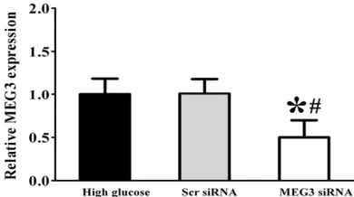
dia-Figure 1. Expression pattern of MEG3 in HUVECs upon high glucose stress. HUVECs were cultured in
[image:4.612.91.288.72.181.2]the medium containing 5 mM D-glucose (control), 30 mM D-glucose (high glucose). Quantitative reverse transcriptase-PCRs (qRT-PCRs) were performed to detect MEG3 levels. MEG3 levels were expressed as the fold change compared with respective control group (mean ± SD, n=3, *P<0.05).
Figure 2. HUVECs were transfected with scrambled
(Scr) siRNA or MEG3 siRNA for 48 h. Quantitative PCR was conducted to detect MEG3 level.
*indicat-ed a significant difference compar*indicat-ed with the high
glucose-treated group (mean ± SD, n=3, *P<0.05).
#indicated a significant difference compared with
[image:4.612.91.286.290.399.2]betic conditions. Our results revealed that high glucose treatment could result in an obvious reduction of MEG3 expression in a time-depen-dent manner (Figure 1).
MEG knockdown affects HUVECs inflammation upon high glucose stress
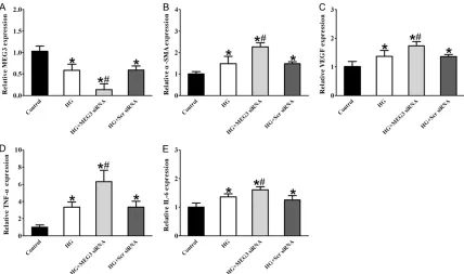
We further investigated the mechanistic aspe- cts and functional significance of MEG3 altera-tion in vitro, HUVCEs cells were transfected with MEG3 small interfering RNA (siRNA). We found that MEG3 siRNA transfection led to a significant reduction in MEG3 level in HUVCEs cells (Figure 2). The following experiments in vitro used MEG3 siRNA for knockdown studies. We performed qRT-PCRs to detect the expres-sion of α-SMA, VEGF, TNF-α, and IL-6, High glucose resulted in a marked increase in the expression of α-SMA, VEGF, TNF-α, and IL-6, whereas MEG3 knockdown significantly increa- sed the expression of α-SMA, VEGF, TNF-α, and IL-6 in HUVECs compared with the high glu-cose-treated group (Figure 3), suggesting that MEG3 knockdown could aggravate endothelial inflammation upon high glucose stress.
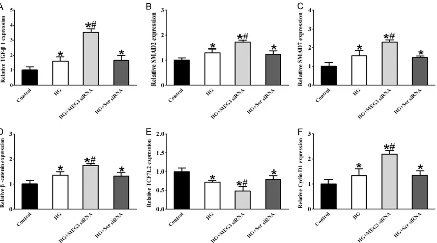
MEG knockdown influenced the expression of TGF-β pathway and Wnt/β-catenin pathway genes upon high glucose stress in HUVECs
There has been an increasing interest in study-ing intracellular signalstudy-ing pathways that partici-pate in endothelial cell function and pathologi-cal angiogenesis in order to apply this knowl-edge for molecular and cellular therapies for DM. qRT-PCR experiments showed that com-pared with the high glucose-treated group, the MEG3 knockdown group had higher expression levels of TGF-β pathway genes TGF-β1, SMAD2, SMAD7 and the Wnt/β-catenin pathway genes β-catenin and Cyclin D1. However, expression of Wnt/β-catenin pathway gene TCF7L2 was significantly decreased in HUVECs after trans-fection with MEG3 siRNA compared with the high glucose-treated group (Figure 4).
The effect of MEG3 knockdown on cell prolifer-ation and apoptosis upon high glucose stress in HUVECs
[image:5.612.92.520.70.323.2]To identify the effects of MEG3 on high glucose induced cell viability. HUVECs were transfected
Figure 3. MEG knockdown affects HUVECs inflammation upon high glucose stress. HUVECs were transfected with
MEG3 siRNA, scramble (Scr) siRNA or left untreated, and then these cells were exposed with or without high glucose (30 mM). The untreated group was taken as the control group. qRT-PCRs were conducted to compare MEG3 levels
(A) and detect the expression of inflammatory genes α-SMA (B), VEGF (C), TNF-α (D) and IL-6 (E). *indicated a sig
-nificant difference compared with the control group (mean ± SD, n=3, *P<0.05). #indicated a significant difference
with MEG3 siRNA, and then high glucose was used to stimulate the cells. Results revealed that cell viability was significantly decreased by high glucose treatment. Subsquent MEG3 knockdown reversed the effects that were induced by high glucose. Compared with the high glucose-treated group, MEG3 knockdown significantly increased cell viability of HUVECs as detected by MTT assay. These data indicate that downregulation of MEG3 expression pro-motes HUVECs proliferation (Figure 5A). In order to explore the potential mechanism between MEG3 and cell apoptosis that was induced by high glucose, HUVECs were treated with MEG3 siRNA, scrambled siRNA, or left untreated, followed by high glucose treatment. The double staining with fluorescein isothiocya-nate (FITC)-Annexin V and propidium iodide (PI) was used to detect the cell apoptosis level. We found that high glucose treatment could signifi-cantly accelerate cell apoptosis. By contrast, MEG3 knockdown significantly reversed cell apoptosis induced by high glucose (Figure 5B). Taken together, these results suggest that MEG3 has a critical role in the regulation of endothelial cell function in vitro.
The effect of MEG3 knockdown on Bax, Bcl-2, Caspase 3, and P53 protein expression upon high glucose stress in HUVECs
We performed Western blot assays to detect expression of apoptosis proteins. Our findings showed that high glucose significantly increa- sed the level of Bax, caspase-3, and P53, whereas MEG3 knockdown could partially reduce the upregulation of Bax, caspase-3, and P53 induced by high glucose. High glucose decreased the level of Bcl-2, whereas MEG3 knockdown partially reversed the downregula-tion of Bcl-2 induced by high glucose (Figure 6).
The effect of MEG3 knockdown on the
β-catenin and SMAD2 protein expression upon high glucose stress in HUVECs
[image:6.612.94.521.71.309.2]We found that the protein expression of β- catenin and SMAD2 was significantly increased in the high glucose-treated group compared with the control. Additionally, the protein ex- pression of β-catenin and SMAD2 was also sig-nificantly increased in high glucose plus MEG3 knockdown stimulated HUVECs rather than
Figure 4. MEG knockdown influenced the expression of TGF-β pathway and Wnt/β-catenin pathway genes upon high glucose stress in HUVECs. HUVECs were transfected with MEG3 siRNA, scrambled (Scr) siRNA or left untreated, and
then these cells were exposed with or without high glucose (30 mM). The untreated group was taken as the control
group. qRT-PCRs were conducted to detect the expression of TGF-β pathway genes TGF-β1, SMAD2 and SMAD7 (A-C), Wnt/β-catenin pathway genes β-catenin, TCF7L2 and Cyclin D1 (D-F). *indicated a significant difference com -pared with the control group (mean ± SD, n=3, *P<0.05). #indicated a significant difference between high
HUVECs with high glucose alone stimulation (Figure 7).
Discussion
[image:7.612.89.374.71.199.2]Diabetes mellitus is one of the most prevalent chronic dis-eases, and has been regarded as a global public health crisis that threatens the economy of all countries. The total popula-tion of worldwide diabetics is close to 285 million, which is expected to reach up to 438 million by 2030, and the popu-lation of Chinese adults with diabetes is more than 100 million [2]. More important, approximately half of the DM patients suffer from major complications, which bring great pain for the diabetic patients. The chronic DM pro-motes injury or dysfunction of many organs, especially the heart, eyes, kidney, and blood vessels [4]. The macrovascu-lar and microvascumacrovascu-lar affec-tions are important character-istics of such complications. DM is usually considered as the vascular disease charac-terized by modified vasoregu-lation, increased generation of reactive oxygen intermedi-ates, inflammatory activation,
Figure 5. The effect of MEG3 knockdown on cell proliferation and apoptosis
upon high glucose stress in HUVECs. HUVECs were transfected with MEG3
siRNA, scrambled (Scr) siRNA or left untreated, and then these cells were treated with or without high glucose (30 mM). The untreated group was taken as the control group. A. Cell viability was detected using MTT method. B. The
apoptotic rates of cells were detected by flow cytometry. This experiment was
independently performed at least three times and the change tendency is
the same. *indicated a significant difference between high glucose-treated
group and control group (mean ± SD, n=3, #P<0.05). #indicated a significant
[image:7.612.92.373.340.457.2]difference between high glucose-treated group and high glucose plus MEG3 knockdown group (mean ± SD, n=3, #P<0.05).
Figure 6. The effect of MEG3 knockdown on Bax, Bcl-2, Caspase 3, and
P53 protein expression upon high glucose stress in HUVECs. HUVECs were
transfected with MEG3 siRNA, scrambled (Scr) siRNA or left untreated, and then these cells were exposed with or without high glucose (30 mM). The un-treated group was taken as the control group. Western blots were conducted to detect the expression of Bax, Bcl-2, Caspase 3, and P53. GAPDH was detected as the loading control. A representative immunoblot was shown
along with the quantitative data (n = 3). *indicated a significant difference
compared with the control group (mean ± SD, n=3, *P<0.05). #indicated a
significant difference between high glucose-treated group and high glucose
plus MEG3 knockdown group (mean ± SD, n=3, #P<0.05).
Figure 7. The effect of MEG3 knockdown on the β-catenin and SMAD2 pro
-tein expression upon high glucose stress in HUVECs. HUVECs were transfect
-ed with MEG3 siRNA, scrambl-ed (Scr) siRNA or left untreated, and then these cells were treated with or without high glucose (30 mM). The untreated group was taken as the control group. Western blots were conducted to detect
the expression of β-catenin, and
SMAD2. GAPDH was detected as the loading control. A representa-tive immunoblot was shown along with the quantitative data (n = 3).
*indicated a significant difference
compared with the control group (mean ± SD, n=3, *P<0.05).
#in-dicated a significant difference
[image:7.612.93.371.599.695.2]and altered barrier function. Endothelial dys-function is essential for the development of DM. Endothelial dysfunction is also recognized as an independent risk factor for cardiovascu-lar disease [16]. Therefore, it is important to increase our understanding of the underlying molecular mechanisms of endothelial dysfunc-tions associated with DM and identify novel potential therapeutic targets for diabetic com- plications.
The ENCODE (encyclopedia of DNA elements) project revealed that 90% of the human geno- me are transcribed to RNA, but only 2.94% are responsible for coding for proteins. Non-coding RNAs (ncRNAs) belong to a newly identified type of RNA molecules with little or no pro-tein-coding capacity [6]. According to their size, ncRNAs are divided into small ncRNAs and long ncRNAs (lncRNAs). Small ncRNAs include siR-NAs, piRsiR-NAs, and miRNAs that have a length of less than 200 nucleotides. Long non-coding RNAs (lncRNAs) were considered to be endog-enous cellular non-coding RNA molecules that are longer than 200 nucleotides. The result of comprehensive surveys showed that lncRNA serve as key molecules affect gene expression, chromatin remodeling, transcription, and post-transcriptional processing [17]. Moreover, ac- cumulated studies suggest that disorders of lncRNA are closely associated with human dis-eases. Moreover, increasing numbers of stud-ies have suggested that aberrant expression of lncRNAs play a critical role in a number of path-ological processes including inflammation and oxidative stress response. Although a large number of lncRNA transcripts are differentially expressed during development where most of them play critical roles, a majority of them have not been clearly identified in mechanistic details. Recently, some studies have demon-strated the roles of lncRNAs in DM, and accu-mulating evidence has revealed that lncRNAs may be implicated in maintaining pancreatic beta cell function and insulin signal transduc-tion, which may influence DM development [18]. Inspired by this evidence, we speculate that lncRNAs are potential regulators of diabe-tes-induced endothelial dysfunction. The emer-gence of lncRNAs provides new insight into the treatment of diabetic complications.
Maternally expressed gene 3 (MEG3), which encoded a long noncoding RNA (lncRNA), is an imprinted gene belonging to the DLK1-MEG3
locus located on chromosome 14q32 in hu- mans [13]. Previous studies showed that MEG3 is expressed in normal human tissue. However, the loss of MEG3 expression has been found in various types of human tumors, including meningioma, colon cancer, nasopharyngeal carcinoma, and leukemia. More importantly, overexpression of MEG3 could inhibit prolifera-tion and promote apoptosis in tumor cells [19]. A number of studies have reported that the MEG3 gene may play an important role in tumor suppression. Previous studies have suggested that the downregulation of MEG3 may be a poor prognostic biomarker in several cancers, such as glioma, hepatocellular cancer, and bladder cancer [14]. Recent studies have found that MEG3 can be regarded as a novel regulator of maintaining beta cells function via affecting insulin synthesis and secretion [20]. Additional- ly, genomic-wide association studies (GWAS) have identified that the SNP rs941576 located in intron 6 of MEG3 is associated with increased risk of type 1 diabetes [21]. Interestingly, MEG3 has also been shown to be downregulated in islets from T2DM donors compared to nondia-betics [22]. Furthermore, upregulation of ME- G3 can induce hepatic insulin resistance by increasing FoxO1 expression [23]. More impor-tantly, MEG3 has obtained attention for its function in progression of DM complications, the expression of MEG3 was significantly down-regulated in diabetic retinas and retinal endo-thelial cells upon high glucose stress. Its knock-down leads to diabetic-induced microvascular dysfunction and results in retinal endothelial cell proliferation, migration, and tube formation [24]. In the present study, we identified whether MEG3 expression was altered by high glucose stress in vitro. We cultured HUVECs in the high glucose medium to mimic diabetic condition. Our results revealed that high glucose treat-ment could result in an obvious reduction of MEG3 expression in a time-dependent manner (Figure 1). We further investigated the mecha-nistic aspects and functional significance of MEG3 alteration in vitro, HUVCEs cells were transfected with MEG3 small interfering RNA (siRNA). We found that MEG3 siRNA transfec-tion led to a significant reductransfec-tion in MEG3 level in HUVCEs cells (Figure 2). The following in vitro
experiments used MEG3 siRNA for knockdown studies.
cyto-kines such as IL-6 and TNF-α have been report-ed to play an important roles in DM. Prolongreport-ed exposure to pro-inflammatory cytokines such as IL-1β, IL-6, and TNF-α can have deleterious effects on beta cell functions, resulting in a decreased capacity to produce and release insulin, and promoting beta cell apoptosis [25]. Endothelial homeostasis is important for all organs and for microvascular and macrovascu-lar. The dysregulated endothelial cells may increase inflammatory cytokines release. More- over, the proliferation and apoptosis of thelial cells are strongly associated with endo-thelial dysfunction in diabetic complications [16].
If we understand the molecular mechanisms implicated in endothelial inflammatory process, we may be able to identify novel strategies for preventing and treating DM and diabetic com-plications. Therefore, we investigated whether MEG3 modulates inflammatory pathways par-ticipating inflammatory cytokines in high glu-cose-induces endothelial dysfunction. In the present study, we observed that expression lev-els of α-SMA, VEGF, TNF-α, and IL-6 in high glu-cose-treated group were significantly increased compared with the control group based on real-time PCR. After treatment with MEG3 siRNA, the expression levels of α-SMA, VEGF, TNF-α, and IL-6 were increased compared to high glu-cose-treated group (Figure 3). These results indicate involvement of MEG3 in high glucose induced inflammation of endothelial cells. ME- G3 siRNA treatment could exacerbate inflam-matory damage in endothelial cells. Therefore, our findings highlight the potential of MEG3 as a new therapeutic target gene to attenuate inflammation in hyperglycemia-related endo-thelial dysfunction. This finding provides a new explanation for a key role of lncRNAs against inflammation, which may be helpful for deepen-ing our understanddeepen-ing of the molecular mecha-nisms involved in the pathogenesis of diabetes-induces endothelial dysfunction.
Transforming growth factor β (TGF β) signaling has multiple roles in different cellular and developmental pathways, beginning with bind-ing TGF β ligands to type II receptors to catalyze phosphorylation of the type I receptors to acti-vate several intracellular signaling cascades, such as small mothers against decapentaple-gic (SMAD) and mitogen-activated protein kina- ses (MAPK) include extracellular regulated kina-
expression of SMAD2 was increased compared to high glucose-treated group (Figure 7). These results suggested that MEG3 knockdown could activate TGF-β signaling pathway via upregulat-ing TGF-β1, SMAD2, and SMAD7.
The Wnt signaling pathway has been reported to be implicated in cell proliferation and cellular differentiation in several organs, including the endocrine pancreas [32]. The Wnt signaling pathway may play an essential role in the pathogenesis of T2DM. The activation of the Wnt signaling pathway could be an etiological factor in the development of hyperinsulinemia and T2DM. Many studies have suggested an important role for the Wnt signaling pathway in pancreatic islet beta cell genesis and prolifera-tion [33]. Wnt signaling pathway is activated in pancreatic islets in the process of prediabetes and may play a fundamental role in the induc-tion of the compensatory beta cell hyperplasia observed at early stage of T2DM. Increasing evidence has revealed that the Wnt signaling pathway plays an important role in various physiological and pathological processes, in- cluding angiogenesis and inflammation. Loss or gain of function of Wnt pathway components results in aberrant vascular development and angiogenesis. Activation of Wnt signaling path-way can lead to pathological processes in dia-betic retinopathy. Nuclear β-catenin accumula-tion has been observed in the retinas of dia-betic retinopathy animal models and in patients with diabetic retinopathy. Inhibition of abnor-mal Wnt signaling pathway exerts antiangio-genic and anti-inflammatory activities in dia-betic retinopathy and therefore become an attractive potential therapies for the treatment of diabetic retinopathy [34]. Further studies into the function of Wnt signaling in diabetes-induced vascular disease will find new thera-peutic target for DM. Genome-wide association studies have directed our attention to the func-tion of the Wnt signaling pathway and its down-stream targets, such as β-catenin and TCF7L2, in regulating hormone gene expression and glu-cose disposal. β-catenin is an important effec-tor of Wnt signaling pathway and has a diverse range of functions in the regulation of cell pro-liferation and differentiation. An accumulation of β-catenin will activate the Wnt signaling pathway [35]. TCF7L2 belongs to the T cell fac-tor (TCF) family of high mobility group box tran-scription factors and is a main effector of the Wnt signaling pathway. TCF7L2 regulates
tran-scription of the proglucagon gene (gcg), which encodes the incretin hormone glucagon-like peptide-1 (GLP-1) [36]. Recently, numerous ge- nome-wide association studies have demon-strated that single nucleotide polymorphisms (SNPs) within intronic regions of TCF7L2 were found to be significantly associated with β-cell dysfunction and the pathogenesis of T2DM. In addition, TCF7L2 positively controls β-cell pro-liferation and glucose-mediated insulin secre-tion. Moreover, overexpression of TCF7L2 pro-tected islets from glucose and cytokine-induc- ed apoptosis [37]. Interestingly, TCF7L2 direct-ly binds to promoters/regulatory elements of a majority of genes that are essential in control-ling hepatic glucose metabolism. Because of TCF7L2 is also expressed in many organs other than pancreatic islets, such as liver, brain, mus-cle, and fat tissues, which also have an essen-tial role in metabolic homeostasis [38], it is important to study the role of TCF7L2 and fur-ther study the metabolic role of Wnt signaling in Endothelial cells. Cyclin D1, the important gene in the downstream of the Wnt signaling path-way, is strongly associated with cell cycle, cell proliferation and cell survival [39]. In the cur-rent study, three key genes of Wnt signaling pathway, including β-catenin, TCF7L2 and Cy- clin D1. In this study, we found that expression levels of Wnt/β-catenin pathway genes β-ca- tenin and Cyclin D1 in high glucose-treated group were higher than those in the control group based on real-time PCR. After treatment with MEG3 siRNA, the expression levels of β- catenin and Cyclin D1 were increased com-pared to the high glucose-treated group. How- ever, the expression level of TCF7L2 in the high glucose-treated group was decreased com-pared to those in the control group. After treat-ment with MEG3 siRNA, the expression levels of TCF7L2 were decreased compared to the high glucose-treated group (Figure 4). Moreover, Western blot results showed that protein ex- pression of β-catenin in the high glucose-treat-ed group were higher than those in the control group. After treatment with MEG3 siRNA, pro-tein expression of β-catenin was increased compared to high glucose-treated group (Figure 7). These results suggest that MEG3 knock-down could activate Wnt/β-catenin signaling pathway via upregulating β-catenin and Cyclin D1 and downregulating TCF7L2.
and developmental biology. The mitochondrial pathway is a classic apoptotic pathway during which the permeability of the outer mitochon-drial membrane increases leading to the open-ing of the permeability transition pore triggeropen-ing the release of apoptogenic molecules from the intermembrane space to the cytoplasm. The opening of the permeability transition pore is controlled by Bcl-2 family members [40]. The two major groups of Bcl-2 family, Bcl-2 and Bax proteins are functionally opposed: Bcl-2 is the important cell apoptosis inhibitory protein, whi- le Bax plays an important role in promoting apoptosis [41]. Anti-apoptotic protein Bcl-2 and proapoptotic protein Bax, on the membrane outer layer of mitochondria are important for cell survival. The pro-apoptotic protein Bax is important for mitochondrial outer membrane permeabilization, leading to cytochrome c re- lease, and resulting in the activation of caspas-es [42]. However, Bcl-2 inhibits this proccaspas-ess via inhibiting the translocation of Bax and there-fore, decreasing the activity of the caspases. Apoptotic stimuli initiated Bax expression, sup-pressed Bcl-2 expression, and also resulted in the release of cytochrome c, which ultimately activated the late stage apoptotic protein, cas-pase-3. As an important tumor suppressor, p53 is capable of regulating the expression of multiple target genes resulting in activation of downstream signal transduction pathways. Moreover, p53 can mediate the mitochondria apoptotic pathways implicating apoptosome formation, and culminating in direct caspase activation [43]. It is obvious that p53 is an important protein in diabetes and the severity of diabetic phenotypes. In particular, it is acti-vation of p53 that was suggested to aggravate diabetic phenotypes [44]. During p53 depen-dent apoptosis, p53 rapidly translocates to the mitochondrial outer membrane, where it inter-acts with the Bcl-2 family members to promote mitochondrial outer membrane permeabiliza-tion leading to cytochrome c release and cas-pase activation. Apoptosis is an essential me- chanism for hyperglycemia-induced endothelial cell death [45]. In our study, we found that ME- G3 plays an important role in regulating HUVECs function. MEG3 knockdown could increase cell proliferation and decrease high glucose indu- ced cell apoptosis (Figure 5). To explore the mechanisms underlying the cell proliferation and apoptosis effects of MEG3 knockdown in HUVECs upon high glucose stress, we exam-ined the expression of Bcl-2, Bax, Caspase-3,
and P53. Western blot results showed that high glucose could promote apoptosis of HUVECs by upregulating the expression of the pro-apoptot-ic protein Bax, caspase-3, and P53, whereas downregulating the expression of the anti-apo- ptotic proteins Bcl-2. After MEG3 siRNA trans-fection, the levels of proapoptotic proteins Bax, caspase-3, and P53 were downregulated, while the levels of anti-apoptotic proteins Bcl-2 were unregulated (Figure 6). In summary, our data indicate that MEG3 may play an important role in high glucose-induces endothelial dysfunc-tion progression, MEG3 knockdown could regu-late endothelial cell proliferation and apoptosis by upregulating Bcl-2 and downregulating Bax, caspase-3 and P53.
In conclusion, this study demonstrates involve-ment of lncRNA MEG3 in high glucose-induced endothelial dysfunction. MEG3 is significantly downregulated in an endothelial cell model of hyperglycemia. In addition, MEG3 knockdown could exacerbate inflammatory damage in en- dothelial cells. Interestingly, MEG3 knockdown in HUVECs significantly induced proliferation and inhibited apoptosis by upregulating Bcl-2 and downregulating Bax, caspase-3, and P53. It should be noted that MEG3 knockdown could activate the TGF-β signaling pathway via upreg-ulating TGF-β1, SMAD2, and SMAD7 and acti-vate the Wnt/β-catenin signaling pathway via upregulating β-catenin and Cyclin D1 and down-regulating TCF7L2. Therefore, MEG3 can be regarded as a novel therapeutic target and molecular biomarker for high glucose-induced endothelial dysfunction. We expect that in the near future study by several groups will provide a better understanding of the role of endotheli-al lncRNAs and how they affect hyperglycemia-related endothelial dysfunction or diabetes-induced vascular disease.
Acknowledgements
This study was supported by a grant from the Science Foundation of Xinjiang Uygur Autono- mous Region (2016D01C296).
Disclosure of conflict of interest
None.
Address correspondence to: Hua Yao and Qi Ma, Xinjiang Key Laboratory of Metabolic Disease Re-
Tel: +86-13999180161; E-mail: yaohua01@sina. com (HY); Tel: +86-18599415157; E-mail: maqi- [email protected] (QM)
References
[1] Fowler MJ. Microvascular and macrovascular complications of diabetes. Clinical Diabetes 2011; 26: 77-82.
[2] Shaw JE, Sicree RA, Zimmet PZ. Global esti-mates of the prevalence of diabetes for 2010 and 2030. Diabetes Res Clin Pract 2010; 87: 4-14.
[3] Hayward RA, Reaven PD, Emanuele NV; VADT
investigators. Follow-up of glycemic control and cardiovascular outcomes in type 2 diabe-tes. N Engl J Med 2015; 373: 978.
[4] Association AD. Diagnosis and classification of
diabetes mellitus. Diabetes Care 2013; 36 Suppl 1: S67-S74.
[5] Xu J, Zou MH. Molecular insights and thera-peutic targets for diabetic endothelial dysfunc-tion. Circulation 2009; 120: 1266-1286. [6] ENCODE Project Consortium. An integrated
en-cyclopedia of DNA elements in the human ge-nome. Nature 2012; 489: 57-74.
[7] Yan H, Yuan J, Gao L, Rao J, Hu J. Long noncod-ing RNA MEG3 activation of p53 mediates ischemic neuronal death in stroke. Neurosci-ence 2016; 337: 191-199.
[8] Jiang GJ, Zhang T, An T, Zhao DD, Yang XY, Zhang DW, Zhang Y, Mu QQ, Yu N, Ma XS, Gao SH. Differential expression of long noncoding RNAs between sperm samples from diabetic and non-diabetic mice. PLoS One 2016; 11: e0154028.
[9] Wang M, Wang S, Yao D, Yan Q, Lu W. A novel long non-coding RNA CYP4B1-PS1-001
regu-lates proliferation and fibrosis in diabetic ne -phropathy. Mol Cell Endocrinol 2016; 426: 136-45.
[10] Arnes L, Akerman I, Balderes DA, Ferrer J,
Sus-sel L. βlinc1 encodes a long noncoding RNA that regulates islet β-cell formation and func -tion. Genes Dev 2016; 30: 502-507.
[11] Yan B, Yao J, Liu JY, Li XM, Wang XQ, Li YJ, Tao ZF, Song YC, Chen Q, Jiang Q. lncRNA-MIAT regulates microvascular dysfunction by func-tioning as a competing endogenous RNA. Circ Res 2015; 116: 1143-56.
[12] Liu JY, Yao J, Li XM, Song YC, Wang XQ, Li YJ, Yan B, Jiang Q. Pathogenic role of lncRNA-MALAT1 in endothelial cell dysfunction in dia-betes mellitus. Cell Death Dis 2014; 5: e1506. [13] Miyoshi N, Wagatsuma H, Wakana S, Shiroishi
T, Nomura M, Aisaka K, Kohda T, Surani MA,
Kaneko-Ishino T, Ishino F. Identification of an
imprinted gene, Meg3/Gtl2 and its human
ho-mologue MEG3, first mapped on mouse distal
chromosome 12 and human chromosome 14q. Genes Cells 2000; 5: 211-220.
[14] Liu B, Shen ED, Liao MM, Hu YB, Wu K, Yang P, Zhou L, Chen WD. Expression and mecha-nisms of long non-coding RNA genes MEG3 and ANRIL in gallbladder cancer. Tumour Biol 2016; 37: 9875-86.
[15] Zhou Y, Zhang X, Klibanski A. MEG3 noncoding RNA: a tumor suppressor. J Mol Endocrinol 2012; 48: R45-R53.
[16] Sun HJ, Hou B, Wang X, Zhu XX, Li KX, Qiu LY. Endothelial dysfunction and cardiometabolic diseases: role of long non-coding RNAs. Life Sci 2016; 167: 6-11.
[17] Yang NQ, Luo XJ, Zhang J, Wang GM, Guo JM. Crosstalk between Meg3 and miR-1297 regu-lates growth of testicular germ cell tumor through PTEN/PI3K/AKT pathway. Am J Transl Res 2016; 8: 1091-1099.
[18] Sun C, Xue L, Zhu Z, Zhang F, Yang R, Yuan X,
Jia Z, Liu Q. Insights from lncRNAs profiling of MIN6 beta cells undergoing inflammation. Me
-diators Inflamm 2016; 2016: 9275106.
[19] Hu D, Su C, Jiang M, Shen Y, Shi A, Zhao F,
Chen R, Shen Z, Bao J, Tang W. Fenofibrate in -hibited pancreatic cancer cells proliferation via activation of p53 mediated by upregulation of LncRNA MEG3. Biochem Biophys Res Com-mun 2016; 471: 290-5.
[20] You L, Wang N, Yin D, Wang L, Jin F, Zhu Y, Yuan Q, De W. Downregulation of long noncoding RNA Meg3 affects insulin synthesis and secre-tion in mouse pancreatic beta cells. J Cell Physiol 2016; 231: 852-862.
[21] Wallace C, Smyth DJ, Maisuria-Armer M, Walk-er NM, Todd JA, Clayton DG. The imprinted DLK1-MEG3 gene region on chromosome 14q32.2 alters susceptibility to type 1 diabe-tes. Nat Genet 2010; 42: 68-71.
[22] Kameswaran V, Bramswig NC, McKenna LB, Penn M, Schug J, Hand NJ, Chen Y, Choi I, Vou
-rekas A, Won KJ, Liu C, Vivek K, Naji A, Fried -man JR, Kaestner KH. Epigenetic regulation of the DLK1-MEG3 microRNA cluster in human type 2 diabetic islets. Cell Metab 2014; 19: 135-45.
[23] Zhu X, Wu YB, Zhou J, Kang DM. Upregulation
of lncRNA MEG3 promotes hepatic insulin re-sistance via increasing FoxO1 expression. Bio-chem Biophys Res Commun 2016; 469: 319-325.
[24] Qiu GZ, Tian W, Fu HT, Li CP, Liu B. Long non-coding RNA-MEG3 is involved in diabetes mel-litus-related microvascular dysfunction. Bio-chem Biophys Res Commun 2016; 471: 135-141.
diabe-tes in NOD mice. Diabetologia 2015; 58: 1827-1835.
[26] Xiao X, Wiersch J, El-Gohary Y, Guo P, Prasadan K, Paredes J, Welsh C, Shiota C, Gittes GK. TGF
β receptor signaling is essential for inflamma
-tion-induced but not β-cell workload-induced β-cell proliferation. Diabetes 2013; 62:
1217-1226.
[27] Toren-Haritan G, Efrat S. TGF β pathway inhibi
-tion redifferentiates human pancreatic islet β
cells expanded in vitro. PLoS One 2015; 10: e0139168.
[28] Böhm A, Hoffmann C, Irmler M, Schneeweiss P,
Schnauder G, Sailer C, Schmid V, Hudemann J, Machann J, Schick F, Beckers J, Hrabě de An -gelis M, Staiger H, Fritsche A, Stefan N, Nieß
AM, Häring HU, Weigert C. TGF-β contributes to
impaired exercise response by suppression of mitochondrial key regulators in skeletal mus-cle. Diabetes 2016; 65: 2849-2861.
[29] Guo C, Zhang Z, Zhang P, Makita J, Kawada H, Blessing K, Kador PF. Novel transgenic mouse models develop retinal changes associated with early diabetic retinopathy similar to those observed in rats with diabetes mellitus. Exp Eye Res 2014; 119: 77-87.
[30] Tulachan SS, Tei E, Hembree M, Crisera C, Prasadan K, Koizumi M, Shah S, Guo P, Bot-tinger E, Gittes GK. TGF-beta isoform signaling regulates secondary transition and mesenchy-mal-induced endocrine development in the embryonic mouse pancreas. Dev Biol 2007; 305: 508-521.
[31] Szabat M, Johnson JD. Modulation of β-cell fate and function by TGF β ligands: a super -family with many powers. Endocrinology 2013; 154: 3965-3969.
[32] Maschio DA, Oliveira RB, Santos MR, Carvalho CP, Barbosa-Sampaio HC, Collares-Buzato CB.
Activation of the Wnt/β-catenin pathway in
pancreatic beta cells during the compensatory islet hyperplasia in prediabetic mice. Biochem Biophys Res Commun 2016; 478: 1534-1540. [33] Hindy G, Mollet IG, Rukh G, Ericson U,
Orho-Melander M. Several type 2 diabetes-associat-ed variants in genes annotatdiabetes-associat-ed to WNT
signal-ing interact with dietary fiber in relation to
incidence of type 2 diabetes. Genes Nutr 2016; 11: 6.
[34] Zhao L, Patel SH, Pei J, Zhang K. Antagonizing Wnt pathway in diabetic retinopathy. Diabetes 2013; 62: 3993-3995.
[35] Qian C, Zhu C, Yu W, Jiang X, Zhang F. High-fat diet/low-dose streptozotocin-induced type 2 diabetes in rats impacts osteogenesis and wnt signaling in bone marrow stromal cells. PLoS One 2015; 10: e0136390.
[36] Shao W, Wang D, Chiang YT, Ip W, Zhu L, Xu F, Columbus J, Belsham DD, Irwin DM, Zhang H, Wen X, Wang Q, Jin T. The Wnt signaling path-way effector TCF7L2 controls gut and brain proglucagon gene expression and glucose ho-meostasis. Diabetes 2013; 62: 789-800. [37] Ip W, Shao W, Chiang YT, Jin T. The Wnt
signal-ing pathway effector TCF7L2 is upregulated by insulin and represses hepatic gluconeogene-sis. Am J Physiol Endocrinol Metab 2012; 303: E1166-E1176.
[38] Essers MA, de Vries-Smits LM, Barker N, Pol -derman PE, Burgering BM, Korswagen HC. Functional interaction between beta-catenin and FOXO in oxidative stress signaling. Science 2005; 308: 1181-1184.
[39] Liu X, Chen D, Wu Z, Li J, Li J, Zhao H, Liu T. Ghrelin inhibits high glucose-induced 16HBE
cells apoptosis by regulating Wnt/β-catenin
pathway. Biochem Biophys Res Commun 2016; 477: 902-907.
[40] Kong WY, Tong LQ, Zhang HJ, Cao YG, Wang GC, Zhu JZ, Zhang F, Sun XY, Zhang TH, Zhang LL. The calcium-sensing receptor participates in testicular damage in streptozotocin-induced diabetic rats. Asian J Androl 2016; 18: 803-808.
[41] Tian Z, Wang J, Xu M, Wang Y, Zhang M, Zhou Y. Resveratrol improves cognitive impairment by regulating apoptosis and synaptic plasticity in streptozotocin-induced diabetic rats. Cell Physiol Biochem 2016; 40: 1670-1677. [42] Liu S, Du F, Li X, Wang M, Duan R, Zhang J, Wu
Y, Zhang Q. Effects and underlying mecha-nisms of irisin on the proliferation and
apopto-sis of pancreatic β cells. PLoS One 2017; 12:
e0175498.
[43] Marchenko ND, Zaika A, Moll UM. Death sig -nal-induced localization of p53 protein to mito-chondria. A potential role in apoptotic signal-ing. J Biol Chem 2000; 275: 16202-16212. [44] Kung CP, Murphy ME. The role of the p53
tu-mor suppressor in metabolism and diabetes. J Endocrinol 2016; 231: R61-R75.
[45] Vaseva AV, Moll UM. The mitochondrial p53