Original Article
Transplanted retinal progenitor cells exhibit long-term
survival and function in a murine model
of laser photocoagulation
Xin Wang1*, Caihui Jiang2*, Mingjiang Liu1, Zhigang Yang3, Chuanbin Liu4, Limin Qin5, Qinjian Peng6
1Department of Ophthalmology, 519 Hospital of Chinese PLA, Xichang, Sichuan Province, China; 2Department of
Ophthalmology, Air Force General Hospital of Chinese PLA, Beijing, China;3307-Ivy Translational Medicine Center,
Beijing, China;4Outpatient Department of The Academy of Military Sciences, Beijing, China;5Department of
Ophthalmology, General Hospital of Chinese People’s Liberation Army, Beijing, China;6Huangsi Aesthetic Surgery
Hospital, Beijing, China. *Equal contributors.
Received January 6, 2016; Accepted March 20, 2016; Epub May 1, 2016; Published May 15, 2016
Abstract: Purpose: Research endeavors over the last decade has shown the potential of retinal progenitor cell transplantation, both autologous and analogous, to have potential in ameliorating retinal degeneration. However, such studies were largely performed on a time period of one month and the long-term survival and functionality of the transplanted retinal progenitor cells have not been investigated, which was the objective of the current study. Methods: Retinal progenitor cells were harvested from the neural retinas of postnatal day 1 enhanced green fluo-rescent protein (GFP) mice and transplanted in host mice induced to have retinal photocoagulation using a diode laser. Cell morphometry and immunofluorescence analyses were conducted 4 and 12 weeks post-transplantation to assess the integration of donor cells. Results: There was numerous retinal progenitor cells that migrated into the recipient outer nuclear layer at both 4 and 12 weeks and there was no observed significant difference between the observed integration numbers at these two times. The majority of the cells developed morphological features as-sociated with mature photoreceptors, expressed and maintained photoreceptor specific proteins. Furthermore, they formed and maintained synaptic connections with bipolar neurons as assessed by expression of bassson and pro-tein kinase C alpha in the transplanted retinal progenitor cells at 12 weeks. Conclusions: Cumulatively our findings indicate that given their long term survival and functionality, autologous donor retinal progenitor cells are optimal for therapeutic approaches to repair retinal degeneration.
Keywords: Retinal degeneration, cell transplantation, mice, survival and functionality
Introduction
The largest cause of untreatable blindness in the developed world is retinal degeneration [1]. Retinal degeneration encompasses an irrevers-ible loss of the sensory photoreceptor cells. Most available therapies aim at slowing the dis-ease progression and research is still ongoing to find permanent cures or reversible strategies for retinal degeneration. On this regard, photo-receptor cell transplantation hold great poten-tial and many studies using laboratory animal models have looked at the potential of retinal progenitor cell transplantation to augment reti-nal degeneration [2-5].
It has been previously shown that rod receptor precursors can integrate within the outer
Some studies have indicated that sub retinal space receiving neonatal retinal allografts can induces immune deviation [6]. Others have shown that transplanted retinal sheets can sur-vive in the sub retinal space for several months, although older grafts presented a loss of reti-nal lamination and structure, and functioreti-nal loss of synaptic connectivity [7-9]. It has been previously shown that laser-induced damage primarily involves the outer layers of the retina [4] and that transplanted retinal progenitor cells (RPCs) can integrate as well as differenti-ate in the sub retinal space in these animals [4].
Hence, the aim of the current work was to determine the ability of transplanted photore-ceptors to integrate and survive within a recipi-ent retina post laser induce damage. We show that transplanted photoreceptor cells integrat-ed and differentiatintegrat-ed within the adult mouse retina and survived for extended periods of time.
Materials and methods Animals
C57BL/6J (B6) mice (8 weeks, Shanghai La- boratory Animal Center, Shanghai, China) and
ously [2] and resuspended at a concen- tration of 400,000 cells/μl. Retinal photoco- agulation and transplantation of retinal pro- genitor cells. Retinal photocoagulation was done as described previously [4]. Animals (8 weeks) received cell transplants (1 μl) via a transcleral injection into the sub retinal space. The intraocular pressure was continuous- ly monitored and intentionally lowered during the procedure by making a small puncture through the cornea.
Cryosections and immunofluorescence
Tissue fixation, sectioning, and immunohisto-chemistry were performed as described previ-ously [10]. Mice were sacrificed 3 months post-transplantation by CO2 inhalation and eyes were fixed immediately in 3.7% PFA in PBS, cryoprotected in serial sucrose solutions, fro-zen in optimal cutting temperature compound (Tissue-Tek, Miles Diagnostic Division, Elkhart, IN) prior to cryosectioning (18 μm sections). All sections were collected for subsequent analy-sis. For immunofluorescence, sections were air dried, rinsed in TBS, and blocked (5% NGS, 1% BSA in TBS) for 2 hours before being incubated with primary antibody overnight at 4°C. After washing, sections were incubated with
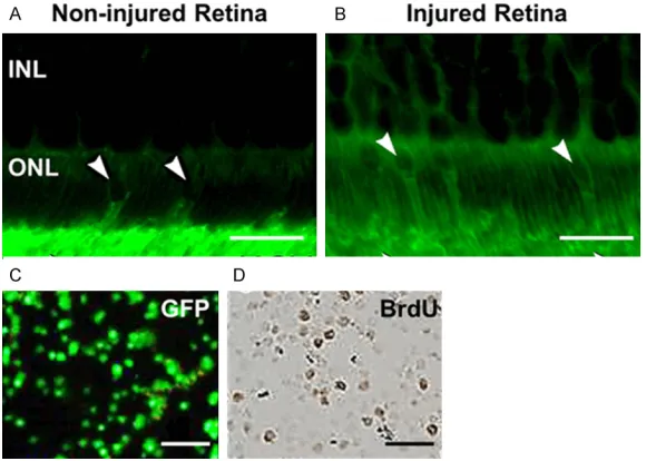
second-Figure 1. Donor rod cells migrate into ONL of recipients and survive post-transplantation. (A, B) GFP-labeled transplanted retinal cells substantially migrate into the ONL of the host retina more promiscuously (B), in compari-son to non-inured retina. (A) (white arrows). Scale bar represents 100 µm. Shown are representative images at 12 weeks post-transplantation. (C, D) BrdU positive staining (D) confirms survivability of the donor rod cells in ONL (green, C). ONL, outer nuclear layer; INL, inner nuclear layer. Scale bar: 20 μm.
enhances green fluorescent protein (eGFP) mice (C57BL/6 background) (Shanghai Labo- ratory Animal Center, Shang- hai, China) were housed in the animal facility at General Hospital of People’s Libera- tion Army in a 12 hours light-dark cycle with water and food provided ad libitum. All experiments were conducted in accordance with the ARVO Statement for the Use of Animals in Ophthalmic and Vision Research, and were approved by the Institute Animal Care and Use Com- mittee of General Hospital of People’s Liberation Army. Dissociation of retinal cells, culture, and transplantation
[image:2.612.89.379.74.281.2]previ-ary antibody for 2 hours at room temperature, washed. Negative controls omitted the pri- mary antibody. Antibodies used: rabbit anti-GFP (1:100; Clontech), mouse anti-rhodopsin (1:100; Chemicon Temecula), rabbit anti-recov-erin (1:1,000; Chemicon), mouse anti-bassoon (Stressgen), and goat anti-protein kinase C alpha (PKCα; 1:100; Santa Cruz). Sections were imaged using a Zeiss LSM510 confocal micro-scope. XY optical sections, ~0.5-μm apart, were taken throughout the depth of the section and built into a stack to give an XY projection image. Cell proliferation levels were detected by bromodeoxyuridine (BrdU). BrdU was admin-istered intraperitoneally at 0.1 mg/g of body-weight into host mice 10 weeks post-trans- plantation (two weeks before they were eutha-nized). BrdU-incorporated cells were subse-quently detected with a BrdU Staining Kit (Life Technologies, Carlsbad, California, USA). Cell counts
Cell count was done as described previously [2, 4] three months after transplantation. Trans- planted GFP+ photoreceptors were counted as integrated if the whole cell body was correctly located within the ONL and at least one of spherule synapse, inner/outer processes and/ or inner segments was visible [2]. The average number of integrated cells per section was determined by counting all the integrated GFP-positive cells in every 1 in 4 serial sections through the site of injection in each eye. This
was multiplied by the total number of sections that encompassed the injection site to give an estimate of the mean number of integrated cells per eye (six eyes were sampled per group). Cell counts for individual eyes were only exclud-ed if there were cells in the vitreous, in turn indicating accidental intravitreal transplanta-tion of the cells.
Statistical analysis
Data are presented as means ± SEM (standard error of the mean), unless otherwise stated; N, number of animals; n, number of eyes or sec-tions examined, where appropriate. A p value of <0.05 was considered statistically significant. Results
Our aim was to confirm that laser-induced reti-nal damage can promote migration and suc-cessful integration of transplanted retinal pro-genitor cells (RPCs) in to the outer retina, which has been previously established [4], and if the integrated RPCs can survive over 12 weeks. For the same expanded passage five donor RPCs from post natal day 1 GFP transgenic were transplanted to the sub retinal space of recipient host adult B6 mice. The total number of integrated GFP+ cells were quantified at 4 weeks (data not shown) and 12 weeks and the increased cell migration and integration was observed in all animals with induced laser pho-tocoagulation; however, we did not observe sig-nificant integration of RPCs into ONL of
unin-Figure 2. Co-localization of rhodopsin and recoverin in the GFP+ donor photoreceptors. The integrated GFP+ donor
[image:3.612.89.529.74.262.2]jured retina at either 4 or 12 weeks post-trans-plantation (102 ± 12 vs. 83 ± 3 cells per eye; P = 4.2138, ANOVA) (Figure 1A, 1B and data not shown). Importantly, the integrated RPCs sur-vived at 12 weeks as assessed by BrdU stain-ing, which showed mitotic activity reminiscent of RPCs (Figure 1C, 1D).
Even though only a portion of integrated cells developed morphological features reminiscent of mature photoreceptors at 4 weeks, their numbers increased significantly at 12 weeks. Almost the entire population (93 ± 4.2%) of surviving integrated cells expressed photore-ceptor associated markers at 12 weeks (Figure 2A-C and Supplementary Figure 1). Rhodop- sin expression was exclusively seen on the outer segments of adult mice retina (Supple- mentary Figure 1).
Finally, to assess if functional synaptic connec-tivity was maintained in the surviving RPCs at 12 weeks, immunofluorescence analysis was performed. As shown in Figure 3A, transplant-ed RPCs elaborattransplant-ed complete morphology as evident by distinct migration of GFP+ cells in the IS and OS, axon, and spherule synapse. Fur- thermore, the integrated cells showed robust expression of the ribbon synapse protein,
bas-are functionally and anatomically rightly posi-tioned within the recipient retina, similar to pre-vious observations [1-5]. Cumulatively, our results indicate that appropriately integrated photoreceptors from autologous sources can survive for extended periods of time, at least up to 12 weeks as tested here, in the adult host retina.
In conclusion, we have shown here that trans-planted rod precursor cells can survive for extended periods of time in the adult murine retina and display all the morphological charac-teristics of correctly integrated rod photorecep-tors. Even though our findings suggest that autologous donor cells are likely to be optimal for therapeutic approaches to repair the neural retina, it would be interesting and important to determine if non-autologous cells may also be effective.
Acknowledgements
This study was supported by Chinese PLA Medical Science Youth Development Program (13QNP090).
Disclosure of conflict of interest
[image:4.612.90.378.73.278.2]None.
Figure 3. Integrated mature rod photoreceptors elaborate complete morphol-ogy and formation of morphological synaptic connections between bassoon protein and PKCα-labeled bipolar cells. Transplanted GFP+, shown in green
throughout, that integrated into the ONL make inner (IS) and outer segments (OS), an axon, and a spherule synapse (Sph) (A, Z-stack) Scale bar: 200 μm. The spherule synapse of integrated cells has a single ribbon that labels for bassoon (B, arrow; 1-μm optical section). Synaptic terminals appose directly to rod bipolar processes immunolabeled for PKCα (C, Z-stack; 1-μm optical section).
soon (Figure 3B) [4]. Finally, the synaptic contact was intact with bipolar neurons as evident by colocalization of protein kinase C alpha (PKCα) (Figure 3C).
Discussion
Photoreceptor transplantation presents a prospective strate-gy of treatment for retinal degeneration [2, 5]. However, the survivality of RPCs has not been previously estab-lished. Our experiments show that the integrated rod photo-receptors can survive for sev-eral months following sub reti-nal transplantation. These ce- lls maintain normal rod mor-phology, including inner/outer segments and spherule syn-apses and express compo-nents of the synaptic ma- chinery.
Address correspondence to: Dr. Qinjian Peng, Hu- angsi Aesthetic Surgery Hospital, 9 Huangsi Street, Beijing 100011, China, Tel: +86-10-66352465; Fax: +86-10-66352465; E-mail: [email protected]
References
[1] West EL, Pearson RA, Barker SE, Luhmann UF, Maclaren RE, Barber AC, Duran Y, Smith AJ, Sowden JC, Ali RR. Long-term survival of photo-receptors transplanted into the adult murine neural retina requires immune modulation. Stem Cells 2010; 28: 1997-2007.
[2] MacLaren RE, Pearson RA, MacNeil A, Douglas RH, Salt TE, Akimoto M, Swaroop A, Sowden JC, Ali RR. Retinal repair by transplantation of photoreceptor precursors. Nature 2006; 444: 203-7.
[3] Klassen H, Kiilgaard JF, Zahir T, Ziaeian B, Kirov I, Scherfig E, Warfvinge K, Young MJ. Progenitor cells from the porcine neural retina express photoreceptor markers after trans-plantation to the subretinal space of allorecipi-ents. Stem Cells 2007; 25: 1222-30.
[4] Jiang C, Klassen H, Zhang X, Young M. Laser injury promotes migration and integration of retinal progenitor cells into host retina. Mol Vis 2010; 16: 983-90.
[5] Pearson RA, Hippert C, Graca AB, Barber AC. Photoreceptor replacement therapy: challeng-es prchalleng-esented by the diseased recipient retinal environment. Vis Neurosci 2014; 31: 333-44. [6] Jiang LQ, Jorquera M, Streilein JW. Subretinal
space and vitreous cavity as immunologically privileged sites for retinal allografts. Invest Ophthalmol Vis Sci 1993; 34: 3347-3354. [7] Ghosh F, Arner K. Transplantation of
full-thick-ness retina in the normal porcine eye: Surgical and morphologic aspects. Retina 2002; 22: 478-486.
[8] Ghosh F, Bruun A, Ehinger B. Graft-host con-nections in long-term full-thickness embryonic rabbit retinal transplants. Invest Ophthalmol Vis Sci 1999; 40: 126-132.
[9] Ghosh F, Engelsberg K, English RV, Petters RM. Long-term neuroretinal full-thickness transplants in a large animal model of severe retinitis pigmentosa. Graefes Arch Clin Exp Ophthalmol 2007; 245: 835-846.