Original Article
Distribution of phosphorylated cyclic AMP response
element binding protein (p-CREB-1) in rat retina
Jianfa Huang*, Pei Chen*, Jingzhi Yu, Lijun Xu, Ying Yang, Nandan Wu, Mingjun Tan, Jian Ge, Keming Yu, Jing Zhuang
State Key Laboratory of Ophthalmology, Zhongshan Ophthalmic Center, Sun Yat-sen University, Guangzhou, Guangdong, P. R. China. *Equal contributors.
Received October 11, 2016; Accepted October 25, 2016; Epub February 1, 2017; Published February 15, 2017
Abstract: Purpose: The phosphorylation of cyclic AMP (cAMP) response element binding protein (CREB) is essential to the physiology of neuron development. In the present study, we sought to determine the expression profile and specific cell distribution of p-CREB-1 in the rat retina. Methods: Double immunofluorescence staining was employed to determine the developmental expression pattern and specific distribution of p-CREB-1 in adult rat retina. Results: Our data implicate a developmental expression pattern of both CREB-1 and p-CREB-1 in rat retina. The p-CREB-1 was prominently expressed in retinal neurons and rarely expressed in retinal glial cells, as evidenced by immuno-fluorescence assay. Conclusion: The developmental phosphorylation pattern and distinct expression localization of p-CREB-1 in rat retina suggest a physiological role for p-CREB-1 in the retinal development.
Keywords: p-CREB-1, retina neurocytes, retina development
Introduction
CREB-1, a pleiotropic leucine zipper transcrip-tion factor, mediates important mechanisms governing several physiological functions of the central nervous system ranging from develop-ment to plasticity and disease [1, 2]. The can- onical pathway that leads to the activation of CREB-1 by phosphorylation of the Ser133 resi-due [3] has been implicated in the regulation of proliferation, differentiation, and neurogenesis in the developing neuron system [4, 5], and mediating synaptic plasticity, long-term memo-ry formation and consolidation in the adult brain [6]. Its activity is triggered by increased intracellular levels of cAMP and Ca2+ influx in response to neurotransmitters, hormone stim-ulationand neuronal activity [7, 8]. Triggering this pathway subsequently initiates the tran-scription of multiple target genes [9]. During mammalian development neuronal activity, and possibly Ca2+ influx, is critical to the forma-tion of synaptic connecforma-tions, suggesting that phosphorylated CREB-1 (p-CREB-1) is involved in this physiological process [7, 10].
In embryonic cortical neurons, neuronal activity induces transcription of neuro-protective
pro-teins such as Bcl-2 and brain-derived neuro-trophic factor (BDNF) by a p-CREB-1-dependent mechanism [11]. In the adult hippocampus, CREB-1 phosphorylation occurs in response to a diverse array of stimuli and has been shown to enhance cell survival and neurogenesis [12]. Moreover, several lines of evidence suggest that p-CREB-1 integrates contextual informa-tion, regulates the repertoire of retinal precur-sor cells and acts as a neuronal survival mod-eratorduring retinal development [13, 14]. In vivo evidence indicates that decreased phos-phorylation levels of CREB-1 are associated with retinal degeneration [15, 16]. Yet the physi-ological role of p-CREB-1 in the regulation of retina development remains unclear.
Distribution of p-CREB-1 in rat retina
Several intracellular signaling cascades that phosphorylated nuclear transcriptional factors are involved in these sophisticated processes [7, 14]. Identifying in which cells the transcrip-tional factors have beenphosphorylated might facilitate the understanding of its biological roles. However, the distribution of these tran-scriptional factors such as p-CREB-1 has not been reported.
Given the importance of p-CREB-1 in neuron development, it is likely that this transcription factor is also involved in the regulation of retina development. Therefore, the expression profile and cell location of p-CREB-1 was immuno-chemically evaluated in rat retina under normal conditions by using double-staining assay. The new insight of this study might shed light on the potential involvement of p-CREB-1 inretinal development.
Methods
Animals
All experiences involving animals strictly adhered to the ARVO Statement for the Use of Animals in Ophthalmic and Vision Research and the procedure was approved by the In- stitutional Animal Care and Use Committee of Zhongshan Ophthalmic Center (Permit Number: SYXK (YUE) 2010-0058). The rats were housed in Ophthalmic Animal Laboratory of Zhongshan Ophthalmic Center, at Sun Yat-sen University, in an air-conditioned room with an ambient tem-perature of 16-26°C, a relative humidity of 40-70% and a 12-hour light-dark cycle. Food and water were available ad libitum and animal
health was monitored by the animal care staff and veterinarian. The Sprague-Dawley (SD) rats were sacrificed by an intra-peritoneal injection of chloral hydrate (P3761, 60 mg/kg) (Sigma,St. Louis, MO) before we harvested the eyes.
Tissue preparation
The rats were anesthetized with chloral hydrate (5 mg/ml) and perfused with physiological saline and then 4% paraformaldehyde. After- wards, the eyes were harvested and fixed in 4% paraformaldehyde overnight. Then, the eyes were dehydrated with graded sucrose solution and embedded in optimal cutting temperature (OCT) compound (Tissue-Tek, California, USA). Subsequently, the transverse sections of the rats’ eyes were cut by using a microtome (Thermo Electron Corporation, Cheshire, UK), and mounted onto glass slide (CITOTEST, Jiangsu, CHINA), at a section thickness of 10 μm, and stored at -20°C until processed.
Immunofluorescence assay
Immunofluorescence was used to determine the specific location of p-CREB-1 in the mature retina. For immunofluorescence staining, the slides with tissue sections were washed with phosphate buffer saline (PBS) and permeabi-lized by 0.5% Triton X-100 (Sigma, St Louis, MO, USA) for 10 min, thenblocked with 10% normal goat serum (Boster, Wuhan, China) for 30 min. The sections were incubated with primary anti-bodies (Table 1) in a humidified chamber at 4°C overnight. After being washed with PBS for 15 min the secondary antibody (CST, Danvers, MA) was added to the sections at room
tem-Table 1. List of primary antibodies used in this study
Antigen Host Antibody source Working concentration
CREB-1 Rabbit CST 9197s 1:200
P-CREB-1 Mouse Millpore 2361835 1:200
Protein kinase C, a isoform (PKC-α) Rabbit CST 2056 1:200
Microtubule-associated protein-2 (MAP-2) Rabbit Abcam ab96378 1:200
THY-1.1 Rabbit Millpore MAB1406 1:200
Calretinin Rabbit Boster BA0681 1:100
Calbindin Rabbit Boster PB0017 1:100
Glutamine synthetase (GS) Rabbit Abcam ab49873 1:200
Glial fibrillary acidic Protein (GFAP) Rabbit Boster BA0056 1:100
[image:2.612.92.526.85.218.2]perature as required, and the nuclei were stained with DAPI. Sections were rinsed with water and mounted with Fluoromount G (eBio-science, San Diego, USA). Photomicrographs
[image:3.612.91.522.71.579.2]were taken by using the LSM 5 Pascal confocal laser scanning microscope (Zeiss, Oberkochen, Germany). Images of the retinal sections were collected at 400× and 800× magnification.
Distribution of p-CREB-1 in rat retina
Results
The developmental expression profile of CREB-1 and P-CREB-CREB-1 in rat retina
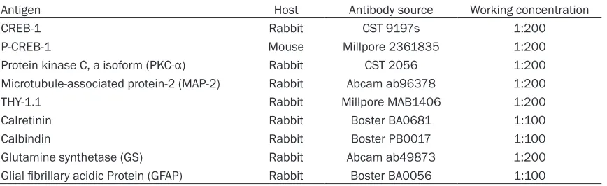
In the central nervous system, almost every neuronal and glial cell appears to exhibit CREB-1 expression [CREB-18]. The activity of CREB-CREB-1 is trig-gered by signaling cascades that result in phos-phorylation of CREB-1 in differentiated cells [19, 20]. Here, we analyzed the developmental expression profile of CREB-1 and p-CREB-1 in therat retinaat different time points by using a double-immunochemical assay. The p-CREB-1 antibody recognizes the peptide sequence con-taining the phosphorylated Ser133. Represen- tative microscopic pictures demonstrated that
both the CREB-1 and p-CREB-1 are expressed in the nuclei of each cell. No staining was observed in the sense controls (datanot shown). Varying intensities of CREB-1 and p-CREB-1 staining were observedin the retina at different developmental stages (Figure 1). As shown in
[image:4.612.93.520.73.409.2]Figure 1A, at postnatal day 1, CREB-1 was con-stitutively distributed in the all layers of the retina; however, with ongoing retina develop-mentits expressionwas down-regulated in the outer nuclear layer (ONL) and gradually restrict-ed to only the ganglion cell layer (GCL) and inner nuclear layer (INL). In contrast, p-CREB-1 was almost exclusively expressed inthe GCL of post-natal day 1 rats and profoundly up-regulated in the GCL and INL of the mature rat retina.
Interestingly, peak expression of p-CREB-1 in the INL was observed at postnatal day 5 and slightly decreased with age (Figure 1A). These temporal and spatial developmental expres-sion patterns of CREB-1 and p-CREB-1 are in accordance with the proposed function as a regulator for retina development signals. Moreover, higher-magnification images of adult rat retina demonstrated that (Figure 1B), about half of the retinal cells in the GCL and INL that expressed CREB-1 were immuno positive for p-CREB-1 regardless of its phosphorylation state. This expression pattern is similar to that observed in cortex [18]. Together, these lines of evidence indicate that the phosphorylation of CREB-1 might contribute to the differentiation of retina neurocytes.
Immunolocalization of p-CREB-1 in the adult
rat retina neurons
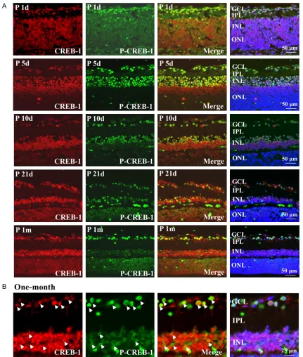
The vertebrate retina consists of seven major neuron cell types. To determine the exact distri-bution of p-CREB-1 in retinal cells, double immunofluorescence staining was performed with specific markers of major neuronal pheno-types in the fully developed postnatal one-month rat retina. Given the lack of p-CREB-1 distribution in the ONL layer, the cellular analy-sis was restricted to the GCL and INL neuro- cytes.
The intensive staining of p-CREB-1 in the GCL suggested positive phosphorylation of CREB-1 in retinal ganglion cells (RGCs). The retinal neu-rons were labeled with microtubule-associated protein-2 (MAP-2) or THY1.1, the specific cellu-lar makers for neurons and retinal ganglion cells, respectively. As shown in Figure 2A and
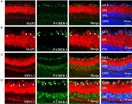
2B, the MAP-2 stained neuronal axons and cell bodies were present in the GCL and IPL of reti-na. Thy1.1 expression was distributed specifi-cally in ganglion cells (Figure 2C and 2D), which were strongly expressed in the membrane and cytoplasm of the cells in the GCL and IPL. It was apparent that the cellular staining of p-CREB-1 co-localized with both MAP-2 and Thy1.1 Anti-protein kinase C-α (PKC-α) was used to label rod ON-bipolar cells and a subpopulation of amacrine cells [21]. As shown in Figure 3A
and 3B, the PKC-α staining was distributed in cytoplasm of the cells in the inner and outer parts of the INL, and the synaptic terminals of the IPL. No clear co-localization was detected between PKC-α and p-CREB-1. Horizontal cells localized in the outer parts of the INL, exhibiting dense plexus formed calbindin labelling, were not p-CREB-1 immunoreactive (Figure 4A and
4B). However, calretinin-positive amacrine cells presented in the inner the INL and GCL, re- vealed only a few p-CREB-1 stained cells (Figure 4C and 4D). Taken together, these observa-tions demonstrate that p-CREB-1 was
[image:5.612.93.519.71.258.2]Distribution of p-CREB-1 in rat retina
nently expressed in the retinal neurons, but not in all neuron cell types of the retina.
Immunolocalization of p-CREB-1 in the adult
rat retina glial cells
To assess the expression of p-CREB-1 in retinal glial cells, double labelling experiments were performed for p-CREB-1 with glial fibrillary acid-ic protein (GFAP) and glutamine synthetase (GS), markers for astrocytes and Muller cells, respectively. In one-month old rat retina, GFAP was expressed in the outer parts of the GCL, and no co-localization between GFAP and p-CREB-1 were observed (Figure 5A and 5B). Additionally, the Muller cells, which were labe- lled using an antibody against GS that span the entire neuro-retina, were not p-CREB-1 immu-noreactive (Figure 5C and 5D). This evidence
indicates that p-CREB-1 was rarely expressed in the rat retinal glial cells.
Discussion
[image:6.612.92.522.73.393.2]In the present study, we first demonstrated the developmental phosphorylation of CREB-1 in rat retina; this suggests a regulator role for p-CREB-1 in retinal developmentand cell class-es. Moreover, in adult rat retina, p-CREB-1 was prominently expressed in the retinal neurons while rarely expressed in the retinal glial cells. The phosphorylation of CREB-1 plays a vital role at a central converging point of pathways mediating the developmental regulated pro-cesses in neurons, including cell differentia-tion, synaptic connections and neurogenesis [1, 2, 22]. Here, we demonstrated that CREB-1 was markedly expressed and extensively
tributed in all layers of the retinathat are still undergoing development in postnatal day one rats, and developmentally down-regulated in the ONL as the rats aged. Conversely, p-CREB-1 expression was predominated in the GCL in the postnatal dayone rat retina, while strong immu-nore activeactivity of p-CREB-1 was observed in the GCL and INL of the one-month old rat retina. This observation is consistent with previ-ously reported patterns of p-CREB-1 staining in adult retina of mouse, dog and cat [23, 24]. As in other parts of the nervous system, various physiological stimuli or cellular stress, such as peptide hormones, growth factors and light exposure [25-27], could induce the phosphory-lation of CREB-1 in retinal neurons. The phos-phorylation of CREB-1 mediates the transcrip-tion of neurotrophic factors and triggers intra-cellular signaling pathways, there by
modulat-ing the proliferation and dictatmodulat-ing the differen-tiation of neurocytes. The developmental phos-phorylation of CREB-1 in the retina observed in the present study indicates that CREB-1 might-be involved in the physiological process of the development of retinal cell classes and archi-tecture as well as maintaining neuronal properties.
[image:7.612.92.520.73.394.2]To identify the differentiation processes of cells that p-CREB-1 has participated in, we analyzed the expression distribution of p-CREB-1 in the adult rat retina. Based on the staining pattern with p-CREB-1 antibody and retinal cell mark-ers antibody, our data indicates that, although every neuronal and glial cell throughout all reti-nal layers appears to exhibit CREB-1 immunore-activity, p-CREB-1 was prominently expressed in the retinal neurons but rarely expressed in
Distribution of p-CREB-1 in rat retina
the retinal glial cells. These observations are consistent with previous reports demonstrating that p-CREB-1 is predominantly expressed in post-mitotic cells [18-20, 28]. Persistent and strong activation of CREB-1 phosphorylation is associated with neuronal survivalsinceit has been suggested to provide neuroprotective sig-nals in times of cellular stress [29]. A close rela-tionship between CREB-1 phosphorylation and neuronal survival has been reported [30, 31]. Drug-induced phosphorylation of CREB-1 pro-motes proliferation and morphological matura-tion of neurons [32, 33]. Applicamatura-tion of lithium, a well-known neuroprotective agent, induces marked enhancement of CREB-1 phosphoryla-tion in the cortex [33]. In line with these stud-ies, our results suggest strong involvement of p-CREB-1 in the development of retinal neuro-cytes. Moreover, in adult rat retina, phosphory-lated-CREB-1 expression in CREB-1 immuno-postive cells was observed. Such moderate activation of CREB-1 may be necessary for the basal maintenance of signal transduction to ensure neuronal integrity [2, 4]. Additionally, given that there is strong phosphorylation of CREB-1 in the retinal neurons, not glial cells, in the GCL and INL during development; it is likely that CREB-1 controls the neurogenic transcrip-tional programs that regulate the repertoire of retinal precursor cells. Neuron cell proliferation and differentiation are sophisticated process-es, coordinated by several extrinsic factors and neurotrophins. Although CREB-1 phosphoryla-tion mediates the transcripphosphoryla-tion signaling of neurotrophin factors, considerable evidence suggests that in developing neurocytes, neuro-trophin factors could enhance the phosphoryla-tion of CREB-1 [26]. Thus, populaphosphoryla-tions of retina cells expressing p-CREB-1 could be either posi-tively or negaposi-tively selected in retinal develop-ment. However, the mechanism underlying the involvement of p-CREB-1 mediated differentia-tion in retina, and whether the phosphoryladifferentia-tion level of CREB-1 is associated with cell direc- tional differentiation, requires further investi- gation.
Zuo et al. suggested that restoring CREB-1 phosphorylation in the retina might be a poten-tial therapeutic strategy to prevent excitotoxic retinal degeneration [33]. Moreover, under the normal circumstances, p-CREB-1 is primarily expressed in the GCL and INL of the adult rat retina, while weakly expressed in the ONL where it is mostly constituted of
photorecep-tors. However, strong activation of CREB-1 phos- phorylation in photoreceptor cells in response to injury stress was observed in previous stud-ies [23, 34]. In photoreceptor degeneration ani-mals and ocular penetrating trauma animal models, p-CREB-1 immunostaining is promi-nent in the ONL [34, 35]. Mónicaet al. reported that Müller-derived progenitors respond to NMDA receptor activation through CREB-1 phosphorylation [36]. Although p-CREB-1 is weakly expressed in the ONL of rat retina, these lines of evidence suggest that p-CREB-1 might be associated with a neuroprotective response in retina cells in the ONL.
In conclusion, the present study reveals the dis-tinct expression localization of p-CREB-1 in rat retina, demonstrating that p-CREB-1 might be involved in the physiological development pro-cesses of retinal cell classes and architecture. Clearly, further investigation is needed to explore the underlying mechanisms by which p-CREB-1 mediates the development process-es of the retina. Collectively, this study not only gives a new insight into the regulation of p-CREB-1 in retinal development, but it might also offer a therapeutic target to currently untreatable retinal diseases.
Acknowledgements
All the studies were funded by the grants from the National Natural Science Foundation (Project: 81070732, 81470626).
Disclosure of conflict of interest
None.
Address correspondence to: Jing Zhuang, State Key Laboratory of Ophthalmology, Zhongshan Ophthal- mic Center, Sun Yat-sen University, 54 Xianlienan Road, Guangzhou 510060, Guangdong, P. R. China. Tel: +86-20-87330290; Fax: +86-20-87330341; E-mail: [email protected]
References
[1] Sakamoto K, Karelina K, Obrietan K. CREB: a multifaceted regulator of neuronal plasticity and protection. J Neurochem 2011; 116: 1-9. [2] Lonze BE, Ginty DD. Function and regulation of
CREB family transcription factors in the ner-vous system. Neuron 2002; 35: 605-23. [3] Lee B, Butcher GQ, Hoyt KR, Impey S, Obrietan
cAMP response element-binding protein (CREB): kinase coupling, stimulus intensity, and temporal regulation of CREB phosphoryla-tion at serine 133. J Neurosci 2005; 25: 1137-1148.
[4] Fujioka T, Fujioka A, Duman RS. Activation of cAMP signalling facilitates the morphological maturation of newborn neurons in adult hippo-campus. J Neurosci 2004; 24: 319-328. [5] Socodato R, Brito R, Calaza KC,
Paes-de-Carvalho R. Developmental regulation of neu -ronal survival by adenosine in the in vitro and in vivo avian retina depends on a shift of sig-naling pathways leading to CREB phosphoryla-tion or dephosphorylaphosphoryla-tion. J Neurochem 2011; 116: 227-39.
[6] Josselyn SA, Shi C, CarlezonWA Jr, Neve RL, Nestler EJ, Davis M. Long-term memory is fa -cilitated by camp response element-binding protein overexpression in the amygdala. J Neurosci 2001; 21: 2404-12.
[7] Shaywitz AJ, Greenberg ME. CREB: a stimulus-induced transcription factor activated by a di-verse array of extracellular signals. Annu Rev Biochem 1999; 68: 821-61.
[8] Kornhauser JM, Cowan CW, Shaywitz AJ, Dolmetsch RE, Griffith EC, Hu LS, Haddad C, Xia Z, Greenberg ME. CREB transcriptional ac-tivity in neurons is regulated by multiple, calci-um-specific phosphorylation events. Neuron 2002; 34: 221-33.
[9] Mayr B, Montminy M. Transcriptional regula-tion by the phosphorylaregula-tion-dependent factor CREB. Nat Rev Mol Cell Biol 2000; 2: 599-609. [10] Blanquet PR, Mariani J, Derer P. A calcium/ calmodulin kinase pathway connects brain-derived neurotrophic factor to the cyclic AMP-responsive transcription factor in the rat hip-pocampus. Neuroscience 2003; 118: 477-90. [11] Giachino C, De Marchis S, Giampetro C, Parlato
R, Perroteau I, Schütz G, Fasolo A, Peretto P. cAMP response element-binding protein regu-lates differentiation and survival of newborn neurons in the olfactory bulb. J Neurosci 2005; 25: 10105-18.
[12] Peltier J, O’Neill A, Schaffer DV. PI3K/Akt and CREB regulate adult neural hippocampal pro-genitor proliferation and differentiation. Dev Neurobiol 2007; 67: 1348-61.
[13] Nagai-Kusuhara A, Nakamura M, Mukumo H, Kanamori A, Negi A, Seigel G. cAMP-responsive element binding protein mediates a cGMP/ protein kinase G-dependent anti-apoptotic sig-nal induced by nitric oxide in retisig-nal neuron-glial progenitor cells. Exp Eye Res 2007; 84: 152-62.
[14] Socodato R, Brito R, Portugal CC, de Oliveira NA, Calaza KC, Paes-de-Carvalho R. The nitric oxide-cGKII system relays death and survival
signals during embryonic retinal development via AKT-induced CREB1 activation. Cell Death Differ 2014; 21: 915-28.
[15] Mali RS, Zhang XM, Chintala SK. A decrease in phosphorylation of cAMP-response element- binding protein (CREBP) promotes retinal de-generation. Exp Eye Res 2011; 92: 528-36. [16] Santos RC, Araujo EG. Cyclic AMP increases
the survival of ganglion cells in mixed retinal cell cultures in the absence of exogenous neu-rotrophic molecules, an effect that involves cholinergic activity. Braz J Med Biol Res 2001; 34: 1585-1593.
[17] Martins RA, Pearson RA. Control of cell prolif-eration by neurotransmitters in the developing vertebrate retina. Brain Res 2008; 1192: 37-60.
[18] Tanaka K, Nagata E, Suzuki S, Dembo T, Nogawa S, Fukuuchi Y. Immunohistochemical analysis of cyclic AMP response element bind-ing protein phosphorylation in focal cerebral ischemia in rats. Brain Res 1999; 818: 520-6. [19] Liu FC, Graybiel AM. Spatiotemporal dynamics
of CREB phosphorylation: transient versus sustained phosphorylation in the develop-ing striatum. Neuron 1996; 17: 1133-44. [20] Nakagawa S, Kim JE, Lee R, Chen J, Fujioka T,
Malberg J, Tsuji S, Duman RS. Localization of phosphorylated cAMP response element-bind-ing protein in immature neurons of adult hip-pocampus. J Neurosci 2002; 22: 9868-76. [21] Huang J, Zhou L, Wang H, Luo J, Zeng L, Xiong
K, Chen D. Distribution of thrombospondins and their neuronal receptor α2δ1 in the rat retina. Exp Eye Res 2013; 111: 36-49. [22] Lonze BE, Riccio A, Cohen S, Ginty DD.
Apop-tosis, axonal growth defects, and degeneration of peripheral neurons in mice lacking CREB. Neuron 2002; 34: 371-385.
[23] Geller SF, Lewis GP, Fisher SK. FGFR1, signal-ing, and AP-1 expression after retinal detach-ment: reactive Muller and RPE cells. Invest Ophthalmol Vis Sci 2001; 42: 1363-1369. [24] Beltran WA, Allore HG, Johnson E, Towle V, Tao
W, Acland GM, Aguirre GD, Zeiss CJ. CREB1/ ATF1 activation in photoreceptor degeneration and protection. Invest Ophthalmol Vis Sci 2009; 50: 5355-63.
[25] Socodato R, Brito R, Calaza KC, Paes-de-Carvalho R. Developmental regulation of neu -ronal survival by adenosine in the in vitro and in vivo avian retina depends on a shift of sig-naling pathways leading to CREB phosphoryla-tion or dephosphorylaphosphoryla-tion. J Neurochem 2011; 116: 227-39.
(CRE)-Distribution of p-CREB-1 in rat retina
binding protein and increases DNA-binding ac -tivity of CRE in rat retina. J Pharmacol Sci 2004; 95: 108-114.
[27] Choi JS, Kim JA, Joo CK. Activation of MAPK and CREB by GM1 induces survival of RGCs in the retina with axotomized nerve. Invest Ophthalmol Vis Sci 2003; 44: 1747-1752. [28] Jagasia R, Steib K, Englberger E, Herold S,
Faus-Kessler T, Saxe M, Gage FH, Song H, Lie DC. GABA-CREB signalling regulates matura -tion and survival of newly generated neurons in the adult hippocampus. J Neurosci 2009; 29: 7966-77.
[29] Tanaka K, Nogawa S, Ito D, Suzuki S, Dembo T, Kosakai A, Fukuuchi Y. Activated phosphoryla-tion of cyclic AMP response element binding protein is associated with preservation of stria-tal neurons after focal cerebral ischemia in the rat. Neuroscience 2000; 100: 345-54. [30] Finkbeiner S. CREB couples neurotrophin
sig-nals to survival messages. Neuron 2000; 25: 11-4.
[31] Nagai-Kusuhara A, Nakamura M, Mukuno H, Kanamori A, Negi A, Seigel GM. cAMP-respon-sive element binding protein mediates a cGMP/protein kinase G-dependent anti-apop-totic signal induced by nitric oxide in retinal neuron-glial progenitor cells. Exp Eye Res 2007; 84: 152-62.
[32] Grimes CA, Jope RS. CREB DNA binding activity is inhibited by glycogen synthase kinase-3 beta and facilitated by lithium. J Neurochem 2001; 78: 1219-32.
[33] Zuo D, Lin L, Liu Y, Wang C, Xu J, Sun F, Li L, Li Z, Wu Y. Baicalin attenuates ketamine-induc- ed neurotoxicity in the developing rats: in- volvement of PI3K/Akt and CREB/BDNF/ Bcl-2 pathways. Neurotox Res 2016; 30: 159-72.
[34] Harada T, Imaki J, Hagiwara M, Ohki K, Takamura M, Ohashi T, Matsuda H, Yoshida K. Phosphorylation of CREB in rat retinal cells af-ter focal retinal injury. Exp Eye Res 1995; 61: 769-72.
[35] Guo XJ, Tian XS, Ruan Z, Chen YT, Wu L, Gong Q, Wang W, Zhang HY. Dysregulation of neuro -trophic and inflammatory systems accompa -nied by decreased CREB signaling in ischemic rat retina. Exp Eye Res 2014; 125: 156-63. [36] Ramírez M, Lamas M. NMDA receptor medi