ABSTRACT
Luciana E. Leopold: Surveillance and Targeting of Aberrant Transcripts in Caenorhabditis elegans
(Under the direction of Shawn Ahmed)
An organism requires mechanisms with which to protect the fidelity and integrity of the genome, transcriptome, and proteome in order not only to remain healthy and alive, but also to pass on viable genetic material to its offspring. To this end, the cell is constantly surveying the transcripts being expressed. When aberrant transcripts are produced, whether they be from endogenous or exogenous sources, mechanisms are required in order to silence or eradicate such messages before they become toxic to the cell. Two such mechanisms are nonsense‐mediated mRNA decay (NMD), which targets transcripts bearing a premature termination codon (PTC), and RNA‐induced epigenetic silencing (RNAe), which silences foreign loci in the genome in a heritable, stable manner.
allele. In the next example, we show that permanent, stable transgene silencing like that generated by RNAe in a single generation, can be attained in certain transgenes only after several generations of successive crossing. These processes are molecularly similar, as they both require the Argonaute protein PRG‐1. However, they differ in that single‐generation RNAe silencing becomes independent of PRG‐1 after initiation, whereas the silencing we observe in multigenerational RNAe requires PRG‐1 in order to maintain its silent state.
ACKNOWLEDGEMENTS
To my family & friends – thank you for putting up with me during this process. I know it hasn’t been easy, but I am very lucky to have so many people in my life who support me. I am grateful for all of you every single day. To my labmates, who are like family and friends combined – thank you for your constant teamwork, I would never have made it without you. To my mentor – Shawn, you are a brilliant man full of ideas. I hope you never lose your zest for science. Finally, to perseverance.
TABLE OF CONTENTS
ABSTRACT...iii
ACKNOWLEDGEMENTS ...v
LIST OF ABBREVIATIONS...x
INTRODUCTION... 1
Figures ... 9
CHAPTER 1: SMG1(YP3) TARGETS UNUSUAL TRANSCRIPTS ...13
Background...13
Results...15
Discussion...23
Materials and Methods ...27
Figures ...30
CHAPTER 2: LACK OF PAIRING DURING MEIOSIS TRIGGERS MULTIGENERATIONAL TRANSGENE SILENCING IN CAENORHABDITIS ELEGANS...37
Background...37
Results...40
Discussion...48
Materials and Methods ...52
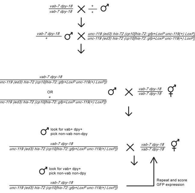
Figure 2.7. Schema used to cross the his72::GFP transgene using
Chromosome III markers vab7 dpy18. ...61
Figure 2.8. Widefield microscopy depicting bright field (left column) and mCherry fluorescent (right column) images of (top‐bottom) in
LIST OF ABBREVIATIONS
bp base pairs
DMD Duchenne muscular Dystrophy dsRNA double‐stranded RNA
EMS ethyl methanesulfonate F1/F2/F3 filial generation 1/2/3 GFP green fluorescent protein miRNA micro RNA
MSUD meiotic silencing by unpaired DNA mRNA messenger RNA
N2 wildtype Bristol strain of C. elegans NMD nonsense‐mediated mRNA decay ORF open reading frame
PI3K phosphatidylinositol 3‐kinase piRNA piwi‐interacting RNA
POT1 protection‐of‐telomeres 1 PTC premature termination codon Pvl protruding vulva phenotype RdRP RNA‐dependent RNA polymerase RNAe RNA‐induced epigenetic silencing RNAi RNA interference
siRNA small interfering RNA snRNA small nuclear RNA snoRNA small nucleolar RNA SL1 splice leader 1
SMG1 suppressor with morphological effect on genetalia 1 uORF upstream open reading frame
INTRODUCTION
In order for an organism to remain viable and fulfill its ultimate evolutionary
purpose of passing along its genetic code, it must be able to protect both itself and the code from harm. While behavioral instincts like the ‘fight‐or‐flight’ response, or the ability to camouflage, were being programmed in via evolution across many millennia, molecular protective mechanisms were also emerging. Cells are equipped with numerous pathways that work to ensure the maintenance of genome, transcriptome, and proteome integrity. Each level of information – DNA, RNA, and protein – is vulnerable to attack from various exogenous sources, such as viruses and radiation, as well as endogenous errors that can occur during transcription and translation. Without protective protocols in place, these attacks can be toxic, and have the potential to cause disease and even death. For example, many cancers are caused by mutations in oncogenes resulting from transcriptional errors.
requires small nuclear RNAs (snRNAs) and RNA nucleotide changes mediated by small nucleolar RNAs (snoRNAs) (4, 5). The ends of eukaryotic chromosomes are maintained by telomerase, which requires an RNA template to replicate the telomeric sequence (6). Many of these pathways have been very well described in both their function and in the identities of the protein players involved. However, it is logical to believe that there may exist
variations and nuances of these pathways in order to achieve the level of specificity required to regulate all of the possible targets. Herein I discuss two different projects that concern RNA stability and investigate mechanisms of RNA surveillance and defense.
In Chapter 1, I address a pathway related to nonsense‐mediated decay (NMD), which is a method the cell has for eradicating endogenous abberantly processed mRNAs in the cytoplasm. NMD is a pathway that is highly conserved among eukaryotes and
canonically identifies RNA transcripts bearing a premature termination codon (PTC) and targets them for degradation, thus alleviating the cell of any toxic stress that may result from a faulty transcript (7). These errors may arise either by a mutation in the DNA source code, or by inaccurate processing. In Caenorhabditis elegans, many of the components of NMD have been identified, notably the seven smg genes (8). The SMG‐2 (Upf1) helicase serves as the first alarm bell of NMD and identifies transcripts containing a PTC. SMG‐2 is then phosphorylated by SMG‐1 and downstream activators of NMD are triggered, and various decay pathways come into play to degrade the message (9, 10).
range of protective mechanisms as well. For instance, NMD components like the SMG/UPF proteins have been shown to be involved in responses to damage induced by UV or γ‐ radiation (12), double strand breaks (13), as well as the oxidative stress response to TNF‐ αinduced apoptosis (14).
I specifically investigate an NMD‐related pathway that targets the mRNA transcript of unc43(r293), which in the past has been thought of as a canonical target of NMD.
However, upon observation, it is clear that the molecular structure of the unc54(r293) RNA transcript is not that of a standard RNA with a PTC. Rather, unc54(r293) contains an extra‐ long 3’ untranslated region (3’UTR) which reads through to the next gene, aex5, and may possibly result in a bicistronic message. We have isolated a novel allele of the smg1 gene, smg1(yp3). While smg1(yp3) functions like other known smg1 alleles to stabilize
messages carrying a standard PTC, a long 3’UTR, and even a bicistron, it still degrades the unc43(r293) transcript, and thus suppresses its function. Therefore, smg1(yp3) is a separation‐of‐function allele that defines a pathway that is related to NMD, but is not identical in the way that it processes this specific transcript. I believe that there is possibly a unique molecular trigger contained within the unc54(r293) transcript that requires a variation adjustment in the way that SMG‐1 is utilized by the NMD process in order to be target the transcript for degradation.
hijacks the cell for its own purposes, and uses the native machinery to transcribe its own RNA, which is then translocated to the cytoplasm to be made into viral protein (Figure 0.1). In order to protect itself from foreign sequences or other rogue elements in the DNA, the cell must have some kind of immunity, and it is only more recently that we are really starting uncover how this is done. My work makes several significant contributions towards understanding how the cell identifies and silences foreign intruders.
mRNA is transcribed from the DNA in the nucleus, but is then translated into protein in the cytoplasm, so at any given time, foreign RNA species can be residing in either of these compartments (Figure 0.1). In order to completely rid itself of the threat of foreign DNA taking over the cell, causing chaos, and maybe even disease, the cell must find ways of degrading unwanted RNAs that is found in both locations.
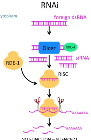
There is a known mechanism which targets and degrades foreign cytoplasmic RNA, called RNA interference, or RNAi (2) (Figure 0.2). Fire and Mello won the Nobel Prize in 2006 for the discovery of RNAi in C. elegans. They uncovered how foreign double stranded RNA in the cytoplasm – which could potentially come from something like a virus, or
something injected in the lab, as Fire and Mello did – is silenced by cellular machinery. First, Dicer, an endoribonuclease, binds to this dsRNA with the help of RDE‐4, a protein known to bind dsRNA, and the RNA is diced into smaller pieces, called siRNAs, which are made
single‐stranded and loaded onto the RDE‐1 Argonaute protein, which is the catalytic component of RISC, a complex of multiple proteins that incorporates this small RNA and is capable of inducing silencing by binding to target mRNA and cleaving it, essentially
This is how the cell takes care of foreign mRNA found in the cytoplasm, but there is another mechanism which targets the mRNA still found in the nucleus. The identification of this process, called RNAe, or RNA‐induced epigenetic silencing, is a newer advance in the field, as it was only discovered in 2012 (Figure 0.3). A series of papers from three different groups describing this pathway were published in close succession (19‐21). In these
studies, a process similar to RNAi was outlined, the difference being that this pathway has the potential to epigenetically mark the DNA itself for silencing, preventing transcription of foreign DNA not only in that particular cell, but any cell it passes its genetic code onto. These groups all saw that the silencing was irreversible and occurred in a single generation. Many of the components of RNAe are familiar as there are several similar players in RNAi (Figure 0.4). I had observed a similar phenomenon and when these studies came out, I realized this mechanism might be related somehow.
Notably the the specific molecular trigger for this pathway is unknown, but it is known that piRNAs, which are expressed in clusters throughout the genome interact with the specialized Argonaute PRG‐1, which participates in the first phase of RNAe, called initiation (20, 22). Over 10000 piRNAs are expressed in the cell and have a broad range of gene silencing functions, the best understood of which is probably the silencing of
help of other factors like MUT‐7, a putative exoribonuclease, and its binding partner RDE‐2 (Figure 0.3).
What makes this pathway particularly unique is that silencing doesn’t simply stop at degrading the RNA message, but ultimately results in epigenetic marks, which prevent the transcription of the foreign DNA from happening in the first place. These marks are
heritable, so even the progeny of an animal with these marks would essentially "remember" this foreign intruder and silence it. It is important to note that these
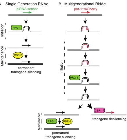
transgenes have a piRNA binding site, and that this process happens in a single generation, and results in an irreversible state of silencing, which requires PRG‐1 for initiation, but it is dispensible for the maintenance phase.
It has been well described in the literature that PRG‐1 is a piwi‐specific Argonaute that binds to piRNAs, and is capable of silencing transgenes that were used in the three studies mentioned above, via this RNAe mechanism. The idea behind these transgene studies is that they serve as a model for how the cell may cope with foreign genes, like those from viruses, inserted into the genome. However, the transgenes that they used contained a piRNA sensor, which has sites of exact homology to piRNAs being made in the cell (Figure 0.3). It is very likely that this specific binding of the piRNA to the piRNA sensor is what is inducing the single‐generation silencing witnessed in these previous studies, but not every invader will have a piRNA binding site. The pathway that I investigate here also implicates PRG‐1 in this silencing, even though our transgenes do not contain a
To summarize, the cell needs two forms of defense, one in the cytoplasm, where mRNA is waiting to be translated into protein, and one in the nucleus where RNA is being actively transcribed from the DNA. The pathway that identifies and degrades mRNA targets in the cytoplasm is called RNAi. The pathway that targets transcripts in the nucleus, and is capable of adding silencing marks to prevent mRNA from even being transcribed from the DNA in the first place, in addition to the degradation in the cytoplasm, is called RNAe (Figure 0.4). In Chapter 2, my investigation identifies a new RNAe pathway called multigenerational RNAe, which is distinct from the single‐generation RNAe described by other groups. Firstly, in single‐generation RNAe, silencing occurs in a single generation, whereas in our multigenerational RNAe process, it takes multiple generations of crossing in order to induce silencing, presumably because there are not piRNA sites in the these genes. In addition, their mechanism shows clearly that PRG‐1 is involved only in the initiation of silencing, and is not required for maintenance. What I see on the other hand is that PRG‐1 is involved in both the initiation and maintenance and that those two steps are not separable, but linked. The silencing that they see is permanent and irreversible. I find that our
silencing is often reversible under certain conditions. Lastly, the molecular trigger for RNAe is unknown, but I have evidence that the molecular trigger of our multigenerational
mechanism may involve a lack of chromosomal pairing and demonstrated by silencing in the zim1 mutant background.
In summary, the identification and eradication of unwanted sequences and
represent advances in the understanding of how vital, conserved processes like NMD and RNAi are able to tailor their machinery to the silencing of various diverse targets. The pathway outlined in Chapter 1 protects against endogenous genetic errors and the processing of this RNA, while the pathway illustrated in Chapter 2 silences foreign
Figures
Figure 0.1. Viruses are capable of injecting their own genetic material into the cell, which in some viruses is then incorporated into the DNA and hijacks the native cellular machinery to transcribe its mRNA. This foreign RNA can then be detected in either the cytoplasm as mRNA or the nucleus precursor RNAs.
a
Figure 0.2. The mechanism RNA interference for silencing induced by small dsRNA.
Figure 0.3. The RNAe silencing mechanism of the piRNA sensor.
Figure 0.4. Silencing mechanisms located in the cytoplasm and the nucleus.
CHAPTER 1: SMG1(YP3) TARGETS UNUSUAL TRANSCRIPTS
Background
For times of stress as well as regular maintenance, the cell is equipped with several surveillance mechanisms that ensure that RNA is being properly transcribed, and that any aberrant messages are quickly identified as they may have deleterious effects and be toxic to the cell. Nonsense‐mediated decay (NMD) is one such mechanism that targets a very specific kind of error – an erroneous premature termination codon (PTC) inserted into the RNA message (7). NMD and its machinery was first identified in yeast, which have three NMD components (25), whereas multicellular eukaryotes have 7 or more, whose identities have largely been determined based on studies in D. melanogaster and C. elegans (26, 27). While NMD has proven to be a vital surveillance mechanism in eukaryotes, particularly in humans where a failure to identify and degrade PTC‐containing mRNAs has been linked to several diseases, it is also know to function on endogenous transcripts (11, 28).
SMG‐1 is a PI3‐kinase‐like kinase (PIKK) and its function in NMD surveillance has been well described (9). For example, it has been shown that upon DNA damage inflicted by UV or γ‐radiation, SMG‐1 is activated. Depleting SMG‐1 increases the cell’s sensitivity to radiation and results in DNA damage (12). SMG‐1 is also implicated in p53 phosphorylation upon G1 checkpoint activation in response to double‐stranded DNA breaks (13). The
oxidative stress response pathway is also mediated by SMG‐1 by protecting against TNF‐α‐ induced apoptosis (14). SMG‐1‐interacting protein UPF‐1 (SMG‐2 in C. elegans) has been shown to be required for the degradation of histone mRNAs in response to replicative stress in response to hydroxyurea‐induction of the S‐phase checkpoint (30).
In C. elegans, mutations in NMD genes were initially discovered by their ability to suppress mutations within genes that function in diverse processes. Seven genes with essential roles in NMD in C. elegans have been identified: smg1, 2, 3, 4, 5, 6, and 7. The smg genes were first identified for their action as recessive allele‐specific suppressors – i.e. ‘suppressor of morphological genitalia’ – which additionally results in a protruding vulva (Pvl) phenotype (8). The key regulatory aspect of NMD processing is the identification of PTC on mRNAs, which is centrally regulated by the SMG‐2 helicase. SMG‐2 is
phosphorylated and dephosphorylated by SMG‐1, which is necessary for the function of NMD (9, 10). There is a high level of conservation of NMD machinery throughout species, although the mechanisms of PTC identification are thought to differ somewhat (31).
suppressors suggests that unc54(293) may not be identified in an identical manner to the canonical PTC target of NMD.
We have identified an allele of the SMG‐1 kinase that is different from any known smg mutation. Exhaustive screens for mutations that affect NMD have been performed using the unc54(r293) mutation, a small deletion in the 3’UTR of unc54 which causes a strong locomotive defect and decreased brood size. Unlike these other NMD‐interacting mutations, smg1(yp3) failed to suppress unc54(r293) and certain satellite and tandem repeat sequences, but can suppress mutations that result in mRNAs with premature stop codons. These results suggest that the NMD machinery may target unusual RNAs, and that their regulatory effects may reach far beyond that of simple surveillance of a single type of target.
Results
smg1 yp3 displays a temperaturesensitive defect in NMD
is a progressive increase of aberrant germlines of every type as the animals are propogated over generations (Figure 1.1 C).
Identification & sequencing of smg1 alleles
Initial mapping the mrt mutation in yp3 suggested that the mutation resides on Chromosome I, and most likely in the smg1 gene. Genomic DNA from the yp3 strain was isolated and sequenced, and a missense mutation was identified. The mutation results in a G1774E amino acid change in the kinase domain of the protein (Figure 1.2 A) – a residue that is highly conserved amongst eukaryotes (Figure 1.2 B). Outcrossing of smg1(yp3) revealed that the Mrt and aberrant germline phenotypes discussed above are not
attributable to the yp3 mutation found in smg1 and must be due to an additional unlinked EMS‐induced mutation.
The identities of other smg1 mutant alleles have been unknown, until now. Sequencing of genomic DNA from both smg1(cc545) and smg1(cc546) revealed that like smg1(yp3), both carry missense mutations resulting in T671I and M1957L conversions respectively. The smg1(cc546) mutation is quite close in proximity to that of smg1(yp3), as they are both harbored in the PI3K domain of SMG‐1 (Figure 1.2 A). The smg1(cc545) mutation resides much farther toward the N‐terminus of the protein, and does not fall in any known domains.
thought of as a substrate of nonsense‐mediated decay (NMD) and has even been used in screens to identify NMD components (8). Under normal circumstances, the unc54(r293) mRNA is a targeted, the message is degraded and the animal displays the unc54 paralyzed phenotype. In certain NMD‐defective backgrounds, including smg mutant alleles, this does not occur ‐ a full‐length mutant unc54 mRNA is made and remains stable, and as a result the animal has little to no motility defects – this effect is termed ‘suppression‐of‐Unc’ (8). I created double mutants of the smg1 alleles ‐ r904, cc545, cc546, yp3 – with unc 54(r293) and subsequently assayed these strains visually for the unc54(r293) phenotype (Figure 1.3 B), as well as quantitatively using a locomotion assay(Figure 1.3 C & D). I found that the unc54(293) phenotype was smg‐suppressible in the presence of the r904, cc545, and cc546 mutations. Animals appeared almost wildtype as observed under the microscope, moving around the plate and taking on a typical sinusoidal shape during locomotion.
However, unlike those canonical targets, yp3 failed to suppress the unc54(r293) phenotype. The animals appeared rigid and failed to move in a wild‐type manner, as evidenced by the small amount of tracks made in the bacteria (Figure 1.3 B).
Quantification of locomotion indicated that movement in the smg1(yp3) unc54(r293) double mutant is not restored and is within the parameters of the unc54(r293) mutation alone (Figure 1.3 D). Thus, the mutation in unc54(r293) is suppressible via the smg1 mutations r904, cc545 and cc546, but not by yp3. This indicates that smg1(yp3) is a separation‐of‐function allele that is proficient in the degradation of unc54(r293) mRNA.
Molecular nature of unc54(r293)
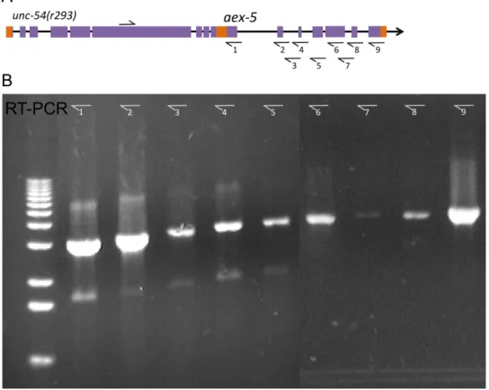
The allele unc54(r293) was identified as a small spontaneous deletion located entirely within the 3’ UTR, including the 3’ cleavage and poly‐adenylation sites (33, 34) (Figure 1.3 A). However, when assayed via Northern Blot, it has been shown that rather than a short mRNA being produced, an extra long message is present (33). I have
determined via reverse transcription polymerase chain reaction (rtPCR) and sequencing that the deletion in unc54(293) actually results in a read‐through to the next gene
downstream – aex5. The sequence contains the full open reading frames (ORFs), including the start and stop codons, of both unc54 and aex5 transcripts, which creates an in‐frame bicistronic message with three stop codons, one at the 3’ ends of unc‐54 and two in a small intergenic linker region (Figure 1.4). The large transcript also contains a linker region between the two ORFs consisting of a 198 bp long track between the stop codon of unc54 and the start codon of aex5. Notably, it contains two small putative ORFs which are 21 bp and 66 bp in length. This linker region is such that aex5 coding region is in frame. If the unc54(r293) mRNA is intact, aex5 is presumably also translated, as null mutants of this gene should produce no viable progeny, according to RNAi experiments (35).
Thus, at the molecular level, the unc54(r293) does not appear to result in the canonical PTC target of NMD, but rather produces a more complex target. While still
regulated by SMG‐1, the lack of suppression via smg1(yp3) indicates that a different type of regulation – governed by NMD machinery, but not identical to NMD – is the real watchdog of this transcript.
smg1(yp3) stabilizes mRNAs with long 3’UTRs
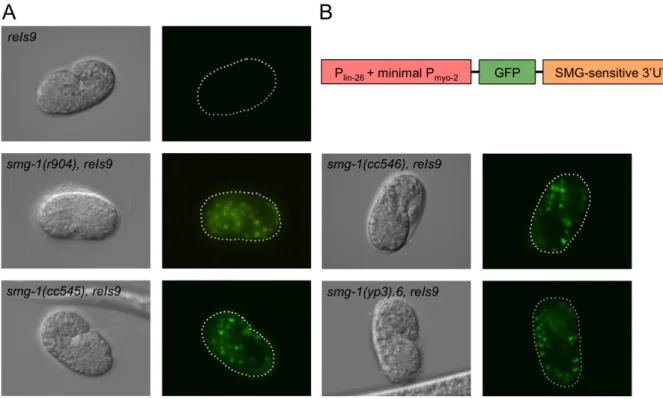
It was previously suspected that a unique property of the unc54(r293) is that it potentially contains an extra‐large UTR – or at least a large portion of sequence on the mRNA after the stop codon of the unc54 gene. I postulated that the aex5 sequence on the end of the unc54(r293) transcript could be behaving simply as an aberrant UTR. D. Reiner contributed the GFP insertion strain pCM1.4 containing a UTR of substantial length that is known to be suppressible in a temperature‐sensitive manner by the smg1(cc546) allele (Figure 1.5) (correspondence, D. Reiner). pCM1.4 consists of a lin26 promoter, a GFP sequence, and smgsuppressible 3’UTR, where neither the poly‐A site nor the stop codon of the GFP sequence are intact and not disrupted by the UTR (Figure 1.5 A). This strain is particularly useful as GFP serves as a visible marker as to whether the transcript is stable or not – if NMD is functioning in a wildtype manner, there will be no GFP expressed, however if the transcript is suppressed, GFP will be evident, particularly in comma stage embryos.
Pairing the pCM1.4 insertion with our novel smg1(yp3) allele would determine whether or not the pattern of suppression was similar to that of the surprising result yielded by the unc54(r293) transcript. Upon making the double mutant with smg1 alleles r904, cc545, cc546 and yp3, I saw equal levels of suppression of degradation of the
transcript resulting in the expression of GFP (Figure 1.5 B). From these results I can determine that the simple inclusion of an extra long‐UTR is not a sufficient target of the alternative smg1mediated mechanism that degrades the unc54(r293) transcript, and does not further illuminate the nature of the yp3 separation‐of‐function.
smg1(yp3) stabilizes the cha1 unc17 gene complex
Few examples of truly bi‐ or multi‐cistronic messages in C. elegans, exist, however the cha1 unc17 complex presents a fairly similar scenario, but is a complicated and unique transcript in the C. elegans genome. cha1 encodes for the structural gene for choline
acetyletransferase, and the unc17 encodes the synaptic vesicle‐associated acetylcholine transporter. Thus, these two genes are related in their function as they are both required for the metabolism acetylcholine. As such, when mutated, both have similar but not identical phenotypes (36).
The molecular structure of the cha1 unc17 complex has been deduced and actually consists of an alternatively spliced transcript that is processed from a pool of one large transcript (36). The cha1 unc17 gene complex is not a true bicistronic message like that of unc54(r293) in that there are not distinct start and stop codons that follow each other sequentially, but rather the two different transcripts arise from alternative splicing – both utilize the splice leader 1 (SL1) at the 5’ end of the complex, and both include the first short exon – it is only then that the transcripts are alternatively spliced into sequential portions of the transcript, as described in previously (36) and additionally shown in Figure 1.6 A.
downstream expression cha1 (37). The unc17(md1447) allele is a deletion of 465 bp – which removes a portion of the 3’UTR, the poly‐adenylated site, and 245 bp of non‐coding genomic sequence (36) (Figure 1.6 A). Both the p1156 and md1447 mutations result in phenotypes with limited locomotion and resistance to cholinesterase inhibitors, which cha 1(p1156) carrying the more severe locomotive phenotype (38).
Testing our novel smg1(yp3) allele against such a target would certainly yield information regarding the type of target necessary to induce the unique pattern of
suppression seen in the smgsuppressibility of unc54(r293). The cause of the dysfunction of the p1156 and md1447 transcripts was previously unknown, however it is now clear that NMD machinery is involved, as the alleles of smg1 – r904, cc545, cc546 and yp3 are all capable of suppressing this degradation (Figure 1.6 B). and as a result, the cha1 unc17 hybrid transcript is produced. The phenotypic consequence of this suppression is an animal which appears more wildtype (Figure 1.6 B). Upon sequencing, the cha1(p1156) mutation was confirmed to be a deletion between the two transcripts. However if NMD‐ mediated degradation is suppressed in the smg1(cc546) cha1(p1156) double mutant, I see that there is read‐through to the downstream cha1 gene. This deletion does cause the insertion of a putative stop codon, but it is unknown whether this is the direct target of degradation. Thus, the cha1 unc17 gene complex is stabilized in an NMD, SMG‐1‐ dependent manner.
not sufficient to illicit the form of degradation targeting the unc54(r293) transcript which results in the yp3 separation‐of‐function phenotype.
smg1(yp3) suppresses a transcript with a canonical PTC
In order to confirm that smg1(yp3) is indeed a functional mutation of smg1 and can suppress the NMD‐mediated degradation of mRNA messages bearing a PTC, I assayed its NMD function against a target bearing a canonical PTC – unc30(e191). unc30 encodes for a transcription factor that controls the terminal differentiation of the GABA‐ergic motor neurons (39), and the mutation unc30(e191) bears a C to T substitution in the first exon, resulting in a Q to amber stop conversion (Figure 1.6 C). This mutation results in a
moderate locomotive defect, which is easily discernable by eye, making it an advantageous choice for analysis.
cc545 and cc546 alleles are temperature‐sensitive, as well as with our expectations regarding the nature of the yp3 mutation, give it’s proximity to cc545 (Figure 1.2 A). Therefore, smg1(yp3) disrupts canonical NMD suppression of an mRNA message containing PTCs in a temperature‐dependent manner.
Tandem and satellite repeats affected by smg1(yp3)
Microarray analysis was performed to identify possible endogenous targets of smg1 that could be stabilized by r904, cc545, and cc546 alleles, but were suppressed by yp3. Our analysis yielded results and I found several satellite and tandem repeats that were suppressed by smg1(yp3). There appear to be two classes of satellite regulation. One, in which satellite repeats are up‐regulated in r904, cc545, and cc546, and down‐regulated in yp3 as compared to wild‐type, and another where repeats are up‐regulated by all smg1 alleles tested, but to a consistently lesser extent in yp3. Further analysis is needed.
Discussion
Rethinking NMD – is it really just about nonsense?
It has become clear in recent years that the mechanism traditionally known as NMD is not merely a simple quality‐control mechanism, but is also an important factor in the regulation of many physiological mRNA targets as a part of their normal function.
NMD mechanism – (1) the canonical PTC inserted into an mRNA regions (43, 44), (2) mRNAs with abnormally long 3’UTRs (45), (3) certain mRNAs that contain small upstream open reading frames (uORFs) (46, 47), (4) pre‐mRNAs that contain introns and have mistakenly entered the cytoplasm (48), and (5) mRNAS where an out‐of‐frame AUG start codon is used for translation initiation (41).
Although higher eukaryotic studies do not go quite as far in attributing these kinds of surveillance to NMD, mammalian experiments do suggest that one‐third of splice events may produce a possible NMD target (49, 50), indicating that NMD is functioning not only as a surveillance mechanism, but as a regulatory system that responds to changes in
transcription environments. For example, it is known that T‐cell receptor genes can form rearrangements that result in their mRNA messages containing PTCs (51). It is highly likely that various components of the NMD machinery are functioning in many different types of regulation and surveillance as in yeast – the system is likely more complex, making it more difficult to elucidate exactly which components and mechanisms are acting on particular breeds of substrate.
A specific molecular target within unc54(r293)
After testing several types of smg‐suppressible targets against our novel smg1(yp3) allele, I can discern that there is something especially unique about the unc54(r293)
are not suppressible via the smg1(yp3) allele ‐ one can contemplate several options for the specific molecular target that lies within unc54(r293) transcript.
A possible molecular target of unc54(r293) is in fact the small 21 bp and 66 bp putative ORFs found in the linker region between unc54 and aex5 in the r293 transcript (Figure 1.4). Initially it was thought that translation machinery would initiate upon locating the most proximal AUG codon (52), but in recent years it has come to light that there exist transcripts with long 5’ regions upstream of the “true” start that contain functional AUG codons that presumably have a regulatory function (53). In fact,
bioinformatic surveys have identified uORFs in as many as 35‐49% of human and rodent transcripts (54‐56). Few of these uORFs have been studied in detail (57, 58) and their mechanism of action is largely unknown. Already found to initiate NMD in yeast (46, 47), uORFs have recently been implicated in functions requiring NMD machinery in human cells. Microarray analysis indicates that when NMD components UPF‐1 (SMG‐2 in C. elegans) and SMG‐6 are knocked down, more than 35.5% of all upregulated genes contain at least one uORF (59).
It is also possible that the linker region in the unc54(r293) transcript (Figure 1.4) is being targeted and degraded via miRNAs. Using miRBase, it was found that four separate miRNAs are predicted to bind to the linker region – cel‐miR‐85, cel‐miR‐65, cel‐miR‐1829a and cel‐miR392. As of yet none of these miRNAs have known targets or functions. However, it is plausible that miRNAs could bind to the small linker region and target the entire unc 54(r293) transcript for degradation in a smg‐mediated manner. This would implicate a new mechanism for miRNA action and could help to elucidate the function and purpose of many miRNAs that are currently known.
Disease implications
The isolation of the separation‐of‐function allele smg1(yp3) may make it possible to further advance therapies that would specifically inactivate NMD, thus rescuing disease phenotypes, leaving other regulation pathways governed by some NMD machinery – like that which degrades the unc54(r293) transcript ‐ intact and functional.
There could also be more that smg1(yp3) could teach us about disease. The novel form of degradation that seems to target the unc54(r293) transcript as well as certain satellite and tandem repeats, could help to explain several kinds of human cancer that have been linked to the de‐repression of these heterochromatic regions. Demethylation at Sat2 repeats was found to be a hallmark in (63), and a genome‐wide profile of lung carcinomas indicated that satellite repeat regions were also hypomethylated (64). It has also been shown that loss of Brca1 in mice results in a de‐repression of tandem satellite repeat DNA and that similar de‐repression was also observed in human BRCA‐1‐deficient breast cancer cells (65).
Delving further into the source of the degradation of the unique transcript unc 54(r293) ‐ and possibly certain satellite and tandem repeats ‐ could be the first small step to uncovering a slew of knowledge pertaining not only to the understanding of basic biological and genetic regulatory mechanisms, but could have a direct impact on disease knowledge that could improve current technology and drug development.
Materials and Methods Strains
1(r904) I, PD8119 smg1(cc545) I, PD8120 smg1(cc546) I, smg1(yp3) I, unc54(r293) I, dpy5(e907) I, CB61 dpy5(e61) I, MT7929 unc13(e51) I, CB450 unc13(e450) I, unc 17(md1447) IV, CB845 unc30(e191) IV, cha1(p1156) IV, pCM1.4
smg1(yp3) was outcrossed versus an outcrossed stock of dpy5 e61, unc13 e450, and freshly isolated homozygous F2 lines were established and outcrossed a further five times by the same method.
Double mutants were typically constructed by marking each mutant with a physical marker, selecting one mutant‐marker strain and crossing with N2 to yield males, and crossing said males with the other mutant‐marker strain. F1 progeny of successful crosses were then isolated, and each marker selected against in subsequent F2 and F3 generations.
Locomotion Assay
As previously described in Reiner et al, 1999 (66). To assay for locomotion, individual animals were placed in the center of a 60 mm plate containing a lawn of Escherichia coli OP50 with the origin marked. The plates were incubated for 15 minutes at room temperature (approximately 23°C), and then briefly incubated at 4°C to stop movement. The radial distance from the origin to the farthest distance away the animal moved was measured to the nearest 0.5mm.
GFP Microscopy
RNA Preparation and cDNA Synthesis
Figures
Figure 1.1. smg1(yp3) displays several unique phenotypes. (A) smg1(yp3) has a
Figure 1.3. unc54(r293) serves as an indicator for NMD function in smg‐1 mutants. (A) Molecular structure of unc54(r293) versus the wild‐type gene. (B) Images of unc54
animals in various smg1 allele backgrounds. (C) Locomotion assay to assess whether NMD is occurring in unc54(r293) animals. (D) Quantification of results of locomotion assay in various smg1 allele backgrounds.
Figure 1.6. Examination of NMD activity in smg1 mutant backgrounds. (A) Structure of the unc17 cha1 complex. (B) Results of examining various smg1 alleles in the cha‐1
Figure 1.7. Model displaying the possible separation‐of‐function occurring in smg1(yp3) due to different mRNA targets..
CHAPTER 2: LACK OF PAIRING DURING MEIOSIS TRIGGERS MULTIGENERATIONAL TRANSGENE SILENCING IN CAENORHABDITIS ELEGANS
Background
Small RNAs can repress expression of endogenous genes as well as parasites such as transposons or viruses. One form of small RNA‐mediated repression is epigenetic silencing of genomic loci that can result in a permanent, heritable state of expression in germ cells.
RNA interference (RNAi) is a conserved biological process in which small non‐ coding RNA molecules promote gene silencing (2). RNAi was originally identified in Caenorhabditis elegans, but has been observed in a large number of eukaryotes ranging from yeast to plants to humans (1, 2, 67‐70). Small interfering RNAs (siRNAs) can be produced from a variety of sources (71, 72) and can be divided into two classes –
siRNAs (17, 73‐76). Secondary Argonaute proteins (SAGOs) then bind with the secondary siRNAs, and it is this complex that directly degrades the target mRNA message (18).
A second class of primary siRNAs in C. elegans is the Piwi‐interacting RNAs (piRNAs) that are highly abundant in the germline and interact with the C. elegans Piwi Argonaute protein PRG‐1 (22, 23). C. elegans piRNAs are termed 21U‐RNAs as they are 21 nucleotides long and possess a 5’ uracil. PRG‐1 and associated piRNAs target transposons and some genes based on imperfect homology, which recruits RdRPs to promote biogenesis of 22G secondary siRNAs that bear perfect homology to their targets. These secondary 22G‐RNAs interact with WAGO‐class Argonaute proteins to promote silencing of germline loci. The vast repertoire of C. elegans piRNAs and their ability to target nucleic acids with
mismatches may allow them to target both endogenous loci as well as foreign nucleic acids such as transposons or viruses (24, 77). Although C. elegans piRNAs can target many endogenous transposons, prg1 mutants displayed transposition for only one out of three DNA transposons tested, even though all three transposons become active if secondary siRNA populations are disrupted with Mutator gene mutations (78). These results imply that small RNA‐mediated epigenetic silencing of many transposons may initially depend on PRG‐1 and associated piRNAs, but then a downstream secondary siRNA system is capable of maintaining silencing of most transposons in the absence of piRNAs.
The diffusible transposon silencing factor depends on Mutator class proteins that promote secondary siRNA production, but not on proteins that initiate the response to exogenous dsRNA such as RDE‐1 or RDE‐4 (80). Cosuppression was originally observed in plants in response to high copy number transgenes and also acts via siRNAs (82‐84). Methylation‐ mediated silencing of multicopy transgenes has also been observed in zebrafish, where transgenes inserted as high copy concatemeric arrays become completely silenced in two to three generations (85, 86). Similar results are seen in mice, where high copy number is inversely correlated to expression (87, 88). Therefore, epigenetic silencing of high copy number transgenes in the C. elegans germline is consistent with a natural defense response to foreign nucleic acids such as transposons that is seen in diverse organisms.
In order to stably express transgenes in C. elegans, an elegant method to create single‐copy transgene insertions was developed with the aid of a unique copy of the Drosophila melanogaster transposon MosI to induce a chromosomal double‐strand break that promotes site‐specific recombination with a plasmid‐derived template (19, 21, 89, 90). Although single‐copy transgenes can be stably expressed in the germline using this method, a GFP transgene with a piRNA target site in its 3’UTR (piRNA sensor) as well as other
transgenes possessing GFP, can be subjected to rapid and permanent epigenetic silencing in the germline (19‐21). Initiation of silencing depends on PRG‐1, as single‐copy transgenes are expressed if created in a prg1 mutant background, but become silenced within a single generation when wildtype PRG‐1 is introduced. Transgene silencing then becomes
production of a secondary small RNA population whose maintenance, in conjunction with siRNA‐directed with histone marks, is sufficient to enforce a stable, heritable silent state. This rapid process is termed ‘RNAe’ (small RNA‐induced epigenetic silencing), though we suggest that the term ‘single‐generation RNAe’ might be an appropriate descriptor.
Here I describe a process that we term ‘multigenerational RNAe’. We identified single‐copy transgenes that become silent if crossed in the hemizygous state for multiple generations. We found that multigenerational transgene silencing can become permanent and that small RNA silencing proteins previously shown to initiate or maintain silencing of transgene epialleles play unexpected roles in this multigenerational process. Our study provides new insights into the genesis and maintenance of epialleles, relevant not only to transgenes but also potentially to dynamic regulation of germline gene expression across generations in the vast majority of metazoans.
Results
Discovery of multigenerational transgene silencing
I repeated the above cross by placing the pot1:mCherry transgene in trans to the rol6 marker mutation and found that a fraction of pot1:mCherry / rol6 animals were mCherry‐negative starting at cross 3, although complete silencing of the pot1::mCherry transgene was not achieved by cross 7 (Figure 2.1 H). Thus, a partially penetrant
multigenerational silencing process occurs when the pot1::mCherry transgene is crossed in trans to the rol6 marker mutation.
I next placed pot1::mCherry in trans to the marker mutations dpy10 unc4, which flank the pot1::mCherry transgene, and repeatedly crossed pot1::mCherry / dpy10 unc4 crossed heterozygous males with dpy10 unc4 hermaphrodites (Figure 2.1 D). A
proportion of silent pot1::mCherry heterozygotes were observed for crosses 2 and 3, followed by uniform silencing for crosses 4 through 6 (Figure 2.1E & I). When F2 animals were revived from starved plates of cross 6 and homozygosed by selecting against the dpy 10 unc4 balancer mutations, the vast majority of pot1::mCherry homozygotes showed POT‐1::mCherry expression (28/30). However, 2/30 pot1::mCherry homozygous lines were completely silent and remained silent for 8 additional generations (Figure 2.1 I columns 6S). I therefore conclude that a hemizygous single‐copy transgene can become strongly silenced following multiple crosses, and that although this silencing reverts for many F3 homozygotes, a fraction of pot1::mCherry homozygotes become permanently epigenetically silenced.
Independent transgenes succumb to multigenerational silencing
integrated into the ttTi5605 MosI locus, Pmex5::mCherry::H2B::tbb2: 3'UTR::gpd2 operon::GFP::H2B::cye1 3'UTR (Figure 2.1 A & E). The mCherry expression in this transgene was highly robust, whereas the GFP expression was substantially weaker,
therefore, I used the mCherry fluorescence to score the animals. Crosses of oxSi487 in trans to dpy10 unc4 mutations resulted in silencing for some progeny in cross 2 and complete silencing by cross 3. F2 animals from starved plates of cross 3 were singled, and I examined the F3 progeny of four oxSi487 homozygotes, all of which displayed uniform silencing of mCherry::H2B (Figure 2.1 E & G columns 3S). These strains displayed fully penetrant silencing for at least 8 generations without reversion, suggesting permanent transgene silencing. I also crossed oxSi487 in trans to rol6 and found that a fraction of animals for four independent crosses were mCherry‐negative from crosses 4 to 11, indicating weaker multigenerational silencing than for the dpy10 unc4 marker mutations. oxSi487
We next asked if multigenerational transgene silencing also occurs for transgenes that only contain GFP‐epitope tags, and if transgene insertion via a double‐strand break created by the Drosophila MosI transposon ttTi5605 has any impact on transgenerational silencing. We therefore tested identical transgenes that express Pmex5::GFP::tbb2 3’UTR that were inserted either via MosI‐mediated gene conversion at ttTi5605, cpSi10[Pmex 5::GFP::tbb2 3’UTR + unc119(+)], or via CRISPR/Cas9‐mediated gene conversion at the identical position on Chromosome II, cpIs10[Pmex5::GFP::tbb2 3’UTR + unc119(+)] (Figure 2.2 A) (92). We found that cpSi10 and cpIs10 underwent multigenerational silencing with very similar kinetics (Figure 2.2 B & C), indicating that the presence of the Drosophila MosI transposon at the ttTi5605 locus prior to transgene insertion does not affect transgene silencing. When cpSi10 and cpIs10 were crossed in trans to our dpy10 unc 4 marker strain, cpSi10 was silenced by cross 3, and cpIs10 at cross 6 (Figure 2.2 B, C & E). Chunking of starved cross plates yielded F3 progeny homozygous for the transgene, where transgene expression was restored (Figure 2.2 B column 3S, 2.2 C column 3S). To
multigenerational silencing process occurs in transgene heterozygotes, and that this silencing can become permanent for a fraction of transgene homozygotes.
We next asked if a GFP insertion at a distinct genomic location, the his72 locus on Chromosome III, could also be subjected to multigenerational silencing (92). his72::GFP was placed in trans to the marker mutations vab7 dpy18 and crossed versus these mutations (Figure 2.7). Similar to our other transgenes tested, a significant fraction of his 72::GFP / vab7 dpy18 heterozygotes displayed transgene silencing for crosses 3 to 22 (Figure 2.2 D & E). Thus, multigenerational transgene silencing is not specific to single‐ copy transgenes at the ttTi5605 locus on Chromosome II, and can regulate the expression of GFP cassettes inserted at endogenous genomic loci.
Outcrossing cpSi10 and cpIs10 with other markers
We decided to test a variety of balancer mutations related to the dpy10 unc4 mutations used, which had been outcrossed numerous times to our laboratory stock of N2 (wildtype). We tested an unoutcrossed dpy10 unc4 obtained from the CGC, dpy2 unc4, and a strain containing an inversion between dpy10 and unc4 (mIn1; unc4 dpy10). When cpSi10 and cpIs10 were crossed in trans to dpy2 unc4 we found a fully penetrant
resulted in multigenerational transgene silencing, it is likely that unidentified secondary mutations are not responsible for this effect.
Coupling of initiation and maintenance of multigenerational transgene silencing via small RNA proteins
RDE‐2 is a component of the Mutator‐class of RNA interference proteins that promote biogenesis of secondary siRNA populations, which were previously shown to be required for silencing of single‐copy transgenes that become silenced in a single cross (19‐ 21), as well as for cosuppression‐mediated silencing of repetitive transgenes (23, 80, 93‐ 95). I found that POT‐1::mCherry fluorescence was partially restored when the silenced pot1::mCherry.9 transgene was placed in an rde2–deficient background (Figure 2.3 A & B). Thus, an siRNA‐mediated process promotes multigenerational transgene silencing. Secondary siRNA proteins such as RDE‐2 have been shown to promote maintenance of single‐generation transgene silencing (78, 80). In contrast, the piRNA‐interacting
Argonaute protein PRG‐1 is required for initiation of single‐generation transgene silencing but is dispensable for maintenance of transgene silencing (20, 96). I found that
As a control, I tested the RSD‐6 Tudor domain protein, which has been previously described to promote spreading of RNAi from the soma to the germline and generation of robust secondary siRNA populations in response to exogenous dsRNA triggers, but is dispensable for single‐generation silencing of the piRNA sensor transgene (21, 97, 98). pot 1::mCherry silencing was maintained when rsd6 was mutant (Figure 2.3 A & B). I also found that the NRDE‐3 Argonaute protein, which promotes transcriptional silencing in response to exogenous dsRNAs (99), was dispensable for pot1::mCherry silencing (Figure 2.3 A & B).
Lack of chromosomal pairing induced rapid silencing
ZIM‐1 is a member of a family of four related C2H2 zinc‐finger domains proteins that promote pairing of specific chromosome homologs during meiosis (100). ZIM‐1 itself promotes pairing of Chromosomes II and III, as meiotic pairing of these chromosomes is completely abrogated in zim1 mutants (101, 102). When pot1::mCherry; zim1 males were crossed with dpy10 unc4; zim1 hermaphrodites, uniform mCherry silencing occurred in the progeny of cross 1 (n=10, F1 hermaphrodites scored) (Figure 2.8). The same result was observed for progeny of oxSi487; zim1 males crossed with dpy10 unc4; zim1 hermaphrodites. Therefore, complete lack of chromosome pairing leads to rapid single‐ generation transgene silencing for transgenes that normally succumb to multigenerational transgene silencing.
Discussion
Lack of a pairing partner in meiosis is known to alert an RNA directed‐silencing mechanism termed Meiotic Silencing of Unpaired DNA (MSUD), which was first identified in Neurospora crassa (103). MSUD occurs when a diploid organism suppresses expression of genes not paired with their homologs during the first prophase of meiosis (104‐106). MSUD requires small RNA factors that include an Argonaute protein and an RNA‐
dependent RNA polymerase that likely act on unpaired meiotic DNA to produce dsRNA that feeds into a siRNA‐mediated silencing system (103, 107‐109).
We found that transgene silencing occurred when singlecopy transgene / marker mutation heterozygotes were crossed with marker mutation homozygotes for multiple generations. While potent, this multigenerational transgene silencing process was
reversible, as transgene activation was commonly observed when transgene homozygotes were isolated from silent singlecopy transgene / marker mutation heterozygotes. These results suggest that the trigger of multigenerational transgene silencing corresponds to unpaired transgene DNA during meiosis, which occurs if one homolog possesses a
transgene and the other does not. Thus, the multigenerational transgene silencing that we report here possesses a physical hallmark of MSUD.
multigenerational silencing process that we define here. We found that although multigenerational silencing of hemizygous transgenes was often reversed when a
transgene becomes homozygous, although permanent transgene silencing was observed for some transgene homozygotes. We term this reversible silencing process that is potent enough to become permanent ‘multigenerational RNAe’.
Maintenance of silencing for transgenes silenced by multigenerational RNAe can require both PRG‐1, which is dispensable for maintenance of single‐generation RNAe, as well as the secondary siRNA protein RDE‐2. Deficiency for either of these proteins led to partial transgene desilencing for the pot1::mCherry.9 transgene, and full desilencing was observed when both PRG‐1 and RDE‐2 were removed. These results imply that
multigenerational RNAe represents a state of epigenetic silencing that is distinct from single‐generation RNAe, where initiation and maintenance phases of transgene silencing can remain mechanistically coupled once the silent state has been established..