UNDERSTANDING THE REGULATORY DYNAMIC OF THE TWO-COMPONENT SYSTEMS, PLRSR AND BVGAS, DURING BORDETELLA COLONIZATION OF THE MAMMALIAN
RESPIRATORY TRACT
Mary Ashley Sobran
A dissertation submitted to the faculty at the University of North Carolina at Chapel Hill in partial fulfillment of the requirements for the degree of Doctor of Philosophy in the Department of Microbiology
and Immunology in the School of Medicine.
Chapel Hill 2019
ABSTRACT
Mary Ashley Sobran: Understanding the Regulatory Dynamic of the Two-Component Systems, PlrSR and BvgAS, During Bordetella Colonization of the Mammalian Respiratory Tract
(Under the direction of Peggy A. Cotter)
Pertussis is a severe respiratory disease caused by the bacterium Bordetella pertussis.
Despite high vaccination coverage, the number of pertussis cases has rebounded in recent years.
To combat this disease, more efficacious vaccines and improved knowledge of Bordetella
virulence is critical. Bacteria often use two-component systems (TCSs) to coordinate essential
cellular processes in response to the diverse environments they encounter. In B. pertussis and the
closely related subspecies B. bronchiseptica, the TCS, BvgAS, controls the expression of almost
all known virulence factor-encoding genes and is considered the main virulence regulatory
system. Recently, another TCS, PlrSR, was identified that is also required for Bordetella
infection within the lower respiratory tract (LRT). However, why PlrSR is important for
colonization is unknown. Using engineered strains of B. bronchiseptica and genetic reporters, we
demonstrated that PlrS is required for the maintenance of BvgAS activity in the lungs of mice,
indicating that PlrSR, along with BvgAS, coordinates virulence specifically in the LRT.
Moreover, our data indicate that PlrSR may regulate genes that are required for persistence in the
LRT independent of BvgAS. Importantly, these genes and their corresponding proteins may
serve as new vaccine components or therapeutic targets. We have also shown that the
Per-Arnt-Sim (PAS) domain of BvgS is required for BvgS inactivation in the absence of PlrS, indicating
that the PAS domain is important for the PlrSR-BvgAS connection and functions as an
PlrSR controls the expression of high-affinity cytochrome oxidases (HACOs) that are required
for bacterial respiration and maintenance of BvgAS activity in the LRT. We have demonstrated
that two of four HACOs contribute to LRT colonization. However, BvgAS activity is unaffected
by the loss of these HACOs and it is unknown if PlrSR regulates the expression of any HACO
encoding genes. Together, this work provides a more detailed understanding of the coordinated
regulation imposed by PlrSR and BvgAS to promote Bordetella survival within the mammalian
TABLE OF CONTENTS
LIST OF TABLES ... xi
LIST OF FIGURES ... xii
LIST OF ABBREVIATIONS ... xiv
CHAPTER 1. INTRODUCTION ... 1
Introduction ... 1
Current pertussis vaccines fail to protect individuals from Bordetella pertussis colonization and pertussis disease ... 1
Despite high rates of vaccination, pertussis remains a burden in the United States and throughout the world ... 2
The progression of pertussis disease is most severe in young patients and can last for many months ... 2
Bordetella pertussis produces many essential virulence factors during human infection ... 3
The complex, two-component phosphorelay system, BvgAS, is essential for Bordetella colonization of the mammalian host and is considered the master regulator of virulence ... 4
Although the importance of BvgAS for Bordetella virulence has been well established, the contributions of other two-component systems to bacterial survival and virulence in the mammalian host is less understood ... 6
PlrSR is a newly identified putative two-component system required for B. bronchiseptica colonization of the lower respiratory tract in rats ... 7
Research Objective ... 7
REFERENCES ... 9
Introduction ... 15
Results ... 18
PlrS is required for enhanced BvgAS-dependent virulence-associated phenotypes in response to elevated CO2 concentrations ... 18
PlrS likely affects BvgAS-dependent phenotypes via PlrR ... 18
B. bronchiseptica lacking plrS modulate to the Bvg– phase within the lower respiratory tract ... 20
B. bronchiseptica lacking plrS fail to activate BvgAS within the lower respiratory tract ... 22
PlrS is required for B. bronchiseptica persistence in the lower respiratory tract, independent of its effects on BvgAS activity ... 23
PlrS is required for survival and persistence of B. pertussis in the lower respiratory tract ... 24
Discussion ... 25
Materials and Methods ... 29
Growth media and bacterial strains ... 29
Construction of cloning plasmids and bacterial strains ... 30
In vitro adherence assays ... 31
Macrophage cytotoxicity assays ... 31
Bacterial adherence to J774 cells and in response to being cultured in media with decreased pH ... 32
Evaluation of B. bronchisepticabvgAS transcription in vitro ... 32
Evaluation of B. bronchiseptica promoter activity in vitro ... 33
Bacterial colonization of the mouse respiratory tract ... 33
Evaluation of B. bronchiseptica phenotypic phase transition in vivo ... 34
Ethics statement ... 34
Statistical analysis ... 35
REFERENCES ... 47
CHAPTER 3. THE BVGS PAS DOMAIN: AN INDEPENDENT SENSORY PERCEPTION MODULE IN THE BORDETELLA BRONCHISEPTICA BVGAS PHOSPHORELAY ... 53
Introduction ... 53
Results ... 57
Mutants used in this study ... 57
Contribution of the PAS domain in regulating BvgS activity in vitro ... 57
Characterization of pGFLIP-PflaA and pBam reporters in BvgS PAS domain mutants in vitro ... 60
The PAS domain is not required for BvgS kinase activity in the LRT in PlrSWT bacteria... 62
The C607S substitution hinders BvgS kinase activity and bacterial colonization in the lower respiratory tract (LRT) of mice ... 62
A role for the BvgS PAS domain is revealed in the absence of PlrS in the LRT ... 63
Discussion ... 65
Materials and Methods ... 69
Bacterial strains, plasmids, and growth conditions ... 69
Construction of B. bronchiseptica BvgS PAS domain mutant strains ... 70
Construction of PptxA-gfpand PfhaB-gfptranscriptional reporters ... 71
Transcription of PptxA-gfpand PfhaB-gfp reporters in vitro ... 71
Characterization of pGFLIP-PflaA and pBam reporters in vitro ... 72
Evaluation of B. bronchiseptica modulation in vivo ... 73
Ethics statement ... 73
Statistical analysis ... 74
Figures and Tables ... 75
CHAPTER 4. TWO HIGH AFFINITY CYTOCHROME OXIDASES ARE REQUIRED FOR MAXIMAL COLONIZATION OF THE MAMMALIAN
LOWER RESPIRATORY TRACT BY BORDETELLA BRONCHISEPTICA ... 87
Introduction ... 87
Results ... 91
High-affinity cytochrome oxidase loci are differentially regulated in ambient air in vitro ... 91
B. bronchiseptica requires high-affinity cytochrome oxidases to robustly colonize the mammalian lower respiratory tract ... 92
Maintenance of BvgAS kinase activity does not depend on bd-type cytochrome oxidases in vivo ... 93
Although required for robust colonization in vivo, the BB4012-BB4011 (cydAB3) promoter is inactive during colonization of the murine respiratory tract ... 94
Discussion ... 95
Materials and Methods ... 98
Bacterial strains, plasmids, and growth conditions ... 98
Construction of high-affinity cytochrome oxidase lacZ transcriptional reporters ... 99
Transcriptional analysis of high-affinity cytochrome oxidase encoding genes in vitro ... 99
Construction of B. bronchiseptica high-affinity cytochrome oxidase deletion strains ... 100
Bacterial colonization of the mouse respiratory tract ... 101
Evaluation of B. bronchiseptica modulation in vivo ... 101
Construction of pGFLIP-PcydAB3 reporter ... 102
Evaluation of cydAB3 expression in vivo ... 102
Ethics statement ... 103
LIST OF TABLES
Table 2.S1. Bacterial Strains and Plasmids Used in this Study ... 46
Table 3.S1. Bacterial Strains and Plasmids Used in this Study ... 81
Table 3.S2. Primers Used in this Study ... 82
Table 4.S1. Bacterial Strains and Plasmids Used in this Study ... 109
LIST OF FIGURES
Figure 2.1. PlrS is required for Bvg-dependent phenotypes in 5% CO2 growth
conditions. ... 36 Figure 2.2. Overexpression of a phosphomimetic plrR allele rescues the ΔplrS L2
cell adherence defect. ... 37 Figure 2.3. Without PlrS, B. bronchiseptica modulates to the Bvg– phase in the
lower respiratory tract. ... 38 Figure 2.4. PlrS is required for persistence of B. bronchiseptica in the LRT
independent of BvgAS activity. ... 39 Figure 2.5. PlrS is required for the persistence of Bordetella pertussis in the lower
respiratory tract. ... 40 Figure 2.6. PlrSR and BvgAS work in tandem to regulate gene expression
required for Bordetella persistence in the lower respiratory tract. ... 41 Figure 2.S1. Bacterial adherence to macrophage-like murine J774 cells. ... 42 Figure 2.S2. Increased adherence of wild-type B. bronchiseptica to L2 lung
epithelial cells does not result from bacteria responding to lowered pH of the
growth media. ... 42 Figure 2.S3. PlrS does not transcriptionally regulate the expression of the bvgAS
in vitro. ... 43 Figure 2.S4. In vitro analysis of wild type and ΔplrS modulation using the
plasmid reporters pGFLIP-flaA and pBam. ... 43 Figure 2.S5. In vitro analysis of wild type and ΔplrS phase transition using the
plasmid reporter pGFLIP-ptxA. ... 44 Figure 2.S6. A constitutively-active bvgS mutation does not prevent rapid
clearance of a ΔplrS strain in the lower respiratory tract. ... 44
Figure 2.S7. In vitro modulation of constitutively-active bvgS strains using the
plasmid reporters pGFLIP-flaA and pBam. ... 45 Figure 3.1. The sensor kinase, BvgS, has a unique PAS domain, which may play a
role in signal perception. ... 75 Figure 3.2. In B. bronchiseptica, the PAS domain influences BvgS kinase activity
and sensitivity to chemical modulation in vitro. ... 76 Figure 3.3. The PAS domain is important for BvgS inactivation in response to
Figure 3.4. A conserved cysteine residue in the PAS domain is important for
maintaining BvgS kinase activity during respiratory tract colonization of mice... 78 Figure 3.5. The PAS domain is required for BvgS modulation in response to
PlrSR dysfunction during LRT colonization. ... 79 Figure 3.S1. The PAS domain of BvgS may sense an activating or inactivating
signal in the LRT. ... 80 Figure 4.1. In vitro, BvgS influences the expression of high affinity cytochrome
oxidase encoding genes in B. bronchiseptica. ... 104 Figure 4.2. Two bd-type high-affinity cytochrome oxidases contribute to B.
bronchiseptica colonization of the trachea in mice. ... 105 Figure 4.3. Without high-affinity cytochrome oxidases B. bronchiseptica is
significantly attenuated for colonization of the trachea and LRT in mice. ... 106 Figure 4.4. Maintenance of BvgS kinase activity does not require bd-type
high-affinity cytochrome oxidases during respiratory tract colonization of mice. ... 107 Figure 4.5. The cydAB3 promoter is not active during respiratory tract
LIST OF ABBREVIATIONS L microliters
-ME Mercaptoethanol
aa amino acid
ACT adenylate cyclase toxin
BG Bordet-Gengou
Bvg+ Bvg-plus phase
Bvg– Bvg-minus phase
Bvgi Bvg-intermediate phase
cfu colony forming units
DAP diaminopimelic acid
DTaP Diphtheria, Tetanus, Acellular Pertussis vaccine
FHA filamentous hemagglutinin
FIM fimbriae
Gm gentamicin
HACOs high affinity cytochrome oxidases
HK histidine kinase
Hpt histidine phospho-transfer domain
HTH helix-turn-helix
Km kanamycin
LACOs low affinity cytochrome oxidases
LB lysogeny broth
LCVs large colony variants
LDH lactate dehydrogenase
LRT lower respiratory tract
MgSO4 magnesium sulfate
MOI multiplicity of infection
NA nicotinic acid
NC nasal cavity
PAS Per-Arnt-Sim
PBS phosphate-buffered saline
PDC PhoP-DcuS-CitA
PfhaB fhaB promoter
PptxA ptxA promoter
PRN pertactin
PT pertussis toxin
REC receiver
ROS reactive oxygen species
SD standard deviation
Sm streptomycin
SpcR spectinomycin-resistant
SS Stainer-Scholte
T1 Time 1
T2 Time 2
TCS two-component system
TM transmembrane
VFT venus fly-trap
vags vir-activated genes
vrgs vir-repressed genes
CHAPTER 1. INTRODUCTION
Introduction
Current pertussis vaccines fail to protect individuals from Bordetella pertussis colonization and pertussis disease
Pertussis is a highly contagious and severe respiratory disease caused by the bacterium
Bordetella pertussis (1). Despite high rates of vaccination, pertussis has reemerged as an urgent
public health threat (2). During the late 1940s, a whole cell pertussis vaccine was introduced that
dramatically reduced the incidence of pertussis (3). However, due to concerns over safety, the
whole cell vaccine was replaced with an acellular pertussis vaccine. The current acellular vaccine
contains only purified bacterial proteins and inactivated toxins, which serve as critical virulence
factors for the bacteria. Since the transition from the whole cell to the acellular vaccine, the
number of pertussis cases has rebounded. Studies have shown that the immunity generated by the
current acellular vaccine is less robust and wanes faster compared to the immunity generated by
pertussis infection or the whole cell vaccine (4–6). As a result, the number of cases of pertussis
reported in adolescence and young adults has increased (2). Recent studies using a baboon
infection model demonstrate that although the acellular and whole cell vaccines provide
protection against overt disease, they fail to prevent colonization of Bordetella within the
respiratory tract (7). Moreover, animals immunized with the acellular vaccine were still able to
transmit Bordetella to naïve animals (7–10). If true for human disease, current vaccination
strategies may be inadvertently increasing the reservoir of Bordetella pertussis in the population,
Despite high rates of vaccination, pertussis remains a burden in the United States and throughout the world
In the Unites States, five doses of the pertussis vaccine (DTap) are recommended for all
individuals by the age of seven and booster doses are recommended for adolescence and adults
(11). Vaccination is also recommended for women during every pregnancy and breastfeeding is
encouraged in hopes of transferring maternal immunity to infants, who are more at risk for fatal
infections (12). In 2017, the median acellular pertussis vaccination coverage in the United States
was 95.1%, almost 10% above the global average (13–15). However, approximately 19,000
cases of pertussis were still reported, including 13 pertussis-associated deaths. Each year since
the 1990s, the total number of pertussis cases has steadily increased, and multiple outbreaks have
been reported across the country. Worldwide, the burden of pertussis is even greater with
approximately 144,000 incidence of pertussis and 90,000 deaths resulting from pertussis
infection reported in 2017 alone (14, 15).
The progression of pertussis disease is most severe in young patients and can last for many months
Pertussis illness can manifest as a range of symptoms from mild upper respiratory tract
disease to severe, persistent, and progressive coughing that can continue for weeks or months (1,
16). Following infection, progression of the disease can be categorized into three consecutive
stages: the catarrhal stage, paroxysmal stage, and convalescent stage. During the catarrhal stage,
which can last 1-2 weeks, symptoms are often mild and mistaken for those of a common cold,
such as malaise, sore throat, nasal congestion, rhinorrhea, sneezing, and mild progressive dry
cough. As a consequence, it often is not until the second stage of disease that pertussis is
diagnosed. In the paroxysmal stage, patients often experience intense periods of severe coughing
reason for its alternative name, ‘Whooping Cough’. Other symptoms associated with this stage
are cyanosis, eye proptosis, tongue protrusion, salivation, and thick oral mucus production. The
paroxysmal stage is the longest in the progression of pertussis and can often last for two months.
For younger patients, this stage is often the most dangerous due to the development of severe
pulmonary hypertension and hypoxia due to apnea, which can lead to respiratory failure or death
(16, 17). Hospitalization is required for severe cases at this stage. Convalescence is the final
stage of pertussis disease and can be characterized by a decrease in coughing spams severity,
duration, and frequency, however, mild coughing can last up to an additional 6 weeks. Moreover,
individuals are more susceptible to secondary viral infections, which can cause a relapse of
severe respiratory symptoms (1). Overall, pertussis disease progression and recovery can last
many months making the burden of this disease immense.
Bordetella pertussis produces many essential virulence factors during human infection
Many virulence factors are required for successful colonization of B. pertussis in the
human respiratory tract. Primarily these include several surface exposed and secreted factors,
such as adhesins and toxins (18, 19). The primary adhesins, filamentous hemagglutinin (FHA)
and fimbriae (FIM), are required for tracheal colonization and help to facilitate localization and
adherence of the bacteria to the large airways, specifically the tracheal epithelium surface (18,
20–22). Similarly, pertactin (PRN), a member of the classical autotransporter family of outer
membrane proteins, has also been implicated as being important for bacterial adherence to
ciliated epithelium (18, 23). Various toxins secreted by Bordetella are also essential for robust
tracheal colonization and can disrupt the immune system response allowing the bacteria to evade
phagocytosis and other bactericidal insults during infection. The most well studied of these
lymphocytosis during infection (18, 24–26), adenylate cyclase toxin (ACT), a
calmodulin-dependent RTX family toxin with dual adenylate cyclase and hemolysin activity (18, 27–29),
and a type three secretion system (T3SS), which injects into host cells toxin effector proteins that
are capable of inducing necrotic cell death and dampening the immune response in the lungs
(30–34). Current formulations of the acellular vaccine used in the United States contain a
combination of purified FHA, FIM, inactivated PT, and PRN (35). B. bronchiseptica, a closely
related sub-species to B. pertussis that can infect a broad range of mammalian hosts, also
produces a very similar, often functionally complementary, set of virulence factors that are
essential for infection, except for PT (18, 19, 36, 37). In fact, due to its ability to naturally to
infect pigs, rabbits, rats, and mice, the importance of many virulence factors has been evaluated
using B. bronchiseptica and natural host animal models (22, 23, 33, 38).
The complex, two-component phosphorelay system, BvgAS, is essential for Bordetella colonization of the mammalian host and is considered the master regulator of virulence
All known protein virulence factor-encoding genes, including those encoding proteins
present in current acellular vaccines, are regulated at the level of transcription by the
two-component system (TCS) BvgAS (39–42). The activity of this system is essential for bacterial
colonization, persistence, and infection within the respiratory tract of the mammalian host and as
such, BvgAS is considered the master regulator of Bordetella virulence (41, 43–47). This is true
for both B. pertussis and B. bronchiseptica, which produce functionally interchangeable BvgAS
systems (48).
The BvgAS system is composed of two proteins. BvgS is a membrane localized,
polydomain, His-Asp-His sensor kinase that phosphorylates its cognate DNA-binding response
regulator, BvgA (42, 49–51). BvgS contains two periplasmic N-terminal sensory perception,
cytoplasmically-located Per-Arnt-Sim (PAS) domain, a histidine kinase (HK) domain, a receiver (REC) domain,
and finally a histidine phospho-transfer (Hpt) domain. When BvgS is activated, protein dimers
undergo autophosphorylation at histidine residue 729 in the HK domain (51). The resulting
phosphoryl group is subsequently transferred down the protein to conserved aspartic acid and
histidine residues in the REC and Hpt domains, respectively. The Hpt domain then
trans-phosphorylates BvgA at aspartic acid residue 54 within the response regulators REC domain.
Once phosphorylated, BvgA~P can dimerize and bind DNA via its C-terminal helix-turn-helix
(HTH) domain to promote or repress gene expression (51–56). The BvgAS phosphorelay can
also be reversed, however, and BvgS can dephosphorylate BvgA~P and inactivate the regulator
protein (57). When BvgS dephosphorylates BvgA~P, the Hpt domain removes the phosphoryl
group from BvgA, which then gets transferred to the REC domain of BvgS. The phosphoryl
group is then donated to water to form inorganic phosphate.
Due to the complex domain structure and function of BvgAS, this system has the ability
to control the expression of four distinct classes of genes and three distinct phenotypic phases of
the bacteria (18, 39, 41, 43, 44, 46, 53). When Bordetella are grown at 37˚C in the laboratory on
Bordet-Gengou (BG) agar or in Stainer-Scholte (SS) broth, BvgS acts as a kinase, resulting in
high levels of BvgA~P, and the bacteria express Bvg+ phase (58, 59). This phase is characterized
by maximal expression of genes that are transcriptionally activated by BvgA~P, termed
virulence activated genes or vags (Class 1 and 2 genes), and by maximal repression of virulence
repressed genes or vrgs (Class 4 genes) (39, 46). All known virulence factor encoding genes are
maximally expressed during the Bvg+ phase, including bvgAS. When BvgS acts as a
phosphatase, resulting in low levels of BvgA~P, the bacteria express Bvg– phase; virulence gene
are expressed in Bvg– phase and this phase, in B. bronchiseptica specifically, has been
hypothesized to be important for bacterial survival in environments outside of a mammalian host
(46, 60, 61). Bordetella express the Bvg– phase in vitro when grown in the presence of
millimolar concentrations of nicotinic acid (NA) or magnesium sulfate (MgSO4) (62–64). When
BvgS is partially active, generating intermediate levels of BvgA~P in the cell, the Bvgi phase is
expressed (44, 52, 65). This phase is characterized by maximal expression of intermediate phase
genes (Class 3 genes), such as bipA, repression of vrgs, and the expression of vags with high
affinity BvgA~P promoter binding sites. Vags with high affinity BvgA~P promoters, termed ‘early’ BvgA-activated genes or Class 2 genes, are expressed upon initial activation of BvgS (52,
53, 55, 66) and once the level of BvgA~P has sufficiently accumulated in the cell, ‘late’ BvgA
activated genes, or Class 1 genes, with low affinity BvgA~P promoter binding sites are activated.
Despite our knowledge of how to manipulate BvgAS activity in vitro, the true signals that
influence BvgS activity in nature are unknown.
Although the importance of BvgAS for Bordetella virulence has been well established, the contributions of other two-component systems to bacterial survival and virulence in the mammalian host is less understood
BvgS kinase activity and thus the Bvg+ phase is essential for Bordetella infection (44, 46,
47, 50, 67, 68). Without BvgS (∆bvgS), bacteria express Bvg– phase and are rapidly cleared from
the upper and lower (LRT) respiratory tract. Experiments with phase-locked and ectopic
expression mutants have led to the conclusion that BvgAS is both necessary and sufficient for
bacterial survival in the host (46, 68). Due to the importance of the BvgAS for Bordetella
pathogenesis, great efforts have been made to characterize the mechanism of signal transduction
approximately 20, putative two-component system encoded in the genome contribute to
Bordetella gene regulation and pathogenesis.
PlrSR is a newly identified putative two-component system required for B. bronchiseptica colonization of the lower respiratory tract in rats
Recently, another putative TCS has been identified that is required for Bordetella
persistence, specifically within the LRT (38). This system is composed of a putative sensor
kinase, PlrS, and a DNA binding response regulator, PlrR. The genes encoding PlrSR are highly
conserved among pathogenic Bordetella and are 99% homologous between B. pertussis and B.
bronchiseptica, suggesting the importance of this system for Bordetella gene regulation. In a rat
model of colonization, B. bronchiseptica containing mutations predicted to abrogate PlrS kinase
activity is rapidly cleared from the LRT similar to ∆bvgS mutants (38). Attempts to delete plrR
have been unsuccessful, suggesting that PlrR is essential (at least under standard laboratory
growth conditions). In addition, our collaborators have observed an increase in
BvgAS-dependent virulence factor production in B. bronchiseptica when grown with 5% CO2 depends
on plrS, suggesting that PlrS influences BvgAS activity (69). However, the contribution of the
PlrSR system to Bordetella colonization of the mammalian host is currently unknown.
Research Objective
For this dissertation, my objective was to understand why the putative PlrSR
two-component system is essential for Bordetella colonization and survival specifically within the
mammalian lower respiratory tract. In Chapter 2, we demonstrate that PlrS is required for the
maintenance of BvgAS activity in the lungs of mice, indicating that PlrSR, and not BvgAS,
coordinates virulence specifically in the LRT. Moreover, our data indicate that PlrSR may
inactivation in the absence of PlrS in the LRT, indicating that the PAS domain is important for
the PlrSR-BvgAS connection and functions as an independent signaling domain. In Chapter 4,
we hypothesize that PlrSR controls the expression of high-affinity cytochrome oxidases
(HACOs) that are required for bacterial respiration and maintenance of BvgAS activity in the
LRT. We demonstrate that two of four HACOs contribute to LRT colonization. However,
BvgAS activity is unaffected by the loss of these HACOs and it is unknown if PlrSR regulates
the expression of any HACO encoding genes. Overall, this body of work contributes to the
REFERENCES
1. Kilgore PE, Salim AM, Zervos MJ, Schmitt H-J (2016) Pertussis: Microbiology, Disease, Treatment, and Prevention. Clinical Microbiology Review 29:449–486.
2. Cherry JD (2012) Epidemic Pertussis in 2012 — The Resurgence of a Vaccine-Preventable Disease. New England Journal of Medicine 367:785–787.
3. Cherry JD (2019) The 112-Year Odyssey of Pertussis and Pertussis Vaccines-Mistakes Made and Implications for the Future. Journal of the Pediatric Infectious Disease Society
piz005, https://doi.org/10.1093/jpids/piz005
4. Warfel JM, Edwards KM (2015) Pertussis vaccines and the challenge of inducing durable immunity. Current Opinion in Immunology 35:48–54.
5. Witt MA, Katz PH, Witt DJ (2012) Unexpectedly Limited Durability of Immunity Following Acellular Pertussis Vaccination in Preadolescents in a North American Outbreak. Clinical Infectious Diseases 54:1730–1735.
6. Edwards KM, Berbers GA (2014) Immune Responses to Pertussis Vaccines and Disease.
The Journal of Infectious Diseases 209:S10–S15.
7. Warfel JM, Zimmerman LI, Merkel TJ (2014) Acellular pertussis vaccines protect against disease but fail to prevent infection and transmission in a nonhuman primate model.
Proceedings of the National Academy of Sciences of the United States of America
111:787–792.
8. Trainor EA, Nicholson TL, Merkel TJ (2015) Bordetella pertussis transmission.
Pathogens and Disease 73:ftv068.
9. Zimmerman LI, Papin JF, Warfel J, Wolf RF, Kosanke SD, Merkel TJ (2018) Histopathology of Bordetella pertussis in the Baboon Model. Infection and Immunity
86:e00511-18.
10. Pinto MV, Merkel TJ (2017) Pertussis disease and transmission and host responses: insights from the baboon model of pertussis. The Journal of Infection 74:S114–S119.
11. Vaccines Help Protect against Whooping Cough | CDC. (2019, January 15). Retrieved June 11, 2019 from https://www.cdc.gov/pertussis/vaccines.html.
12. Mazzilli S, Tavoschi L, Lopalco P (2018) Tdap vaccination during pregnancy to protect newborns from pertussis infection. Annali di igiene: medicina preventia e di comunita
30:346–363.
14. VanderEnde K, Gacic-Dobo M, Diallo MS, Conklin LM, Wallace AS (2018) Global Routine Vaccination Coverage - 2017. MMWR Morbidity and Mortally Weekly Report
67:1261–1264.
15. Pertussis | WHO (2018, November 06). Retrieved June 11, 2019, from
https://www.who.int/immunization/monitoring_surveillance/burden/vpd/surveillance_type /passive/pertussis/en/.
16. Mattoo S, Cherry JD (2005) Molecular Pathogenesis, Epidemiology, and Clinical
Manifestations of Respiratory Infections Due to Bordetella pertussis and Other Bordetella
Subspecies. Clinical Microbiology Reviews 18:326–382.
17. Vittucci A, Vennarucci V, Grandin A, Russo C, Lancella L, Tozzi A, Bartuli A, Villani A (2016) Pertussis in infants: an underestimated disease. BMC Infectious Diseases 16:414.
18. Melvin JA, Scheller EV, Miller JF, Cotter PA (2014) Bordetella pertussis pathogenesis: current and future challenges. Nature Reviews Microbiolgy 12:nrmicro3235.
19. Mattoo S, Foreman-Wykert A, Cotter P, Miller J (2001) Mechanisms of Bordetella
pathogenesis. Frontiers in Bioscience: aJournal and Virtual Library 6:E168-86.
20. Cotter P, Yuk M, Mattoo S, Akerley B, Boschwitz J, Relman D, Miller J (1998) Filamentous hemagglutinin of Bordetella bronchiseptica is required for efficient establishment of tracheal colonization. Infection and Immunity 66:5921–9.
21. Scheller EV, Cotter PA (2015) Bordetella filamentous hemagglutinin and fimbriae: critical adhesins with unrealized vaccine potential. Pathogens and Disease 73:ftv079.
22. Scheller EV, Melvin JA, Sheets AJ, Cotter PA (2015) Cooperative Roles for Fimbria and Filamentous Hemagglutinin in Bordetella Adherence and Immune Modulation. Mbio
6:e00500-15.
23. Nicholson TL, Brockmeier SL, Loving CL (2009) Contribution of Bordetella
bronchiseptica Filamentous Hemagglutinin and Pertactin to Respiratory Disease in Swine.
Infection and Immunity 77:2136–2146.
24. Carbonetti NH (2016) Pertussis leukocytosis: mechanisms, clinical relevance and treatment. Pathogens and Disease 74:ftw087.
25. Carbonetti NH (2015) Contribution of pertussis toxin to the pathogenesis of pertussis disease. Pathogens and Disease 73:ftv073.
27. Cannella SE, Enguéné V, Davi M, Malosse C, Pérez A, Chamot-Rooke J, Vachette P, Durand D, Ladant D, Chenal A (2017) Stability, structural and functional properties of a monomeric, calcium–loaded adenylate cyclase toxin, CyaA, from Bordetella pertussis.
Scientific Reports-uk 7:42065.
28. Ahmad J, Cerny O, Linhartova I, Masin J, Osicka R, Sebo P. 2016. cAMP signalling of
Bordetella adenylate cyclase toxin through the SHP‐1 phosphatase activates the BimEL‐ Bax pro‐apoptotic cascade in phagocytes. Cell Microbiol 18:384–398.
29. Masin J, Osicka R, Bumba L, Sebo P (2015) Bordetella adenylate cyclase toxin: a unique combination of a pore-forming moiety with a cell-invading adenylate cyclase enzyme.
Pathogens and Disease 73:ftv075.
30. Abe A, Nishimura R, Kuwae A (2017) Bordetella effector BopN is translocated into host cells via its N‐terminal residues. Microbiology and Immunology 61:206–214.
31. Kuwae A, Momose F, Nagamatsu K, Suyama Y, Abe A (2016) BteA Secreted from the
Bordetella bronchiseptica Type III Secetion System Induces Necrosis through an Actin Cytoskeleton Signaling Pathway and Inhibits Phagocytosis by Macrophages. PLoS One
11:e0148387.
32. Ahuja U, Shokeen B, Cheng N, Cho Y, Blum C, Coppola G, Miller JF (2016) Differential regulation of type III secretion and virulence genes in Bordetella pertussis and Bordetella bronchiseptica by a secreted anti-σ factor. Proceedings of the National Academy of Sciences of the United States of America 113:2341–2348.
33. Nicholson TL, Brockmeier SL, Loving CL, Register KB, Kehrli ME, Shore SM (2014) The Bordetella bronchiseptica Type III Secretion System Is Required for Persistence and Disease Severity but Not Transmission in Swine. Infection and Immunity 82:1092–1103.
34. Yuk M, Harvill ET, Cotter PA, Miller JF (2000) Modulation of host immune responses, induction of apoptosis and inhibition of NF‐κB activation by the Bordetella type III secretion system. Molecular Microbiology 35:991–1004.
35. Diphtheria, Tetanus, and Whooping Cough Vaccination | What You Should Know | CDC (2018, December 17). Retrieved June 11, 2019, from
https://www.cdc.gov/vaccines/vpd/dtap-tdap-td/public/index.html.
36. Diavatopoulos DA, Cummings CA, houls L, Brinig MM, Relman DA, Mooi FR (2005)
37. Parkhill J, Sebaihia M, Preston A, Murphy LD, Thomson N, Harris DE, Holden MT, Churcher CM, Bentley SD, Mungall KL, Cerdeño-Tárraga AM, Temple L, James K, Harris B, Quail MA, Achtman M, Atkin R, Baker S, Basham D, Bason N, Cherevach I, Chillingworth T, Collins M, Cronin A, Davis P, Doggett J, Feltwell T, Goble A, Hamlin N, Hauser H, Holroyd S, Jagels K, Leather S, Moule S, Norberczak H, O’Neil S, Ormond D, Price C, Rabbinowitsch E, Rutter S, Sanders M, Saunders D, Seeger K, Sharp S,
Simmonds M, Skelton J, Squares R, Squares S, Stevens K, Unwin L, Whitehead S, Barrell BG, Maskell DJ (2003) Comparative analysis of the genome sequences of Bordetella pertussis, Bordetella parapertussis and Bordetella bronchiseptica. Nature Genetics
35:ng1227.
38. Kaut CS, Duncan MD, Kim J, Maclaren JJ, Cochran KT, Julio SM (2011) A Novel Sensor Kinase Is Required for Bordetella bronchiseptica To Colonize the Lower Respiratory Tract. Infection and Immunity 79:3216–3228.
39. Moon K, Bonocora RP, Kim DD, Chen Q, Wade JT, Stibitz S, Hinton DM (2017) The BvgAS Regulon of Bordetella pertussis. Mbio 8:e01526-17.
40. Decker KB, James TD, Stibitz S, Hinton DM (2012) The Bordetella pertussis model of exquisite gene control by the global transcription factor BvgA. Microbiology 158:1665– 1676.
41. Kinnear SM, Marques RR, Carbonetti NH (2001) Differential Regulation of
Bvg-Activated Virulence Factors Plays a Role in Bordetella pertussis Pathogenicity. Infection and Immunity 69:1983–1993.
42. Bock A, Gross R (2001) The BvgAS two-component system of Bordetella spp.: a versatile modulator of virulence gene expression. International Journal of Medical Microbiology
291:119–130.
43. Vergara-Irigaray N, Chávarri-Martínez A, Rodríguez-Cuesta J, Miller JF, Cotter PA, de Tejada G (2005) Evaluation of the Role of the Bvg Intermediate Phase in Bordetella pertussis during Experimental Respiratory Infection. Infection and Immunity 73:748–760.
44. Cotter PA, Miller JF (1997) A mutation in the Bordetella bronchiseptica bvgS gene results in reduced virulence and increased resistance to starvation, and identifies a new class of Bvg‐regulated antigens. Molecular Microbiology 24:671–685.
45. Beier D, Fuchs T, Graeff-Wohlleben H, Gross R (1996) Signal transduction and virulence regulation in Bordetella pertussis. Microbiologica (Madrid, Spain) 12:185–96.
46. Cotter P, Miller J (1994) BvgAS-mediated signal transduction: analysis of phase-locked regulatory mutants of Bordetella bronchiseptica in a rabbit model. Infection and Immunity
47. Merkel T, Stibitz S, Keith J, Leef M, Shahin R (1998) Contribution of regulation by the
bvg locus to respiratory infection of mice by Bordetella pertussis. Infection and Immunity
66:4367–73.
48. de Tejada G, Miller JF, Cotter PA (1996) Comparative analysis of the virulence control systems of Bordetella pertussis and Bordetella bronchiseptica. Molecular Microbiology
22:895–908.
49. Uhl MA, Miller JF (1995) Bordetella pertussis BvgAS virulence control system. In Hoch J, Silhavy T (ed). Two-component signal transduction. American Society for
Microbiology. 333-349. 10.1128/9781555818319.ch21.
50. Cotter PA, Jones AM (2003) Phosphorelay control of virulence gene expression in
Bordetella. Trends in Microbiology 11:367–373.
51. Uhl M, Miller J (1994) Autophosphorylation and phosphotransfer in the Bordetella pertussis BvgAS signal transduction cascade. Proceedings of the National Academy of Sciences of the United States of America 91:1163–1167.
52. Williams CL, Boucher PE, Stibitz S, Cotter PA (2005) BvgA functions as both an
activator and a repressor to control Bvgi phase expression of bipA in Bordetella pertussis.
Molecular Microbiology 56:175–188.
53. Jones AM, Boucher PE, Williams CL, Stibitz S, Cotter PA (2005) Role of BvgA
phosphorylation and DNA binding affinity in control of Bvg‐mediated phenotypic phase transition in Bordetella pertussis. Molecular Microbiology 58:700–713.
54. Perraud A-L, Rippe K, Bantscheff M, Glocker M, Lucassen M, Jung K, Sebald W, Weiss V, Gross R (2000) Dimerization of signaling modules of the EvgAS and BvgAS
phosphorelay systems. Biochimica et Biophysica Acta.1478:341–354.
55. Zu T, Manetti R, Rappuoli R, Scarlato V (1996) Differential binding of BvgA to two classes of virulence genes of Bordetella pertussis directs promoter selectivity by RNA polymerase. Molecular Microbiology 21:557–565.
56. Steffen P, Goyard S, Ullmann A (1996) Phosphorylated BvgA is sufficient for
transcriptional activation of virulence-regulated genes in Bordetella pertussis. The EMBO Journal 15:102–9.
57. Uhl AM, Miller JF (1996) Central Role of the BvgS Receiver as a Phosphorylated Intermediate in a Complex Two-component Phosphorelay. The Journal of Biological Chemistry 271:33176–33180.
59. Hulbert RR, Cotter PA (2009) Current Protocols in Microbiology. Current Protocols in Microbiology Chapter 4:4B.1.1-4B.1.9.
60. Akerley B, Monack D, Falkow S, Miller J (1992) The bvgAS locus negatively controls motility and synthesis of flagella in Bordetella bronchiseptica. Journal of Bacteriology
174:980–990.
61. Taylor-Mulneix DL, Bendor L, Linz B, Rivera I, Ryman VE, Dewan KK, Wagner SM, Wilson EF, Hilburger LJ, Cuff LE, West CM, Harvill ET (2017) Bordetella
bronchiseptica exploits the complex life cycle of Dictyostelium discoideum as an amplifying transmission vector. PLoS Biology 15:e2000420.
62. Dupré E, Lesne E, Guérin J, Lensink MF, Verger A, de Ruyck J, Brysbaert G, Vezin H, Locht C, Antoine R, Jacob-Dubuisson F (2015) Signal Transduction by BvgS Sensor Kinase Binding of modulator nicotinate affects the conformation and dynamics of the entire periplasmic moiety. The Journal of Biological Chemistry 290:23307–23319.
63. Prugnola A, Aricò B, Manetti R, Rappuoli R, Scarlato V (1995) Response of the bvg
regulon of Bordetella pertussis to different temperatures and short-term temperature shifts.
Microbiology 141:2529–2534.
64. Scarlato V, Rappuoli R (1991) Differential response of the bvg virulence regulon of
Bordetella pertussis to MgSO4 modulation. Journal of Bacteriology 173:7401–7404.
65. Stockbauer KE, Fuchslocher B, Miller JF, Cotter PA (2001) Identification and characterization of BipA, a Bordetella Bvg‐intermediate phase protein. Molecular Microbiology 39:65–78.
66. Scarlato V, Prugnola A, Aricó B, Rappuoli R (1990) Positive transcriptional feedback at the bvg locus controls expression of virulence factors in Bordetella pertussis. Proceedings of the National Academy of Sciences of the United States of America 87:6753–6757.
67. Miller J, Johnson S, Black W, Beattie D, Mekalanos J, Falkow S (1992) Constitutive sensory transduction mutations in the Bordetella pertussis bvgS gene. Journal of Bacteriology 174:970–979.
68. Akerley BJ, Cotter PA, Miller JF (1995) Ectopic expression of the flagellar regulon alters development of the Bordetella-host interaction. Cell 80:611–620.
CHAPTER 2. THE BORDETELLA PLRSR REGULATORY SYSTEM CONTROLS
BVGAS ACTIVITY AND VIRULENCE IN THE LOWER RESPIRATORY TRACT1
Introduction
Caused by the human-specific, Gram-negative bacterium Bordetella pertussis, whooping
cough (aka pertussis) is reemerging in the United States and other developed countries despite
high vaccine coverage (1, 2). Increased incidence in recent years coincides with the switch to
acellular vaccines, which induce immunity that is less durable than that induced by whole cell
vaccines or by infection with B. pertussis (3-5) . Although closely related to B. pertussis,
Bordetella bronchiseptica infects nearly all mammals and typically causes more chronic,
long-term respiratory infections (6). Despite these differences, B. pertussis and B. bronchiseptica
produce a nearly identical set of virulence factors that includes adhesins, such as filamentous
hemagglutinin (FHA) and fimbriae (FIM), and toxins, such as adenylate cyclase toxin (ACT) and
a Type III Secretion System (T3SS) (2).
The BvgAS phosphorelay-type two component regulatory system (TCS) is considered the ‘master virulence control system’ in Bordetella. BvgAS differentially regulates (either directly or
indirectly) hundreds of genes and at least three distinct phenotypic phases (7, 8). The Bvg+ phase
occurs when the bacteria are grown at 37°C in Stainer-Scholte (SS) broth or on Bordet-Gengou
1This chapter previously appeared as an article in PNAS. The original citation is as follows: M. Ashley Bone, Aaron
(BG)-blood agar and correlates with BvgAS activity. The Bvg+ phase is characterized by
expression of all currently known protein virulence factor-encoding genes (referred to
collectively as vags) and lack of expression of BvgAS-repressed genes (called vrgs), which
includes those encoding flagella in B. bronchiseptica. The Bvg– phase occurs when the bacteria are grown at ≤26°C or when millimolar concentrations of MgSO4 or nicotinic acid are added to
the growth medium (referred to as ‘modulating conditions’). The Bvg– phase is characterized by
expression of vrg loci and lack of expression of vags. The Bvg-intermediate (Bvgi) phase occurs
at intermediate temperatures or in the presence of low concentrations of MgSO4 or nicotinic acid
(9). It is characterized by expression of vags that contain high affinity BvgA binding sites at their
promoters (such as fhaB, encoding FHA, fimBCD, encoding the FIM biogenesis proteins, and
bvgAS itself), lack of expression of vags with low-affinity BvgA binding sites (such as
cyaABDE, encoding ACT and ptxA-E, encoding pertussis toxin), lack of expression of vrgs, and
maximal expression of bipA, which encodes an outer membrane protein of unknown function
(9-11). Although BvgAS activity is altered by temperature, MgSO4 and nicotinic acid in vitro, the
true signals it senses in nature are unknown.
B. pertussis and B. bronchiseptica strains containing loss-of-function mutations in bvgAS
are avirulent, while strains containing mutations that render BvgAS active even under
modulating conditions in vitro are indistinguishable from wild-type bacteria in their ability to
cause respiratory tract infections (12-14). Characterization of antibody responses following
infection and analyses using recombination-based reporters of gene expression indicate that
modulation to the Bvg– phase does not occur during infection (12, 15, 16). Studies with strains
that produce Bvg– phase factors ectopically in the Bvg+ phase have demonstrated the importance
have been interpreted to indicate that the Bvg+ phase is both necessary and sufficient for the
development of respiratory infection by Bordetella. Moreover, these data, together with the fact
that the phenotypic profile of wild-type bacteria grown at 37°C in SS medium or on BG agar
(Bvg+ phase conditions) is identical to that of mutants containing bvgS mutations that render
BvgS insensitive to modulating conditions, have led to the conclusion that these in vitro growth
conditions mimic, at least to some extent, those experienced by the bacteria in the respiratory
tract.
In a survey of putative two component regulatory systems in B. bronchiseptica, we
discovered a gene (BB0264) predicted to encode an NtrY-like sensor kinase that is essential for
B. bronchiseptica to colonize the trachea of rats following low-dose, low-volume intranasal
inoculation and to persist in the lungs of mice following high-dose, large-volume intranasal
inoculation (18). We named BB0264 plrS, for persistence in the lower respiratory tract sensor.
Subsequently, Hester et al. reported the identification of a CO2 responsive regulon in Bordetella.
They hypothesized the existence of a CO2 responsive regulator and suggested that increased CO2
may be sensed by the bacteria as a cue indicating their localization in the lower respiratory tract
(LRT) (19). Here, we show that plrS is required for increased virulence factor production in
response to CO2, and that plrS is required for BvgAS to be fully active in the LRT. We also show
that plrS is required for persistence in the LRT even when BvgAS is constitutively active,
indicating that although BvgAS is necessary in vivo, it is not sufficient. Moreover, our data
suggest the existence of genes that are expressed only in the LRT that encode previously
unknown virulence factors, which may serve as new therapeutic or diagnostic targets and/or
Results
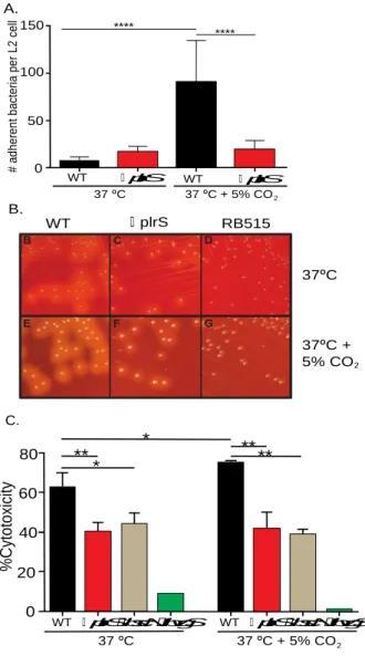
PlrS is required for enhanced BvgAS-dependent virulence-associated phenotypes in response to elevated CO2 concentrations
BvgAS-regulated virulence-associated phenotypes include adherence to epithelial cells
and macrophages, which is mediated by FHA (20, 21), hemolysis on blood-containing agar,
which is mediated by ACT (22), and toxicity to various eukaryotic cell types in culture, which is
mediated by the T3SS (23). All of these phenotypes were enhanced when wild-type (WT) B.
bronchiseptica was grown at 37°C in 5% CO2 compared to growth at 37°C in ambient air (Fig.
2.1, Fig. 2.S1, and(19)). These virulence-associated phenotypes did not increase in response to
5% CO2 in the ∆plrS mutant, indicating that PlrS is required for this effect. The effect was not
due to acidification of the growth medium because acidification alone, without increased CO2,
did not result in increased adherence (Fig. 2.S2). These results suggest the possibility of a
functional interaction between PlrS and BvgAS.
PlrS likely affects BvgAS-dependent phenotypes via PlrR
A putative response regulator, which we are naming PlrR, is encoded immediately 3’ to
plrS. Attempts to disrupt or delete plrR (BB0265) were unsuccessful, suggesting plrR is essential
for cell viability under the growth conditions tested. As an alternate approach to determine if
PlrS and PlrR function as a cognate TCS, we delivered a plrR allele (plrRD52E) encoding a PlrR
protein in which the predicted site of phosphorylation, Asp52, was replaced with Glu, a predicted
phosphomimetic (24), to the attTn7 site in WT, ∆plrS, and ∆bvgS (which harbors a deletion in
bvgS)B. bronchiseptica. When grown in 5% CO2, the level of adherence of the ∆plrS strain
expressing plrRD52E to L2 cells was as high as that of WT B. bronchiseptica (and WT expressing
plrRD52E), demonstrating that the plrRD52E allele could complement a ∆plrS mutation and,
(Fig. 2.2). Increased adherence in the WT and ∆plrS strains expressing plrRD52E in bacteria
grown in ambient air is also consistent with the D52E substitution functioning as a
phosphomimetic, rendering PlrR constitutively active (Fig. 2.2). Lack of adherence by the ∆bvgS
strain with and without the plrRD52E allele in the presence or absence of 5% CO2 confirms that
bvgS is required for adherence to host cells and that PlrRD52E does not induce a
BvgAS-independent adherence activity (Fig. 2.2). These data provide evidence (but do not prove) that
PlrS and PlrR function as a canonical TCS, and that increased adherence in response to CO2 is
mediated by PlrS via PlrR. However, these data do not rule out the possibility that BvgA and
PlrR interact synergistically to affect expression of some genes or that PlrS directly interacts with
BvgS while also affecting the phosphorylation state of PlrR.
Positive autoregulation of the bvgAS operon is well-documented (25-28). Nonetheless, to
determine if PlrS affects BvgAS activity by controlling bvgAS transcription, we introduced a
PbvgA-gfp transcriptional reporter into the attTn7 site of WT and ∆plrS B. bronchiseptica and
measured GFP activity in bacteria grown at 37°C with and without 5% CO2. Under all conditions
tested, the levels of GFP, and therefore the expression of bvgAS, was not significantly different between WT and the ∆plrS strains (Fig. 2.S3). GFP levels in the ∆bvgS strain reflect activity
from the bvgA P2 promoter, which is expressed at a low level under Bvg– phase conditions
(providing the cell with a low amount of BvgAS so that it can respond, via positive
autoregulation, when Bvg+ phase conditions are encountered) (29). This low level expression of
PbvgA was not affected by the ∆plrS mutation. Together, these data suggest that PlrS, via PlrR,
B. bronchiseptica lacking plrS modulate to the Bvg– phase within the lower respiratory tract
Based on the functional link between PlrS and BvgAS (Fig. 2.1) and the similarly rapid
clearance of B. bronchiseptica ∆plrS and ∆bvgS mutants from the LRT of mice (13, 18), we
hypothesized that PlrS may influence BvgAS activity within the host. To test this hypothesis, we
used two reporter systems developed in our lab. pGFLIP contains a recombinase-based reporter
system, similar to RIVET (15). Expression of flp, encoding FLP recombinase, results in excision
of gfp and nptII (encoding kanamycin resistance, Kmr) genes located between FLP recombinase
target (FRT) sites and conversion of the bacteria from GFP+ and Kmr to GFP– and Kms. In
pGFLIP-flaA, the Bvg– phase-specific PflaA promoter is located 5’ to flp and conversion to GFP–
(and Kms) indicates that the bacteria have expressed the Bvg– phase at some point during the
experiment.
By contrast with pGFLIP, which reports on the history of the bacteria, the pBAM plasmid
reports on the status of the bacteria at the time of plating (29). The pBAM plasmid integrates
within the bvgAS promoter region and causes the P2 promoter to be expressed at a
lower-than-normal level such that, when bacteria are growing under Bvg– phase conditions, the amount of
BvgAS in a small proportion of cells in the population (~5%) is below the threshold required for positive autoregulation, “trapping” these cells in the Bvg– phase (29). When WT B.
bronchiseptica containing the pBAM plasmid are grown under Bvg– phase conditions and then
plated onto BG-blood agar and incubated at 37°C (Bvg+ phase conditions), the “trapped” bacteria
yield colonies that are larger, flatter, and less hemolytic than colonies formed by Bvg+ phase
bacteria because these colonies contain ~5% phenotypically Bvg– phase bacteria (as the colony
grows, ~95% of the daughter cells produce enough BvgAS to convert to the Bvg+ phase, while
~5% remain phenotypically Bvg– phase). We previously referred to these colonies, which are
because these colonies result from phenotypic bistability and not a genetic change, it is more
appropriate to refer to them as large colony phenotypes (LCPs), which we will do henceforth.
While all LCPs result from a founder bacterium that was Bvg– phase at the time of plating, ~95%
of bacteria that contain pBAM and are Bvg– phase at the time of plating form typical Bvg+
phase-appearing (non-LCP) colonies (29). Hence, although the presence of LCPs indicates that
bacteria have modulated to the Bvg– phase, it vastly underestimates the number of bacteria that
are Bvg– phase at the time of plating.
When grown at 37°C without addition of MgSO4 to the medium (Bvg+ phase conditions),
wild-type and ∆plrS strains containing both reporter systems maintained GFPfluorescence and
no LCPs formed (Fig. 2.S4A and 2.S4B), indicating that neither strain modulated to the Bvg–
phase. When switched from Bvg+ phase growth conditions to Bvg– phase growth conditions, both
strainslost GFP fluorescence and a small proportion of LCPs formed after 24 hours (Fig. 2.S4A
and 2.S4B). Both reporters, therefore, can accurately report that the bacteria modulated to the
Bvg– phase and ∆plrS mutants modulate to a similar extent as wild-type bacteria when grown in
vitro.
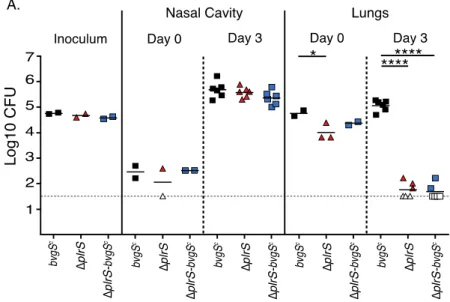
Following intranasal inoculation of BALB/cJ mice, the numbers of colony forming units
(cfu) recovered from the nasal cavities and lungs of wild-type and ΔplrS strains containing the
reporters were similar to cfu recovered of the parental strains (without reporters), indicating that
the reporters do not influence virulence (Fig. 2.3A). The proportion of GFP– and LCP cfu
recovered from the nasal cavities was extremely low for both strains, indicating that the bacteria
did not modulate to the Bvg– phase at this site (Fig. 2.3B and 2.3C). The proportion of GFP– and
LCP cfu recovered from the lungs of mice inoculated with wild-type bacteria was also extremely
strain had lost GFP fluorescence and 10-80% formed LCPs by day 1 post-inoculation (Fig. 2.3B
and 2.3C), indicating that a majority of these bacteria had modulated to the Bvg– phase and that a
majority of these bacteria were in the Bvg– phase at the time of recovery from the lungs. These
findings demonstrate that in strains lacking plrS, the BvgAS phosphorelay fails to remain active
specifically within the LRT. Moreover, the fact that 10-80% of the bacteria recovered from the
lungs formed LCPs (i.e., much more than 5%) indicates that the physiology of ∆plrS bacteria in the LRT is substantially different from the physiology of ∆plrS bacteria that have modulated to
the Bvg– phase in vitro by chemical modulators such as MgSO4 (see discussion).
B. bronchiseptica lacking plrS fail to activate BvgAS within the lower respiratory tract
To determine if BvgAS can transition from an inactive (Bvg– phase) to an active (Bvg+
phase) state in the absence of PlrS activity within the LRT, we inoculated mice with wild-type and ∆plrS mutants containing a pGFLIP reporter in which the Bvg+ phase-specific ptxA promoter
from B. pertussis was cloned 5’ to flp. Both wild-type and ∆plrS strains activated the ptxA
promoter when switched from Bvg– phase to Bvg+ phase growth conditions in vitro (Fig. 2.S5).
Similar to what has been shown in B. bronchiseptica and B. pertussis previously (15, 16), 100%
of the bacteria recovered from both the nasal cavity and the lungs of mice inoculated with Bvg–
phase wild-type bacteria activated PptxA and therefore transitioned to Bvg+ phase within 24 hours
post-inoculation (Fig. 2.3D and 2.3E). By contrast, although all of the cfu recovered from the nasal cavities of mice inoculated with the ∆plrS strain were GFP– by 24 hours post-inoculation,
only ~80% of those recovered from the lungs were GFP– (Fig. 2.3D and 2.3E). While, based on this reporter, only a seemingly small proportion of the ∆plrS mutants failed to switch to the Bvg+
phase in vivo, it is important to note that the numbers of cfu of the ∆plrS mutant recovered from
present (day 0). It is impossible to determine if the bacteria cleared from the lungs at these time
points had lost GFP. However, if they remained GFP+ (indicating that they did not switch to the
Bvg+ phase) then the proportion of ∆plrS bacteria that had transitioned to the Bvg+ phase in the
LRT would in fact be far less than 1%. Together, our data indicate that full activation and
maintenance of BvgAS activity in the LRT requires PlrS.
PlrS is required for B. bronchiseptica persistence in the lower respiratory tract, independent of its effects on BvgAS activity
Lack of production of BvgAS-dependent virulence factors could be the reason that plrS
mutants are cleared rapidly from the LRT. To test this hypothesis, we introduced the bvgS-C3
mutation, which encodes a BvgS protein that is active even under modulating conditions in vitro
(12), into the plrS mutant, and compared this ∆plrS-bvgSc strain with the bvgSc mutant in vitro
and in vivo. Like the bvgSc strain, the ∆plrS-bvgSc strain formed small, domed, hemolytic
colonies characteristic of the Bvg+ phase on BG-blood agar containing 50 mM MgSO4 (i.e., Bvg–
phase conditions), indicating that BvgAS was constitutively active in the absence of plrSin vitro.
In vivo, the ∆plrS-bvgSc strain colonized and persisted in the nasal cavity similarly to the bvgSc
and ΔplrS strains(Fig. 2.S6). However, the ∆plrS-bvgSc strain was cleared from the lungs as rapidly as the ∆plrS mutant (Fig 2.S6), indicating that the ∆plrS mutation is epistatic to the
bvgS-C3 mutation with regard to persistence in the LRT.
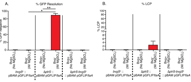
To investigate BvgAS activity in the ∆plrS-bvgSc strain, we used our pGFLIP-flaA and
pBAM reporters. None of the bvgSc or ∆plrS-bvgSc colonies containing these reporters were
GFP– or displayed the LCP phenotype after growth in medium containing 50 mM MgSO4 (Fig.
2.S7), indicating their insensitivity to modulating conditions in vitro. Numbers of cfu of each
strain recovered from the nasal cavities and lungs of BALB/cJ mice were similar to those of the
GFP– or LCP colonies were recovered from the nasal cavity for any strain, indicating that no
bacteria had modulated to the Bvg– phase in the nasal cavity (Fig. 2.4B and 2.4C). As expected, a
significant proportion of GFP– colonies and LCPs were recovered from the lungs of mice
inoculated with the ∆plrS mutant on days 1 and 3, consistent with our previous results (Fig. 2.4B
and 2.4C). By contrast, no GFP– or LCP colonies were recovered from the lungs of mice
inoculated with the bvgSc or ∆plrS-bvgSc mutants (Fig. 2.4B and 2.4C), indicating that neither
strain modulated to the Bvg– phase in the LRT. Therefore, modulation to the Bvg– phase and
lack of BvgAS-activated virulence factors is not the only reason that ∆plrS mutants fail to persist
in the LRT. These data indicate that PlrS is required for bacterial persistence in the LRT even
when BvgAS remains active, likely because PlrR (presumably phosphorylated PlrR, PlrR~P)
activates expression of one or more genes encoding proteins that are required in this environment
or because PlrR~P represses expression of one or more genes that encode proteins that are
detrimental to survival in this environment.
PlrS is required for survival and persistence of B. pertussis in the lower respiratory tract
The plrSR locus is highly conserved (≥99% identical) among all strains of the classic
bordetellae (B. pertussis, B. bronchiseptica and B. parapertussis). We constructed a derivative of
B. pertussis strain BP536 with a large in-frame deletion mutation in plrS and compared it with
wild-type BP536 in BALB/cJ mice. Both strains colonized the nasal cavity at similar levels, and
were cleared from this site by day 3 post-inoculation (Fig. 2.5). However, while approximately
103 cfu of BP536 were recovered from the lungs on days 1 and 3 post inoculation, significantly
fewerBP536ΔplrS cfu were recovered from this site at these time points (Fig. 2.5). These data
indicate that similar to B. bronchiseptica, plrS is requiredfor the survival and persistence of B.
Discussion
BvgAS has been considered the master virulence regulator in Bordetella since its
identification in 1983 (30) and demonstration of its penetrance by subsequent mutagenesis and
genome-wide analyses (8, 31, 32). Our new data indicate, however, that in the LRT, BvgAS
activity depends on PlrS, likely via the activity of PlrR. Moreover, PlrS(R) is required for
bacterial survival in the LRT even when BvgS is rendered constitutively active, strongly
suggesting that one or more PlrSR-dependent, BvgAS-independent genes (or genes that require
both PlrSR and BvgAS) is required for bacterial survival at this site. PlrS(R) is therefore at least
as important for Bordetella virulence as BvgAS.
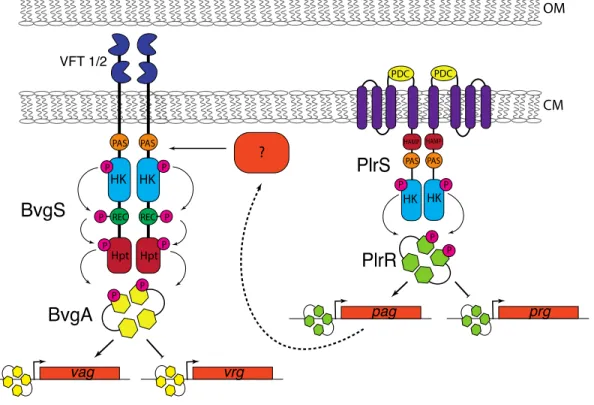
Why is PlrS(R) required for bacterial persistence in the LRT? PlrSR belongs to the NtYX
family of TCS. NtrY family proteins, including PlrS, are predicted to contain three
transmembrane domains at their N-termini, followed by a periplasmically-located
PhoP-DcuS-CitA (PDC) domain, another transmembrane domain, then cytoplasmically-located HAMP,
Per-Arnt-Sim (PAS) and HisKA-type histidine kinase domains (Fig 2.6). NtrY of Brucella abortus
has been shown to bind heme via its PAS domain and to function as a redox sensor, becoming
active as a kinase in response to anaerobiosis (33). NtrX family response regulators contain
N-terminal receiver and C-N-terminal DNA binding domains. In Neisseria gonorrhoeae and
Rhodobacter capsulatus, the ntrX genes are required for induction of high-affinity cytochrome
oxidases, which are required for bacterial growth under low oxygen conditions (34). Our data
indicate that PlrS is required specifically in the LRT, an environment that is low in oxygen and
high in CO2. Although we have been unable to delete plrRin vitro, our experiments using
PlrRD52E indicate that PlrS affects BvgAS-dependent phenotypes in vitro via PlrR (providing
far, we hypothesize that PlrS senses low oxygen (and perhaps increased CO2) in the LRT,
phosphorylates PlrR, and that PlrR~P activates expression of one or more of the high-affinity
cytochrome oxidase-encoding loci present in B. bronchiseptica (and B. pertussis) (35), allowing
the bacteria to respire in this environment (Fig. 2.6). We note, however, that NtrY family
members, including PlrS, contain HisKA-type DHp domains with ExxN motifs that suggest that
these proteins possess both kinase and phosphatase activities (36-38). Although our data suggest
that PlrS may function to phosphorylate PlrR in vitro, especially in 5% CO2, the contributions of
its predicted kinase and phosphatase activities in vivo cannot be predicted from our current data.
We are currently investigating the role of PlrS activities in vitro and in vivo.
Why is PlrS(R) required for BvgAS activity in the LRT? Positive autoregulation by
BvgAS has been well-characterized (25-28, 39) and we showed here that bvgAS expression in
both ambient air and 5% CO2 is not dependent on plrS. Our experiments with the bvgScmutant
further indicate that bvgAS expression is not dependent on PlrS(R) in vivo because BvgAS in the ∆plrS-bvgSc strain, which contains a single nucleotide substitution in bvgS, rendering the BvgS
protein constitutively active, was active in the LRT. If bvgAS transcription required PlrSR, the ∆plrS mutation would be epistatic to the bvgS-C3 mutation. Our data therefore rule out the
possibility that PlrS(R) controls BvgAS activity by controlling bvgAS transcription. We
hypothesize that instead, PlrSR controls expression of one or more genes that encode proteins
required for BvgS activity specifically in the LRT. BvgS contains three predicted signal-sensing
domains; two periplasmically-located venus fly-trap (VFT) domains and a
cytoplasmically-located PAS domain (40, 41). While the VFT domains appear to convert BvgS into a
phosphatase that dephosphorylates BvgA in response to modulating conditions (i.e., MgSO4 or
biochemical analyses using the cytoplasmic portion of BvgS suggested that the redox state of
ubiquinone could affect BvgS kinase activity (45). PlrSR-dependent production of high-affinity
cytochrome oxidases would both allow electron transport-coupled oxidative phosphorylation to
occur in the LRT, which is required for both ATP production (and hence cell viability), and
prevent the accumulation of reduced ubiquinone, which could inactivate BvgS. This model is
consistent with that proposed for the ArcB protein of Escherichia coli, which is activated by low
oxygen conditions (46, 47). If true, this model would predict that a role for the PAS domain in
sensing signals in vitro may have been missed because the activity of low-affinity (high
efficiency) cytochrome oxidases present under all of the conditions tested would keep reduced
ubiquinone levels at a minimum. These low-affinity cytochrome oxidases are also likely present
and sufficient for respiration in the URT, making BvgS activity and bacterial survival at this site
independent of PlrS.
Why was the proportion of LCPs recovered from the lungs of mice inoculated with the ∆plrS strain so much higher than the proportion obtained after switching the bacteria from Bvg–
phase conditions to Bvg+ phase conditions in vitro? Our previous studies showed that the reason
that ~5% of bacteria containing the pBAM plasmid integrated at the bvgAS promoter region form
LCPs when shifted from Bvg– phase conditions to Bvg+ phase conditions in vitro, is that activity
of the P2 promoter in this strain is decreased such that ~5% of the bacteria have BvgAS levels
below the threshold required to respond to Bvg+ phase conditions when they are encountered. It
appeared, therefore, that the maximum proportion of LCPs that would form from a population of
pBAM-containing bacteria in which 100% of the bacteria had modulated to the Bvg– phase was