Balancing Trained Immunity with
Persistent Immune Activation and the
Risk of Simian Immunodeficiency Virus
Infection in Infant Macaques Vaccinated
with Attenuated
Mycobacterium
tuberculosis
or
Mycobacterium bovis
BCG
Vaccine
Kara Jensen,
aMyra Grace dela Pena-Ponce,
aMichael Piatak, Jr.,
b†
Rebecca Shoemaker,
bKelli Oswald,
bWilliam R. Jacobs, Jr.,
cGlenn Fennelly,
dCarissa Lucero,
bKatie R. Mollan,
eMichael G. Hudgens,
fAngela Amedee,
gPamela A. Kozlowski,
gJacob D. Estes,
bJeffrey D. Lifson,
bKoen K. A. Van Rompay,
hMichelle Larsen,
cKristina De Paris
aDepartment of Microbiology and Immunology and Center for AIDS Research, School of Medicine, University of North Carolina, Chapel Hill, North Carolina, USAa; AIDS and Cancer Virus Program, Leidos Biomedical Research,
Inc., Frederick National Laboratory for Cancer Research, Frederick, Maryland, USAb; Albert Einstein College of
Medicine, New York, New York, USAc; Rutgers New Jersey Medical School, Newark, New Jersey, USAd;
Lineberger Cancer Center and Center for AIDS Research, University of North Carolina, Chapel Hill, North Carolina, USAe; Gillings School of Global Public Health and Center for AIDS Research, University of North
Carolina, Chapel Hill, North Carolina, USAf; Department of Microbiology, Immunology, and Parasitology,
Louisiana State University Health Sciences Center, New Orleans, Louisiana, USAg; California National Primate
Research Center, University of California, Davis, Davis, California, USAh
ABSTRACT
Our goal is to develop a pediatric combination vaccine to protect the
vulnerable infant population against human immunodeficiency virus type 1 (HIV-1)
and tuberculosis (TB) infections. The vaccine consists of an auxotroph
Mycobacterium
tuberculosis
strain that coexpresses HIV antigens. Utilizing an infant rhesus macaque
model, we have previously shown that this attenuated
M. tuberculosis
(AMtb)-simian
immunodeficiency virus (SIV) vaccine is immunogenic, and although the vaccine did
not prevent oral SIV infection, a subset of vaccinated animals was able to partially
control virus replication. However, unexpectedly, vaccinated infants required fewer
SIV exposures to become infected compared to naive controls. Considering that the
current TB vaccine,
Mycobacterium bovis
bacillus Calmette-Guérin (BCG), can induce
potent innate immune responses and confer pathogen-unspecific trained immunity,
we hypothesized that an imbalance between enhanced myeloid cell function and
immune activation might have influenced the outcome of oral SIV challenge in
AMtb-SIV-vaccinated infants. To address this question, we used archived samples
from unchallenged animals from our previous AMtb-SIV vaccine studies and
vacci-nated additional infant macaques with BCG or AMtb
only. Our results show that
vac-cinated infants, regardless of vaccine strain or regimen, had enhanced myeloid cell
responses. However, CD4
⫹T cells were concurrently activated, and the persistence
of these activated target cells in oral and/or gastrointestinal tissues may have
facili-tated oral SIV infection. Immune activation was more pronounced in BCG-vaccinated
infant macaques than in AMtb-vaccinated infant macaques, indicating a role for
vac-cine attenuation. These findings underline the importance of understanding the
in-terplay of vaccine-induced immunity and immune activation and its effect on HIV
acquisition risk and outcome in infants.
Received11 July 2016Returned for modification29 July 2016 Accepted12 September 2016
Accepted manuscript posted online21 September 2016
CitationJensen K, dela Pena-Ponce MG, Piatak M, Jr, Shoemaker R, Oswald K, Jacobs WR, Jr, Fennelly G, Lucero C, Mollan KR, Hudgens MG, Amedee A, Kozlowski PA, Estes JD, Lifson JD, Van Rompay KKA, Larsen M, De Paris K. 2017. Balancing trained immunity with persistent immune activation and the risk of simian immunodeficiency virus infection in infant macaques vaccinated with attenuated
Mycobacterium tuberculosisorMycobacterium bovisBCG vaccine. Clin Vaccine Immunol 24: e00360-16. https://doi.org/10.1128/CVI.00360-16.
EditorHelene F. Rosenberg, IIS/LAD/NIAID/NIH
Copyright© 2017 American Society for Microbiology. All Rights Reserved.
Address correspondence to Kristina De Paris, [email protected].
†Deceased.
For a commentary on this article, see https:// doi.org/10.1128/CVI.00509-16.
KEYWORDS
pediatric HIV/SIV, vaccine, immune activation, myeloid cells
H
uman immunodeficiency virus (HIV) and tuberculosis (TB) infections remain highly
prevalent in many parts of the world and often show substantial geographical
overlap. Infants represent one of the populations most vulnerable to both diseases. An
HIV vaccine is not yet available, and although a vaccine to prevent TB infection, the live
attenuated
Mycobacterium bovis
bacillus Calmette-Guérin (BCG) vaccine, is routinely
administered to
⬎
80% of neonates globally and is effective at preventing the most
severe complications of TB, BCG efficacy wanes with age (1–3). In HIV-infected infants
and infants at risk for HIV, the BCG vaccine is contraindicated due to the potential of
inducing disseminated BCG disease (4). Thus, both a new TB vaccine and a pediatric HIV
vaccine are urgently needed to reduce infant mortality and morbidity in areas of high
HIV and TB prevalence. Toward this goal, we aimed to develop a pediatric combination
HIV-TB vaccine that could protect infants against HIV and TB infections. Our vaccine was
intentionally designed to retain the immunogenicity of BCG but was more attenuated
to exhibit an improved safety profile, while expressing HIV antigens to induce both
HIV-specific and TB-specific immune responses. Further, we opted to utilize an
atten-uated human-adapted
Mycobacterium tuberculosis
for improved immune memory,
rather than further edit the
M. bovis
backbone of BCG.
Our pediatric TB-HIV combination vaccine is based on an auxotroph mutant of
Mycobacterium tuberculosis
that has deletions in the
panCD,
leuCD, and
secA2
loci that
render this attenuated
M. tuberculosis
(AMtb) strain mc
26208 replication incompetent
(5–7). In fact, because these bacteria are unable to synthesize essential nutrients
(panthothenate and leucine), replication in unsupplemented conditions, such as a
mammalian host, can occur only temporarily until the cell reserves are depleted. In
contrast, BCG does not harbor auxotroph mutations and can replicate freely until
thwarted by the host immune system. Using experimental simian immunodeficiency
virus (SIV) infection of infant rhesus macaques, an animal model of pediatric HIV
infection, we have previously demonstrated that this AMtb
vaccine, in contrast to BCG,
does not cause mycobacterial dissemination or TB tissue pathology (8). Further
mod-ification of the mc
26208 vaccine strain to coexpress SIVgag (mc
26435) or SIVenv
(mc
26439) rendered this vaccine capable of inducing both TB- and SIV-specific T cell
and antibody responses in infant macaques (9, 10). Although these vaccine-induced
SIV-specific immune responses were not sufficient to prevent oral SIV acquisition, a
subset of vaccinated infant macaques with higher-avidity plasma SIV Env-specific IgG
antibodies and SIV Env-specific mucosal IgA antibodies showed better control of SIV
replication upon infection (9). At the same time, unexpectedly, the group of vaccinated
infant macaques had a twofold-higher SIV infection risk per exposure compared to
unvaccinated infants (9). Despite not being statistically significant, the difference in the
per exposure risk of SIV infection was reminiscent of increased CD4
⫹T cell activation,
and thereby a potentially increased risk of HIV infection, seen in South African infants
after BCG vaccination (11, 12).
candidates should aim to preserve these unique features of BCG. In fact, among the
new TB vaccine candidates under development, several are based on additional
attenuations of
M. bovis
within the existing BCG vaccine or on attenuated
M.
tubercu-losis
strains similar to the auxotroph AMtb
vaccines used here (17–21). Recombinant
BCG strains expressing HIV antigens are also in development as potential HIV vaccines
and have been tested in mice (22–24) and in adult (25–29) and neonatal (30) macaques.
Considering the high risk of HIV and TB infection in infants, especially in sub-Saharan
Africa, we deemed it important to understand how novel TB vaccines or combination
HIV-TB vaccines might impact HIV acquisition.
In a first attempt to better define the beneficial effects of mycobacterial vaccines
and to weigh them against potentially negative outcomes on infant HIV infection risk,
we report here studies conducted on archived tissue samples from animals immunized
with the AMtb-SIV prime/modified vaccinia Ankara (MVA)-SIV boost regimen that were
not challenged with SIV (10) or samples from animals included in our recent efficacy
studies that were collected prior to or at the time of the first SIV challenge (9). To
exclude any effects of the vaccine booster component or the SIV inserts on oral SIV
acquisition, we expanded upon those historical studies by vaccinating infant macaques
with AMtb
vaccines only (AMtb-SIV prime plus a homologous boost) or animals primed
only with AMtb-SIV, the AMtb
strain without SIV cassettes, or the human infant regimen
of BCG. Specifically, the current study examined the potential of our AMtb
vaccine to
increase functional responses of infant monocytes/macrophages and whether
en-hanced myeloid cell function was associated with immune activation that could
potentially predispose infants to SIV infection.
RESULTS
Increased functional responses in myeloid cell population of A
Mtb
-vaccinated
infant macaques.
In human adults, the persistence of heightened monocyte function
has been described as a hallmark of BCG-induced trained immunity (15, 31, 32). To
determine whether our AMtb
vaccine mc
26435 (expressing SIVgag) could also enhance
monocyte responses and whether it could do so in the pediatric setting, we utilized
archived peripheral blood mononuclear cell (PBMC) and tissue samples from
SIV-uninfected infant macaques that were vaccinated with strain mc
26435 at birth, boosted
tissues are presumed sites of entry and early SIV replication after oral exposure and our
goal is to develop a pediatric HIV-TB combination vaccine, we deemed it important to
test whether the AMtb
vaccination had also resulted in CD4
⫹T cell activation.
Prolonged CD4
ⴙT cell activation after A
Mtb
vaccination of infant macaques.
Indeed, 16 to 18 weeks after vaccination with AMtb, the frequencies of CD4
⫹CCR5
⫹T
cells were significantly higher in PBMC, retropharyngeal LN, and colons of
AMtb-vaccinated animals than in naive controls (Fig. 2). In addition, frequencies of Ki-67
⫹,
PD-1
⫹, and CD69
⫹CD4
⫹T cells were generally higher in retropharyngeal LN and colon
cell suspensions of cells from AMtb-vaccinated infants than in those from naive controls
FIG 1Increased functional responsiveness of monocytes/macrophages and mDC. (A and B) Frequencies of IL-12- or TNF-␣-producing monocytes/macrophages and mDC, respectively, in PBMC from naive controls and AMtb-vaccinated infant macaques at 16 to 18 weeks after vaccination. (C) Frequencies of CCR5⫹or
CD69⫹peripheral blood monocytes/macrophages from the same animals. (D and E) Frequencies of TNF-␣- or IL-12-producing monocytes/macrophages (D) and
(Fig. 2). Although only PBMC, retropharyngeal LN, and colons were available for this
retroactive analysis, these data further confirmed that the AMtb-induced immune
activation persisted for several weeks to months after vaccination and provided
evi-dence that SIV target cell frequencies at presumed sites of viral entry and/or early
replication were concurrently increased. These findings of persistent AMtb
vaccine-induced immune activation in myeloid cell and CD4
⫹T cell populations in blood and
tissues reiterated the important question of whether vaccine-induced immune
activa-tion could enhance the risk for HIV/SIV infecactiva-tion, as suggested by the results of the STEP
HIV vaccine trial (35–37).
Study design to test the association between A
Mtb
or BCG vaccines and oral
SIV challenge outcome.
We recently reported that an AMtb-SIV prime/MVA-SIV boost
regimen did not protect infant macaques against oral SIV infection; furthermore,
vaccinated animals appeared to require fewer oral SIV exposures to become infected
compared to naive controls (9). The current study was designed to include additional
control groups to more conclusively determine whether mycobacterial vaccines would
predispose infant macaques to SIV infection. Vaccine-induced adaptive immune
re-sponses were previously reported (9, 10) and were not assessed in these cohorts. Here,
we tested whether mycobacterial vaccines could induce innate immune responses and
immune activation. As outlined in Fig. 3, control animals consisted of nine infant
macaques that received placebo (phosphate-buffered saline [PBS]) immunization
(group C2) and also included the six historical control animals (group C1) from our
previous study (9). Similar to the vaccinees in the former study (group D), infant
macaques in group E were immunized at birth with mc
26435 and mc
26439, but instead
of receiving heterologous MVA-SIV boosts, the animals received the same mc
26435 and
mc
26439 vaccines (homologous boost) at week 3 by the intradermal (i.d.) route, the
current route of BCG vaccination. To exclude other confounding factors, such as SIV
antigen expression by AMtb
vaccines, group F infant macaques were vaccinated with
a single dose of mc
26208, the parental vaccine strain of mc
26435/mc
26439 that does
not contain SIV expression cassettes. A group of single-dose BCG-vaccinated infants
was also included (group G) as a control for vaccine-induced enhanced myeloid cell
function and to determine whether any of the effects described here for experimental
AMtb
vaccines may have translational potential for novel TB vaccine in human infants.
Weekly repeated low-dose oral SIV
mac251challenges were started at week 9 (Fig. 3).
On the basis of our observations of persistent immune activation in blood and tissues
at 16 to 18 weeks after AMtb
vaccination of SIV-unexposed infant macaques, we asked
whether we could detect activated myeloid cell populations or increased numbers of
CCR5-positive CD4
⫹T cells (preferred targets for SIV infection) at the time of challenge
(week 9).
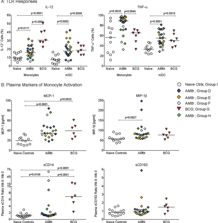
Increased function of peripheral blood monocytes/macrophages and mDC of
infant macaques vaccinated with A
Mtb
or BCG vaccine at the start of oral SIV
challenges.
To address this question, we stimulated PBMC that were collected at the
time of the start of repeated low-dose oral SIV exposures (week 9) with R848. To
increase our sample size, we included a group of infant macaques that received a single
immunization with mc
26435 at birth (group H [Fig. 3]); this group was not included in
the SIV challenge analysis, because these infants were challenged with a high dose of
SIV
mac251. Indeed, the frequencies of IL-12-producing monocytes were significantly
higher in PBMC from AMtb- or BCG-vaccinated infant macaques than from naive control
animals, whereas TNF-
␣
responses were increased only in AMtb-vaccinated infants (Fig.
4A). Infant macaques vaccinated with AMtb
or BCG also had significantly higher
frequencies of IL-12- or TNF-
␣
-producing mDC than naive controls (Fig. 4A).
We next tested whether these enhanced functional responses of
monocytes/mac-rophages and mDC were associated with systemic immune activation. Week 9 plasma
samples were analyzed for a panel of cytokines and chemokines that included several
proinflammatory markers. However, none of the cytokines typically associated with
inflammation and immune activation, e.g., TNF-
␣
, IL-6, IL-1, and IL-8, were elevated in
vaccinated infant macaques. In fact, out of 37 cytokines, chemokines, and growth
factors tested, only macrophage chemoattractant 1 (MCP-1) and macrophage
inflam-matory protein 1 (MIP-1

) were significantly increased in the plasma of vaccinated
animals (Fig. 4B). Both of these cytokines represent markers of monocyte activation.
Therefore, we performed further testing for soluble CD14 (sCD14) and sCD163 that are
produced by activated monocytes. To account for the large variation in absolute levels
between animals and to focus our analysis on vaccination-related changes, we used the
ratio of plasma sCD14 and sCD163 levels at week 9 normalized to those at week 0 for
each animal. Animals vaccinated with AMtb
or BCG had a significant increase in sCD14
plasma levels, with the increase in sCD14 being significantly higher in BCG-vaccinated
compared to AMtb-vaccinated infant animals (Fig. 4B). Vaccination was not associated
with increased sCD163 plasma levels (Fig. 4B). Consistent with our conclusion that the
mycobacterial vaccine component(s), and not MVA boosting or SIV antigen expression,
was driving myeloid cell activation, we observed increased functional responses and
elevated plasma cytokine levels when we compared each of the AMtb
vaccine groups
separately to the naive control animals at week 9 (see Fig. S1 in the supplemental
material).
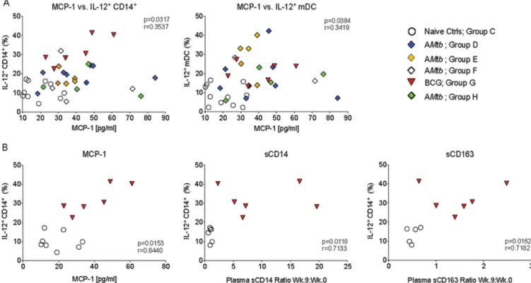
To determine whether the elevated plasma cytokine levels were at least partially due
to myeloid cell activation, we tested whether changes in plasma cytokine levels were
correlated with functional measurements of myeloid cells at week 9. Indeed, the
frequencies of IL-12-producing peripheral blood monocytes/macrophages and mDC
were positively correlated with plasma MCP-1 levels (Fig. 5A), but not MIP-1

level (data
not shown). This correlation was maintained when we analyzed the relationship
between IL-12-positive (IL-12
⫹) monocytes/macrophages of BCG-vaccinated animals
and naive control animals to plasma MCP-1 (Fig. 5B), but it was not statistically
significant when we tested naive controls and AMtb-vaccinated infants (P
⫽
0.0940)
(data not shown). We also observed positive correlations between plasma sCD14 and
sCD163 week 9/week 0 ratios and week 9 IL-12
⫹monocyte/macrophage frequencies in
BCG-vaccinated infants, but not in AMtb-vaccinated infants (Fig. 5B).
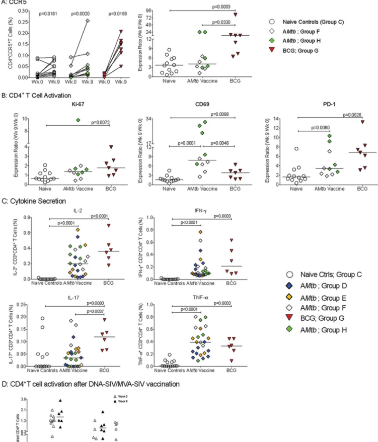
Vaccine-induced CD4
ⴙT cell activation at the start of oral SIV challenges.
Analysis of longitudinal blood samples showed that all AMtb- and BCG-vaccinated
infant animals experienced an increase in CD4
⫹CCR5
⫹T cells after vaccination. However,
consistent with the changing ratio of naive to antigen-experienced CD4
⫹T cells as part
of normal immune development, the frequencies of CCR5
⫹CD4
⫹T cells increased in
all groups between week 0 and week 9 (Fig. 6A). Thus, it was important to distinguish
between changes in peripheral blood immune cell populations that reflect normal
FIG 6PBMC CD4⫹T cell activation at week 9. (A) Frequencies of CCR5⫹CD4⫹T cells for individual animals in each group at week 0 (time of immunization)
and week 9 (start of SIV challenge);Pvalues were determined by Wilcoxon signed-rank test. The right graph shows the ratio of CD4⫹CCR5⫹T cell frequencies
postnatal immune development (38) and vaccine-induced immune activation.
There-fore, we report all changes in activated CD4
⫹T cell frequencies as ratios of week 9
values compared to week 0 values for each animal. This analysis revealed that
BCG-vaccinated infants had significantly higher frequencies of CD4
⫹CCR5
⫹T cells than
naive controls at 9 weeks of age (Fig. 6A). Although CD4
⫹CCR5
⫹T cells were not
increased in AMtb-vaccinated infants, vaccine-induced CD4
⫹T cell activation was
suggested by significantly higher frequencies of CD69
⫹and PD-1
⫹CD4
⫹T cells
compared to the naive controls (Fig. 6B). BCG-vaccinated infants showed a significant
rise in Ki67
⫹, CD69
⫹, and in PD-1
⫹CD4
⫹T cells (Fig. 6B).
Immune activation of CD4
⫹T cells was further substantiated by higher levels of
endogenous cytokines in
ex vivo
unstimulated CD4
⫹T cells from vaccinated animals
compared to unvaccinated infant macaques (Fig. 6C). Compared to naive controls,
CD4
⫹T cells from both AMtb- or BCG-vaccinated infants had significantly increased
levels of IL-2, gamma interferon (IFN-
␥
), and TNF-
␣
at week 9, while IL-17A levels were
increased only in the BCG-vaccinated infants (Fig. 6C). It should be pointed out that this
analysis was performed on
ex vivo
isolated cells without any stimulation and that the
measured frequencies of cytokine-producing CD4
⫹T cells exceeded those of
antigen-specific (both SIV and TB) CD4
⫹T cells (9, 10), and thus are consistent with immune
activation as opposed to memory responses. Furthermore, an analogous analysis of
PBMC samples collected from infant macaques vaccinated with a DNA-SIV
prime/MVA-SIV boost regimen did not reveal similar CD4
⫹T cell activation (Fig. 6D). These results
were confirmed by the statistical significance of separate comparisons of individual
AMtb
vaccine groups to naive control animals (Fig. S2), indicative of mycobacterial
vaccine-driven CD4
⫹T cell activation.
These combined data show that early vaccination with highly replication-attenuated
auxotroph
M. tuberculosis
vaccine strains or with attenuated, but replication-competent
BCG, induced activation in myeloid cells and in CD4
⫹T cells that was clearly evident at
9 weeks after vaccination, the time of the first oral SIV challenge. While we focused our
discussion of mycobacterial-vaccine-induced immune activation on myeloid cells and
CD4
⫹T cells as the cell types most relevant to SIV acquisition, vaccine-induced immune
activation was also observed in CD8
⫹T cells and in B cells (Fig. S3), underlining the
global nature of systemic immune perturbation, even months after mycobacterial
vaccination. Functional differences of CD8
⫹T cells and B cells between naive controls
and AMtb- or BCG-vaccinated infants could not be further explored within the scope of
the current study.
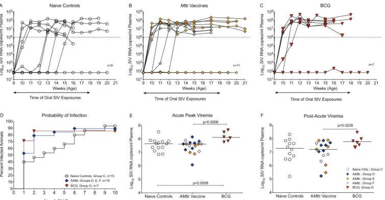
Challenge outcome in A
Mtb
- or BCG-vaccinated infant macaques using a
repeated low-dose oral SIV exposure model.
Similar to our recent study (9), in
which unvaccinated infants had an SIV infection risk per exposure of 0.24, the SIV
infection risk per exposure for naive controls was 0.25 in the current study. Thus, 4
of 9 naive controls (44%) became infected after 1 SIV exposure, with 1 animal each
requiring 3, 4, 5, or 6 exposures, and 1 animal failing to become persistently
infected after 10 oral SIV exposures (not persistently infected [NPI]) (Fig. 7A). In
contrast, AMtb-vaccinated infants in groups E and F tended to become infected
after fewer SIV exposures, with 8 of 11 animals (73%) becoming infected after only
one or two exposures (Fig. 7B) and 1 group F animal requiring four exposures. As
one infant each in groups E and F remained uninfected after 10 SIV exposures, the
SIV infection risk per exposure (0.34) in infant macaques vaccinated with AMtb
vaccines was lower than in our previous study using the AMtb-SIV prime/MVA-SIV
FIG 6Legend (Continued)
expressing Ki-67, CD69, or PD-1 from week 9 to week 0 (exact Mann-WhitneyPvalues). (C) Percentages of IL-2-, IFN-␥-, IL-17-, and TNF-␣-producing CD4⫹T
cells in animals from each experimental group at week 9 (exact Mann-WhitneyPvalues). (D) Percentages of CCR5-, Ki-67-, CD69-, PD-1-, and TNF-␣-positive CD4 T cells at week 12 in infant macaques who received an oral DNA-SIV vaccine at weeks 0 and 3 and were vaccinated with MVA-SIV at weeks 6 and 9 by both the intramuscular (i.m.) and sublingual route (DNA expressing SIVmac239was encapsulated in cationic liposomes and administered at 3 mg/animal into the
mouth; MVA expressing SIV Gag, Pol, and Env was administered at 108PFU by both the i.m. and sublingual route; K. De Paris, unpublished data). No statistically
boost regimen (exposure risk of 0.5) (9). Similar to AMtb, five of seven
BCG-vaccinated infants (71%) became infected after a single oral SIV exposure, while one
animal required two exposures and one animal remained uninfected for an
esti-mated probability of infection per oral SIV exposure of 0.35 (Fig. 7C).
Given the overall similarity in challenge outcome between the previous (9) and
current studies, we combined the animals of both studies to assess the risk of SIV
infection. These results translated into a per exposure risk of SIV infection of 0.25 for
naive controls (groups A and C) and 0.34 for infant animals receiving any kind of AMtb
vaccine (groups D, E, and F). As one unvaccinated infant (group C), two
AMtb-vaccinated infants (groups D to F) and one BCG-AMtb-vaccinated infant (group G) had to be
categorized as NPI, there was only a 1.4-fold increased risk of SIV infection per exposure
in vaccinated animals compared to naive control animals. Comparing the
AMtb-vaccinated animals (n
⫽
19) or the BCG-vaccinated animals (n
⫽
7) to the control
animals (n
⫽
15), we did not detect statistically significant differences in the number of
SIV exposures until infection (P
⫽
0.6 and
P
⫽
0.8, respectively; Fig. 7D). Yet,
descrip-tively, after only two low-dose oral SIV exposures, 79% of AMtb-vaccinated and 88% of
BCG-vaccinated infants were infected compared to 47% of naive controls. Furthermore,
BCG-vaccinated animals (group G) exhibited significantly higher acute viremia (weeks
1 to 3 postinfection) than AMtb-vaccinated animals or naive controls (Fig. 7E), and they
tended to have higher viremia during the period after the acute phase of infection
(weeks 5 to 7 postinfection; Fig. 7F).
To exclude genetic factors associated with increased susceptibility or resistance to
SIV infection or with disease outcome, we determined the tripartite motif-containing
protein 5 (TRIM5
␣
) and major histocompatibility complex (MHC) class I genotypes of
SIV-challenged animals. The
TRIM5
␣
genotypes
TFP/TFP
and
TFP/Cyp
confer resistance
to SIV infection, while the genotypes
Q/Q
and
Q/CypA
are associated with increased
susceptibility to infection (39, 40). The MHC alleles
Mamu A
*
01,
B
*
08, and
B
*
17
alleles
9 10 11 12 13 14 15 16 17 18 19 20 21 101 102 103 104 105 106 107 108 109 Weeks (Age) Log 10 SIV RNA copies/ml Plasma Naive Controls
Time of Oral SIV Exposures
n=9
A
0 1 2 3 4 5 6 7 8 9 10
0 10 20 30 40 50 60 70 80 90 100
No. of SIV Exposures
Percent I
n
fect
ed
Animals
Probability of Infection
Naive Controls; Group C, n=15 AMtb; Groups D, E, F, n=19 BCG; Group G, n=7
D
9 10 11 12 13 14 15 16 17 18 19 20 21 101 102 103 104 105 106 107 108 109 Weeks (Age) Log 10 SIV RNA copies/ml Plasma
Mtb Vaccines
n=11
Time of Oral SIV Exposures
B
Naive Controls AMtb Vaccine BCG 4 5 6 7 8 9 Log 10 SIV RNA copies/ml Plasma
Acute Peak Viremia
p=0.0006
p=0.0058
E
9 10 11 12 13 14 15 16 17 18 19 20 21 101 102 103 104 105 106 107 108 109 Weeks (Age) Log 10 SIV RNA copies/ml Plasma BCG n=7
Time of Oral SIV Exposures
C
Naive Controls AMtb Vaccine BCG 4 5 6 7 8 9 Log 10 SIV RNA copies/ml Plasma Post-Acute Viremia p=0.0235
Naive Ctrls.; Group C
AMtb ; Group F BCG; Group G AMtb ; Group E AMtb ; Group D
F
FIG 7Challenge outcome using a repeated low-dose oral SIV exposure model. (A) Plasma viremia of naive controls (group C2) after weekly low-dose oral SIVmac251exposures, starting at 9 weeks of age. (B) SIV challenge outcome for AMtb-vaccinated infants of group E (orange diamonds) and group F (empty
have been associated with improved control of viral replication and better disease
outcome, whereas allele
B
*
01
is associated with higher viremia (41–46). Genotypes
could not be evaluated prior to enrollment because animals were assigned to the
different groups within 3 days of birth. Animals that required more challenges or were
resistant to SIV infection showed no bias toward protective
TRIM5
␣
alleles or MHC class
I alleles, as calculated in the susceptibility score (Table 1). Conversely, animals infected
after fewer SIV exposures were not more likely to express alleles associated with greater
susceptibility to infection or with reduced control of viremia. Although none of the
animals in groups D and E expressed the protective
Mamu A
*
01
allele, “MHC protection”
refers to the ability to control virus replication better in infected animals; there are no
reports, to our knowledge, that conclusively show that the
Mamu A
*
01
genotype
protects against SIV infection. Similarly,
TRIM5
␣
alleles have not been associated with
resistance to infection with the pathogenic strains SIV
mac239or SIV
mac251(47). Therefore,
TABLE 1MHC class I andTRIM5␣genotypesGroup and
animal no. Sexa
MHC class I typeb
TRIM5␣ genotypec
Susceptibility scored
No. of SIVmac251
exposures
Age (wk) at euthanasia
Group Ce
44586 F B*01,B*17 Q/CypA ⫺1 4 16
44618 M B*01 TFP/TFP 0 3 16
44684 F A*02,B*08 TFP/TFP ⫹2 NPIf 20
44694 F B*01 TFP/TFP 0 1 15
44696 M B*17,B*29 TFP/TFP ⫹2 1 15
44698 F B*01,B*08 TFP/TFP ⫹1 1 14
44718 M A*01 TFP/Q ⫹1 6 20
44739 M Negativeg TFP/Q 0 5 20
44746 F A*01 TFP/Q ⫹1 1 19
Group Dh
Group E
42903 F Negative TFP/Q 0 1 21
42918 M A*01,B*01 TFP/TFP ⫹1 NPI 34
42925 F Negative TFP/Q 0 1 21
42943 M A*01 TFP/Q ⫹1 2 21
42947 F A*01 TFP/CypA ⫹2 1 20
42950 M Negative TFP/Q 0 1 20
Group F
44146 F B*17,B*29 TFP/Q ⫹1 1 17
44151 F Negative TFP/Q 0 2 17
44155 F B*01,B*17,B*29 TFP/CypA ⫹1 1 17
44159 F A*11,B*17,B*29 Q/Q 0 4 17
44160 F Negative TFP/Q 0 NPI 41
Group Gi
44165 M A*02,B*01 TFP/TFP 0 NPI 41
44166 M B*01 TFP/TFP 0 1 16
44169 M B*01 TFP/TFP 0 1 16
44176 M Negative TFP/TFP ⫹1 2 18
44180 F A*08 Q/Q ⫺1 1 18
44186 F A*02 TFP/TFP ⫹1 1 17
44195 M A*08 TFP/Q 0 1 17
aF, female; M, male.
bTheMamu A*01,B*08, andB*17alleles have been associated with better disease outcome, whereas alleleB*01is associated with higher viremia.
cTheTRIM5␣genotypesTFP/TFPandTFP/CypAconfer resistance to SIV, while the genotypesQ/QandQ/CypAare associated with increased susceptibility to infection. dProtective class I alleles and resistantTRIM5␣alleles (A*01,B*08,TFP/TFP, andTFP/CypA) were assigned a value of 1. Genotypes associated with increased
susceptibility or disease outcome received a score of⫺1. All other alleles were assigned a score of 0. The susceptibility score represents the sum of all scores for an individual animal (e.g., animal 42376 has MHC 1⫹0⫹1⫹0 and TRIM5␣of 0 for a score of⫹2).
eIn addition, group C includes the six naive control animals (group C1) described by Jensen et al. (9).
fNPI, not persistently infected.
gNegative, not positive for any of theMamuclass I alleles tested (A*01,A*02,A*08,A*11,B*01,B*03,B*04,B*08,B*17, andB*29).
hMHC class I andTRIM5␣genotypes are published in the study by Jensen et al. (9).
iNote that group H is not included in this table, because animals received a high-dose SIV challenge and were used for week 9 immune activation measurements, but
we concluded that the genotype of each animal was not a principal cause of enhanced
SIV
mac251acquisition in vaccinated animals.
Collectively, although statistically not significant, the SIV challenge results suggested
that infant macaques vaccinated with AMtb
vaccines or BCG showed a trend toward
enhanced SIV acquisition compared to vaccine-naive controls.
Persistence of vaccine-induced immune activation post-SIV challenge.
Al-though BCG-vaccinated animals had higher viremia after infection, CD4
⫹T cell loss did
not differ significantly between these animals, naive controls, and
AMtb-vaccinated
infant animals (data not shown). Histological analysis also revealed no differences in the
integrity of the mucosal epithelium between the naive controls and AMtb- or
BCG-vaccinated groups (data not shown). It is also important to note that mycobacteria
could not be recovered from the lung or tracheobronchial lymph nodes of any of the
animals at the time of euthanasia. Despite similar clinical outcome, differences in
activation of myeloid cells and CD4
⫹T cells between naive controls and AMtb- or
BCG-vaccinated infants were still apparent post-SIV infection. BCG-vaccinated animals
had higher frequencies of CCR5
⫹monocytes/macrophages in the submandibular
lymph nodes and in bronchoalveolar lavage (BAL) fluid than naive control infants did
(Fig. 8A). HIV and SIV infections have been associated with increases in CD14
⫹CD16
⫹cells (48, 49), and while the frequency of activated CD14
⫹CD16
⫹peripheral blood
monocytes in naive controls increased from a median of 4.2% at week 9 to 24.5% after
SIV infection (Fig. 8B), even higher frequencies of this cell population were observed in
blood and tissues of AMtb- and BCG-vaccinated infants. At the time of euthanasia,
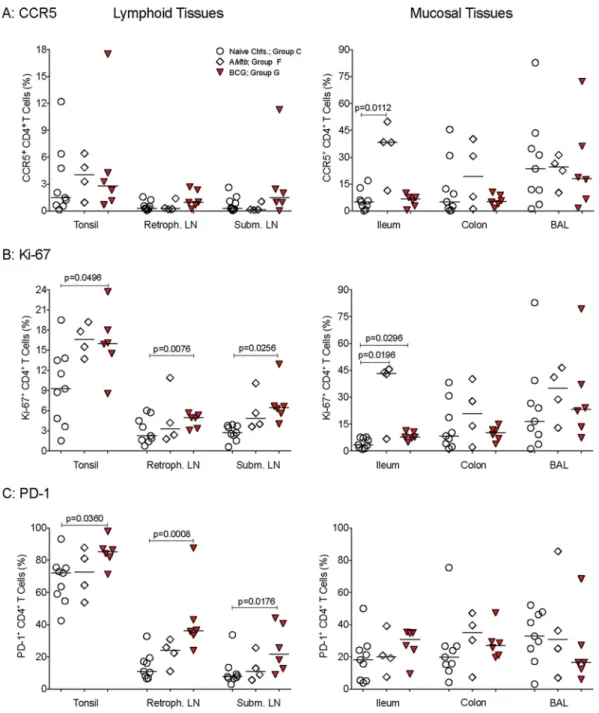
BCG-vaccinated animals also had higher frequencies of activated CD4
⫹T cells in
lymphoid tissues than naive controls did (Fig. 9), but differences were not consistently
observed in mucosal tissues. In contrast, tissue CD4
⫹T cell activation generally did not
FIG 8Myeloid cell activation in blood and tissue samples from vaccinated and SIV-infected infants. (A) Frequencies of CCR5⫹CD14⫹cells in lymphoid and
mucosal tissues at the time of euthanasia (Table 1) for animals in group C, F, and G. (B) Frequencies of CD16⫹CD14⫹cells in PBMC and tissues. ThePvalues
were determined by Wilcoxon signed-rank test for CD16⫹CD14⫹cells in PBMC from naive controls at week 9 compared to week 16 and by exact Mann-Whitney
differ significantly between AMtb-vaccinated infants and naive controls, except for
higher frequencies of CCR5
⫹and Ki-67
⫹CD4
⫹T cells in the ilea of AMtb-vaccinated
infants (Fig. 9). Similarly, the activation of CD8
⫹T cells and B cells was more
pro-nounced in BCG-vaccinated animals (Fig. S4 and S5).
Differential immune activation of human myeloid cells by BCG and strain
mc
26435
in vitro
.
Our data suggested that immune activation of myeloid cell
popu-lations might be more pronounced after BCG than AMtb
vaccination, and thus, we
wanted to confirm this effect in human myeloid cells. This direct comparison was also
important because the AMtb
and BCG vaccines were administered at different doses
and routes in the infant macaque studies, making a quantitative comparison of immune
responses between these two groups difficult. However, the main goal of our studies
was to determine whether enhanced myeloid cell function was induced by both
FIG 9CD4⫹T cell activation in SIV-infected infant macaques. (A to C) Frequencies of CCR5-, Ki-67-, and PD-1-positive CD4⫹
vaccine regimens, and thus likely mediated by mycobacteria. To assess for common
signaling properties, we first used the HEK-Blue human TLR reporter cell assay to
evaluate the TLR2, TLR4, or TLR8 signaling by
M. tuberculosis-SIV vaccine strain
mc
26435, BCG, and
Mycobacterium smegmatis
mc
2155. The highest TLR2 signal was
observed after stimulation with
M. smegmatis, which is thought to be the least
immunogenic of the mycobacteria tested (Fig. 10). However, in contrast to the other
strains, the replication of
M. smegmatis
is not attenuated, and the higher replication
rate may have resulted in stronger signaling. None of the mycobacterial strains induced
signaling via TLR4 or TLR8 in a 24-h time period.
We then incubated human PBMC with strain mc
26435 or BCG and measured the
induction of activation markers and cytokines. Heat-killed mycobacteria (heat-killed
M.
tuberculosis
[HK-Mtb]) and R848 were included as controls; all responses were corrected
for baseline values in unstimulated cultures. Peripheral blood monocytes/macrophages,
but not mDC, stimulated with BCG showed higher induction of the costimulatory
molecules CD40, CD80, and CD86 than mc
26435-stimulated cells did (Fig. 11).
Consis-tent with stronger myeloid cell activation, frequencies of IL-12- and TNF-
␣
-producing
monocytes/macrophages and mDC were also higher after BCG stimulation than after
mc
26435 stimulation (Fig. 12A), and these results were confirmed by increased
proin-FIG 10Mycobacteriumstrains induce TLR2-mediated signaling. (A) Image of the reporter cell line culture plate after 15 h of incubation and the plate template. Blue color is indicative of SEAP secretion and subsequent substrate hydrolysis in culture supernatants.M. smeg,
M. smegmatis. (B, left) Relative increase in NF-B induction by heat-killedM. tuberculosis(HK-Mtb), LPS, R848, or TNF-␣compared to media in HEK reporter cell lines for TLR2, TLR4, or TLR8. (Right) Relative increase in TLR2-mediated NF-B activation induced by mc26435, BCG, orM. smegmatismc2155. The sample optical density (OD) readings were first corrected for background by subtracting
flammatory cytokine secretion into culture supernatants, reiterating the possible
im-pact of vaccine attenuation on cellular activation (Fig. 12B).
These
in vitro
results with adult PBMC confirm that mycobacterial exposure also
induces myeloid cell activation in human cells and recapitulates the differences in the
degree of activation observed in infant macaques between BCG and our auxotroph
FIG 11In vitroactivation of human monocytes/macrophages by strain mc26435 or BCG. The graphs show thefrequencies of CD16-, CD40-, CD80-, or CD86-positive peripheral blood monocytes/macrophages or mDC after a 5-h
in vitroculture of human PBMC with mc26435, BCG, HK-Mtb, orM. smegmatismc2155. R848 stimulation served as
vaccine strain mc
26435. Further, these experiments were normalized for bacterial dose,
suggesting that the
in vivo
studies using different dose and route immunization
regimens did not artificially bias immune activation readouts.
DISCUSSION
Vaccines to prevent HIV-1 and TB infections are especially needed for vulnerable
infant populations in resource-poor countries where limited access to therapies results
FIG 12Cytokine induction in human PMBC. (A) Frequencies of IL-12- or TNF-␣-producing myeloid cells were determined by flow cytometry after a 5-h culture with strain mc26435, BCG, HK-Mtb, or R848. (B)Change in IL-6, TNF-␣, IL-12p70, or IFN-␥in 24-h culture supernatants of mc26435 or BCG-stimulated
in high morbidity and mortality rates. Several new TB vaccine candidates are currently
being pursued, many of which take advantage of the potent adjuvant activity of
mycobacteria and the nonspecific activation of innate and adaptive immune cells,
termed “trained immunity,” which can provide bystander protection against other
pathogens (32, 50). These advantages of mycobacterial vaccinations are especially
attractive features for pediatric vaccines that need to stimulate the immature infant
immune system (51).
Trained immunity was originally described in BCG-vaccinated adults and refers to
the persistence of enhanced monocyte function to mycobacterial antigens and
heter-ologous antigens for several months (15). Recently, it was demonstrated that NK cells
and T cells of BCG-vaccinated adults can also mediate heterologous immunity (15, 16,
52). While enhanced monocyte function in BCG-vaccinated human adults was
associ-ated with NOD2-dependent epigenetic changes (15), the mechanisms mediating
trained immunity in NK cells and T cells of BCG-vaccinated adults have not been
defined. Multiple mechanisms contribute to heterologous immunity. In fact, the
phe-nomenon of vaccine-induced heterologous immunity has been known for many years
and is not limited to BCG, but has also been reported for measles vaccine (31, 53). So
far, only live attenuated vaccines have been shown to confer trained immunity (54),
suggesting that persistent antigen expression might be essential for its induction.
Among the current TB vaccine candidates, the MTBVAC (a mutant of
M. tuberculosis)
and the recombinant BCG vaccine VPM1002 represent live attenuated vaccines with the
potential to confer “trained immunity,” and both vaccines have already been shown to
exhibit improved safety compared to BCG, while maintaining immunogenicity (55, 56).
Our AMtb
vaccine strain mc
26435 (ΔpanCD
ΔleuCD
ΔsecA2) is similar in concept to
MTBVAC and VPM1002, but it undergoes only limited replication in the mammalian
host (8, 57). However, we cannot exclude the possibility that certain bacterial
by-products persisted
in vivo
and stimulated innate immune responses. The data of the
current study clearly demonstrated that our AMtb
vaccines enhanced the functional
responses of monocytes/macrophages and mDC and that these heightened responses
persisted for at least 16 to 18 weeks after a single AMtb
immunization at birth. These
results are consistent with a recent study showing that African infants in Guinea-Bissau
had greater responses to various TLR agonists at 4 weeks after BCG vaccination (58).
Consistent with findings in BCG-vaccinated adults (16), we also observed
vaccine-induced activation of CD4
⫹and CD8
⫹T cells and of B cells. Our results in infant
macaques, which mirrored results from the human pediatric study (58), imply that the
phenomenon of mycobacterial-vaccine-induced trained immunity in adults (16) might
also be applicable to infants. However, we have not specifically tested for the induction
of heterologous immunity and limited cell numbers did not allow us to directly test
whether our AMtb
vaccines induced epigenetic modifications in myeloid cell
popula-tions similar to those observed in BCG-vaccinated adults (15). Given our narrow
mechanistic understanding of trained immunity in myeloid and other cell populations,
additional research is needed to enhance our understanding of the complex
interac-tions between vaccine components, innate immune cell activation, and adaptive
immune responses. Such knowledge will be essential to design more-effective and safe
vaccine strategies, both for infant and adult populations.
The potent effects of BCG, and similarly AMtb, vaccines likely explain the improved
immune responses to pediatric vaccines administered to previously BCG-immunized
infants (13, 14). Not surprisingly though, the increased functional capacity of myeloid
cell populations was associated with persistent immune activation. In comparison to
control animals, vaccinated infant macaques had increased levels of MCP-1 in plasma
that positively correlated with
in vitro
TLR responses, suggesting that systemic immune
activation was driven by myeloid cells, which are target cells for mycobacteria present
in live attenuated TB vaccines.
immune activation could potentially increase the risk of HIV infection. In fact, it has
been reported that BCG vaccination in South African newborns induced an increase in
CCR5
⫹and in HLA-DR/CD38
⫹-expressing CD4
⫹T cells (11, 12), raising the question of
whether BCG-induced CD4
⫹T cell activation might increase the risk of HIV-1 acquisition
in the pediatric population. Further, immune activation in the intestine or other sites of
virus entry has been associated with increased HIV or SIV acquisition in humans and in
nonhuman primates (59–64). This conclusion is supported by our findings that infant
macaques vaccinated with AMtb
or BCG vaccines had increased CD4
⫹CCR5
⫹T cells in
blood and in tissues that serve as presumed entry sites for SIV after oral exposure.
Indeed, as in our recent SIV challenge study (9), AMtb- or BCG-vaccinated infants in the
current study on average required fewer oral SIV exposures to become systemically
infected than unvaccinated animals did, and they had a 1.4-fold higher risk of SIV
infection per exposure than naive controls of similar age. The differences in per
challenge probability of infection did not reach statistical significance between naive
controls and AMtb- or BCG-vaccinated infant macaques. To conclusively demonstrate a
1.4-fold increased risk of SIV infection would require 100 animals per group to achieve
80% power assuming a 25% per challenge probability of SIV infection in naive controls
(http://www.scharp.org/tools/RLDwebCalc/RLD.php), a prohibitive study design for
multiple reasons. Further, each experimental group included one animal that remained
uninfected after 10 exposures, which is not unusual in studies with outbred rhesus
macaques, especially protocols like the present one employing low-dose SIV
chal-lenge regimens (65, 66). Thus, despite the lack of a formally demonstrated statistically
significant difference in acquisition rates between vaccinees and controls, the facts that
(i) both AMtb- and BCG vaccine-primed infants needed fewer SIV exposures to become
infected, (ii) this challenge outcome was observed regardless of vaccine boost,
admin-istration route, or known genetic susceptibility factors for SIV, and (iii) compelling
statistically significant differences in viremia and in immune activation, the trend
toward enhanced SIV acquisition in vaccinated infant macaques may have high
bio-logical significance for human infants who receive the BCG vaccine at birth and are at
risk for HIV acquisition from breast-feeding.
The importance of vaccine-induced immune activation in relation to HIV infection
risk was brought to the forefront by the unexpected results of the STEP HIV vaccine trial
(35). Since then, there have been numerous investigations of the mechanisms
respon-sible for increasing HIV susceptibility in STEP vaccine recipients. Some studies have
suggested that vaccination with the adenovirus 5 (Ad5) vector activated Ad5-specific
memory T cells, increasing the number of target cells for HIV (63, 67–69). However,
others have not been able to confirm these results (70, 71). Two recent nonhuman
primate studies also suggested that the outcome of SIV challenge is associated with
immune activation at the time of challenge. One study showed that PBMC IFN-
␥
enzyme-linked immunosorbent spot assay (ELISPOT) responses inversely correlated
with the number of SIV exposures needed to establish infection in adult macaques (72).
Another study comparing various vaccine modalities showed that regardless of the
vaccine strategy, the number of activated CD4
⫹T cells in the rectal mucosa predicted
early viremia and risk for rectal SIV infection (73). Both studies established a link
between immune activation at the time of challenge and SIV exposure risk, although a
statistically significant difference in challenge outcome between the groups was not
demonstrated (72, 73). Similarly, in a recent low-dose rectal SIV infection study, animals
with higher numbers of Ki67-positive CD4
⫹T cells in the colorectal mucosa required
fewer SIV exposures to become infected (74). These results are particularly reminiscent
of our findings, namely, increased activated CD4
⫹T cells at SIV entry sites and SIV
infection after fewer exposures in vaccinated infants.
Recent
in vitro
studies provide further evidence that exposure of human CD4
⫹T
TLR2, it was beyond the scope of our study to test whether AMtb
or BCG vaccination
in infant macaques altered TLR2 expression or TLR2 signaling in CD4
⫹T cells. We are
planning to explore this mechanism in future studies because TLR2-dependent
mech-anisms have been implicated in enhanced HIV infectivity (76, 77). The latter study also
showed that exposure of human CD4
⫹T cells to nonpathogenic
M. smegmatis
mc
2155
did not result in increased HIV susceptibility (75). Thus, the identification of the specific
mycobacterial components that are responsible for immune activation could lead to
the design of safer pediatric TB vaccines that do not contain these mycobacterial
components (78). Alternatively, genetic factors known to influence TB pathogenesis,
such as single nucleotide polymorphisms in CCL2 (chemokine [C-C motif] ligand 2) (79),
TLR (80), or cytokine genes (81), could play a role. These are important questions, but
they were beyond the scope of the current study that was focused on risk factors for
oral SIV acquisition. It is noteworthy that we observed differences between AMtb
and
BCG vaccine-induced immune activation in the
in vivo
studies and could confirm these
differences in myeloid cell activation in human PBMC. Mycobacterial strain-specific
differences in response magnitudes suggest that the attenuation of the mycobacterial
vaccine strains might influence host immune responses, HIV/SIV susceptibility, and
disease outcome.
Combined, the data from human studies, adult macaque studies, and the infant
macaque studies here, strongly suggest the need to conclusively determine the risk of
HIV infection in human infants that are vaccinated with BCG or novel TB vaccine
candidates. In areas with a high incidence of TB, even a small increase in the rate of HIV
acquisition would have a major impact on pediatric HIV infections. Importantly, a
recently published study reported that higher frequencies of activated CD4
⫹T cells in
BCG-vaccinated human infants from the MVA85A TB vaccine trial had an increased risk
for TB disease (82). This is the first report that CD4
⫹T cell activation is associated not
only with higher HIV infection risk but also with predisposition to TB disease.
Consid-ering the high susceptibility of neonates and infants in many resource-poor countries
to both HIV and TB, our data reiterate the need to include pediatric populations in
clinical trials testing novel TB vaccines and to incorporate immune activation
measure-ments into vaccine safety assessmeasure-ments. Several of our analyses imply that
vaccine-induced immune activation was less pronounced in infants vaccinated with the
auxo-troph AMtb
vaccine than in those vaccinated with BCG. Therefore, an important goal for
future pediatric TB vaccine development should include the design of AMtb
or BCG
vaccine strains with reduced nonspecific immune activation to achieve a balance
between potent priming of innate immune responses to induce long-lasting
vaccine-induced memory T and B cell responses while avoiding persistent immune activation
that could enhance susceptibility to HIV. Concern for such risk might be reduced once
(i) new therapeutics, such as long-lasting broadly neutralizing antibodies, become
available as treatment options for breast-feeding infants with HIV-infected mothers, (ii)
more-frequent HIV testing of mother and child during the breast-feeding period
becomes the standard of care, and/or (iii) better adherence of HIV-infected mothers to
antiretroviral drug regimens is achieved.
MATERIALS AND METHODS
Animals.Infant rhesus macaques (Macaca mulatta) were vaginally delivered by colony dams from the SIV-negative and type D retrovirus-free colony at the California National Primate Research Center (CNPRC; Davis, CA) and reared in the nursery. Infant macaques of the same experimental group were cohoused in groups of two or three animals, depending on the group size. We strictly adhered to the
Guide for Care and Use of Laboratory Animals(83) and the standards outlined by the American Association for Accreditation of Laboratory Animal Care; all animal protocols were reviewed and approved by the University of California, Davis Institutional Animal Care and Use Committee prior to study initiation. Animals were randomly assigned to groups and were between 3 and 7 days of age at the first immunization (week 0 in Fig. 3). For all vaccinations and blood draws, animals were immobilized by 10 mg of ketamine-HCl (Parke-Davis, Morris Plains, NJ) per kg of body weight, injected intramuscularly. Complete blood counts (CBC) were performed on each peripheral blood sample by the CNPRC Clinical Laboratory.
Vaccine strains.The vaccine strains described in this study were derived from the human-adapted
engineered withpanCDandleuCDlocus deletions to produce a highly attenuated double auxotroph strain mc26206 unable to synthesize the essential nutrients pantothenate and leucine (57). The mc26206
strain was further attenuated by deletion of thesecA2locus, encoding components of a nonessential secretion system important for bacterial growth, host immune response restriction, and secretion of virulence factors, resulting in strain mc26208. Strain mc26208 was further engineered to coexpress
full-length SIVmac239 Gag (mc26435) or Env (mc26439) multimer cassettes that were codon optimized
for expression by mycobacteria. MVA-SIV was kindly provided by B. Moss and P. Earl (NIAID, NIH, Bethesda, MD) (84–86).
Immunizations.Figure 3 schematically shows the vaccine regimens applied to historical animals (groups A, B, C1, and D) and animals of the current study. Briefly, naive controls (groups A, C1, and C2) were immunized at weeks 0, 3, and 6 with sterile phosphate-buffered saline (PBS) (9). Group B (n⫽8) and group D animals (n⫽8) were orally immunized with 109CFU each of strains mc26435 and mc26439
at birth before receiving systemic boosts intramuscularly (i.m.) (divided over four injection sites) at weeks 3 and 6 with recombinant modified vaccinia virus Ankara expressing SIVmac239 Gag and Pol (MVA vJH4) and MVA-SIV Env (MVA-SIV; each at 108infectious units) as described previously (9). The per os (p.o.)
immunizations were done by slowly releasing vaccine with a needleless 1-ml syringe at different sites within the mouth, including the cheek pouches, the sublingual mucosa, and the top of the tongue. Therefore, vaccine uptake could occur locally and after swallowing. In the current study, group E animals (n⫽6) were primed using the same regimen but received an intradermal (i.d.) boost of a divalent cocktail of 107CFU each of strains mc26435 and mc26439 at week 3. We chose to boost group E animals
by the i.d. route because the current TB vaccine BCG is administered by this route. Group F infants (n⫽
5) were orally immunized at birth with 109CFU of mc26208 without the SIV expression plasmid. Group
G animals (n⫽7) received 105CFU of BCG Danish by the i.d. route (Statens Serum Institute, Denmark,
Copenhagen) as clinically advised for human newborns. Group H infants received a single p.o. immu-nization of mc26435 at birth. In order to assess the effects of mycobacterial immunizations alone, animals
in groups F, G, and H did not receive booster immunizations.
SIV challenge.As described previously (9), low-dose exposures to virulent SIVmac251(500 TCID50)
were administered orally under light anesthesia beginning at week 9 to naive controls (group C) and vaccinated infant macaques (3 or 6 weeks after the last immunization in group D or group E infants, respectively, and 9 weeks after vaccination of infants in groups F and G). The p.o. SIV exposures were performed analogously to the p.o. immunizations in volumes of 1 ml. Animals were challenged weekly until SIV viral RNA levels in plasma indicated persistent infection. We defined persistent infection as 3 or more consecutive weeks of plasma viremia greater than the highest limit of detection over the course of the study (ⱖ65 copies of SIV RNA/ml of plasma). Due to the low-dose nature of the challenge model, some animals exhibited episodes of very low transient viremia or measurable viral RNA levels at or below the limit of detection. Animals that failed to become infected after 10 low-dose SIV exposures were right censored for statistical analysis. All animals were monitored for a minimum of 10 weeks after SIV infection and were euthanized before they met clinical criteria established for retrovirus-infected animals (87).
Sample collection and processing.EDTA-anticoagulated blood samples were longitudinally col-lected prior to all interventions throughout the study period. Infant blood sample volumes were based on infant weight and ranged from 0.5 to 3.5 ml per bleed over the course of the study. Plasma was isolated from whole blood by centrifugation and stored in multiple small aliquots at ⫺80°C. At euthanasia, bronchoalveolar lavage (BAL) fluid samples were collected. Recovered BAL fluid samples were centrifuged to pellet cells which were resuspended in complete RPMI 1640 medium for flow cytometric analysis, while supernatant was aliquoted and stored separately at⫺80°C.
At euthanasia, multiple tissues, including tonsil, lymph nodes (LN) (axillary, mesenteric, retropharyn-geal, and submandibular), spleen, and intestinal tissues (colon and ileum) were also collected. Tissues were divided into multiple aliquots and (i) formalin fixed/paraffin embedded, (ii) preserved in RNAlater, or (iii) placed in complete media for fresh tissue analysis as previously described (10, 65). Single-cell suspensions for cellular immunity assays were prepared using density gradient centrifugation (whole blood, spleen), gentle homogenization (lymph nodes, tonsil), or collagenase digestion and gradient centrifugation (mucosal tissues) as described previously (10, 65). For the analysis of archived samples, cryopreserved peripheral blood mononuclear cell (PBMC) and tissue cell suspensions from historical animals (9, 10) were quickly thawed at 37°C, immediately washed two times in 37°C serum-free AIM V medium (Thermo Fisher Scientific) supplemented only with antibiotics and allowed to rest overnight at 37°C in 5% CO2. The cells were washed the next day prior to staining for flow cytometric analysis.
SIV RNA analysis. Longitudinal plasma samples were used for virological analysis by reverse transcription-PCR (RT-PCR) for SIV RNA essentially as described but with manual RNA extraction for samples with limited volumes (88). Note that limits of detection depended on input plasma volume and ranged from 3 to 65 copies/ml, with a mean of 26 and median of 30. Samples showing transient low viremia followed by SIV RNA-negative time points were retested to confirm the initial PCR results. Data are reported as the number of SIV RNA copy equivalents per milliliter of plasma.
MHC class I allelic typing.DNA from cryopreserved splenic cell suspensions was typed for common major histocompatibility complex (MHC) class I variantsMamu-A*01, -A*02, -A*08, -A*11, -B*01, -B*03, -B*04, -B*08, -B*17, and -B*29chosen for their possible roles in restricting SIV infection and/or control of viremia (Table 1). Animals that did not express any of the assayed allelic variants were defined as “negative” (Table 1). Genotyping was completed by the MHC Genotyping Service at the University of Miami Miller School of Medicine using previously reported methods (41, 42, 89–92).