ROLE OF KINASES IN KAPOSI’S SARCOMA-ASSOCIATED HERPESVIRUS PATHOGENESIS
Jason P. Wong
A dissertation submitted to the faculty at the University of North Carolina at Chapel Hill in partial fulfillment of the requirements for the degree of Doctor of Philosophy in the Department
of Microbiology & Immunology in the School of Medicine.
Chapel Hill 2019
Approved by:
Blossom A. Damania
Dirk P. Dittmer Cary Moody
ABSTRACT
Jason P. Wong: Role of kinases in Kaposi’s sarcoma-associated herpesvirus pathogenesis (Under the direction of Blossom A. Damania)
Kaposi’s sarcoma-associated herpesvirus (KSHV) is associated with four diseases:
Kaposi’s sarcoma (KS), primary effusion lymphoma (PEL), multicentric Castleman’s disease (MCD), and KSHV-associated inflammatory cytokine syndrome (KICS). KS is the most
common AIDS defining malignancy, and even though long term remission is possible, KS often relapses and there is no evidence that suggests KS can be cured. PEL is a B-cell non-Hodgkin lymphoma (NHL) that has a poor prognosis with a median survival time of about six months.
Therefore, there is a need to develop alternative therapies for KSHV-associated malignancies. KSHV modulates several cellular signal transduction pathways, some of which play a role in
tumorigenesis. Kinases often control the propagation and signaling within these pathways. In this dissertation we examine how both host (Chapter 2) and viral kinases (Chapter 3) modulate KSHV pathogenesis, which may lead to the development of targeted therapies to treat
KSHV-associated diseases. Characterization of host kinases which have upregulated activity in PEL in comparison to healthy B-cells and other B-cell NHLs led to the discovery that the receptor
tyrosine kinase, Tyro3 is uniquely upregulated in PEL. Tyro3 drove survival in PEL cells by upregulating the phosphoinositide-3-kinase (PI3K) pathway. We developed an inhibitor against Tyro3, which inhibited growth and PI3K signaling in PEL cells, and resulted in a decrease in
KSHV known as viral protein kinase (vPK). Since previous work in our lab has shown vPK
upregulates protein synthesis and overexpression of vPK in mice results in an increased rate of developing lymphomas, we were interested in characterizing possible host protein interactors of
ACKNOWLEDGEMENTS
I would like to acknowledge my research mentor, Blossom Damania, for her guidance
and direction throughout my graduate studies. She gave me the freedom to pursue many ideas, while keeping me on the right path. I am also grateful that she encouraged me to pursue many opportunities that I would have not pursued if left to my own devices. Without her mentorship, I
would not be where I am as a scientist today.
I would also like to acknowledge my committee members: Drs. Cary Moody (chair), Dirk
Dittmer, Albert Baldwin, and Nancy Raab-Traub. I am grateful for their feedback and help throughout my graduate studies. I would also like to thank them for finding time in their
schedules to meet a tight deadline at the end. To Dr. Bob Bourret, Dixie Flannery, and Michelle
Hightower, thank you for your help during my graduate studies. To Lorrie Crammer and Gina Donato, thank you for the wonderful TA experience. I would also like to acknowledge the
Virology training grant (and the people who run it) for providing support and training opportunities throughout my graduate studies.
I would also like to acknowledge the Damania lab as a whole for their help and for
providing an enjoyable environment in lab. Special thanks to Aadra Bhatt and Louise Giffin, who made my rotation wonderful; and to my fellow graduate students (past and present) in both
Chapel Hill Bible Church with special thanks to my friend Tyler Farnsworth and to my life
group.
I also want to thank my collaborators for without them my project would not be possible.
Special thanks to Drs. Tim Stuhlmiller, Noah Sciaky, and Gary Johnson for performing the MIB/MS analysis; and Drs. Xiaodong Wang and Stephen Frye for developing UNC3810A and providing expertise regarding pharmacology-related questions.
Lastly, I would like to acknowledge my family. To my father, who sacrificed a lot to raise both my brother and I; to my brother, who is gracious in dealing with me throughout life;
TABLE OF CONTENTS
LIST OF TABLES ... x
LIST OF FIGURES ... xi
LIST OF ABBREVIATIONS ... xiii
LIST OF SYMBOLS ... xxi
CHAPTER 1: REGULATION OF SIGNALING PATHWAYS BY KINASES IN THE LIFE CYCLE OF KAPOSI’S SARCOMA- ASSOCIATED HERPESVIRUS ... 1
Kaposi’s sarcoma-associated herpesvirus (KSHV)-associated diseases ... 1
KSHV life-cycle ... 2
Modulation of signal transduction by kinases ... 4
Common cancer-associated pathways dysregulated by KSHV ... 6
Dysregulation of the phosphoinositide-3-kinase (PI3K)/protein kinase B (Akt)/mammalian target of rapamycin (mTOR) pathway by KSHV ... 10
Autophagy ... 16
Mitophagy ... 20
Conclusions... 23
CHAPTER 2: KINOME PROFILING OF NON-HODGKIN LYMPHOMA IDENTIFIES TYRO3 AS A THERAPEUTIC TARGET IN PRIMARY EFFUSION LYMPHOMA ... 25
Introduction ... 25
Materials and methods ... 26
Discussion ... 62
CHAPTER 3: VIRAL PROTEIN KINASE (VPK) MODULATION OF HOST SIGNALING PATHWAYS ... 66
Introduction ... 66
Materials and methods ... 70
Results... 78
Discussion ... 91
CHAPTER 4: CONCLUSIONS AND FUTURE DIRECTIONS ... 95
Summary ... 95
Upregulation of several pathways in KSHV-associated PEL by Tyro3 ... 97
Targeting of mitochondria during the KSHV lytic cycle ... 100
Elucidating the vPK and PPP6 interaction ... 103
Conclusions... 104
LIST OF TABLES
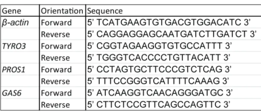
Table 2.1 Primers used for RT-PCR analysis ... 32 Table 2.2 IC50 values of UNC3810A against top kinase hits
from the kinome profiling. ... 53 Table 3.1 Peptide counts of proteins identified to interact with
LIST OF FIGURES
Figure 1.1 Activation of the PI3K/Akt/mTOR and MAPK/ERK
Pathway in KSHV Infection. ... 11 Figure 1.2 Generation of the phagophore in mitophagy. ... 21 Figure 2.1 Replicates between MIB/MS runs are consistent with
each other. ... 41 Figure 2.2 The cellular kinome is more similar within subtypes
than across subtypes. ... 42 Figure 2.3 A common set of kinases are activated within NHL
lymphomas compared to primary B-cells. ... 43 Figure 2.4 Tyro3 is uniquely upregulated in PEL... 44 Figure 2.5 Trends in expression data are similar to a previous
literature report... 46 Figure 2.6 Inhibition of Tyro3 results in attenuation of PI3K signaling. ... 47 Figure 2.7 Tyro3 knockdown attenuates survival and signaling in
PEL cells. ... 49 Figure 2.8 Synthesis of UNC3810A. ... 50 Figure 2.9 Specificity of UNC3810A to Tyro3. ... 52 Figure 2.10 Inhibition of Tyro3 results in a decrease of cell survival
and colony formation in PEL. ... 55 Figure 2.11 UNC3810A treatment of PEL cells results in cell death. ... 57 Figure 2.12 UNC3810A sensitizes PEL cells to treatment with
PI3K inhibitors. ... 58 Figure 2.13 Pharmacokinetic analysis of UNC3810A. ... 59 Figure 2.14 UNC3810A retards PEL tumor growth in vivo. ... 60 Figure 2.15 UNC3810A treatment does not reduce tumor burden
Figure 3.1 vPK binds and phosphorylates several subunits of the
PPP6 complex. ... 80 Figure 3.2 Examining the role of PPP6C in multiple pathways and
cellular processes relevant in the KSHV life cycle. ... 83 Figure 3.3 Ingenuity pathway analysis (IPA) of host interacting
partners of vPK identified using mass spectrometry. ... 86 Figure 3.4 Autophagic markers are induced but not the integrated
stress response (IRS) in vPK-HUVECs. ... 88 Figure 3.5 vPK expression promotes mitophagy. ... 90 Figure 4.1 Unresolved questions regarding the role of Tyro3 in PEL. ... 98 Figure 4.2 Potential questions regarding the role of vPK-induced
LIST OF ABBREVIATIONS
4EBP1 Eukaryotic translation initiation factor 4E-binding protein 1
ABL Abelson murine leukemia viral oncogene homolog ACSR AIDS and Cancer Specimen Resource
AIDS Acquired immunodeficiency syndrome
AMPK AMP activated protein kinase ANKRD28 Ankyrin repeat domain 28
ANKRD48 Ankyrin repeat domain 48 ANKRD52 Ankyrin repeat domain 52 ANOVA Analysis of variance
AP-1 Activator protein 1 ART Antiretroviral therapy
ATF4 Activating transcription factor 4
ATG Autophagy related)
ATP Adenosine triphosphate
AURKA Aurora kinase A
BCR B-cell receptor
BCR Breakpoint cluster region (BCR)
BL Burkitt’s lymphoma
BNIP3 BCL2/Adenovirus E1B 19 kDA interacting protein 3
BTK Bruton’s tyrosine kinase
CDK Cyclin-dependent kinase
CFA Colony formation assays CHOP C/EBP homologous protein
CI Combination index
CK2 Casein kinase II
CML Chronic myeloid leukemia
Co-IP Co-immunoprecipitation
CPCSEA Committee for the Purpose of Control and Supervision of Experiments on Animals (CPCSEA)
CTG CellTiter-Glo
CXCR2 C-X-C chemokine receptor type 2 DAB 3,3′-diaminobenzidine
DC-SIGN Dendritic cell-specific intercellular adhesion molecule-3-grabbing non-integrin
DDR DNA damage response
DE Delayed early
DHX9 DExH-box helicase 9 DIPEA Diisopropylethylamine
DLBCL Diffuse large B-cell lymphoma
DMSO Dimethyl sulfoxide
DNA-PK DNA-protein kinase
E. coli Escherichia coli
EBV Epstein-Barr virus
eIF4B eukaryotic translation initiation factor 4B
eIF4E eukaryotic translation initiation factor 4E
ER Endoplasmic reticulum
ERK Extracellular-signal-regulated kinase
EV Empty vector
FL Follicular lymphoma
FUNDC1 Fun14 domain containing 1
GAPDH Glyceraldehyde 3-phosphate dehydrogenase
GEF Guanine nucleotide exchange factor GFP Green fluorescent protein
GRK2 G protein-coupled receptor kinase 2
GST Glutathione S-transferase HBV Hepatitis B virus
HCV Hepatitis C virus HDAC Histone deacetylase HHV8 Human herpesvirus 8
HIV Human immunodeficiency virus HTLV Human T-lymphotropic virus
HUVEC Human umbilical vein endothelial cell
I.P. Intraperitoneal
I.V. Intravenous
IE Immediate-early
IFN Interferon
IFNAR Type 1 interferon receptor
IHC Immunohistochemistry
IKK IκB kinase
IL Interleukin
IMDM Iscove’s modified Dulbecco’s medium IPA Ingenuity Pathway Analysis
IRAK1 Interleukin 1 receptor associated kinase 1 IRF3 Interferon regulatory factor 3
IRIS Immune reconstitution inflammatory syndrome
ISR Integrated stress response
JAK Janus kinase
JNK c-jun N-terminal kinase
KICS KSHV-associated inflammatory cytokine syndrome
KS Kaposi’s sarcoma
KSHV Kaposi’s sarcoma associated herpesvirus LANA Latency-associated nuclear antigen
LC Liquid chromatography
LC3B Isoform B of LC3 LIR LC3 interacting region
LMP2A Latent membrane protein 2A
MAPK Mitogen-activated protein kinase
MAPKK MAPK kinase
MAPKKK MAPKK kinase
MAVS Mitochondrial antiviral signaling protein MCD Multicentric Castleman’s disease
MCL Mantle cell lymphoma
MEK Mitogen-activated protein kinase kinase MerTK MER proto-oncogene tyrosine kinase
Met Methionine
MIB Multiplexed kinase inhibitor-conjugated bead
miRNA MicroRNA
MK2 MAPK activated protein kinase 2 MPP Matrix processing peptidase
MS Mass spectrometry
MTCO2 Mitochondrially encoded cytochrome c oxidase II mTOR Mammalian target of rapamycin
mTORC1 mTOR complex 1
MTS (3-(4,5-dimethylthiazol-2-yl)-5-(3-carboxymethoxyphenyl-2-(4-sulfophenyl)-2H-tetrazolium)
MWF Monday/Wednesday/Friday
NBR1 Neighbor of BRCA1 gene NDP52 Nuclear domain 10 protein 52
NF-κB Nuclear factor kappa-light-chain-enhancer of activated B-cells
NIK NF-κB-inducing kinase
NIX Nip3-like protein X
NKT NK T-cells
NLS Nuclear localization signal
NSG Nod scid gamma
NTC Non-targeting control
NUSAP1 Nucleolar and spindle associated protein 1 OMM Outer mitochondrial membrane
Optn Optineurin
ORF Open reading frame
PARL PINK1/PGAM5-associated rhomboid-like protease
PARP Poly (ADP-ribose) polymerase PAS Phagophore assembly site
PCR Polymerase chain reaction
PDK1 Phosphoinositide dependent kinase 1
PE Phosphatidylethanolamine
PEL Primary effusion lymphoma PERK PKR-like ER kinase
PGAM5 Phosphoglycerate mutase 5 PI3K Phosphoinositide-3-kinase PIC Pre-initiation complex
PINK1 PTEN-induced kinase 1
PK Pharmacokinetic
PKCδ Protein kinase C-delta
PLC Phospholipase C
pNPP p-nitrophenyl phosphate PP2A Protein phosphatase 2A PP4 Protein phosphatase 4
PPP6 Protein phosphatase 6 PPP6C PPP6 catalytic subunit
PPP6R1 PPP6 regulatory subunit 1 PPP6R2 PPP6 regulatory subunit 2 PPP6R3 PPP6 regulatory subunit 3
RIG-I Retinoic acid-inducible gene 1
RSK p90 S6 kinase
RT Real-time
RTA Replication and transcription activator RTK Receptor tyrosine kinase
S6KB1 p70 S6 kinase
shRNA Short-hairpin RNA
STAT Signal transducer and activator of transcription STING Stimulator of interferon genes
TAK1 Transforming growth factor beta-activated kinase 1
TAM Tyro3/Axl/MerTK
TBK1 Tank binding kinase 1
Tbkbp1 TBK1 binding protein 1
TEM Transmission electron microscopy
TRIM28 Tripartite motif-containing protein 28
tRNA Transfer RNA
TSC2 Tuberous sclerosis complex 2
uORF Short upstream open reading frame UTR Untranslated region
vBCL-2 Viral homolog of B-cell lymphoma 2 vCyclin Viral cyclin protein
VEGF Vascular endothelial growth factor
vFLIP Viral Fas-associated death domain-like interleukin-1β-converting enzyme-like inhibitory protein
vGPCR Viral G protein-coupled receptor
vIL-6 Viral interleukin 6
vIRF1 Viral interferon regulatory factor 1 vPK Viral protein kinase
WNV West Nile virus
LIST OF SYMBOLS
α Alpha
β Beta
δ Delta
γ Gamma
κ Kappa
CHAPTER 1: REGULATION OF SIGNALING PATHWAYS BY KINASES IN THE
LIFE CYCLE OF KAPOSI’S SARCOMA-ASSOCIATED HERPESVIRUS1
Kaposi’s sarcoma-associated herpesvirus (KSHV)-associated diseases
Kaposi’s sarcoma-associated herpesvirus (KSHV), also known as human herpesvirus 8
(HHV8), is the etiological agent of three cancers: Kaposi’s sarcoma (KS), primary effusion lymphoma (PEL), and multicentric Castleman’s disease (MCD) (1). KS is the most common
cancer associated with KSHV, and is the most common malignancy in men in some Sub-Saharan African countries. KS tumors are of endothelial origin and endothelial cells within the tumor are infected with KSHV (2-4). There are four different types of KS that are characterized by the
patients who present the disease (1). Classical KS occurs in elderly men of Mediterranean and eastern European descent, while African endemic KS presents itself in children and middle-aged
adults from sub-Saharan Africa. The other two classes, AIDS-KS and iatrogenic/post-transplant KS, occur in the context of immunosuppression from either viral infection (HIV) or drugs (to prevent allograft rejection). The two other KSHV-associated cancers, PEL and MCD, are of
B-cell origin. PEL is a clonal lymphomatous effusion generally present in the body cavities, while MCD is a polyclonal B-cell hyperplasia characterized by systemic inflammatory symptoms (1,
5). PEL and MCD often exhibit rapid disease progression, and PEL patients have a median survival time of six months post-diagnosis (6). A recently characterized KSHV-linked disease is
Kaposi’s sarcoma-associated herpesvirus inflammatory cytokine syndrome (KICS), which has
symptoms similar to MCD (1). Patients have systemic inflammation characterized by high viral titers, viral interleukin 6 (vIL-6), human IL-6, and human IL-10, but lack the lymphadenopathy
seen in MCD patients. KSHV-associated diseases generally occur in the context of immune deficiency such as in AIDS, transplant, or elderly patients. Demonstrating the role the immune system plays in KSHV malignancies, immune reconstitution due to antiretroviral therapy (ART)
in AIDS patients often result in clinical regression of AIDS-KS (1, 7). However, paradoxically and in rare cases, the pro-inflammatory environment generated following ART initiation known
as immune reconstitution inflammatory syndrome (IRIS) can lead to the development of KS, termed IRIS-KS (7). Current treatment regimens of all of these diseases consist of some form of chemotherapy, radiation, and/or immunotherapy.
KSHV life-cycle
Like all herpesviruses, infection with KSHV is life-long. Transmission of the virus often occurs through bodily fluids such as saliva and blood (1). KSHV infects a plethora of cell types ranging from monocytes, fibroblasts, endothelial, epithelial, dendritic, and B-cells (8-10). Viral
entry is mediated by binding of different KSHV glycoproteins to a variety of host receptors. The ubiquitous cell surface molecule heparan sulfate aids in the binding of KSHV to multiple cell
types (8, 9). There are several entry receptors that KSHV uses and these differ based on cell type. In endothelial cells, the ephrin receptor tyrosine kinase A2, the transmembrane
cysteine/glutamine exchange transporter protein xCT, and several integrins (α3β1, αVβ3, αVβ5)
are important for KSHV entry (8). In macrophages, dendritic, and B-cells, KSHV binds the dendritic cell-specific intercellular adhesion molecule-3-grabbing non-integrin (DC-SIGN)
release the capsid and tegument layer. The viral capsid is then transported along microtubules
and is delivered into the nucleus. Once in the nucleus, KSHV can enter into either the lytic or latent lifecycle. KSHV infection is primarily latent with spontaneous bouts of lytic replication. In
KS tumor biopsies, ~99% of KSHV positive tumor cells remain latent (1). In contrast, MCD biopsies display the highest degree of lytic replication.
No virions are produced during latency; instead KSHV persists within host cells utilizing
viral proteins to pass its genome to daughter cells following cell division. Only a few viral genes are expressed during latency to prevent detection by the host immune system. The latency locus
encodes the latent proteins, ORF71 to ORF73, the kaposins, and twelve microRNAs (miRNAs) (2, 11). During latency, lytic gene expression is suppressed; however, several proteins highly expressed in the lytic cycle are present at low levels during latency such as K1 and vIL-6 (12,
13). The viral genome is tethered as an episome to the host chromosome by ORF73/LANA (latency-associated nuclear antigen) and is replicated in coordination with the host genome using
host replication machinery (2). LANA also binds multiple lytic promoters, such as ORF50, to suppress lytic gene expression. Many of the latent proteins upregulate cell proliferation and survival to promote persistence of the infected cell. For instance, ORF71 can bind and activate
the IκB kinase (IKK), thereby activating the nuclear factor kappa-light-chain-enhancer of activated B-cells (NF-κB) pathway to upregulate cell survival genes (2). Another latent gene,
ORF72/vCyclin is a homolog of cellular cyclin D and can bind to cyclin-dependent kinase 6 (CDK6) to phosphorylate retinoblastoma (Rb) and other targets to drive cell cycle progression and proliferation (2). Many of the latent genes are expressed in KSHV malignancies. Indicative
Although latency is the dominant program in the KSHV life cycle, spontaneous lytic
reactivation does take place (10). While the physiological stimuli that reactivates KSHV from latency is not known, environmental stresses, histone deacetylase (HDAC) inhibitors, or phorbol
esters can cause KSHV to reactivate in vitro (1, 2). Expression of genes during the lytic phase is separated into three groups: the immediate-early (IE), delayed early (DE), and late (2). During the lytic phase, KSHV replicates its genome and produces infectious progeny to spread to other
cells. After viral progeny are produced the infected cell subsequently dies (1). Proteins encoded during both the latent and lytic phases of the lifecycle are thought to play a role in
KSHV-associated diseases. Highlighting the importance of lytic gene expression, use of a viral
replication inhibitor, ganciclovir reduced the risk of KS by at least 75% in AIDS patients (14). In a group of KSHV-MCD patients, treatment with zidovudine and valganciclovir resulted in a
greater than 80% clinical response (5). Zidovudine and ganciclovir (the active form of valganciclovir) are phosphorylated into their toxic moieties by the lytic kinases, ORF21 and
ORF36, respectively. It is proposed that lytic cells can exhibit a paracrine effect on neighboring cells by secreting inflammatory or angiogenic factors that are highly prevalent in
KSHV-associated diseases. For instance, endothelial cells treated with media harvested from viral G
protein-coupled receptor (vGPCR)/ORF74 expressing cells had activated extracellular signal-regulated kinases 1/2 (ERK1/2), which is a kinase upsignal-regulated in KSHV malignancies (15).
Modulation of signal transduction by kinases
The ability of a cell to respond to different internal and external environmental cues is
important to the development, growth, survival, and/or function of the cell. In order to properly respond to these environmental cues, receptors in the cell sense and then trigger a signaling
include infection by a pathogen, presence or absence of energy sources or nutrients, and crosstalk
between different cells. Improper responses either by the activation or inactivation of these signaling cascades can result in localized or gross pathology of the cell or organism, respectively.
Therefore, signaling cascades tend to be tightly controlled to prevent improper signaling. Signal transduction within the cell can typically be simplified into three steps: (1) recognition of an environmental cue by the cell, (2) the initiation and amplification of a signal, and (3) a resulting
‘activated’ factor that then turns on, off, or modifies cellular processes (typically by gene expression) (16). One of the major role players in the second step are proteins called kinases,
which are enzymes that catalyze the transfer of a phosphate group from ATP to a substrate (17). The addition of the phosphate group to a substrate can alter its activity or functionality, which then results in further transmission of the signaling cascade. Since kinases are enzymes, they are
able to catalyze many reactions over a short period of time, thereby amplifying the initial signal (18). This allows the cell to quickly respond to environmental cues. The activity of a kinase is
typically regulated by its phosphorylation and its ability to access and bind its substrate. As kinases are integral parts in signaling cascades, many kinases in cancer cells have dysregulated activity (19). In a sequencing study of multiple cancers (which include breast, lung, colorectal,
gastric, melanoma, etc.) containing 210 samples, there were approximately 119 kinase-encoding genes containing a driver mutation in 66 of the samples (19). Therefore, while not all cancers
contain a driver mutation within a kinase, a sizable portion of them may harbor a dysregulated kinase that drives tumorigenesis. For example, the Philadelphia chromosome, which is present in almost all patients with chronic myeloid leukemia (CML), results in the fusion between the
kinase activity. Treatment of CML patients with imatinib, an ATP competitive inhibitor of the
BCR-ABL kinase, resulted in major and/or complete responses in a large portion of patients (20). As seen in this example, targeting kinases as a therapeutic target in cancer is an attractive option.
Indeed, kinase inhibitors are the fastest growing class of FDA approved oncology drugs (21). Common cancer-associated pathways dysregulated by KSHV
A common set of signaling pathways are frequently dysregulated in cancer. Many of these dysregulated pathways contribute to cellular processes that are considered ‘hallmarks of
cancer’ including sustained proliferation, resistance to cell death, and evasion of immune cells (22). A certain characteristic of herpesvirus infection is the establishment of life-long infection within the host. Therefore, many of the proteins encoded by KSHV facilitate viral persistence
and replication. In accordance with this goal, the virus aims to keep its host cell alive. Therefore, many pro-survival and proliferative pathways are upregulated during the KSHV life cycle, which
include the NF-κB, Janus kinase (JAK)/signal transducer and activator of transcription (STAT), mitogen-activated protein kinase (MAPK), and the phosphoinositide 3-kinase (PI3K)/ protein kinase B (Akt)/mammalian target of rapamycin (mTOR) pathways (6, 12, 23, 24). In the
following paragraphs, we will briefly review each of these pathways (with a more thorough review on the PI3K/Akt/mTOR pathway), and how KSHV regulates the host cellular kinases
within them to alter their signaling.
In humans, the NF-κB pathway involves a set of transcription factors (p50, p52, p65,
RelB, and c-Rel) that bind the κB enhancer in DNA to induce target gene expression in response
to injury or stress (25, 26). NF-κB signaling is split into two pathways: the classical and non-canonical pathway. In the classical pathway, NF-κB is bound to IκB during unstimulated
phosphorylates IκB, which results in the eventual proteasomal degradation of IκB, and
subsequent translocation of NF-κB to the nucleus. The NF-κB dimer then binds to its target DNA sequences, thereby inducing gene transcription of its target genes, which have roles in cell
survival, proliferation, and inflammation (26, 28). In the non-canonical pathway, the unprocessed form of p52 (known as p100) is bound to RelB in an inactive complex within the cytoplasm (29). Stimulation results in the activation of NF-κB-inducing kinase (NIK), which further activates
IKKα. IKKα then phosphorylates p100, which results in the ubiquitination and proteasomal processing of p100 to p52. The p52/RelB complexes can then translocate to the nucleus to
activate its target genes, which affect processes including cell development and the activation of several immune cells (29). NF-κB is frequently dysregulated in cancers as it can induce
proliferation, prevent apoptosis, induce angiogenesis, and regulate cellular metabolism (30).
Highlighting the importance of the NF-κB pathway in KSHV-associated diseases, PEL cells require activated NF-κB for survival (31-34). Two KSHV proteins, K13 and K15, can bind and
activate IKK and NIK, respectively. K13, also known as viral Fas-associated death domain-like interleukin-1β-converting enzyme-like inhibitory protein (vFLIP), is able to bind the gamma
subunit of IKK (IKKγ – also known as NEMO) to activate classical NF-κB signaling (35-37).
On the other hand, K15 can activate non-canonical NF-κB signaling by recruiting NIK and IKKα
to its cytoplasmic tail (38). NF-κB activation by K15 is NIK-dependent as knockdown of NIK
resulted in ablation of NF-κB activation (38).
Another pathway that responds to cytokine and growth factors is the Janus kinase
(JAK)/signal transducer of activators of transcription (STAT) pathway. This pathway consists of
pathway consists of three players, the aforementioned JAK and STAT proteins, and a receptor
that recognizes ligands in the extracellular environment (such as cytokines and growth factors) (39). Since the different JAKs are associated with certain receptors, each family member tends to
respond to certain cytokines and growth factors (39). Signal transduction in this pathway begins when a receptor engages its ligand (39). The receptor-bound JAK becomes activated, which phosphorylates a neighboring JAK (i.e. transphosphorylation) and the cytoplasmic tail of the
receptor. This allows for the recruitment of a STAT to the complex, which recognizes and binds the phosphorylated cytoplasmic tail of the receptor. The JAKs subsequently phosphorylate the
STATs, which result in their dimerization and translocation to the nucleus, where they can act as a transcription factor (39). As the JAK/STAT pathway regulates proliferation, survival, and inflammation, it is frequently dysregulated in many cancers (40). Latent KSHV infection of
endothelial cells results in constitutive activation of STAT3; and in PEL cells STAT3 and STAT6 are constitutively active (41-44). Part of this activity is mediated by kaposin B, which is
able to bind and activate MAPK activated protein kinase 2 (MK2), which inhibits the tripartite motif-containing protein 28 (TRIM28), a suppressor of STAT3 (44, 45). On the other hand, KSHV seems to inhibit STAT signaling in the context of interferon induction (46, 47). The RIF
protein encoded by open reading frame 10 is able to bind JAK1 and TYK2-associated type 1 interferon receptor (also known as IFNAR) and prevent the activation of STAT1 and STAT2
upon interferon alpha (IFN-α) treatment. Furthermore, several KSHV-encoded miRNAs target
kinases (such as PKCδ and IRAK1) that can phosphorylate STAT3 (47). Nevertheless, treatment of PEL cells with STAT3 or STAT6 inhibitors resulted in inhibition of cell proliferation and
life cycle is complex and may be dependent on the type of ligand- receptor interaction that is
activated.
Another pathway that is activated by ligands such as growth factors and chemokines is
the mitogen-activated protein kinase (MAPK) pathways. There are four canonical MAPKs: the p38 MAPK, c-jun N-terminal kinase (JNK), ERK1/2, and ERK5 kinases (48, 49). The canonical MAPK pathways transduce signals by a similar mechanism. A stimulus leads to the activation of
a MAPKK kinase (MAPKKK) (50). The activated MAPKKK then phosphorylates and activates a MAPK kinase (MAPKK), which subsequently phosphorylates and activates its MAPK. One of
the most common signaling pathways dysregulated in cancer is the MAPK/ERK pathway (51). In the MAPK/ERK pathway, there are three MAPKKKs from the Raf family, two MAPKKs, MEK1 and MEK2, and two MAPKs, ERK1 and ERK2 which propagate the signaling cascade.
The activation of the MAPK/ERK pathway promotes cell growth and proliferation. ERK1/2 achieves this by activating the p90 S6 kinase (RSK) leading to the phosphorylation of S6 and
eIF4B, or by promoting the activation of the transcription factor activator protein 1 (AP-1) (50, 52). The signaling cascade starts by the binding of growth factors and chemokines to their respective receptors. Some of these receptors activate Ras, which then recruits Raf to the cell
membrane to initiate the signaling cascade. The MAPK pathway is difficult to target in cancer due to feedback within itself and crosstalk with other survival signaling pathways such as the
PI3K pathway (50, 53). In KSHV infection and in PEL cells, the MAPK/ERK, JNK, and p38 MAPK pathways are activated (Figure 1.1) (24, 48, 49, 52, 54, 55). KSHV-encoded ORF45 can bind to RSK, thereby stabilizing the association of RSK with ERK. This complex maintains the
phosphorylate and activate MKK4 and MKK7, which are the MAPKK proteins in the JNK
signaling cascade (56). Highlighting the importance of the MAPK pathways, treatment of PEL cells with chemical inhibitors that attenuate MAPK/ERK and p38 MAPK signaling resulted in
apoptosis (57, 58).
Dysregulation of the phosphoinositide-3-kinase (PI3K)/protein kinase B (Akt)/mammalian target of rapamycin (mTOR) pathway by KSHV
There are four different classes of PI3K kinases (10). The class IA and 1B PI3K kinases
generate phosphatidylinositol-3,4,5-trisphosphate (PIP3), which recruits phosphoinositide
dependent kinase 1 (PDK1) and Akt to the membrane, where PDK1 phosphorylates and activates
Akt (Figure 1.1). The activation of Akt leads to the upregulation of several cellular processes including glycolysis, angiogenesis, cell survival, protein synthesis, and proliferation (10, 59, 60). Regarding protein synthesis, Akt inhibits tuberous sclerosis complex 2 (TSC2), leading to the
activation of mTORC1. The activation of mTORC1 promotes protein synthesis by activating several translation initiation factors. The phosphorylation of the eukaryotic translation initiation
factor 4E-binding protein 1 (4EBP1) by mTORC1 releases the eukaryotic translation initiation factor 4E (eIF4E) leading to cap-dependent mRNA translation. The phosphorylation of p70 S6 kinase (S6KB1) by mTORC1 also leads to activation of the ribosomal protein S6 and the
eukaryotic translation initiation factor 4B (eIF4B) (50, 61). Overall the activation of mTORC1 leads to the upregulation of anabolic processes to promote cell growth. Normally, energy
deficiencies and stress signals feedback into mTORC1 thereby inhibiting cell proliferation. In non-viral cancers many genes within the PI3K/Akt/mTOR pathway are mutated to prevent the shutdown of this pathway, while in viral cancers, oncogenic viral proteins activate
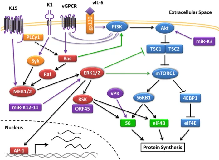
Figure 1.1 Activation of the PI3K/Akt/mTOR and MAPK/ERK Pathway in KSHV
Infection. KSHV activates the PI3K and MAPK/ERK pathways during infection, which are
heavily reliant on kinases (drawn as circles) to propagate the signaling cascade. Multiple viral proteins (colored in purple) activate the PI3K/Akt/mTOR pathway (in blue) to promote protein translation by activating subunits of the translation initiation complex. The MAPK/ERK pathway (in red) is similarly activated by viral proteins to promote protein translation through RSK. Some cross-talk (shown in green) occurs between both pathways. Activation of ERK1/2 also leads to the transcription of genes regulated by AP-1. Genes under the control of AP-1 include cytokines, which after being transcribed and translated are secreted out of the cell.
generation in the cell. Since mTORC1 is the master regulator of energy metabolism, KSHV
modulates this pathway to keep key biosynthesis pathways activated during the viral life cycle. Many KSHV proteins regulate the PI3K/Akt/mTOR pathway including vIL-6, vGPCR, K1, K15, ORF45, and ORF36 (Figure 1.1) (10, 23, 52, 61). Interestingly, these proteins are
low levels during latency. A latent miRNA, miR-K3 also regulates the PI3K/Akt pathway (4). As
mentioned before, paracrine signaling from lytic cells is thought to help drive KSHV pathogenesis by stimulating neighboring latent cells in a paracrine fashion. In the following
paragraphs below, we describe how several viral proteins or microRNAs (miRNAs) affect the PI3K/Akt/mTOR pathway, and how their contribution in KSHV-associated malignancies may be dependent on paracrine and autocrine signaling.
KSHV encodes several transmembrane proteins, including a chemokine receptor vGPCR, which is a cellular homologue of the interleukin 8 receptor (15). Two other KSHV-encoded
transmembrane proteins include K1, which mimics the B-cell receptor (BCR), and K15 (62). K1 and K15 are partial functional homologues to the latent membrane protein 2A (LMP2A) encoded by a related gammaherpesvirus, Epstein-Barr virus (EBV). Cells infected with a mutant KSHV
virus lacking K1 had attenuated activation of Akt following reactivation (63). Both K1 and vGPCR exhibit transformative properties when expressed in cells (10). The expression of
vGPCR immortalizes endothelial cells, which is dependent on PI3K/Akt activation as addition of a PI3K inhibitor results in apoptosis (64). Furthermore, nude mice present vascular tumors when injected with stably expressing vGPCR-NIH3T3 cells (54). Both vGPCR- and K1-expressing
transgenic mice develop tumors (10, 65). In vGPCR-transgenic mice, vascular KS-like tumors express CD31, an endothelial cell marker present in KS (65). However, many of the cells within
the tumor did not express the vGPCR protein and only some tissues harvested from mice with tumors expressed ORF74 transcripts. Knockdown of vGPCR also severely attenuated tumor growth and secretion of VEGF in a mouse model where endothelial cells transfected with a
observations would suggest paracrine signaling by vGPCR-expressing cells to neighboring cells
play a role in the development of KSHV malignancies.
KSHV also encodes a cellular homologue of IL-6 known as vIL-6; however unlike IL-6
which requires binding to the gp130 and IL-6 receptor, vIL-6 can transduce a signal by binding to gp130 alone (10, 12, 66). Similar to human IL-6, vIL-6 activates the JAK/STAT, MAPK, and PI3K/Akt pathways upon binding to gp130. In endothelial cells, vIL-6 activates gp130 leading to
the upregulation of several lymphatic markers including podoplanin in a Akt- and STAT3-dependent manner (10). Stably-expressing vIL-6 NIH3T3 cells can also induce tumor formation
in nude mice and transgenic mice expressing vIL-6 exhibit MCD-like disease (10, 66). However, expressing vIL-6 in IL-6 deficient mice resulted in no MCD-like disease suggesting that human IL-6 is important for MCD pathogenesis (10). Recently it was shown that the HIV-1
Nef protein synergistically augments the angiogenesis of vIL-6 in an Akt-dependent manner (66). Endothelial cells stably expressing vIL-6 showed a significant increase in cell growth,
angiogenesis, and levels of activated Akt upon addition of soluble Nef. Highlighting the
importance of PI3K signaling, nude mice injected with NIH3T3 cells co-expressing Nef and vIL-6 failed to develop any tumor growth upon treatment with a PI3K inhibitor. These results suggest
a novel mechanism on how HIV-infection can play a role in the development of KSHV malignancies besides immune suppression.
KSHV ORF36 encodes a serine/threonine viral protein kinase (vPK) conserved among the herpesviruses, and has been shown to activate JNK, a MAPK family member (56, 61, 67). Though vPK is classified as a late lytic gene, it can be expressed in hypoxic conditions. Knock
deficient or kinase-dead vPK viruses. We recently reported that vPK can mimic S6KB1 within
the mTOR pathway and residues lining the catalytic pocket of vPK are conserved to S6KB1 (61). Similar to S6KB1, vPK can phosphorylate S6, which was inhibited upon addition of a S6KB1
specific inhibitor, but not a JNK or Aurora kinase inhibitor. Overexpression of vPK in cells augmented anchorage-independent growth, angiogenesis, and cell proliferation as well. Indicative of vPK’s possible role in KSHV-associated malignancies, transgenic vPK mice in a
C57BL/6 background resulted in an eight-fold higher incidence of B-cell lymphomas in aged mice (68). Lymphomas harvested from these mice were of germinal center and post-germinal
center origin, and exhibited activation of S6, which is downstream of mTORC1. Furthermore, IL-1β and IL-12 p40 were elevated in the serum of vPK transgenic mice in comparison to the wild-type (WT) control mice. Both of these inflammatory cytokines have been implicated in
different cancers and may contribute to the development of these lymphomas in the vPK transgenic mice (69-72).
The microRNAs (miRNA) encoded by KSHV are transcribed from the latent
Kaposin/K12 promoter and are highly expressed in KSHV-associated malignancies (4). One miRNA, miR-K3 was shown to activate Akt by downregulating the G protein-coupled receptor
kinase 2 (GRK2) which suppresses expression of an IL-8 receptor, C-X-C chemokine receptor type 2 (CXCR2) (4). Knockdown of CXCR2 led to decreased activation of Akt in endothelial
cells similar to what was shown previously in skin keratinocytes. Furthermore, GRK2 can interact and inhibit Akt signaling in CXCR2-independent manner. The expression of miR-K3 resulted in enhanced cell migration and invasion of endothelial cells in an Akt-dependent
As seen by the many proteins encoded by KSHV that regulate the PI3K/Akt/mTOR
pathway, this pathway plays an important role in the KSHV life cycle. Indeed, the
PI3K/Akt/mTOR pathway is important for cell survival in KSHV-infected cells. Treatment of
PEL cells with rapamycin, an inhibitor of mTORC1, inhibited cell growth in vitro and
significantly attenuated tumor growth in mice (73). Low doses of NVP-BEZ235, a dual inhibitor that targets both PI3K and mTOR resulted in apoptosis in PEL cells (6). Inhibition of the
PI3K/Akt/mTOR pathway also attenuated cytokine secretion from PEL cells as well (6, 73). In renal-transplant patients, KS biopsies at the time of diagnosis showed elevated levels of activated
Akt and S6KB1, and increased vascular endothelial growth factor (VEGF) expression (74). Treatment of these patients with Sirolimus (rapamycin) resulted in remission of KS in all patients by six months. Therefore, inhibiting PI3K signaling has shown promise in a variety of
KSHV-associated diseases.
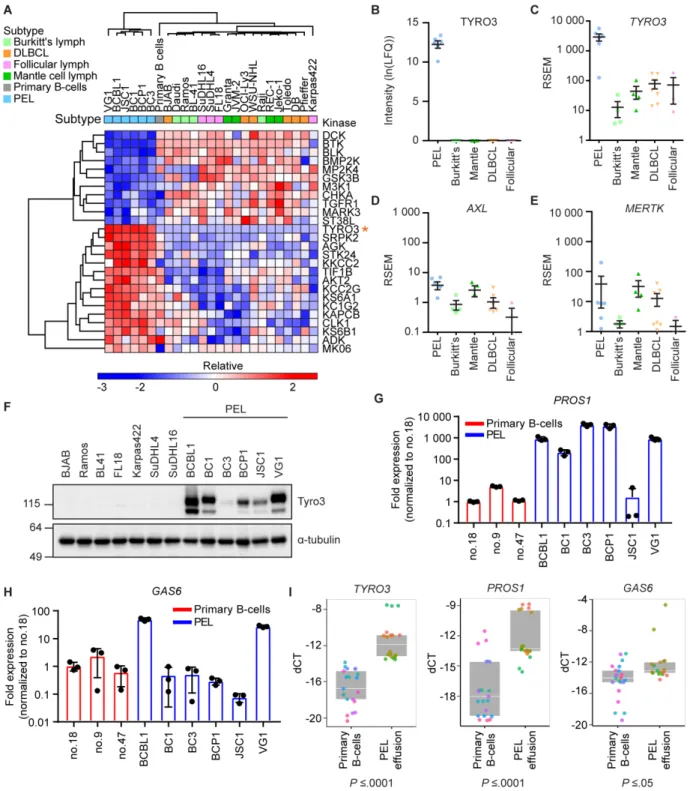
In this dissertation (Chapter 2), we expand on how PEL cells have upregulated Tyro3, a
host receptor tyrosine kinase. Tyro3 is a member of the TAM receptor family consisting of Tyro3, Axl, and MerTK, whose activation can lead to the upregulation of several signaling pathways including the PI3K/Akt/mTOR pathway (75). The TAM receptors have been shown to
be upregulated in many different cancers, thereby promoting cell survival and chemoresistance (76-78). A group previously showed that Axl was upregulated in Kaposi’s sarcoma (KS), but not
MerTK or Tyro3 (79). The expression of Axl in KS promoted cellular proliferation by
upregulating PI3K signaling. Furthermore, in a KS xenograft mouse model, treatment of mice with a monoclonal antibody against Axl reduced tumor growth. We utilized multiplexed
MIB/MS screen, we identified Tyro3, but not Axl or MerTK, as being uniquely upregulated in
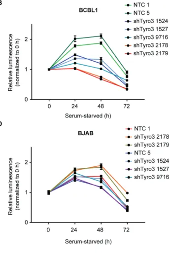
PEL. Knockdown of Tyro3 resulted in an attenuation of PI3K signaling, cell survival, and colony formation in PEL cells. Furthermore, treatment of PEL cells with a Tyro3 inhibitor, UNC3810A,
resulted in a decrease in tumor burden in a PEL xenograft mouse model. Overall, this work identifies a new mechanism by which the PI3K pathway is modulated in PEL, and that targeting Tyro3 may provide clinical benefits in the treatment of patients with PEL.
Autophagy
As mentioned above, there are four classes of PI3K kinases. There is one class III PI3K kinase in humans known as VPS34 (encoded by the PIK3C3 gene), whose role is to regulate macroautophagy (80). Autophagy is the process by which proteins and organelles in cells can be
degraded into their building blocks, which can then be recycled to build other proteins and organelles. Macroautophagy is a non-selective process by which bulk cargo in the cell is
degraded. The converse is selective autophagy, where different organelles and proteins are selectively targeted for degradation. Types of selective autophagy include mitophagy
(degradation of mitochondria), pexophagy (degradation of peroxisomes), ribophagy (degradation
of ribosomes), and reticulophagy (degradation of the endoplasmic reticulum) (81). Proper
induction of autophagy is important to maintain a healthy cell by preventing the buildup of old or
defective proteins and organelles. In humans, autophagy is an important cellular process by which the cell can respond to stress such as nutrient starvation, hypoxia, ER stress, microbial infection, and low energy levels (82). The maturation of the phagosome to engulf cellular cargo
can be defined by four steps: (1) induction and nucleation of the phagophore assembly site (PAS), (2) expansion of the phagophore, (3) completion of the phagosome and fusion with a
The most well-studied mechanism by which macroautophagy (henceforth called
autophagy) is induced is during nutrient starvation (83). The Autophagy related 1 (Atg1) complex, which consists of Atg1, Atg13, Atg17, Atg29, and Atg31 controls the recruitment of
other Atg proteins to the phagophore assembly site (PAS) in a process known as nucleation to initiate autophagy (82). The mammalian homologues to Atg1 are the ULK proteins - with the main family members being ULK1 and ULK2 (83). AMP activated protein kinase (AMPK) and
mTORC1 are the main regulators of the ULK1 complex (83). Under normal conditions, mTORC1 phosphorylates ULK1 resulting in its inactivation (84). However under nutrient
starvation, AMPK phosphorylates and activates ULK1 (84). The activation of the ULK1 complex in turn allows the recruitment and activation of the class III PI3K complex, which includes VPS34, Beclin-1 (the mammalian homolog of Atg6), and Atg14L (82, 83). The
activation of this complex promotes nucleation, and thus the formation of the autophagosome. The expansion of the phagophore is dependent on the recruitment of Atg8 (also known as LC3 in
mammals), Atg9, and Atg12 to the PAS (82). Both LC3 and Atg12 undergo ubiquitin-like conjugation reactions (in a reminiscent manner similar to the E1-, E2-, and E3-like enzyme chain reaction as seen in the priming and attachment of ubiquitin to substrates). Atg12 is conjugated to
Atg5 by Atg7 and Atg10 (81). Conjugated Atg5-Atg12 forms a complex with Atg16, which is recruited to the phagophore. The Atg5-Atg12-Atg16 complex helps recruit the lipidated form of
LC3 (known as LC3-II) to the phagophore (81). The generation of LC3-II from LC3 also involves a similar ubiquitin-like conjugation system. LC3 is first cleaved by Atg4 on the C-terminus in the cytoplasm generating LC3-I (82). The conjugation of LC3-I to
autophagosome. The fusion of a lysosome to the autophagosome results in an autolysosome,
where the cargo originally enclosed in the autophagosome is degraded.
The expression of several Atg proteins is upregulated in response to stress. A mechanism
by which this occurs is the translation of a differential set of mRNAs under stress conditions. One of the central proteins in the integrated stress response (ISR) is the eukaryotic initiation
factor 2 subunit alpha (eIF2α) (85, 86). The eukaryotic initiation factor 2 (eIF2) complex -
consisting of eIF2α, eIF2β, and eIF2γ - initiates mRNA translation by recognizing the AUG start codon, and by forming the 43S pre-initiation complex (PIC) with other translation initiation
factors (85). The 43S PIC is then recruited to mRNA containing a 5’ N7 methylated guanosine cap (known as the 5’ cap) (87). The loading of the Met-tRNA to the AUG start codon results in the hydrolysis of GTP to GDP on eIF2. After placement of the methionine (Met)-transfer RNA
(tRNA), the 43S PIC disassociates, and the complexes are recycled to initiate another round of translation. The association of GTP with eIF2 determines its ability to associate with the 43S PIC
(85). Therefore after a round of translation initiation, the guanine nucleotide exchange factor
(GEF), eIF2B, must exchange GDP with GTP on eIF2. However, under stress conditions, eIF2α
becomes phosphorylated on the serine 51 residue, which inhibits the ability of eIF2B to
exchange out GDP to GTP (85). This prevents the formation of the 43S PIC and ultimately shuts down cap-dependent translation in the cell. On the other hand, the translation of mRNAs
containing a short upstream open reading frame (uORF) in the 5’ untranslated region (UTR) occurs instead as they are not dependent on 5’ cap-dependent translation (85). A couple of these mRNAs include the transcription factors, ATF4 and CHOP (88). Synthesis of these transcription
isoform B of LC3 (LC3B) (89, 90). Therefore, regulation of autophagy is two-fold, and involves
the production of the autophagy machinery, and the maturation of the autophagosome. Due to the importance of autophagy in maintaining cell homeostasis, dysregulation of
autophagy has been tied to several neurodegenerative diseases that are characterized by the buildup of toxic proteins such as in Alzheimer’s and Parkinson’s disease (91). Autophagy has also been linked to cancer, but its role in tumor development is less clear (92). Autophagy seems
to confer a more tumor suppressive phenotype during early stages of tumor progression, but is more protective during late stages of tumor progression (92). The role of autophagy in the
herpesvirus life cycle is complex with many different viral proteins encoded by several herpesvirus either inhibiting or inducing autophagy (which is reviewed in depth by Lussignol and Esclatine, see reference (93)). Many of the herpesvirus induce autophagy early on in the lytic
cycle, but block the process in later stages of the lytic cycle (93). Several of the herpesviruses block the formation of the autolysosome, and utilize the autophagosomes generated as a
precursor for the generation of viral particles (93-97). KSHV upon reactivation from latency induces autophagy early on in the lytic cycle (97). Interestingly, the autophagic process during the lytic cycle may be independent of mTORC1 regulation as treatment with the mTOR
inhibitor, Torin, did not affect autophagic flux or induction of autophagy markers (98).
Expression of KSHV-encoded RTA, the main protein which controls reactivation from latency,
is able to increase autophagic vesicle formation as seen by transmission electron microscopy (TEM) (99). In reactivated BC3 PEL cells, viral particles were also observed in autophagic vesicles (97). However when examining autophagic flux in reactivated BC3 cells at 36 hours
upregulation of autophagy markers, yet the inhibition of autophagic flux indicate that the
autophagic process is initiated, but the cargo is not degraded. KSHV-encoded K7 protein binds Rubicon, which is a cellular protein that inhibits autophagosome maturation (101). Expression of
K7 did not alter autophagosome formation, but decreased the amount of autolysosomes.
Furthermore, KSHV encodes a viral homolog of B-cell lymphoma 2 (vBCL-2), which can bind and inhibit Beclin-1 similar to cellular Bcl-2 to inhibit nucleation (102-104). On the other hand,
KSHV inhibits autophagy during latency. While, the viral cyclin protein (vCyclin) induces autophagy due to activating the DNA damage response (DDR), co-expression of vCyclin with
vFLIP, demonstrated that vFLIP was able to inhibit vCyclin-mediated autophagy (105-107). Highlighting the potential role of autophagy in herpesvirus-associated cancers, the EBV-encoded latent membrane protein 2A (LMP2A) is able to induce autophagy in nonmalignant breast
epithelial cells to prevent anoikis, a type of cell death that occurs when adherent cells become detached to the extracellular matrix (108).
Mitophagy
A selective form of autophagy, which degrades mitochondria, is known as mitophagy
(109). The recycling of old and defective mitochondria is important for the health and proper energy homeostasis in the cell. Highlighting the importance of maintaining healthy
mitochondria, 1 in 2,000 individuals are affected with diseases related to mitochondria-dysfunction (110). Mitophagy occurs under basal conditions, but can be upregulated under stress-conditions such as in hypoxia conditions or nutrient starvation (111). The central step in
mitophagy is the recruitment of mitophagy-associated receptors, which helps trigger the recruitment of the autophagy machinery including ULK1 (109). These receptors have an LC3
induced kinase 1 (PINK1)-Parkin ubiquitin-dependent or hypoxia-mediated mitophagy (Figure
1.2).
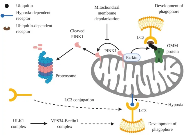
Figure 1.2 Generation of the phagophore in mitophagy. The generation of the phagophore
during mitophagy primarily occurs through either hypoxia-dependent mitophagy or ubiquitin-dependent mitophagy. During hypoxia-ubiquitin-dependent mitophagy, hypoxic conditions lead to the upregulation of certain mitophagy receptors that localize to the mitochondria to recruit LC3. In contrast, ubiquitin-dependent mitophagy requires the stabilization of PINK1 in the mitochondrial membrane. Stabilized PINK1 then activates Parkin, which ubiquitinates outer mitochondrial membrane (OMM) proteins. Ubiquitin acts as docking sites for ubiquitin-dependent mitophagy receptors, which upon localizing to the mitochondria recruit LC3. In both cases, the recruitment of LC3 results in the development of the phagophore. Figure 1.2 is modeled from Figure 1 from Palikaras et. al., 2018 (111).
During normal conditions, the matrix processing peptidase (MPP) processes PINK1,
it is degraded. However, under mitochondrial membrane depolarization, PINK1 is stabilized and
becomes activated (109). Afterwards, PINK1 phosphorylates ubiquitin present on the
mitochondria on residue serine 65 (112). Parkin, an E3 ubiquitin ligase, is able to bind
phospho-ubiquitin on the mitochondria, and becomes phosphorylated by PINK1. This results in the activation of Parkin, which leads to Parkin’s ubiquitination of mitochondrial proteins (109). The ubiquitinated mitochondrial proteins, then act as docking sites for the mitophagy receptors. The
five main receptors are optineurin (Optn), sequestosome-1 (p62), nuclear domain 10 protein 52 (NDP52), TAX1 binding protein (TAX1BP1), and neighbor of BRCA1 gene (NBR1) (112). Of
the five, Optn and NDP52 are the primary receptors as cells with all five receptors knocked out were only rescued by Optn and NDP52 (113). After receptors bind to the mitochondria, the autophagy machinery is recruited to begin phagophore formation, which leads to the eventual
degradation of the mitochondrion (113). In hypoxia-mediated mitophagy, the receptors are regulated by hypoxia (112). These receptors are BCL2/Adenovirus E1B 19 kDA interacting
protein 3 (BNIP3), Nip3-like protein X (NIX – also known as BNIP3L), and Fun14 domain containing 1 (FUNDC1) (112). Both BNIP3 and NIX expression levels increase in hypoxic conditions (109). BNIP3 is able to stabilize PINK1 to promote mitophagy (114). On the other
hand, FUNDC1 is phosphorylated by Src and casein kinase II (CK2), which prevents its interaction with LC3 (112). In hypoxic conditions, phosphoglycerate mutase 5 (PGAM5)
dephosphorylates FUNDC1, thereby relieving this suppression (112).
The role of mitophagy is not well-studied in herpesviruses. Recently, a group showed that mitophagy is induced during lytic reactivation in KSHV in a viral interferon regulatory factor 1
induction of mitophagy was shown to be dependent on vIRF1-mediated activation of NIX.
Inhibiting mitophagy resulted in an attenuation of viral protein expression and replication. Interestingly, depletion of NIX had little effect on viral replication and ORF45 protein
expression. This suggests there may be alternative mechanisms that KSHV uses to regulate mitophagy. In this dissertation, we examine how vPK, a kinase encoded by ORF36 in KSHV, may regulate mitophagy (Chapter 3). Expression of vPK in human umbilical vein endothelial
cells (HUVECs) results in an increase in phosphorylated-eIF2α and LC3B-II expression.
However, protein expression of alternative transcripts such as ATF4 and CHOP was not induced.
A group that pulled down all the open reading frames (ORFs) in KSHV to identify host cellular interacting partners to KSHV proteins noted that vPK heavily interacted with mitochondrial proteins (116). When fractionating mitochondria, we found that vPK was present in
mitochondria, and that mitochondrial content decreased in vPK-expressing cells. Our work suggests that vPK may induce mitophagy in the KSHV life cycle.
Conclusions
The modulation of signaling pathways by viral proteins is critical to the survival and
persistence of KSHV in the human host. These pathways rely on kinases to induce a signaling cascade by phosphorylating proteins to recruit and/or alter activity of target substrates. Inhibition
of these pathways by biochemical means or drug treatment often attenuates viral replication and/or induces cell death in KSHV-infected cells (6, 24, 73). Therefore, kinase inhibitors have represented an attractive target in treating KSHV malignancies (74, 117). Currently there are
clinical trials to test the efficacy of Selumetinib, an inhibitor of the dual specificity mitogen-activated protein kinase kinase 1/2 (MEK1/2) in KS patients, and Sirolimus (rapamycin), an
(74, 117). Active drug discovery aimed at cellular kinases comprises a large proportion of
pharmaceutical drug development. Therefore, novel specific kinase inhibitors identified by drug discovery programs may be viable treatment options for KSHV associated diseases. In this
dissertation, we aim to further characterize how both host (Chapter 2) and virally-encoded (Chapter 3) kinases play a role in KSHV-infection and pathogenesis. This work allows us to gain a better understanding of how kinases modulate pathways that are important in the KSHV
CHAPTER 2: KINOME PROFILING OF NON-HODGKIN LYMPHOMA IDENTIFIES
TYRO3 AS A THERAPEUTIC TARGET IN PRIMARY EFFUSION LYMPHOMA2
Introduction
Non-Hodgkin lymphoma (NHL) consists of many subtypes covering a wide range of
characteristics that include differences in cell-of-origin, aggressiveness, and viral association. One of these NHL subtypes is primary effusion lymphoma (PEL), which is associated with
Kaposi’s sarcoma-associated herpesvirus (KSHV). Patients with PEL have a median survival time of 6 months after diagnosis, as standard chemotherapy is generally ineffective in treating PEL (118, 119). Several clinical studies have taken different approaches to treat PEL, and while
some have shown promise, there is still no effective treatment for PEL (120). For example, treatment of PEL with bortezomib, a proteasome inhibitor that inhibits NF-κB activity, has
shown variable results in clinical trials (121-123). Therefore, there is a need to identify new targeted therapies to treat PEL.
Signaling pathways are often dysregulated in cancer, and kinases that control the
propagation of these signaling cascades frequently have aberrant activity. The use of kinase inhibitors to treat cancers has shown clinical efficacy as a single agent or in the adjuvant setting.
For example, imatinib, an inhibitor of the BCR-ABL kinase, is efficacious in treating chronic
2This chapter previously appeared as an article in the journal PNAS. The original citation is as follows: Wong JP,
myelogenous leukemia (20). We were interested in identifying kinases that were uniquely
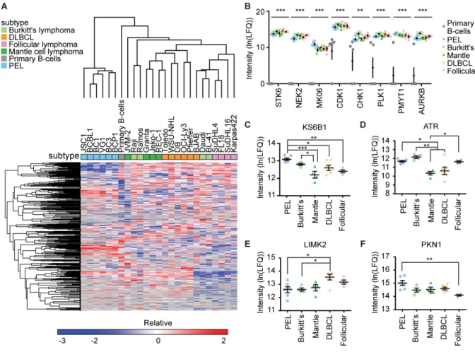
activated in PEL in comparison to other NHL subtypes. A kinase with unique or significantly higher activity in PEL compared to other NHL subtypes may indicate that PEL is dependent on
the signaling activity of that particular kinase compared to other NHL subtypes.
We decided to compare PEL to four other B cell NHL subtypes: follicular lymphoma (FL), diffuse large B-cell lymphoma (DLBCL), mantle cell lymphoma (MCL), and Burkitt’s
lymphoma (BL). These subtypes are derived from different stages of B-cell development that include the mantle zone, germinal center, and post-germinal center (124-129). As different B-cell
NHL subtypes may arise during different stages of normal B-cell development, there are likely differences in the expressed kinome (130). Two of the subtypes (BL and PEL) are commonly associated with viral infection. BL is associated with Epstein-Barr virus (EBV) infection, while
PEL is distinguished by Kaposi’s sarcoma-associated herpesvirus (KSHV) infection (131-133). Co-infection with EBV is also common in PEL (134-136). By examining an array of NHL
subtypes that are derived from different stages of B-cell development, and being
virally-associated or not, we intended to capture a wide enough spread in cellular phenotypes to identify distinct kinases which are activated or repressed in PEL in comparison to other B-cell NHL
subtypes.
Materials and methods Mice
Nod scid gamma (NSG) mice used in the PEL and BJAB xenograft studies were
maintained in pathogen-free conditions and the studies were approved by the Institutional Animal Care and Use Committee (IACUC) at the University of North Carolina at Chapel Hill.
analysis was completed by Sai Life Sciences Limited. The study was conducted in accordance to
guidelines provided by the Committee for the Purpose of Control and Supervision of
Experiments on Animals (CPCSEA) as published in The Gazette of India (1998) and approved
by the Institutional Animal Ethics Committee (IAEC). Eight to twelve-week old Swiss Albino male mice weighing approximately 25 to 35 grams used in the PK study was purchased from Global, India.
Cell Culture
All cell lines are grown at 37oC at 5% CO
2 levels. NHL cell lines except for OCl-Ly1,
OCl-Ly3, and PEL cell lines were grown in RPMI 1640 medium supplemented with 10% FBS, 1% penicillin-streptomycin, and 1% L-glutamine. PEL lines were grown in the same complete RPMI media as described above supplemented additionally with 0.075% sodium bicarbonate
(Gibco, 25080-094) and 0.05 mM β-mercaptoethanol (Gibco, 21985-023). Ly1 and OCl-Ly3 were grown in Iscove’s modified Dulbecco’s medium (IMDM), 10% human AB serum
(Sigma-Aldrich, H4522), 1% penicillin-streptomycin, and 75 µM β-mercaptoethanol (Gibco, 21985-023). 293FT and B16-F10 cells were grown in DMEM supplemented with 10% FBS, 1% penicillin-streptomycin, and 1% L-glutamine. 500 µg/ml of G418 (Gibco, 10131-027) was also
added for selection in 293FT cells. HT-29 cells were grown in McCoy’s 5a medium supplemented with 10% FBS, 1% penicillin-streptomycin, and 1% L-glutamine. Human
umbilical vein endothelial cells (HUVEC) were grown in endothelial cell basal medium 2 (PromoCell, C-22211) supplemented with 1% L-glutamine, 10% FBS, 1%
penicillin-streptomycin, and the growth medium 2 supplement pack (PromoCell, C-39211) excluding the
were grown in RPMI 1640 medium supplemented with 10% Tet-free FBS (Clontech, 631107),
1% penicillin-streptomycin, 1% L-glutamine, 20 µg/ml hygromycin B (Corning, 30-240-CR), and 1.25 µg/ml puromycin (Corning, 61-385-RA). All cell lines were tested free of mycoplasma
contamination using the LookOut Mycoplasma PCR Detection kit. All PEL cell lines were authenticated by STR profiling using the GlobalFiler STR panel.
Lentiviral infection of cell lines
To generate Tyro3 knockdown cell lines, plasmids containing MISSION pLKO.1-puro control (Sigma, SHC002), TRC2 pLKO.5-puro control (Sigma, SHC202), or the following
Tyro3 shRNA (Sigma, SHCLNG-NM_006293): #1524 (Clone NM_006293.2-1664s21c1), #1527 (Clone NM_006293.2-2755s21c1), #9716 (Clone NM_006293.2-1718s21c1), #2178 (Clone NM_006293.x-2490s1c1), and #2179 (Clone NM_006293.x-743s1c1) were used. Clones
#2178 and #2179 contain the pLKO.1 backbone; clones #1524, #1527, and #9716 contain the pLKO.5 backbone. Lentiviruses containing these plasmids were then made in 293FT using
ViraPower (Invitrogen, K497500) according to the manufacturer’s instructions. B-cells were centrifuged with lentiviruses containing either the control plasmid or shTyro3 for 90 minutes at 2,500 rpm at 30oC in serum-free medium containing 10 µg/ml polybrene (Sigma, 107689). After
centrifugation, cells were incubated overnight. The next day, cells were spun down and fresh complete media was added. Two days after infection, 1 µg/ml puromycin (Corning, 61-385-RA)
selection was added. The concentration of puromycin was doubled after each subsequent split to a final concentration of 4 µg/ml. Trex-RTA Luc BCBL1 cells were made from Trex-RTA BCBL1 cells (obtained as a gift from J. Jung) that were infected with RediFectTMLuc
Primary B-cell isolation
Human B-cells were isolated from whole human blood purchased from Gulf Coast Regional Blood Center (E5318). Blood was tested by Gulf Coast for human immunodeficiency
virus 1 (HIV-1) RNA, HIV-1 Group M RNA, HIV-1 Group O RNA, HIV-2 RNA, hepatitis C virus (HCV) RNA, hepatitis B virus (HBV) DNA, West Nile virus (WNV) RNA, Zika RNA, hepatitis B surface antigen, and for antibodies to HIV-1/2+O, hepatitis B core antigen, HCV,
human T-lymphotropic virus I/III (HTLV-I/III), CMV, and syphilis. Trypanosoma cruzi was also tested for if patient history indicated an exposure risk since previous testing, otherwise noted as
historical non-reactive. B-cells were only used from blood that tested non-reactive or negative to all of the above. Briefly, 20 mL of blood was transferred to a 50 mL conical tube and diluted with 20 mL of 2 mM EDTA-PBS. The diluted blood was then layered slowly over 15 mL of
Ficoll-Paque (GE Healthcare, 17-5442-02) in SepMate-50 tubes (StemCell Technologies, 85450). The tubes were centrifuged at 1500 rpm for 20 minutes, after which the top layer
containing PBMCs was isolated. Cells were washed with 2 mM EDTA-PBS, and pelleted at 1500 rpm at 4oC for 5 minutes. Remaining blood cells were lysed by resuspending the cell pellet
in ACK lysing buffer (Gibco, A10492-01) and was incubated for 10 to 15 minutes at room
temperature. The suspension was further washed with 2 mM EDTA-PBS and resuspended in 2 mM EDTA, 0.5% BSA (Sigma-Aldrich, A7906) in PBS. B-cells were isolated with the B-cell
isolation kit II (Miltenyi Biotec, 130-091-151) according to manufacturer’s instructions. Cells were stained with CD19 (Miltenyi Biotec, 130-091-247) and ran on a MACSQuant VYB cytometer to determine purity. Analysis was performed with FlowJo v10.2 software. B-cell
MIB/MS affinity chromatography
Suspension cells were pelleted and washed three times in cold PBS. Pellets were lysed on ice in MIB lysis buffer [50 mM HEPES (N-2-hydroxyethylpiperazine-N'-2-ethanesulfonic acid)
pH 7.5, 0.5% Triton X-100, 150 mM NaCl, 1 mM EDTA, 1 mM EGTA, 10 mM sodium fluoride, 2.5 mM sodium orthovanadate, 1X Roche protease inhibitor cocktail (Sigma-Aldrich, 11836145001), and 1% each of phosphatase inhibitor cocktail 2 (Sigma-Aldrich, P5726) and 3
(Sigma-Aldrich, P0044)]. Cell lysates were sonicated 3×10 seconds on ice and centrifuged at 16,000×g in microfuge tubes for 10 minutes at 4°C. The supernatant was syringe-filtered through
a 0.2 µm SFCA membrane. Protein concentration was determined by Bradford assay (Bio-Rad, 500-0006) and equal amounts of protein, around 2.5 mg, were used for each sample.
The filtered lysate was brought to 1 M NaCl and passed through a column of multiplexed
kinase inhibitor-conjugated beads (MIBs) consisting of Sepharose-conjugated Purvalanol B, PP58, CTx-0294885, VI-16832, and two custom-synthesized pan-kinase inhibitor compounds,
UNC-8088A and UNC-2142A (53). The MIBs were washed with 5 mL of high-salt buffer and 5 mL of low-salt buffer [50 mM HEPES (pH 7.5), 0.5% Triton X-100, 1 mM EDTA, 1 mM EGTA, and 10 mM sodium fluoride, and 1 M NaCl or 150 mM NaCl, respectively]. The
columns were washed a final time with 1 mL 0.1% SDS before elution in 1 mL of 0.5% SDS (100°C, 5 min). Eluted kinases were reduced (dithiothreitol) and alkylated (iodoacetamide) prior
to being concentrated with Amicon Ultra centrifugal filters (Millipore, UFC801096) and
detergent was removed from the concentrated eluate by chloroform/methanol extraction. Protein pellets were resuspended in 50 mM HEPES (pH 8.0) and were digested overnight with
and cleaned with C-18 spin columns (Thermo Scientific, 89870) according to the manufacturer
instructions.
Peptides were resuspended in 5% acetonitrile and 0.1% formic acid, and 25-50% injected
onto a Thermo Easy-Spray 75uM x 25cm C-18 column via an Easy nanoLC-1000. Peptides were separated as a single fraction on a 300 minute gradient (5-40% acetonitrile) and identified by a Q-Exactive mass spectrometer. Parameters: 3e6 AGC MS1, 80ms MS1 max inject time, 1e5
AGC MS2, 100ms MS2 max inject time, 20 loop count, 1.8 m/z isolation window, 45s dynamic exclusion. Spectra were identified using MaxQuant software and the Uniprot/Swiss-Prot
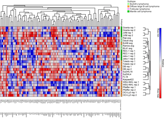
database (138). Peptide abundance was calculated using label-free quantification (LFQ). Mass spectrometry runs were normalized by natural log-transformed and median-centering LFQ values on a common-captured set of 151 kinases. Heat maps were generated using the Euclidian
hierarchical clustering program from GENE-E (https://software.broadinstitute.org/GENE-E/). RNA-sequencing (seq)
RNA was harvested from cell lines using the RNeasy Plus Mini Kit (Qiagen, 74136) according to manufacturer’s instructions. For sequencing of the NHL cell lines, mRNA-Seq libraries were generated using the Stranded mRNA-Seq kit (KAPA Biosystems, KK8421) and
multiplexed with Illumina TruSeq adapters. Samples were run on two 75-cycle single-end sequencing runs with an Illumina NextSeq-500. QC-passed reads were aligned to hg19 using
MapSplice (139). Picard Tools v1.64 was used to determine the alignment profile. Aligned reads were sorted and indexed using SAMtools and translated to transcriptome coordinates and filtered for indels, large inserts, and zero mapping quality using UBU v1.0 (140). Transcript abundance