MECHANISMS AND CONSEQUENCES OF STAPHYLOCOCCUS AUREUS LEUKOCIDIN AB-MEDIATED ACTIVATION OF THE HOST NLRP3 INFLAMMASOME
Jason H. Melehani
A dissertation submitted to the faculty at the University of North Carolina at Chapel Hill in partial fulfillment of the requirements for the degree of Doctorate of Philosophy in the
Department of Pharmacology in the School of Medicine
Chapel Hill 2016
iii ABSTRACT
Jason H. Melehani: Mechanisms and consequences of Staphylococcus aureus Leukocidin AB-mediated activation of the host NLRP3 inflammasome
(Under the direction of Joseph A. Duncan)
The NLRP3 inflammasome is a critical innate immune sensor implicated in the pathogenesis of dozens of infectious and non-infectious diseases. Activation of the NLRP3 inflammasome causes IL-1β and IL-18 secretion and necrotic cell death. Staphylococcus aureus is a common cause of infections in humans. S. aureus produces a family of pore-forming toxins that are cytotoxic to human immune cells. One recently discovered pore-forming toxin,
Leukocidin AB, is the focus of studies herein.
Leukocidin AB is a human-specific, pore-forming toxin that binds CD11b to initiate pore formation. In order to characterize the mechanism of Leukocidin AB cytotoxicity and determine its significance, we evaluated the effects of Leukocidin AB on primary human monocytes and THP1 monocytic cells. Leukocidin AB was one of the most potent toxins in killing primary human monocytes. In THP1 cells, knockdown of NLRP3 or ASC by shRNA diminished Leukocidin AB-induced cytotoxicity and prevented secretion of IL-1β and IL-18. We also characterized the NLRP3 inflammasome in bacterial survival during phagocytosis. When S. aureus was
iv
cell death when Leukocidin AB binds CD11b on the phagosome membrane instead of the plasma membrane.
We also initiated studies to characterize the role of kinases and phosphorylation in the response to Leukocidin AB. Using multiplex inhibitor bead chromatography and quantitative mass spectrometry, we identified eight kinases that rapidly decrease in activity during Leukocidin AB exposure. We demonstrated a role for Death-Associated Protein Kinase in the response to Leukocidin AB by showing that its inhibition suppressed Leukocidin AB-induced cytokine secretion and cytotoxicity. And finally, we used a novel transfection method for overexpressing mutant proteins in THP1 cells to show that the NLRP3 S198D/S201D mutant could spontaneously activate NLRP3 inflammasome signaling.
v
vi
ACKNOWLEDGEMENTS
vii
TABLE OF CONTENTS
LIST OF TABLES ... xiii
LIST OF FIGURES ... xiv
LIST OF ABREVIATIONS... xvi
CHAPTER 1: Discovery, activation and regulation of the NLRP3 inflammasome ... 1
1.1 A general overview of NLRP3 inflammasome form and function ... 1
1.2 Clinical relevance of NLRP3 inflammasome signaling ... 3
1.3 Interleukin-1β-driven monogenic hyperinflammatory fever disorders are caused by mutations in the NLRP3 inflammasome pathway ... 4
1.4 The NLRP3 inflammasome is a master innate immune sensor ... 7
1.5 Cautions in interpreting NLRP3 inflammasome research... 8
1.5.1 Studies frequently overlook important outcomes of NLRP3 inflammasome activity ... 8
1.5.2 Dozens of activators and regulators complicate a comprehensive model for NLRP3 inflammasome activation ... 11
1.5.3 Commonly used Caspase 1 knockout mice are also Caspase 11 deficient ... 12
1.6 Priming the NLRP3 inflammasome for activation ... 13
1.6.1 Pattern-recognition receptor signaling upregulates transcription of NLRP3 inflammasome components ... 13
1.6.2 Negative feedback loops respond to prolonged priming to reign in NLRP3 inflammasome signaling ... 18
1.6.3 Kinase-mediated post-translational priming regulates NLRP3 inflammasome activity ... 22
viii
1.6.5 Multiple ubiquitination signals regulate NLRP3 inflammasome priming and
protein degradation ... 28
1.6.6 Post-transcriptional regulation of NLRP3 inflammasome mRNA stability ... 32
1.6.7 NLRP3 inflammasome signaling without priming ... 33
1.7 Critical features of NLRP3 inflammasome activation ... 34
1.7.1 Potassium efflux is a universal requirement for NLRP3 inflammasome formation ... 35
1.7.2 Mitochondrial disruption triggers ROS production ... 37
1.7.3 Lysosome dysfunction liberates the cysteine protease Cathepsin B ... 43
1.7.4 Endoplasmic reticulum dysfunction disrupts protein folding ... 47
1.7.5 Calcium acts as a second messenger ... 49
1.7.6 Ubiquitination acts as a platform for regulator binding ... 58
1.7.7 Phosphorylation controls inflammasome oligomerization ... 62
1.7.7.1 Syk kinase couples C-type lectin domain family members to NLRP3 inflammasome signaling ... 62
1.7.7.2 RIPK1/RIPK3- and NLRP3-dependent necrosis overlap ... 65
1.7.7.3 Kinases have a wide ranging role in regulating the NLRP3 inflammasome ... 68
1.8 Canonical inflammasome activators: nigericin, ATP and monosodium urate ... 74
1.8.1 Nigericin is a potassium ionophore that activates the NLRP3 inflammasome ... 75
1.8.2 Extracellular ATP triggers NLRP3 activation by binding P2RX7 ... 85
1.8.3 Monosodium urate disrupts lysosomes to trigger the NLRP3 inflammasome ... 97
1.9 Consequences of NLRP3 inflammasome signaling in pulmonary diseases ... 105
1.9.1 The NLRP3 inflammasome is a sentinel in pulmonary infections... 108
1.9.1.1 NLRP3 inflammasome activation helps clear respiratory viruses ... 108
1.9.1.2 The NLRP3 inflammasome is differentially required for clearing extracellular and intracellular bacterial infections ... 110
ix
1.9.2 Chronic obstructive pulmonary disease is influenced by long term NLRP3
inflammasome stimulation ... 115
1.9.3 NLRP3 may contribute to airway hyperreactivity in asthma, allergy and obesity ... 117
1.9.4 The NLRP3 inflammasome is critically activated in acute lung injury ... 122
1.9.5 The NLRP3 inflammasome drives fibrotic changes in pulmonary fibrosis ... 126
1.9.6 NLRP3 inflammasome signaling may predict risk of bronchopulmonary dysplasia in preterm infants ... 129
1.10 Concluding remarks ... 130
REFERENCES ... 131
CHAPTER 2: Inflammasome activation mediates tissue-specific pathogenesis in Staphylococcus aureus infection ... 154
2.1 Introduction ... 154
2.2 S. aureus factors that activate inflammasomes ... 155
2.3 Inflammasomes that are activated by S. aureus ... 160
2.4 The role of inflammasomes in S. aureus infections ... 169
2.4.1 The NLRP3 inflammasome responds to hemolysins to control S. aureus dermal infections ... 169
2.4.2 S. aureus hijacks the NLRP3 inflammasome to exacerbate lung infection pathology ... 173
2.4.3 Microglia activate NLRP3 in vitro but depend on AIM2 for survival in S. aureus brain abscesses ... 177
2.4.4 IL-1β signaling is critical in soft tissue infections ... 179
2.5 How host inflammasomes can affect other immune processes ... 179
2.6 Integrating inflammasome studies to improve patient care ... 182
REFERENCES ... 184
CHAPTER 3: Staphylococcus aureus Leukocidin A/B (LukAB) kills human monocytes by co-opting host NLRP3 and ASC when extracellular, but not intracellular ... 192
x
3.2 Materials and Methods ... 195
3.2.1 Ethics statement ... 195
3.2.2 Mammalian cell lines ... 195
3.2.3 Purifying primary CD14+ human monocytes ... 196
3.2.4 Bacterial strains, culture conditions and generating mutants ... 196
3.2.5 Purifying toxins form S. aureus ... 197
3.2.6 Collecting culture filtrates ... 197
3.2.7 Transmission electron microscopy ... 198
3.2.8 Evaluating cell death by lactate dehydrogenase release ... 198
3.2.9 Evaluating cell death by propidium iodide staining ... 199
3.2.10 Measuring cytokine secretion by AlphaLISA ... 199
3.2.11 Measuring Caspase 1 activation with FLICA ... 200
3.2.12 Immunoblot analysis ... 200
3.2.13 Measuring THP1 surface CD11b levels ... 201
3.2.14 Evaluating infection assays by flow cytometry ... 201
3.3 Results ... 205
3.3.1 S. aureus kills human monocytes in a LukAB dependent manner ... 205
3.3.2 LukAB kills human monocytes by engaging its cellular receptor ... 209
3.3.3 LukAB-induced cell death displays necrotic features ... 213
3.3.4 LukAB induces activation of Caspase 1 in human monocytes ... 217
3.3.5 NLRP3 and ASC are necessary for LukAB-induced cytokine secretion and necrotic cell death ... 220
3.3.6 Caspase 1 is required for LukAB-induced cytokine secretion ... 223
3.3.7 LukAB promotes S. aureus escape from within human monocytes independent of NLRP3 or ASC, but dependent on CD11b ... 226
3.4 Discussion ... 230
xi
CHAPTER 4: Novel approaches to studying kinase-dependent regulation of
the NLRP3 inflammasome ... 239
4.1 Introduction ... 239
4.2 Materials and Methods ... 242
4.2.1 Mammalian cell lines ... 242
4.2.2 Purifying LukAB from S. aureus ... 243
4.2.3 Evaluating cell death by propidium iodide staining ... 243
4.2.4 Measuring cytokine secretion by AlphaLISA ... 244
4.2.5 Measuring Caspase 1 activation with FLICA ... 244
4.2.6 Multiplex inhibitor bead chromatography ... 244
4.2.7 iTRAQ labeling ... 245
4.2.8 Mass spectrometry ... 245
4.2.9 Exploratory statistical analysis of MIB/MS results ... 246
4.2.10 DAPK inhibition experiments ... 246
4.2.11 Site directed mutagenesis of NLRP3 ... 246
4.2.12 Cloning of NLRP3 mutants into T7 expression vector ... 247
4.2.13 In vitro transcription of capped and poly-adenylated mRNA ... 247
4.2.14 Transfection of mRNA into THP1 cells ... 248
4.3 Results ... 248
4.3.1 LukAB causes rapid NLRP3 inflammasome activation and cell death ... 248
4.3.2 MIB/MS identifies 344 kinases expressed in THP1 cells ... 252
4.3.3 Eight kinases are modulated in response to LukAB intoxication ... 252
4.3.4 Inhibiting DAPK suppresses LukAB cytotoxicity and IL-1β and IL-18 secretion ... 255
4.3.5 mRNA transfection of NLRP3 S198D/201D into THP1 cells causes spontaneous IL-18 secretion not seen in S198D single mutant ... 256
4.4 Discussion ... 259
xii
CHAPTER 5: Concluding thoughts and future directions ... 267
APPENDIX A: Functional amyloid signaling via the inflammasome, necrosome and signalosome: new therapeutic targets in heart failure ... 271
A.1 Abstract ... 271
A.2 Introduction ... 272
A.3 Misfolded proteins in cardiac disease ... 272
A.4 Protein folding, preamyloid oligomers, and aggregation ... 275
A.5 Protein aggregates, proteotoxicity, and heart failure ... 278
A.6 How endogenous systems prevent protein misfolding ... 281
A.7 Pharmacological chaperone therapies ... 282
A.8 Amyloid proteins, heart failure, and activation of the inflammasome ... 295
A.9 Amyloid proteins, heart failure, and activation of “functional amyloid”: Necrosome ... 299
A.10 The COP9 signalosome enhances protein degradation to reduce the misfolded protein burden ... 300
A.11 Summary: The structural continuum between functional and pathological amyloids and the significance in heart failure ... 304
xiii
LIST OF TABLES
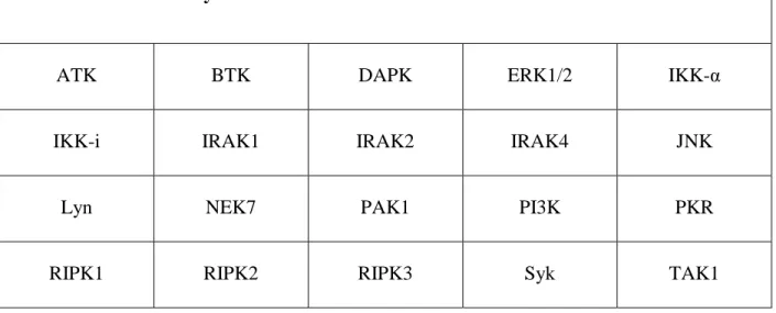
Table 1.1 – Kinases analyzed in NLRP3 inflammasome research ...74
Table 3.1 – Staphylococcus aureus strains ...203
Table A.1 – Clinical studies investigating tafamidis and siRNA TTR in
transthyretin-associated (amyloid) cardiomyopathy ...285
Table A.2 – Clinical studies investigating doxycycline in transthyretin-associated
(amyloid) cardiomyopathy ...291
Table A.3 – Clinical studies investigating ECGG in primary cardiac amyloidosis
and Alzheimer’s disease ...293
Table A.4 – Proteins forming amyloid as part of their functional role in cellular
xiv
LIST OF FIGURES
Figure 1.1 – TLR signaling triggers NLRP3 inflammasome transcriptional priming ...17
Figure 1.2 – Negative feedback loops limit NLRP3 inflammasome priming ...21
Figure 1.3 – Kinases in non-transcriptional priming of the NLRP3 inflammasome ...24
Figure 1.4 – Caspase 8 is critical for transcriptional and post-translational NLRP3 inflammasome priming ...27
Figure 1.5 – Ubiquitination regulates NLRP3 protein levels and activity ...31
Figure 1.6 – Accumulation of mitochondrial damage activates the NLRP3 inflammasome ...42
Figure 1.7 – Lysosomal disruption contributes to NLRP3 inflammasome signaling ...46
Figure 1.8 – TLR signaling may shift TAK1 between priming and NLRP3 inflammasome roles ...103
Figure 2.1 – S. aureus pathogen-associated molecular patterns and virulence factors activate inflammasome priming and signaling ...156
Figure 2.2 – NLRP3, NLRC5, NLRP7, AIM2 and other inflammasome-like signaling is activated in response to S. aureus...161
Figure 2.3 – NLRP3 inflammasome signaling plays unique rolls in skin and lung infections....176
Figure 2.4 – Inflammasome signaling upregulates γδ-T cells and a TH17 response ...181
Figure 3.1 - Staphylococcus aureus LukAB induces cell death in human monocytic cells ...207
Figure 3.2 – LukAB recognizes CD11b on human monocytes to potently induce cell death ...211
Figure 3.3 – LukAB induces necrotic cell death and secretion of pro-inflammatory cytokines IL-1β and IL-18 ...215
Figure 3.4 – LukAB is a potent activator of Caspase 1 ...218
Figure 3.5 – LukAB activates the NLRP3 inflammasome leading to cell death and cytokine secretion...221
Figure 3.6 – NLRP3 and ASC contribute to S. aureus culture filtrate-mediated cytokine secretion and THP1 cell death...222
Figure 3.7 – Media supplementation with KCl inhibits LukAB-mediated FLICA-1 activation and cell death...223
xv
Figure 3.9 – LukAB produced by extracellular or phagocytized S. aureus kills
human monocytes ...228
Figure 4.1 – LukAB intoxication leads to rapid NLRP3 inflammasome activation,
mitochondrial depolarization and cell death ...250
Figure 4.2 – Mitochondrial depolarization is not blocked by extracellular potassium
or shRNA-mediated depletion of NLRP3 or ASC ...251
Figure 4.3 – Kinome analysis by MIB/MS identifies 344 kinases, 8 of which are
significant changed at 10 separate time points ...254
Figure 4.4 – DAPK inhibition blocks LukAB-mediated IL-1β and IL-18 secretion
and cell death in THP1 cells ...255
Figure 4.5 – GFP mRNA transfection efficiency is high and well tolerated in THP1 cells ...258
Figure 4.6 – Phosphomimetic mutation of NLRP3 S198 and S201 leads to
spontaneous NLRP3 inflammasome activity ...259
Figure A.1 – Native, non-native, aggregates, and amyloid protein structures and
the stressors that drive them ...276
Figure A.2 – Protein aggregate and amyloid stimulate cellular dysfunction ...280
Figure A.3 – The NLRP3 inflammasome is an amyloid-like fibrillar cytokine-processing
platform, which senses amyloid and contributes to worsening heart failure ...296
Figure A.4 – Cullin-RING ubiquitin ligase (CRL) activity is regulated via
neddylation and deneddylation ...303
xvi
LIST OF ABBREVIATIONS
2-APB 2-aminoethoxydiphenyl borate
3-MA 3-methyladenine
ADAM10 A disintegrin and metalloprotease 10
ADCY Adenylate cyclase
AIM2 Absent in melanoma 2
AL Amyloid light-chain amyloidosis
ALI Acute lung injury
ANOVA Analysis of variance
AQP Aquaporin
ASC Apoptosis-associated speck-like protein
BAL Bronchoalveolar lavage
BLM Bleomycin
BMDC Bone marrow dendritic cells
BMDM Bone marrow-derived macrophages
BPD Bronchopulmonary dysplasia
BSA Bovine serum albumin
BTK Bruton’s tyrosine kinase
C5aR Complement component 5a receptor 1
CA-MRSA Community-acquired methicillin-resistant S. aureus
CaM Ca2+/Calmodulin domain
cAMP Cyclic AMP
CANTOS Canakinumab anti-inflammatory thrombosis outcomes study
CaOx Calcium oxalate
xvii
CARD Caspase activation and recruitment domain
CAS Casamino acids
CASR Calcium sensing receptor
CCCP Cyanide m-chlorophenyl hydrazine
CCR C-C motif receptor
CD11b Cluster of differentiation 11b
CFR Code of Federal Regulations
CHOP C/EPB homologous protein
CINCA Chronic infantile neurological cutaneous and articular syndrome
Cm Chloramphenicol
COP9 Constitutive photomorphogenesis mutant 9
COPD Chronic obstructive pulmonary disease
CR3 Complement receptor 3
CREB Cyclic AMP-responsive element-binding protein
CRL Cullin-RING-ligase
CS Cyclic stretching
CSN COP9 signalosome
CTSB Cathepsin B
CXCR CXC chemokine receptor
DAPK Death-associated protein kinase
DPI Diphenyliodonium
dsRNA Double-stranded RNA
DUB Deubiquitinating enzyme
EGCG (-)-epigallocatechin-3-gallate
xviii
FACS Fluorescence-activated cell sorting
FADD Fas-associated protein with death domain
FAP Familial amyloidotic polyneuropathy
FBS Fetal bovine serum
FCAS Familial cold autoinflammatory syndrome
FEV1 Forced expiratory volume, 1 second
FLICA-1 FLICA-FMK Alexa Fluor 660
FMK Fluoromethyl ketone
GFP Green fluorescent protein
HALI Hyperoxic acute lung injury
HBSS Hank’s balanced salt solution
HDAC Histone deacetylase
HEPES 4-(2-hydroxyethyl)-1-piperazineethanesulfonic acid
HIES Hyper IgE Syndrome
HIF1α Hypoxia-inducible factor 1-alpha
HKSA Heat-killed S. aureus
Hla Alpha hemolysin
Hlb Beta hemolysin
Hld Delta hemolysin
HlgAB Gamma hemolysin AB
HlgCB Gamma hemolysin CB
HMGB1 High mobility group box 1 protein
HRP Horseradish peroxidase
HSP Heat shock protein
xix
IAP Inhibitor of apoptosis proteins
IFN Interferon
IKBKE Inhibitor of NF-κB kinase subunit epsilon
IL Interleukin
iNOS Inducible nitric oxide synthase
IRAK IL-1 receptor-associated kinase
IRB Institutional review board
IP3 Inositol 1,4,5-trisphosphate
LAC Los Angeles County
LDH Lactate dehydrogenase
LLMe Leu-Leu-OMe
LLO Listeriolysin
LOF Loss of function
LRR Leucine-rich repeat
LT Lethal toxin
LTA Lipoteichoic acid
LUBAC Linear ubiquitin chain assembly complex
LukAB Leukocidin AB
LukED Leukocidin ED
LV Low tidal volume
MAC Membrane attack complex
Mac-1 Macrophage-1 antigen
MAPK Mitogen-activated protein kinase
MAVS Mitochondrial antiviral signaling
xx
Mdm2 Mouse double minute 2 homolog
MDP Muramyl dipeptide
MOI Multiplicity of infection
MRSA Methicillin-resistant S. aureus
MSU Monosodium urate
mtDNA Mitochondrial DNA
MVK Mevalonate kinase
MWS Muckle-Wells syndrome
NAC N-acetylcysteine
NOX2 NADPH oxidase 2
Nec-1 Necrostatin-1
NHS Normal human serum
NLRP3 NLR family, pyrin domain containing 3
NO Nitric oxide
NOD Nucleotide-binding oligomerization domain
NSA Necrosulfonamide
NTA Nickel-nitrilotriacetic acid
OVA Ovalbumin
p53 Tumor protein 53
PAK p21-activated kinase
PAMP Pathogen-associated molecular pattern
PARP Poly(ADP-ribose) polymerase
PBMC Peripheral blood mononuclear cell
PBS Phosphate buffered saline
xxi
PFT Pore-forming toxin
PGAM5 Phosphoglycerate mutase family member 5
PGN Peptidoglycan
PI Propidium iodide
PKR RNA-dependent protein kinase
PLC Phospholipase C
PML Promyelocytic leukemia protein
PMN Polymorphonuclear cell
Poly-Q19 Poly-glutamine (19 repeats)
Poly-Q83 Poly-glutamine (83 repeats)
PP2A Protein phosphatase 2A
PRR Pattern recognition receptor
PSM Phenol-soluble modulin
PVL Panton-Valentine leukocidin
PYD Pyrin domain
RA Rheumatoid arthritis
RHIMs RIP homotypic interaction motifs
RIPK Receptor interacting protein kinase
ROS Reactive oxygen species
RPMI Roswell Park Memorial Institute
SAA Serum amyloid A
SDS Sodium dodecyl sulfate
SERCA Sarco/endoplasmic reticulum Ca2+ ATPase
shRNA Short hairpin RNA
xxii
SNAP S-nitroso-N-acetylpenicillamine
SOCE Store-operated Ca2+ entry
STIM Stromal interaction molecule
Syk Spleen tyrosine kinase
TAB TAK-1 binding protein
TBS Tris-buffered saline
TRIM33 Tripartite motif 33
TRPM2 Transient receptor potential cation channel subfamily M
TRX Thioredoxin
TSB Tryptic soy broth
TTR Transthyretin
TXNIP Thioredoxin-interacting protein
TUDCA Tauroursodeoxycholic acid
UNC University of North Carolina
UPR Unfolded protein response
UTR Untranslated region
VILI Ventilator-induced lung injury
XIAP X-linked inhibitor of apoptosis protein
1
Chapter 1. Discovery, activation and regulation of the NLRP3 inflammasome
1.1 A general overview of NLRP3 inflammasome form and function
The NLRP3 inflammasome is the most well characterized of a recently discovered family
of sensors that activate Caspase 1 to initiate immune signaling. NLR proteins [1] (ex.
Nucleotide-binding domain and leucine-rich repeat containing, pyrin-domain 3 – NLRP3)
respond to pathogens and cellular stresses known as danger signals. Upon activation, NLR
proteins interact with Caspase 1, either directly in those that contain a Caspase Activation and
Recruitment Domain (CARD), such as in the NLRC family, or indirectly through pyrin-pyrin
domain (PYD) interactions with Apoptosis-associated speck-like protein containing a CARD
(ASC) [2]. Activation of Caspase 1 leads to proteolytic processing of critical inflammatory
signals, including interleukin-1β (IL-1β) and interleukin-18 (IL-18), to their mature form.
Additionally, activation of inflammasomes, and NLRP3 in particular, can cause necrotic-type
cell death. This cell death has been referred to as pyroptosis or pyronecrosis, largely depending
on the requirement for Caspase 1. However, the use of these terms in the literature is sometimes
inexact so they will be avoided here.
Activation of inflammasomes is also often times preceded by a “priming” step (or signal
1). Toll-like receptor (TLR) signaling primes inflammasome activation by: (1) upregulating
NF-κB-dependent transcription of inflammasome components and pro-IL-1β and (2) initiating a
2
some cells, usually those with high levels of expression of inflammasome components,
such as THP1 cells, this step can be bypassed, though usually without IL-1β secretion.
Hundreds of unique agents activate the NLRP3 inflammasome. Because of the large number
of different activating agents, a detailed comprehensive model of inflammasome activation has
yet to emerge. Instead, the field is littered with disparate findings pertaining to different
activators that are rarely confirmed system-wide. In this introduction I will summarize the
current state (as of the end of 2015) of the NLRP3 inflammasome field. I will start by discussing
the discovery of the NLRP3 inflammasome as the cause of heritable IL-1β-driven
hyperinflammatory periodic fever disorders. Then, I will review the dominant hypotheses of how
the NLRP3 inflammasome is regulated across different activators. Following that, I will discuss
how each of these hypotheses fit in with each one another by reviewing the requirements for the
three classical NLRP3 agonists: nigericin, extracellular ATP and monosodium urate (MSU). And
lastly, I will highlight the role of NLRP3 in infectious and non-infectious pulmonary diseases, a
personal and professional interest of mine, to demonstrate the wide ranging effects of the NLRP3
inflammasome. My dissertation research will follow. This will include:
a comprehensive literature review of S. aureus and inflammasome interactions with a
focus on the unique tissue specific roles of different inflammasomes
primary research comparing the cytotoxicity of S. aureus exotoxins in killing human
monocytes and identifying that Leukocidin AB (LukAB) activates the NLRP3
inflammasome
primary research on the role of kinases broadly and DAPK specifically in regulating
3
a review of novel therapeutic targets in heart failure, including specifics on a role for the
NLRP3 inflammasome (this previously published paper will be included as an appendix)
At the end, I will conclude with my thoughts on significance of my work and the future of
NLRP3 inflammasome research.
1.2 Clinical relevance of NLRP3 inflammasome signaling
The NLRP3 inflammasome is increasingly being recognized as a source of pathology in a
wide range of infectious and non-infectious diseases, and at the same time, critical for clearing
many infections. Staphylococcus aureus is a highly drug-resistant and clinically important
pathogen responsible for a growing number of skin and lung infections. Early research into the
interaction between S. aureus and inflammasomes has revealed interesting insights into the
pathogenesis of infections. In pneumonia, S. aureus pore-forming toxins (PFTs) such as
alpha-hemolysin (Hla) activate the NLRP3 inflammasome leading to severe lung damage and death. In
dermatitis, S. aureus PFTs and phenol-soluble modulins (PSMs) activate the NLRP3
inflammasome allowing the bacteria to infiltrate the keratinocyte barrier but also promote
bacterial clearance by neutrophils. Additionally, the NLRP3 inflammasome is activated in
human monocytes in response to PFTs that were only recently discovered because of their
specificity for human cells over mice. As a deeper understanding of S. aureus-inflammasome
interactions begins to emerge, the inflammasome signaling node may become an attractive target
for clinical intervention to treat infections or reduce inflammation-associated tissue damage in
4
1.3 Interleukin-1β-driven monogenic hyperinflammatory fever disorders are caused by
mutations in the NLRP3 inflammasome pathway
NLRP3 (also referred to in early literature as CIAS1, PYPAF1, NALP3 and Cryopyrin)
was identified as a critical protein in inflammation when it was found to be the causal gene
mutated in familial cold autoinflammatory syndrome (FCAS) and Muckle-Wells syndrome
(MWS) [3]. Shortly thereafter, NLRP3 was also show to be mutated in chronic infantile
neurological cutaneous and articular (CINCA) syndrome (also known as neonatal-onset
multisystem inflammatory disease [NOMID]) [4]. Mutations in NLRP3 have been confirmed in
dozens of patients with these diseases [5], now collectively known as Cryopyrin-Associated
Periodic Syndromes (CAPS) or Cryopyrinopathies.
FCAS is an autosomal-dominant systemic hyperinflammatory disease in which patients
experience recurrent episodes of rash, arthralgia, fever and conjunctivitis after exposure to cold
[6-8]. MWS is also an autosomal-dominant syndrome of periodic fevers, rash, arthralgia,
sensorineural hearing loss, and sometimes amyloidosis [9,10]. CINCA syndrome is a severe
early onset chronic inflammatory disease characterized by cutaneous symptoms, central nervous
system involvement and arthropathy [11-13]. The mutations in NLRP3 that cause these three
diseases are sometimes overlapping, suggesting that secondary modifying factors contribute to
their distinct presentations. However, such modifying factors have yet to be identified.
Insight into the function of NLRP3 quickly followed discovery of CAPS causing
mutations. A mammalian two-hybrid screen showed that ASC interacts with NLRP3 and
5
was also found to interact directly with pro-Caspase 1 [15] and, in conjunction with NLRP3,
formed an IL-1β processing inflammasome [16].
When NLRP3 with common disease-associated mutations (R260W, D303N, and E637G)
was expressed in THP1 cells, these mutants constitutively associated with ASC and induced
spontaneous IL-1β secretion, whereas wildtype NLRP3 did not [16,17]. Macrophages from
MWS patients also spontaneously secreted active IL-1β [16]. Monocytes from patients with
FCAS were cold sensitive and responded to incubation at 32°C by secreting IL-1β [18],
mimicking the sensitivity of FCAS patients generally. Knockin mice expressing the D301N
NLRP3 mutation (the ortholog of D303N in humans) exhibited widespread neutrophilia and high
levels of serum inflammatory markers. They also exhibited skeletal abnormalities seen in
CINCA patients including knee joint deformity, growth retardation and severe postnatal
osteopenia resulting from increased osteoclastogenesis, accelerated bone resorption and loss of
chondrocytes [19].
Early hints that IL-1β dysregulation may drive pathology in CAPS led to therapeutic
trials of the recombinant human IL-1-receptor (IL-1R) antagonist anakinra (brand name:
Kineret). Anakinra led to a dramatic, rapid, and sustained resolution of symptoms in patients
with MWS [20] that has been reproduced in hundreds of patients [21], including patients with
FCAS [22] and CINCA [23]. The clinical experience with anakinra has become the basis for
clinical guidelines [24]. Clinical trials have also shown success with Rilonacept, an IL-1 Trap
that acts as a long-acting and potent inhibitor of IL-1β, in patients with FCAS and MWS [25,26].
A third IL-1β disrupting therapy, Canakinumab, a human anti-IL-1β monoclonal antibody, has
6
Additional periodic fever syndromes have been identified with casual genes that relate to
inflammasome signaling providing greater insight into the activity and regulation of the NLRP3
inflammasome-IL-1β signaling axis. Sterile Multifocal Osteomyelitis with Periostitis and
Pustulosis (OMPP) [28,29] is caused by a deficiency in the IL-1R antagonist (IL1-RN) that leads
to unopposed IL-1β signaling and life-threatening systemic inflammation [30]. Familial
Mediterranean Fever (FMF) is an autosomal recessive disorder caused by mutations in the
MEFV gene that encodes for Pyrin [31]. Pyrin negatively regulates the NLRP3 inflammasome
by disrupting the interaction between NLRP3 and ASC, and thus, when mutated, leads to
increased inflammasome activation [32]. Hyper-IgD and Periodic Fever Syndrome is caused by
mutations in mevalonate kinase (MVK) [33,34] that disrupt autophagy, which is thought to
negatively regulate the NLRP3 inflammasome and IL-1β secretion, leading to a
hyperinflammatory state [35]. Pyogenic Sterile Arthritis, Pyoderma Gangrenosum, and Acne
(also known as PAPA Syndrome - PAPAS) [36], is caused by mutation in the PSTPIP1 gene
[37]. PSTPIP1 binds to Pyrin and disease-associated mutations exert a dominant-negative effect
on Pyrin leading to increased IL-1β production in patients [38].
It is interesting to note that in all Mendelian disorders affecting the NLRP3
inflammasome pathology results from increased activation of this pathway, not decreased. This
makes the NLRP3 inflammasome fairly unique among critical immune signaling pathways in
that outright disease resulting from absent NLRP3 inflammasome signaling has not yet been
identified. It is likely that low or absent function NLRP3 alleles do exist in the human
population, but their susceptibility to infection may be so minimal so as to not be easily
7
1.4 The NLRP3 inflammasome is a master innate immune sensor
Activation of the NLRP3 inflammasome typically proceeds in two stages. First, a
“priming” step upregulates transcription and translation of inflammasome components and
pro-IL-1β through the well-studied TLR-NF-κB signaling pathway. Additionally, priming prepares
the NLRP3 inflammasome for activation through a series of post-translational modifications.
Second, Caspase 1 catalytic activity is triggered through NLRP3 inflammasome oligomerization
leading to processing of cytokines to their mature form for secretion and necrotic cell death. In
the absence of priming, cells that constitutively express NLRP3 inflammasome components can
trigger IL-18 secretion and cell death, without IL-1β secretion. However, the physiologic
significance of this partial activation is still unclear.
Early functional studies of the NLRP3 inflammasome drew comparisons to the Toll-like
receptor system, a family of pattern recognition receptors (PRRs) for sensing
pathogen-associated molecular patterns (PAMPs). While other NLR proteins detect a narrow range of
PAMPs (ex. NLRC4 binds flagellin and NLRP1 binds muramyl dipeptide - MDP), NLRP3 is
activated by hundreds of pathogen-associated molecules, endogenous danger signals, and
environmental and man-made pollutants. The only common feature of these NLRP3-activating
molecules identified thus far is that their activity is inhibited by increasing the extracellular
concentration of potassium. As such, the NLRP3 inflammasome would be more accurately
placed in a category of its own – as a master innate immune sensor – to distinguish its high
8
As a result of this promiscuity, the NLRP3 inflammasome research field is full of gaps.
Before discussing the NLRP3 inflammasome in more detail, the reader will be warned in section
1.5 of these gaps and how they hamper generalizability of important data.
1.5 Cautions in interpreting NLRP3 inflammasome research
Research on the NLRP3 inflammasome has been expanding rapidly with each passing
year and with no signs of slowing down. A search of “NLRP3” on PubMed at the end of 2015
returned 2182 results, of which, nearly half, 1035 (47.4%), were published in 2014 or 2015.
Because NLRP3 inflammasome research is still a nascent field, many new discoveries are
disjointed and little effort is made to integrate new findings into a coherent and comprehensive
model for NLRP3 inflammasome activity. This issue has been exacerbated by the large number
of different activating stimuli. Below, as a brief aside, I will highlight some of the common faults
in the field in order to prepare the reader for some of the unfortunate confusion and shortcomings
that exist in the data.
1.5.1 Studies frequently overlook important outcomes of NLRP3 inflammasome activity
The vast majority of research on the NLRP3 inflammasome focuses on two readouts of
NLRP3 activity: cleavage of pro-Caspase 1 into its active form and secretion of mature IL-1β. As
previously mentioned, activation of the NLRP3 inflammasome actually triggers Caspase 1
activation, IL-1β and IL-18 secretion, and necrotic cell death. These are also only the most
9
IL-33 [39] and cleavage of possibly up to 41 other cellular proteins including glycolytic enzymes
[40].
While activation of Caspase 1 and maturation of IL-1β are appropriate readouts for
NLRP3 inflammasome activation, ignoring these other downstream events is certain to result in
faulty assumptions and missed opportunities. For example, the secreted Listeria monocytogenes
(Lm) p60 protein activated the NLRP3 inflammasome to trigger IL-1β and IL-18 secretion but
not cell death. Diphenyliodonium (DPI) treatment to block production of reactive oxygen species
(ROS) blocked secretion of IL-1β but not IL-18. Additionally, Caspase 11 deficient mice failed
to secrete IL-1β in response to Lm p60 but IL-18 secretion was fully intact [41]. Of course, it
would be unreasonable to evaluate forty different targets of Caspase 1 cleavage, but by more
rigorously evaluating downstream results of NLRP3 inflammasome activity this one study
demonstrated at least two unique aspects of Lm p60 that would have otherwise been
unappreciated. First, that Lm p60-induced NLRP3-dependent IL-1β and IL-18 secretion are
regulated by separate mechanisms, and second, that NLRP3 inflammasome activation in
response to Lm p60 does not cause cell death.
Numerous studies also support a larger role for Caspase 1 beyond IL-1β signaling
(reviewed [42]). Interestingly, despite early studies establishing a role for Caspase 1 in
NLRP3-mediated cell death, many have since demonstrated NLRP3-dependent, Caspase 1-independent
cell death, most notably the pore-forming toxins of S. aureus [43-45]. By just measuring IL-1β
secretion, studies miss an opportunity to characterize the mechanism of NLRP3-mediated cell
death in their system. My work, detailed in Chapter 3, also serves as an example showing that
even when the NLRP3 inflammasome is activated, other pathways may contribute to the
10
activated NLRP3 and killed THP1 cells. shRNA-mediated depletion of NLRP3 blocked
LukAB-induced IL-1β secretion but did not diminish LukAB-LukAB-induced cell death, suggesting that a
NLRP3-independent cell death pathway is responsible [45].
Most importantly, though, when studies of the NLRP3 inflammasome focus exclusively
on IL-1β, there exists a potential to do harm to patients. As mentioned previously, disruption of
IL-1β signaling has successfully been used as a treatment for CAPS. However, evidence from
mouse models suggests that both IL-18 and cytokine-independent processes play an important
role in disease pathogenesis as well. Expression of NLRP3-activating mutations A352V or
L353P (commonly associated with MWS and FCAS respectively) in mice resulted in neonatal or
perinatal lethality. Breeding each mutation onto an IL-1R knockout background resulted in an
incomplete phenotypic rescue. The A352V MWS mutation combined with IL-1R knockout mice
still had a diminished growth rate and limited cutaneous inflammation when compared to the
parental strain mice. The L353 FACS mutation combined with IL-1R knockout was more severe
with eighty percent of mice dying by 35 days after birth [46]. These data raised the possibility of
non-IL-1β-mediated effects in CAPS.
The MWS and FCAS mutations were also crossed onto an IL-18 receptor (IL-18R)
knockout background in a subsequent study. FCAS IL-18R knockout mice had equivalent
survival to FCAS IL-1R knockouts and MWS IL-18R knockout mice had vastly improved
survival compared to MWS IL-1R knockouts. Knockout of Caspase 1 was completely protective
against the FCAS mutation. However, knockout of both IL-1R and IL-18R does not fully protect
against the lethality of the FCAS mutation. These data suggest that there are IL-1β- and
IL-18-independent processes resulting from Caspase 1 activation that are important for disease
11
humans is not exact. While there have not been widespread reports of treatment failure with
anti-IL-1 therapy in CAPS, these studies point to a theoretical shortcoming that must be investigated
further.
1.5.2 Dozens of activators and regulators complicate a comprehensive model for NLRP3
inflammasome activation
Studies of the NLRP3 inflammasome typically focus on identifying new activators or
new regulators of NLRP3 inflammasome signaling. Mechanistic discoveries are being made
across a wide range of cellular processes including organelle perturbation, calcium signaling,
ubiquitinating enzymes and kinases. The breadth of these discoveries has created tremendous
difficulty in relating these discoveries to one another. Additionally, because new regulators are
often identified with only a small subset of activators, it is not clear that each regulatory element
is critical across the range of all NLRP3 activating molecules.
Compounding this difficulty, studies usually only validate mechanistic discoveries across
activators using a narrow range of assays, rather than the full extent used to prove the original
requirement. For example, in a typical publication, one that shows a role for Xanthine
oxidoreductase (XOR) in regulating the NLRP3 inflammasome in response to crystalline
activators, only one experiment using an inhibitor of XOR in ATP- and nigericin-induced IL-1β
secretion is shown to extend the regulatory role of XOR to those activators. This was presented
as sufficient evidence in light of the experiments on crystalline activators to substantiate an
12
Additionally, this makes it unlikely that future research will comprehensively address those
claims because of the “first to publish” mentality.
In some cases, when a well-studied protein or molecule is found to regulate the NLRP3
inflammasome, the data provide no mechanistic hints except to rule out involvement of
previously characterized pathways. One example of this is a study that showed the
anti-inflammatory eicosanoid 15-deoxy-delta-12,14-PGJ2 (15d-PGJ2) inhibits inflammasomes,
including the NLRP3 inflammasome, but not through its known actions on PPARγ, NRF2, or
COX-1 [49]. While these studies are important starting points, they must be followed up on in
order to avoid creating too many holes in the field.
1.5.3 Commonly used Caspase 1 knockout mice are also Caspase 11 deficient
Caspase 1 activation is the most immediate result of NLRP3 inflammasome activation.
The Caspase 1 knockout mouse strain commonly used in published studies in the field was
produced using strain 129 embryonic stem cells. Strain 129 mice harbor a mutation in the
Caspase 11 locus that attenuates Caspase 11 expression and segregates with Caspase 1 because
of close proximity in the genome, approximately 1,500 base pairs. As such, the C57BL/6
Caspase 1 knockout mouse first published in Li et al in 1995 [50] was determined to be deficient
in both Caspase 1 and Caspase 11 activity in 2011 [51]. Unfortunately, many studies that
preceded this discovery, and even some that have followed it, have not been clarified to
determine the role of Caspase 1 and Caspase 11 separately. Caspase 11 activating stimuli are
now commonly referred to as non-canonical inflammasome stimuli, but the failure to recognize
13
these two Caspases. When possible, in the discussion below, these mice will be called “Caspase
1/Caspase 11 deficient”to highlight this important distinction.
1.6 Priming the NLRP3 inflammasome for activation
Priming of the NLRP3 inflammasome is often required for the full complement of
NLRP3 inflammasome activity. Classically, priming involves transcriptional upregulation of
NLRP3 inflammasome components and pro-IL-1β through NF-κB signaling. It has more
recently been expanded to include non-transcriptional events including post-translational
modifications that enhance eventual NLRP3 inflammasome activity. In this section, I will review
current data on priming of the NLRP3 inflammasome.
1.6.1 Pattern-recognition receptor signaling upregulates transcription of NLRP3
inflammasome components
Toll-like receptor ligand binding activated NF-κB leading to transcriptional upregulation
of pro-IL-1β and components of the NLRP3 inflammasome. Ligand binding to plasma
membrane-based TLR2 or LTR4, or endosomal membrane-based TLR3 or TLR7, are all
sufficient to induce priming of the NLRP3 inflammasome. TLR4 binding to LPS signaled
through two adapters, MyD88 and TRIF. Macrophages deficient in MyD88 or TRIF responded
normally to LPS priming but macrophages deficient in both MyD88 and TRIF did not,
suggesting that these two downstream adaptors can compensate for each other [52]. IRAK4, a
14
TLR ligands that signal exclusively through MyD88 [52]. Bay11-7082, an inhibitor of IκB-α
phosphorylation and NF-κB signaling [53], and cycloheximide, an inhibitor of protein synthesis,
both blocked transcriptional priming [52].
RNA from both Gram-positive and Gram-negative bacteria can prime NLRP3
inflammasome activity in bone marrow-derived macrophages (BMDM) and bone marrow
dendritic cells (BMDC). This response was independent of established nucleic acid sensing
endosomal TLRs, including TLR3, 7 and 9, and the TLR-adaptor TRIF. Bacterial RNA-based
priming required UNC93B, an endoplasmic reticulum protein that has a known role in delivery
of TLRs to the endosome, and MyD88. Knockout of UNC93B or TLR2/3/7/9 had no impact on
LPS-induced priming but blocked R848, CpG or polyI:C induced priming, as would be expected
for these ligands. These data suggest that there is a yet unidentified intracellular nucleic acid
receptor involved in bacteria RNA-mediated NLRP3 inflammasome priming that signals through
MyD88 [54]. Despite the Toll-like receptor system being very well studied, this study highlights
the importance of thoroughly evaluating the role of previously worked out pathways in the
context of NLRP3 inflammasome priming.
NOD2, the intracellular receptor for muramyl dipeptide, a component of peptidoglycan,
also promoted transcriptional priming of the NLRP3 inflammasome through a receptor
interacting protein kinase 2 (RIPK2)-dependent mechanism [52]. TNFR1 or TNFR2 binding to
TNF-α was also sufficient for transcriptional priming of the NLRP3 inflammasome as loss of
both these receptors blocked priming by TNF-α [52,55]. Likewise, exposure to IL-1α or IL-1β
induced a lower levels of transcription as compared to TNF-α, but was sufficient to prime the
15
Interestingly, whereas murine macrophages required priming for NLRP3 inflammasome
activation, transcriptional priming was not required for AIM2 or NLRC4 activation in response
to poly(dA:dT) and Flagellin, respectively. Cycloheximide had no effect on AIM2 or NLRC4
activation but significantly reduced NLRP3-dependent Caspase 1 activation in response to LPS
and nigericin. The AIM2 inflammasome is an ASC- and Caspase 1-containing inflammasome,
suggesting that neither of these components are critically regulated by transcriptional priming.
Transcriptional priming was necessary for transcription and translation of NLRP3, as constitutive
expression of NLRP3 from an exogenous promoter was able to overcome the priming
requirement [56]. In a second study, while constitutive expression of NLRP3 in BMDM was able
to overcome the requirement for priming, LPS-induced priming still enhanced Caspase 1
activation further, suggesting that other factors may also be induced or modified by priming [57].
DPI, an inhibitor of Flavin-containing cofactors that blocks NOX2-dependent ROS
production, and N-acetylcysteine (NAC), an ROS scavenger, both blocked induction of NLRP3
and IL-1β mRNA. Treatment of macrophages with DPI or NAC prior to LPS exposure
suppressed IL-1β secretion but had no effect on IL-1β secretion when DPI was added after
priming of the NLRP3 inflammasome was complete [56,58]. DPI treatment blocked
phosphorylation of IκB-α and prevented downstream NF-κB activation [58]. Constitutive
expression of NLRP3 from an exogenous promoter overcame sensitivity to DPI, further
supporting the notion that DPI was blocking transcriptional priming [56]. It is important to note
that in this series of experiments, DPI and NAC both also affected LPS-induced TNF-α secretion
[56], consistent with a role for ROS in NF-κB-dependent transcriptional priming.
In addition to activation of NF-κB, TLR signaling activates MAPK signaling pathways
16
(CREB) and activator protein 1 (AP1) [59]. Inhibitors of MAPK signaling, including the MEK1
inhibitor PD98059 and the JNK1/2 inhibitor SP600125, but not the p38 inhibitor SB203580,
blocked the LPS-induced increases in NLRP3 expression. The JAK2 inhibitor AG490 and the
PI3-kinase inhibitors LY294002 and wortmannin also inhibited LPS-induced upregulation of
NLRP3 protein [58]. This collection of inhibitor experiments suggests that multiple signaling
paths downstream from TLRs all coordinate to promote induction of NLRP3 mRNA during LPS
17
Figure 1.1. TLR signaling triggers NLRP3 inflammasome transcriptional priming. This
figure summarizes the discussion in section 1.6.1. LPS binding to TLR4 trigged activation of
both TRIF and MyD88 which compensated for each other to transduce signaling through IκB-α
to activate NF-κB. Signaling through MyD88 also required IRAK4. Treatment of cells with the
18
transcriptional priming. MAPK and PI3K inhibitors blocked transcription of NLRP3 from the
endogenous locus. Use of cyclohexamide demonstrated a requirement for transcriptional priming
by blocking all protein translation. Solid lines represent direct interactions and dotted lines
represent indirect interactions. Only relationships investigated in the context of NLRP3
inflammasome priming are including, not all previously known TLR signaling components.
Other pattern recognition receptors, such as NOD2 binding to MDP, and immune receptors such
as TNFR binding to TNF-α can trigger transcriptional priming. A requirement for a new bacterial
RNA sensor was also identified, demonstrating the importance of comprehensively evaluating
these well-studied pathways in the context of NLRP3 inflammasome signaling. Solid lines
represent direct interactions and dotted lines represent indirect interactions.
1.6.2 Negative feedback loops respond to prolonged priming to reign in NLRP3
inflammasome signaling
Activation of the NLRP3 inflammasome and secretion of inflammatory mediators
dramatically changes tissue structure and physiology. Just as the activation of the NLRP3
inflammasome is critical for defense against many pathogens, it must be carefully regulated to
avoid detrimental effects of inflammation. In this section, I will focus on mechanisms that
translate prolonged TLR signaling into decreases in NLRP3 inflammasome activity.
NLRP3 inflammasome transcriptional priming seems to integrate signals into
time-sensitive negative feedback loops. Murine macrophages stimulated with LPS for 12 hours
secreted significantly less IL-18 in response to ATP and nigericin as compared to murine
19
knockout strain, IL-18 secretion was nearly equivalent in both four and twelve hour-long LPS
incubations. Long-term (twelve hour) stimulation of macrophages with LPS induced type I
interferon (IFN) production leading to upregulation of iNOS and nitric oxide (NO) synthesis.
Endogenous NO and the NO donor S-nitroso-N-acetylpenicillamine (SNAP) inhibited the
NLRP3 inflammasome. S-nitrosylation of NLRP3 and Caspase 1 was detected in long-term
LPS-treated cells and was proposed, but not confirmed, to be responsible for iNOS-dependent
suppression of NLRP3 inflammasome activity [60]. A second study investigating the
immunopathology of tuberculosis showed that IFN-γ, a type II IFN, suppressed IL-1β secretion
in an iNOS-dependent manner. They also observed s-nitrosylation of NLRP3 and Caspase 1 and
demonstrated that ascorbate, a reducing agent, reversed the modification and release inhibition of
IL-1Β secretion in a dose dependent manner [61].
Long term stimulation of macrophages with LPS also required inhibitor of NF-κB kinase
subunit epsilon (IKBKE) for negative feedback control of NLRP3 inflammasome priming. Loss
of IKBKE enhanced LPS-induced NLRP3 and IL-1β mRNA transcription in both a four-hour
and twenty four-hour LPS exposure. Ultimately, this led to IKBKE deficient cells secreting more
IL-1β relative to wildtype controls. Similar results were seen in adipose tissues [62]. IKBKE is
also involved in activating production of type I IFNs in response to viral infections, suggesting
the possibility that IKBKE could upregulate iNOS as well. However, the exact mechanism by
which IKBKE decreased NLRP3 and IL-1β mRNA remains unexplored.
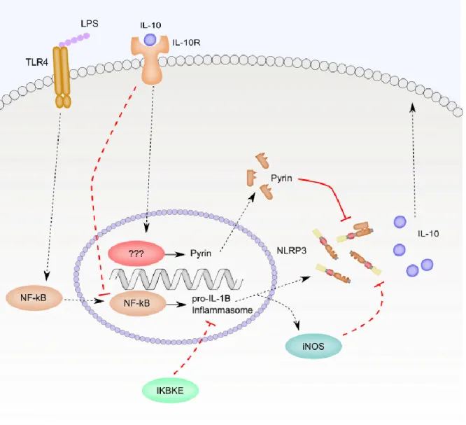
A study on hemorrhagic shock highlights how multiple different signals can be integrated
to balance NLRP3 inflammasome priming. Mice subjected to hemorrhagic shock then given LPS
intratracheally experienced an increase in NLRP3 expression, Caspase 1 activation and IL-1β
20
with LPS as in hemorrhagic shock mice, but IL-1β secretion was much higher in hemorrhagic
shock, leading to the suggestion that hemorrhagic shock induces a NLRP3 inflammasome
modifying factor. Hemorrhagic shock suppressed IL-10 secretion and these mice had lower
levels of Pyrin, a negative regulator of the NLRP3 inflammasome. Blocking IL-10 with an
antibody in sham treated mice decreased Pyrin expression and increased IL-1β secretion, while
administering IL-10 exogenously to hemorrhagic shock treated mice increased Pyrin expression
and decreased IL-1β secretion. Knockdown of Pyrin in mouse lung vascular endothelial cells
(MLVECs) led to an increase in IL-1β secretion that could not be blocked by administration of
exogenous IL-10 [63]. The ability of IL-10 to suppress NLRP3 inflammasome activity during
chronic LPS stimulation was confirmed in a subsequent study of LPS and ATP-treated BMDMs.
In this setting, IL-10 decreased expression of NLRP3 leading to decreased Caspase 1 activation
21
Figure 1.2. Negative feedback loops limit NLRP3 inflammasome priming. This figure
summarizes the discussion from section 1.6.2. Long term stimulation of cells with LPS leads to
production of iNOS which regulates NLRP3 inflammasome signaling through production of
nitric oxide and s-nitrosylation of NLRP3. IKBKE also negatively regulates NLRP3
inflammasome signaling in long term LPS exposures. Solid lines represent direct interactions
and dotted lines represent indirect interactions. Only relationships investigated in the context of
22
1.6.3 Kinase-mediated post-translational priming regulates NLRP3 inflammasome activity
Careful time course analysis of shorter than typical priming exposure followed by ATP
treatment demonstrated unique outcomes that depended on the TLR signal adapter used.
Specifically, priming with Pam3CSK4, a TLR2 ligand that signals through MyD88, resulted in
biphasic activation of Caspase 1, with levels of active Caspase 1 higher at 10 minutes and 180
minutes of Pam3CSK4 stimulation and lower following 30 minutes and 60 minutes. Priming
with poly I:C, a TLR3 ligand that signals through TRIF, induced maximal Caspase 1 activation
following 30 and 60 minutes of poly I:C stimulation and almost none seen at 10 minutes and 180
minutes. In contrast, when priming with LPS, a TLR4 agonist that signals through MyD88 and
TRIF, robust Caspase 1 activation was observed at all four time points. Interestingly, knockout
of MyD88 or TRIF led to LPS-induced priming that mirrored Pam3CSK4 or poly I:C, depending
on the signaling adaptor present. Cells treated with just ATP did not activate Caspase 1 [65].
These priming adaptations are more rapid than one would expect for transcriptional
changes. Actinomycin D, an inhibitor of RNA chain elongation, was used to test for a
requirement for transcription directly. Actinomycin D treatment had no effect on Pam3CSK4-,
poly I:C- or LPS-induced priming of Caspase 1 activation at any time point but blocked
upregulation of NLRP3 and pro-IL-1β. IRAK1 and IRAK4 were critically required for NLRP3
inflammasome priming during a 10-minute exposure of Pam3CSK4 or LPS. Loss of IRAK1 or
IRAK4 resulted in decreased ASC oligomer formation during simultaneous treatment with
Pam3CSK4 and ATP. NLRP3 and IRAK1 co-immunoprecipitated together in macrophages and
the interaction appeared to be enhanced by Pam3CSK4 treatment [65]. A subsequent study
23
and NLRP3 inflammasome co-localization leading to IRAK, NLRP3-, and Caspase
1-dependent IL-18 secretion and cell death [66].
NOX2- and proteasome-dependent phosphorylation of ERK was also required for
post-translational priming of the NLRP3 inflammasome in human monocytes. Co-stimulation with
LPS and ATP triggered Caspase 1 activation and IL-18 secretion from human monocytes in as
little as 5 minutes whereas ATP treatment alone was insufficient. LPS induced phosphorylation
of ERK, but ATP did not. Treatment of monocytes with U0126, an inhibitor of MEK1/2 that
prevents phosphorylation of ERK, blocked IL-18 secretion in response to rapid LPS and ATP
exposure. siRNA specifically targeting ERK1, but not ERK2, confirmed this finding. ERK was
phosphorylated in response to LPS and treatment with MG132 or bortezemib, both inhibitors of
the proteasome, blocked ERK phosphorylation, Caspase 1 activation and IL-18 secretion.
Treatment of cells with DPI to block ROS production also suppressed ERK1/2 phosphorylation
and IL-18 secretion. The deubiquitinating (DUB) enzyme inhibitors G5 and WP1130 blocked
IL-18 secretion but had no effect on ERK1/2 phosphorylation, suggesting that they acted
downstream or separately from this pathway. Wortmannin, a PI3K inhibitor, SB203580, a p38
MAPK inhibitor, and SP600125, a JNK inhibitor, had no effect on IL-18 secretion. Loss of
NLRP3 or Caspase 1 also had no effect on ERK phosphorylation in response to LPS [67]. The
requirement for proteasome activity strongly suggests that priming of the NLRP3 inflammasome
initiated destruction of some factor that blocked ERK phosphorylation, though the identity of the
24
Figure 1.3. Kinases in non-transcriptional priming of the NLRP3 inflammasome. This
figure summarizes the discussion in section 1.6.3. Rapid priming by Pam3CSK4 binding to TLR
2 trigged MyD88-dependent activation of IRAK1. IRAK1 co-immunoprecipitated with the
NLRP3 inflammasome and this association was enhanced by Pam3CSK4 priming. IRAK1
promoted ASC oligomerization. LPS binding to TLR4 promoted activation of MEK1/2 and
25
U1026, NOX2 with DPI or the proteasome with MG132 or bortezemib, suppressed ERK
phosphorylation and IL-18 secretion. The DUB inhibitors G5 and WP1130 had no effect on ERK
phosphorylation, but blocked IL-18 secretion. Solid lines represent direct interactions and dotted
lines represent indirect interactions. Only relationships investigated in the context of NLRP3
inflammasome priming are including.
1.6.4 Caspase 8 contributes to transcriptional and post-translational NLRP3 inflammasome
priming
Caspase 8 played a bifunctional roll in priming the NLRP3 inflammasome through both
transcriptional and post-translational means. Previous research has noted that deletion of
Fas-associated protein with death domain (FADD), the activator of Caspase 8, leads to defects in
myeloid cell differentiation [68]. Differentiation can be normalized with additional deletion of
RIPK3, allowing for the study of FADD and Caspase 8 in BMDMs. LPS-primed and ATP or
nigericin-stimulated BMDMs required TLR4, TRIF, MyD88 and FADD for Caspase 8
maturation. Deletion of Caspase 8 resulted in decreased ATP- and nigericin-induced Caspase 1
activation, IL-1β secretion and cell death, suggesting that Caspase 8 regulates NLRP3
inflammasome activity [69,70]. Deletion of FADD significantly decreased LPS induction of
NLRP3 and pro-IL-1β mRNA. Similar results were observed with Caspase 8 deletion as well
[69].
Interestingly, Caspase 8 maturation also required NLRP3 and ASC, but not Caspase 1 or
Caspase 11, suggesting a possible interaction between Caspase 8 and the core of the NLRP3
26
and Caspase 8 in LPS and ATP-stimulated macrophages. Pharmacologic inhibition of Caspase 8
with Ac-IETD-fmk was equally as effective in suppressing Caspase 1 activation in response to
LPS and ATP when used both as a pretreatment and following LPS priming [69]. Activation of
the NLRP3 inflammasome as measured by dsRNA also required Caspase 8 but, contrary to the
previous study, could not be blocked by another Caspase inhibitor, the pan Caspase inhibitor
27
Figure 1.4. Caspase 8 is critical for transcriptional and post-translational NLRP3
inflammasome priming. This figure summarizes the discussion in section 1.6.4. Caspase 8
maturation in response to LPS stimulation required TLR4, TRIF, MyD88, FADD, NLRP3 and
ASC. Caspase 8 co-localized with Caspase 1 in BMDM stimulated with LPS plus ATP. Loss of
Caspase 8 impacted both production of NLRP3 and IL-1β mRNA and Caspase 1 activation,
28
and dotted lines represent indirect interactions. Only relationships investigated in the context of
NLRP3 inflammasome priming are including.
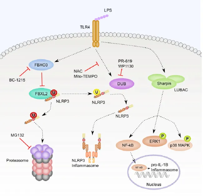
1.6.5 Multiple ubiquitination signals regulate NLRP3 inflammasome priming and protein
degradation
TLR receptor signaling also primes the NLRP3 inflammasome through ubiquitination
and degradation of the NLRP3 inflammasome and its regulators. It was noted that NLRP3
protein levels increased in response to LPS priming in U937 human monocytic cells even in the
presence of cycloheximide, a protein translation inhibitor. Blocking protein degradation with the
proteasome inhibitor MG132, but not the lysosomal inhibitor leupeptin, prevented steady-state
degradation of NLRP3 in untreated cells, leading to increased NLRP3 protein levels. These data
suggest that the ubiquitin-proteasome targeting of NLRP3 is regulated by TLR priming to
control the half-life of NLRP3.
FBXL2 catalyzed NLRP3 ubiquitination in vitro, and interacted with NLRP3 when
overexpressed in cells. LPS treatment decreased the interaction between FBXL2 and NLRP3 and
also decreased ubiquitination of NLRP3. Additionally, depletion of FBXL2 by siRNA in human
macrophages led to increased levels of NLRP3. Mutation of NLRP3 lysine 689 to arginine
blocked ubiquitination, suggesting that K689 was the ubiquitin acceptor site [72].
FBXO3 is an E3 ligase subunit that regulates activity of FBXL2 when FBXL2 is
phosphorylated GSKβ [73]. The FBXO3 inhibitor BC-1215 increased FBXL2 levels and
29
accumulations in NLRP3 but had no effect on mutant K689R NLRP3, suggesting that FBXO3
was responsible for degrading FBXL2 leading to increased NLRP3 protein levels [72].
A loss-of-function (LOF) polymorphism was identified in FBXO3 (V221I) that has an
allele frequency of 6.2% in European Caucasians. Peripheral blood mononuclear cells (PBMCs)
from patients carrying the LOF allele produced less IL-1β when treated with LPS. Mice injected
with a lentivirus encoding for wildtype FBXO3, but not FBXO3 V221I, had increased cellular
infiltration and lung damage in response to Pseudomonas aeruginosa pneumonia, suggesting
increased activity from the NLRP3 inflammasome [73]. The discovery of a naturally occurring
allele that would result in decreased NLRP3 inflammasome activity is particularly interesting.
Mendelian disorders of NLRP3 are the result of activating mutations, but now with a reasonably
abundant allele causing decreased NLRP3 activity, it would be interesting to compare patient
outcomes in NLRP3-driven processes such as S. aureus pneumonia or acute lung injury.
However, this would not be a perfect correlation with NLRP3 activity as FBXO3 and FBXL2
also regulate TRAF [73].
Deubiquitination of NLRP3 is also required for LPS-mediated priming. The general DUB
enzyme inhibitors PR-619 and WP1130 blocked deubiquitination of NLRP3 in response to LPS.
Similarly, treatment with ROS scavenging NAC or mitochondrial ROS specific Mito-TEMPO
inhibited LPS-induced deubiquitination of NLRP3. ROS scavenging blocked LPS plus
ATP-induced Caspase 1 activation, but not Caspase 1 activation resulting from ATP treatment alone,
suggesting a relationship between TLR signaling, ROS production and
deubiquitination-mediated priming. This deubiquitination did not result in accumulation of NLRP3, suggesting
that ubiquitin is acting to suppress NLRP3 activity in this case and not acting to target NLRP3
30
The linear ubiquitin chain assembly complex (LUBAC) plays a role in priming of the
NLRP3 inflammasome. Knockout of Sharpin, one of the three components of the LUBAC -
along with HOIP and HOIL-1, in BMDMs leads to defective pro-IL-1β synthesis in response to
LPS stimulation. Loss of Sharpin decreases LPS-induced NF-κB activation, phosphorylation of
ERK and phosphorylation of p38 MAPK [75]. The exact point of intersection between the
LUBAC and LPS-induced priming is unclear, but the broad effects on these downstream
31
Figure 1.5. Ubiquitination regulates NLRP3 protein levels and activity. This figure
summarizes the discussion in section 1.6.5. LPS priming triggered numerous changes through
ubiquitination-deubiquitination. First, LPS activated FBXO3 to prevent FBXL2 mediated
ubiquitination and proteasomal degradation of NLRP3. Treatment with the FBXO3 inhibitor
BC-1215 opposed LPS-induced activation of FBXO3. Treatment with the proteasome inhibitor
MG132 increased NLRP3 protein levels. LPS also activated deubiquitinating enzymes that
32
suppress ROS blocked deubiquitination of NLRP3. Additionally, the deubiquitinating enzyme
inhibitors PR-619 and WP1130 blocked LPS-mediated priming of NLRP3 by preventing
deubiquitination of NLRP3. Lastly, LPS treatment required Sharpin for full NF-κB signaling,
ERK phosphorylation and p38 MAPK phosphorylation. “P” and “U” indicate the
post-translational modifications phosphorylation and ubiquitination, respectively. The color of the “P”
and “U” reflects the effect of the post-translational modification: green is activating, yellow is
inhibiting, and red is targeted for degradation. Solid lines represent direct interactions and dotted
lines represent indirect interactions.
1.6.6 Post-transcriptional regulation of NLRP3 inflammasome mRNA stability
While TLR-induced priming upregulated transcription, other mechanisms exists to
facilitate tight control of NLRP3 inflammasome mRNA. The myeloid-specific microRNA
miR-223 was a critical regulator of NLRP3 inflammasome expression. miR-miR-223 interacted with the 3’
untranslated region (UTR) of NLRP3 transcripts to target them for degradation. Overexpression
of miR-223 led to decreased NLRP3 protein levels and sequestration of miR-223, by expression
of GFP with a 3’ UTR containing multiple miR-223 binding sites, led to increased NLRP3
protein levels. The consequence of these changes in NLRP3 protein levels carry through to
differences in IL-1β secretion as well. miR223 is not induced during priming, however. It has
been suggested to act as a threshold regulator of transcriptional priming – requiring a strong
enough activating signal to overcome endogenous miR-223-mediated repression [76,77].
Interestingly, Epstein-Barr virus (EBV) produced a microRNA that binds to the same site
33
secretion. When EBV-infected B cells and THP1 cells were cultured in the same media divided
by a 3-μm pore-containing filter, miR-BART15 was secreted from infected B cells via exosomes
that were ultimately taken up by the THP1 cells leading to decreased NLRP3 protein levels and
inhibition of IL-1β secretion [77].
1.6.7 NLRP3 inflammasome signaling without priming
THP1 cells are able to activate the NLRP3 inflammasome independent of priming. This
is thought to be the result of sufficiently high expression of NLRP3, ASC and pro-Caspase 1,
allowing THP1 cells to respond to NLRP3-activating stimuli without priming. In this case,
NLRP3 inflammasome activation led to secretion of IL-18 and necrotic cell death without IL-1β
secretion. THP1 cells are a human leukemic cell line established from a patient with monocytic
leukemia [78], so it is unclear how relevant this response is to physiologically normal cells.
However, in experiments presented in Chapter 3, I have found that primary human CD14+
monocytes responded to LukAB stimulation with production of IL-18 and necrotic cell death,
even in the absence of priming, similar to THP1 cells.
There are likely to be differences in the wide range of phagocytic cell phenotypes,
including not just dendritic cells vs monocytes and macrophages, but alveolar macrophages of
the lung versus Kupffer cells of the liver and Langerhans dendritic cells versus plasmacytoid
dendritic cells, for example. The Immunological Genome Project [79], an effort to catalog gene
expression profiles across a wide range of immune cells using microarrays, suggested that
tissue-specific phenotypes may influence NLRP3 inflammasome activity. Expression of NLRP3 was