effects, angiogenesis, mitogenesis, and polypeptide hor-mone secretion in multiple cell types (1, 2). Both EETs and HETEs are synthesized by cytochrome P450 (CYP450) mo-nooxygenases, but their presence in cells does not entirely depend on acute de novo synthesis. Although initially be-lieved to be transiently acting short-lived metabolites, both EETs and HETEs can replace acyl groups in membrane phospholipids; thus, the cellular content of these eicos-anoids may also be influenced by reciprocal esterification and phospholipase pathways that have not been well-characterized (3–6). We reported that long-chain acyl-CoA synthetase (ACSL) isoform 4 (ACSL4) can use EETs as a substrate (7), but because EET- and HETE-containing phospholipids are present in tissues that express low levels of ACSL4 (8, 9), we wondered whether one or more of the other ACSL isoforms might also activate these molecules to form EET- or HETE-CoAs that could be esterified to spe-cific phospholipids.
Five ASCLs (ACSL1, -3, -4, -5, and -6) (EC 6.2.1.3) con-vert FAs of 12–20 carbons to their corresponding FA-CoAs. Each ACSL isoform has been functionally differentiated by its FA chain length preference, tissue distribution, and sub-cellular location, attributes that alter the ability of an indi-vidual ACSL to channel FAs toward disparate metabolic fates (10).
Once activated, acyl-CoAs have multiple metabolic fates, including the synthesis of triacylglycerol, cholesterol esters, retinal esters, and phospholipids, -oxidation, FA elonga-tion and desaturaelonga-tion, protein acylaelonga-tion, and use as signal-ing molecules (10). Members of the ACS family require Abstract Because the signaling eicosanoids,
epoxyeicosatri-enoic acids (EETs) and HETEs, are esterified to membrane phospholipids, we asked which long-chain acyl-CoA synthe-tase (ACSL) isoforms would activate these molecules and whether the apparent FA substrate preferences of each ACSL isoform might differ depending on whether it was as-sayed in mammalian cell membranes or as a purified bacte-rial recombinant protein. We found that all five ACSL isoforms were able to use EETs and HETEs as substrates and showed by LC-MS/MS that ACSLs produce EET-CoAs. We found differences in substrate preference between ACS as-says performed in COS7 cell membranes and recombinant purified proteins. Similarly, preferences and Michaelis-Menten kinetics for long-chain FAs were distinctive. Substrate preferences identified for the purified ACSLs did not cor-respond to those observed in ACSL-deficient mouse mod-els. Taken together, these data support the concept that each ACSL isoform exhibits a distinct substrate preference, but apparent substrate specificities depend upon multiple factors including membrane character, coactivators, inhibitors, protein interactions, and posttranslational modification.— Klett, E. L., S. Chen, A. Yechoor, F. B. Lih, and R. A. Coleman. Long-chain acyl-CoA synthetase isoforms differ in prefer-ences for eicosanoid species and long-chain fatty acids. J. Lipid Res. 2017. 58: 884–894.
Supplementary key words arachidonic acid • cytochrome P450 • fatty acid/metabolism • phospholipids • acyl-coenzyme A
The arachidonate (AA) metabolites, epoxyeicosatrie-noic acids (EETs) and HETEs, are potent mediators of vasodilation, ion channel activation, anti-inflammatory
This work was supported in part by National Institutes of Health Grants DK090141 and DK107481 (E.L.K.) and DK59935 (R.A.C.) and by the Intra-mural Research Program of the National Institutes of Health, National Institute of Environmental Health Sciences. The content is solely the responsibility of the authors and does not necessarily represent the official views of the National Insti-tutes of Health.
Manuscript received 6 October 2016 and in revised form 16 February 2017. Published, JLR Papers in Press, February 16, 2017
DOI https://doi.org/10.1194/jlr.M072512
Long-chain acyl-CoA synthetase isoforms differ in
preferences for eicosanoid species and long-chain
fatty acids
Eric L. Klett,1,*,† Shufen Chen,*,† Alekhya Yechoor,† Fred B. Lih,§ and Rosalind A. Coleman†
Department of Medicine,* Division of Endocrinology, University of North Carolina School of Medicine, Chapel Hill, NC 27599; Department of Nutrition,† University of North Carolina School of Public Health, Chapel Hill, NC 27599; and Division of Intramural Research,§ Epigenetics and Stem Cell Biology Laboratory (ESCBL), National Institute of Environmental Health Sciences, National Institutes of Health, Research Triangle Park, NC 27709
Abbreviations: AA, arachidonate; ACS, acyl-CoA synthetase; ACSL, long-chain acyl-CoA synthetase; CYP450, cytochrome P450; EET, ep-oxyeicosatrienoic acid; F-ACSL, FLAG-long-chain acyl-CoA synthetase; MBOAT, membrane-bound O-acyltransferase.
1 To whom correspondence should be addressed. e-mail [email protected]
Co-A and substrates with a carboxylic acid group in order to catalyze the thioesterification reaction. The thioesterifi-cation two-step reaction is energetically costly, using the equivalent of two high-energy bonds (11, 12). This high energetic cost emphasizes the importance of the ACS-catalyzed reaction as a regulatory node for the metabo-lism of FAs.
The five ACSL isoforms were identified, cloned, and characterized by Yamamoto and his colleagues (8, 13–15), and most of our knowledge regarding their substrate pref-erences comes from these initial studies. However, the sub-strate preferences reported in these studies cannot be directly compared with each other because the enzymes were expressed differently and the methods used for activ-ity assays varied. For activactiv-ity studies of ACSL1, ACSL4, ACSL5, and ACSL6 [initially known as ACSL2 (16)], the enzymes were expressed in and purified from Escherichia coli, whereas ACSL3 was expressed and studied in COS7 (monkey kidney) cells, but in each instance, the substrate concentration ranges and the specific Vmax and Km values were unclear (8, 13–15). Although substrate specificities for the ACSL isoforms were reported, direct and systematic comparisons of substrate preferences for each ACSL iso-form were not periso-formed.
The importance of substrate preference for a specific ACSL isoform suggests how and where a specific substrate will be metabolized. We report here the ACS enzyme kinet-ics with different FA and eicosanoid substrates of the rat ACSL isoforms overexpressed in bacterial and mammalian cells. Further, we provide validation of the indirect spectro-photometric ACS activity assay by showing LC-MS/MS evi-dence that the product of the reaction produces an acyl thioester.
MATERIALS AND METHODS
Materials
AA, (±)-8,9-EET, (±)-11,12-EET, and (±)-14,15-EET were pur-chased from Cayman Chemical (Ann Arbor, MI). Arachidonoyl-CoA (20:4-Arachidonoyl-CoA) was purchased from Avanti Polar Lipids (Alabaster, AL). All other FAs and reagents were purchased from Sigma (St. Louis, MO).
Construction of recombinant pFLAG-ACSLs and mammalian ACSL plasmid
cDNA was synthesized from either rat liver or brain total RNA (extracted using TRIzol; Invitrogen) and used as a template to amplify the ACSL open reading frames (high capacity cDNA re-verse transcription kit; Applied Biosystems, Foster City, CA). Prim-ers for amplification of ACSL1, ACSL3, ACSL4, ACSL5, and ACSL6 were designed to include the entire open reading frames, based on nucleotide sequences obtained from the GenBank data-base (supplemental Table S1). ACSL amplification was performed by PCR with the designed primers. The amplified ACSL PCR products were ligated into either pFLAG-CTC vector (Sigma) or pcDNA3.1 vector (Invitrogen) digested with the same restriction enzymes. The sequences of pFLAG-ACSL and pcDNA3.1-ACSL fusion constructs were verified by the University of North Caro-lina Automated Sequencing Facility.
Expression of recombinant F-ACSL proteins in E. coli Recombinant F-ACSLs were expressed in E. coli DH5 after in-duction with 1 mM IPTG at an A600 of 1.0. DH5 was grown in Terrific Broth (Life Technologies, Inc.) supplemented with ampi-cillin (60 g/ml) at 30°C and shaken at 250 rpm. After a 12 h induction, cells were harvested by centrifuging at 4,800 g for 10 min in a Sorvall HS-4 rotor. The cell pellet was resuspended in 10 ml of 10 mM Tris (pH 7.4), 0.5 mM EDTA (TE) buffer. Resus-pended cells were incubated with 100 g/ml lysozyme for 30 min on ice and then sonicated with six 10 s bursts, each followed by a 10 s rest on ice. Cellular debris was removed from the cell lysates by centrifugation at 3,000 g for 10 min. Part of the supernatant was saved (cell extract), and the remainder was layered over a 2 ml cushion of 55% (w/w) sucrose topped with 0.5 ml of 5% (w/w) sucrose in TE buffer. After centrifuging in a Beckman SW41 rotor at 210,000 g for 3 h, the supernatant was removed (soluble frac-tion). The membrane band at the interface was collected with a 19 gauge needle and syringe. Protein concentrations were deter-mined by the BCA method (Pierce).
Purification of the recombinant F-ACSL proteins
F-ACSLs were purified by Flag M2 column chromatography. The Flag M2 antibody affinity matrix (1 ml) (Sigma) was activated with 0.1 M glycine (pH 3.5), 50 mM Tris (pH 7.4), and 150 mM NaCl (TBS) buffer. DH5 membrane fractions containing over-expressed F-ACSLs were solubilized in TBS containing 1% Triton X-100 and passed over the column four times. The column was washed three times with 12 ml of TBS (pH 7.4), and then eluted with five 1 ml aliquots of 100 g/ml Flag peptide (Sigma) dissolved in TBS buffer (pH 7.4).
Transient transfection of pcDNA3.1-ACSL1 and ACSL4 COS7 cells were routinely cultured in Dulbecco’s Modified Eagle’s Medium containing 10% fetal bovine serum. COS7 cells were plated at a cell density of 2.0 × 10⁶ in 10 cm dishes and trans-fected for 18 h after plating with 10 g of plasmid carrying rat ACSL1 or ACSL4 (XtremeGene HP; Roche). Cell homogenates were collected 48 h posttransfection in ice-cold medium A [10 mM Tris (pH 7.4), 250 mM sucrose, 1 mM EDTA, 1 mM dithiothreitol, and protease inhibitor mixture (Sigma)]. Homogenates were then centrifuged at 1,000 g for 10 min at 4°C. Total membranes were prepared by subjecting the supernatant to ultracentrifuga-tion at 100,000 g for 1 h at 4°C. The resulting supernatant was removed and the membrane pellet was resuspended in ice-cold medium A. Aliquots were stored at 80°C until use.
Spectrophotometric ACS activity assay
intercept created from an NADH standard curve with concentra-tions from 0 to 0.2256 M.
Identification of AA-CoA and EET-CoAs by LC-MS/MS Methods were adapted from Magnes et al. (18). Solid phase extraction was performed on Hypersep Retain PEP 60 mg, 3 ml cartridges (Thermo Fisher Scientific). Elution solvent was pre-pared by mixing 50 ml water with 50 ml acetonitrile plus 100 l 28% NH4OH solution and 5 l formic acid. Cartridges were con-ditioned with 3 ml acetonitrile and then 3 ml water. Two hundred fifty microliters of reaction mixture were diluted with 2 ml water and loaded onto the cartridge. The cartridge was rinsed with 6 ml water before eluting acyl-CoAs in 2.5 ml of elution solvent.
Analysis was performed on an Ultimate 3000 UHPLC and Quantiva tandem mass spectrometer (Thermo Fisher Scientific.) The chromatography column was an Xselect CSH C18, 2.1 × 50 mm, 3.5 m particles (Waters). Mobile phase A consisted of 1 ml 28% NH4OH solution, 50 l formic acid, and 1,000 ml water. Mobile phase B was acetonitrile. The starting mobile phase was 20% B and was held for 0.5 min before ramping to 40% B over 3.5 min. The column was held at 40% B for 2 min and then returned to 20% B for 3 min for re-equilibration. The injection volume was 4 l.
Samples were introduced to the mass spectrometer via an ESI source. The vaporizer temperature was 300°C and the ion transfer tube temperature was 325°C. Sheath, auxiliary, and sweep gas were set to 35, 20, and 1 arbitrary units, respectively. Spray voltage was 4,250 V for positive ion experiments and 3,750 V for nega-tive ion experiments. Acyl-CoAs were detected via posinega-tive ion neutral loss scanning from m/z 900 to 1,400 with neutral loss set to 507.1. The collision energy was set to 34 V and collision cell pres-sure was 2 mTorr. Positive product ion spectra of acyl-CoAs were also obtained.
Western blot
FLAG column purified E. coli F-ACSLs (F-ACSLs) (6 g) and membranes from COS7 cells (50 g) were separated by electro-phoresis on a 10% polyacrylamide gel and transferred to nitrocel-lulose membranes (Bio-Rad). Protein expression of F-ACSLs was detected by anti-FLAG monoclonal antibody (Sigma; F1804). For ACSL1 and ACSL4 expressed in mammalian cells, rabbit anti- human ACSL1 (Cell Signaling, Danvers, MA; 4047) and rabbit anti-human ACSL4 (19), a generous gift from S. M. Prescott (University of Utah, Salt Lake City, UT), were used to detect ACSL1 and ACSL4, respectively. GAPDH was used as the loading control (Ab-cam, Cambridge, MA; ab8245). To determine the purity of FLAG-column purified F-ACSLs, both E. coli lysates (40 g) and purified F-ACSLs (6 g) were separated by electrophoresis on a 10% poly-acrylamide gel and stained with Coomassie brilliant blue.
Data analyses
Data are expressed as mean ± SEM from three separate experi-ments. Michaelis-Menten kinetic enzyme activity curves were drawn and Vmax and Km values derived using GraphPad Prism® 6.0 h
(San Diego, CA).
RESULTS
Bacterial and mammalian overexpressed ACSL isoforms are able to activate EETs and HETEs
EETs and HETEs are oxidative products of CYP450 ep-oxygenases, using AA as the substrate. EETs and HETEs function as autocrine and paracrine signals that regulate vasodilation, ion channel activation, anti-inflammatory
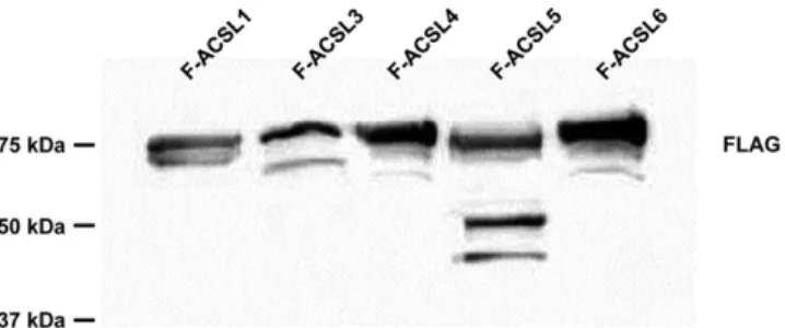
Fig. 1. FLAG column purified F-ACSL isoforms overexpressed in
E. coli were resolved by PAGE and immunoblotted with anti-FLAG antibody.
effects, angiogenesis, mitogenesis, and polypeptide hor-mone secretion (2). They are incorporated into phospho-lipids by a CoA-dependent process that plays a significant role in regulating EET actions on either a membrane re-ceptor or as a direct interactor with intracellular effectors (2). We previously reported that purified bacterial F-ACSL4 can use (±)-8,9-EET, -11,12-EET, and -14,15-EET as sub-strates in an indirect ACS activity assay (7). Because AA is the precursor for all eicosanoids and because eicosanoids contain a terminal carboxyl group, we hypothesized that the ACSL isoforms would differ in their activation of eico-sanoid substrates. Using the sequences of rat ACSL iso-forms (supplemental Table S1), we constructed bacterial plasmids containing the cDNA for each isoform linked to a C terminus FLAG tag (FLAG). Bacterial pACSL-FLAG plasmids were transfected and induced in E. coli and the resulting recombinant proteins were purified by col-umn chromatography. To ensure expression of the recom-binant proteins, we resolved 6 g of FLAG column purified F-ACSLs by PAGE, blotted, and probed with anti-FLAG an-tibody (Fig. 1). Because there were doublet bands observed in the immunoblot, we wanted to ensure the purity of the FLAG-column purified recombinant F-ACSLs and again re-solved each purified F-ACSL isoform (6 g) and the E. coli
lysates (40 g) by PAGE and stained with Coomassie bril-liant blue (supplemental Figs. S1, S2). Each purified F-ACSL showed a double band with the upper band (80–75 kDa) being the predicted molecular mass for each F-ACSL isoform. We suspect that the lower bands (60 kDa) are degradation products. Using purified bacterial expressed F-ACSL enzymes, we performed indirect ACS activity assays using 1 g protein with a fixed (5 M) final concentration of EETs and HETEs as substrates. To validate our indirect ACS activity assay and ensure that eicosanoid-CoAs were produced, we performed the indirect ACS activity assay with F-ACSL4 using 5 M AA, 8,9-EET, 11,12-EET, and 14,15-EET as substrates. Upon reaction termination, the reaction products were subjected to LC-MS/MS analy-ses. Comparing the authentic reference standard, arachi-donoyl-CoA (20:4-CoA) (Fig. 2B), to the reaction product of F-ACSL4 and AA (Fig. 2C), we found a distinct peak at
m/z 1,054 corresponding to arachidonoyl-CoA [M+H]+.
(supplemental Fig. S3) (18). We next analyzed the reaction products of F-ACSL4 and the EETs, with the expected m/z
of the EET-CoAs to be 1,070 (AA → 1,054 plus oxygen →
16). Indeed, for each of the EETs tested, we observed
strong peaks at m/z 1,070 corresponding to EET-CoAs (Fig. 2D–F). Further, positive product ion scans of reaction products showed ion peaks that were consistent with the formation of EET-CoAs (supplemental Fig. S4). Taken
together, the LC-MS/MS analyses of the indirect ACS activ-ity assay reaction products provide strong evidence that EET-CoA products are formed.
Following validation that the indirect ACS activity assay produced EET-CoA products, we assayed each of the F-ACSL isoforms with EETs and HETEs as substrates. Each of the F-ACSL isoforms was able to convert both substrates to acyl-CoA products, but we observed differences in substrate preference among the isoforms (Fig. 3). For example, F-ACSL4 exhibited the greatest activity with EETs, whereas F-ACSL1 showed the greatest activity with HETEs (Fig. 3A, B). Interestingly, compared with the parent molecule, AA, F-ACSL1 had greater activity with all three HETE species tested (Fig. 3B). ACS activity with F-empty vector using the substrates tested was not observed (data not shown).
Previous reports describing the substrate preferences of ACSL isoforms were based on proteins expressed and puri-fied from recombinant bacteria (8, 14, 15). Because ACSLs are normally membrane bound proteins, we wondered whether there were differences between purified ACSL iso-forms expressed in bacteria versus the ACSL isoiso-forms over-expressed in membranes of mammalian cells. To answer this question, we cloned rat ACSL1 and ACSL4 into the mammalian pcDNA3.1 expression vector and transformed COS7 cells to overexpress ACSL1 and ACSL4 isoforms (supplemental Table S1). We selected ACSL1 and ACSL4 because they had the greatest ACS activity from the bacte-rial expressed F-ACSLs using the eicosanoids as substrates. To ensure overexpression, we resolved 50 g of mem-branes by PAGE, blotted and probed with anti-ACSL1 or anti-ACSL4 antibody (Fig. 4). Both ACSL1 and ACSL4 were overexpressed compared with control COS7 cells transfected with the empty vector. Interestingly, in COS7 cells, the basal expression of ACSL4 appeared higher than the overexpressed ACSL1. Despite the high amount of basal ACSL4 protein expression, the indirect ACS activity using palmitate was 1,000-fold lower compared with what we observed with the overexpressed ACSL isoform indirect
ACS activities (nanomoles per minute per milligram protein vs. micromoles per minute per milligram protein, respectively) (supplemental Fig. S7). We performed indi-rect ACS activity assays using 1 g protein from total mem-brane preparations using the same fixed (5 M) final concentration of EETs and HETEs as substrates. To vali-date that EET-CoAs were produced with COS7 overex-pressed ACSL4, we again performed indirect ACS activity assays and subjected the reaction products to LC-MS/MS analyses. As with the F-ACSL4 reaction products, the COS7-ACSL4 reactions with 5 M AA, 8,9-EET, 11,12-EET, and 14,15-EET as substrates produced AA-CoA and EET-CoA products, respectively (Fig. 5). Further, chromatographic retention times of the COS7-ACSL4 reaction products for AA and EETs corresponded to those from the F-ACSL4 re-action products (supplemental Figs. S5, S6). However, the
m/z 1,054 and 1,070 peaks were much less intense com-pared with the F-ACSL4 reaction products, probably because the F-ACSLs were purified enzymes, unlike the
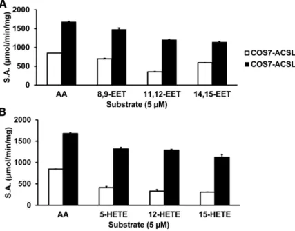
Fig. 3. Purified F-ACSLs can activate either EETs (A) or HETEs (B) as substrates. ACS activity with different substrates at 5 M (final concentration). Error bars reflect SEM from three separate experiments. S.A., specific activity.
COS7-ACSLs, which were membrane bound and surrounded by lipids, proteins, and cofactors. It is likely that these mem-brane constituents impact the activity of the ACSL. Despite these less intense peaks, overexpressed COS7-ACSL4 was able to synthesize EET-CoAs.
Comparing the ACS activities between COS7-ACSL1 and COS7-ACSL4 with EET and HETE as substrates, we found that ACSL4 had greater activity with both EETs and HETEs compared with ACSL1 (Fig. 6). Further, the mammalian expressed ACSL1 and ACSL4 had greater activity with the
parent AA than with EETs or HETEs, which was not seen with the bacterial F-ACSL1 and F-ACSL4 assays. Again, these differences in ACS activity are likely due to the mem-brane constituents retained in mammalian preparations, but not in the purified F-ACSLs.
FA substrate preferences of ACSL isoforms expressed in mammalian cells differ from those expressed in bacteria
Because of previous reports describing the substrate preferences of ACSL isoforms from purified recombinant bacteria and because we identified substrate preference
differences using EETs and HETEs between bacterial and mammalian expressed ACSL isoforms, we wondered whether there were differences in substrate preference for FAs based on whether the ACSL isoform was expressed in bacteria or expressed in mammalian membranes. Using FAs of differing chain length and degree of unsaturation (palmitate, 16:0; oleate, 18:1; stearate, 18:0; linoleate, 18:2; AA, 20:4) at concentrations ranging from 2.5 M to 100 M, we performed indirect ACS activity assays (17) using the purified F-ACSL isoforms to characterize the enzyme kinetics. The plotted Michaelis-Menten kinetic
Fig. 6. ACSL1 and ACSL4 overexpressed in COS7 cells use both EETs and HETEs as substrates. ACS ac-tivity with 5 M final substrate concentrations of AA, 8,9-EET, 11,12-EET, 14,15-EET, 5-HETE, 12-HETE, and 15-HETE. Error bars reflect SEM from three separate experiments. S.A., specific activity.
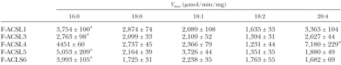
enzyme activity curves (Fig. 7) showed that each F-ACSL isoform had activity with each substrate, but that substrate preferences were distinct. For F-ACSL1, -3, -5, and -6, the apparent Vmax values were highest with palmitate (Table 1); however, similar to a previous report (8), the Vmax and Km values of F-ACSL4 were greatest with AA [7,180 ± 229 mol/min/mg and 11.4 ± 1.3 M, respectively (Table 1)]. The apparent Vmax and Km values obtained from recombi-nant proteins show different orders of FA substrate prefer-ences in Table 1 and Table 2.
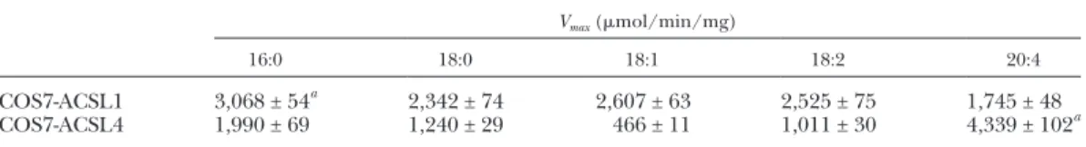
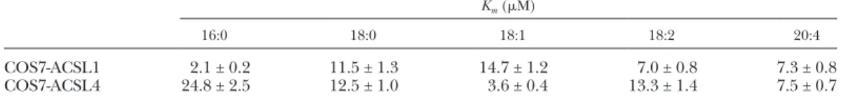
Because we noted differences in substrate preference be-tween loss-of-function rodent models of ACSL1 (20) and the purified bacterial expressed ACSLs, we suspected that substrate preference would differ in the mammalian ex-pressed ACSL isoforms. Indeed, similar to the overex-pressed purified F-ACSL isoforms in bacteria, the substrate preferences, based on the apparent Vmax and Km values, dif-fered between COS7 cell overexpressed ACSL1 and ACSL4 isoforms (Fig. 8, Tables 3, 4).
Activities with 16:0 were highest for both purified re-combinant F-ACSL1 and the COS7 cell overexpressed ACSL1, but marked differences were observed for sub-strates with chain lengths longer than 16 carbons and for unsaturated FAs. For example, recombinant F-ACSL1 preferred 20:4, whereas COS7-ACSL1 did not (Tables 1, 3). Major differences in substrate preference were not observed between recombinant F-ACSL4 and COS7-ACSL4, but we confirmed the previous findings that ACSL prefers 20:4 (8), a preference that was not af-fected by ACSL4 expression in bacteria compared with expression in mammalian cells. Taken together, these data suggest that ACSL activity and substrate preference depend on multiple factors, including the substrate, mem-brane character, coactivators, inhibitors, interactions with
other enzymes, ACSL cellular location, and ACSL tissue expression.
DISCUSSION
AA is converted to potent eicosanoid signaling mole-cules by the cyclooxygenase, lipoxygenase, and CYP450 monooxygenase pathways, which produce both EETs and HETEs by CYP epoxygenases and CYP -oxidases, respec-tively (21). EETs and HETEs have multiple biologic func-tions that have been primarily defined in vascular and renal systems, including vasodilation, ion channel activa-tion, anti-inflammatory effects, angiogenesis, mitogenesis, regulation of coagulation, and polypeptide hormone se-cretion. Despite the focus on the free eicosanoids, most EETs and HETEs are in cell membranes, esterified in glyc-erophospholipids (4, 5, 22). The mechanism by which EETs and HETEs exert their biological effects is not en-tirely known, but the presence of esterified EETs and HETEs in phospholipids suggests that they might either alter membrane dynamics that change receptor or channel activity or serve as a preformed reservoir that can be re-leased by activated phospholipases potentially leading to downstream receptor activation (1, 2, 23). Further, the es-terification of EETs and HETEs into glycerophospholipids could serve as a means to “de-activate” these signaling mol-ecules. The incorporation of EETs and HETEs into phos-pholipids requires a CoA-dependent process (5, 24). Once the EET or HETE is activated by an ACS, it can be esterified by a phospholipid acyltransferase, such as membrane-bound O-acyltransferase (MBOAT)5 (MBOAT5/LPCAT3) or MBOAT7 (MBOAT7/LPIAT1) (Fig. 9) (25). Indeed, we show here that each of the ACSL isoforms is able to activate TABLE 1. Vmax values from purified F-ACSLs with various substrates
Vmax (mol/min/mg)
16:0 18:0 18:1 18:2 20:4
F-ACSL1 3,754 ± 100a 2,874 ± 74 2,089 ± 108 1,635 ± 33 3,363 ± 104 F-ACSL3 2,763 ± 98a 2,099 ± 33 2,109 ± 52 1,394 ± 31 2,627 ± 44 F-ACSL4 4451 ± 60 2,737 ± 45 2,366 ± 79 1,231 ± 44 7,180 ± 229a F-ACSL5 5,053 ± 209a 2,164 ± 39 3,726 ± 44 1,351 ± 35 1,880 ± 49 F-ACLS6 3,993 ± 105a 1,725 ± 31 2,238 ± 35 1,763 ± 55 1,682 ± 69
Vmax values derived from kinetics data from Fig. 7 where data points are the mean of determinations from proteins obtained in three independent experiments.a
Indicates the substrate with which the greatest ACS activity is achieved for each F-ACSL.
TABLE 2. Km values from purified F-ACSLs with various substrates
Km (M)
16:0 18:0 18:1 18:2 20:4
F-ACSL1 2.7 ± 0.4 6.6 ± 0.7 6.7 ± 1.4 5.0 ± 0.4 6.3 ± 0.8 F-ACSL3 4.0 ± 0.7 3.1 ± 0.2 5.3 ± 0.6 7.3 ± 0.6 4.3 ± 0.3 F-ACSL4 4.5 ± 0.3 2.9 ± 0.2 16.7 ± 1.9 4.0 ± 0.6 11.4 ± 1.3 F-ACSL5 6.5 ± 1.1 4.4 ± 0.4 6.3 ± 0.3 5.8 ± 0.6 1.8 ± 0.3 F-ACLS6 3.0 ± 0.4 4.4 ± 0.4 4.4 ± 0.3 6.0 ± 0.8 2.9 ± 0.6
EETs and HETEs to produce EET-CoAs and HETE-CoAs, respectively.
The CYP450 monooxygenases that produce EETs and HETEs are expressed in the heart, vasculature, kidney, brain, liver, lung, spleen, blood cells, testis, and pancreas (26). Because the different ACSL isoforms differ in their EET and HETE substrate specificities, the expression of specific ACSL isoforms (27) may directly influence EET and HETE metabolism and action in a tissue-specific man-ner and suggest why these eicosanoids might differ in their biological functions and metabolism.
ACSs catalyze the thioesterification of FAs with CoA to yield fatty acyl-CoAs. After activation, the fatty acyl-CoA products have multiple metabolic fates, including mem-brane synthesis, energy production, storage, and the pro-duction of signaling molecules. Despite the fact that each ACSL isoform performs the identical reaction, the isoforms differ in their tissue and subcellular distribution and likely substrate preference. We show here that FA preferences of the ACSL isoforms are similar, but not identical, when determined with purified E. coli prepara-tions versus COS7 cell membranes. The reason for the dif-ferences in substrate preference between the purified bacterial and membrane bound mammalian expressed ACSL isoforms is multifactorial, including membrane character, coactivators, inhibitors, interactions with other
enzymes, specific ACSL cellular location, specific ACSL expression, and posttranslational modification (6). In-deed, we have previously shown that ACSL1 is differen-tially phosphorylated and acetylated in different cell types (28). Furthermore, in cardiac muscle, where the ACSL1 isoform predominates, ACSL activity is 3.5 times greater with 18:2 than with 16:0, 18:0, 18:1, or 20:4 (20), strongly suggesting that tissue-specific composition of membranes may also influence ACSL substrate prefer-ence. Collectively, the substrate preferences presented here and those from previous animal model experiments indicate that when describing ACSL substrate prefer-ence, it must be placed into the context of the model system.
Our results with recombinant expressed ACSL isoforms are comparable to substrate preferences that were previ-ously reported (8, 14, 15) in that ACSL1, -5, and -6 pre-ferred 16:0, whereas ACSL4 prepre-ferred 20:4. Because the ACS activity of ACSL3 was originally reported from ex-pression in COS7 cells, we were not able to directly com-pare our substrate preference from bacterial recombinant F-ACSL3. However, our results that F-ACSL3 prefers 16:0 agree with the preferences reported for ACSL3 ex-pressed in COS7 cells (13). We also observed higher ACS specific activities from recombinant expressed ACSL isoforms than previously reported. While there are multiple
Fig. 8. Michaelis-Menten enzyme activity curves from membranes of COS7 cells overexpressing ACSL1 (A) or ACSL4 (B) with different FAs. Data points rep-resent the mean of determinations from three inde-pendent experiments.
TABLE 3. Vmax values from overexpressed ACSL1 and ACSL4 COS7 membranes with various substrates
Vmax (mol/min/mg)
16:0 18:0 18:1 18:2 20:4
COS7-ACSL1 3,068 ± 54a 2,342 ± 74 2,607 ± 63 2,525 ± 75 1,745 ± 48 COS7-ACSL4 1,990 ± 69 1,240 ± 29 466 ± 11 1,011 ± 30 4,339 ± 102a
Vmax values derived from kinetics data from Fig. 8 where data points are the mean of determinations from proteins obtained in three independent experiments.
explanations for these differences, the most likely relate to how we made the ACSL isoform constructs, which con-tained a FLAG epitope, and our method of purification. We likely obtained purer enzyme preparations because we used FLAG affinity column purification. It seems less likely that the fusion of FLAG to the C terminus enhances ACSL activity.
Our measures of ACS-specific activity between the dif-ferent ACSL isoforms and substrates have several limita-tions. First, three different methods have been used historically to measure ACS activity; these include direct HPLC measurements (29) or 3H-labeled CoA (30), and the indirect coupled assay measuring the reduction of NADH (17). Each method has its own specific pitfalls, but in all cases the methods measure only relative activi-ties for a given experimental condition. As a result, it is not possible to compare ACS activities obtained with dif-fering methods. In this study, to allow comparisons of substrate preference, we used the same assay for recom-binant and cell expressed ACSL isoforms, and we main-tained the same experimental conditions. A second limitation concerns substrate concentration; the amount of substrate used in these assays far exceeds the intracel-lular concentration of unbound free FA, calculated to be less than 1 M (31). Furthermore, we cannot know the FA concentration at the enzyme’s active site. These diffi-culties make it difficult to relate in vitro to in vivo ACS activities. Until we have more sensitive methods to measure
the in vivo fluxes of free FAs and acyl-CoAs simultaneously, we are forced to rely on the current measures of ACS activity. Finally, our in vitro ACS activity assays do not account for the mixtures of FAs that are normally pre-sented to ACSLs. To define a true substrate preference, an ACSL must select a FA from a cellular pool of FAs and FA metabolites.
We have shown, using a validated indirect assay for ACS activity, that whereas each ACSL isoform catalyzes the same reaction, individual ACSL isoforms differ in sub-strate preferences that appear to be modified depending on the model system in which the ACSL is expressed. These differences likely allow specificity in FA and FA me-tabolite partitioning toward different metabolic fates in a cellular or tissue-dependent manner. We have also dem-onstrated that each of the ACSL isoforms, whether ex-pressed in bacterial or mammalian cells, can use both EETs and HETEs as substrates. Given the importance of these signaling eicosanoids and the ability of the ACSL isoforms to activate them, further efforts should be directed toward determining whether the functions of esterified eicosanoids differ from those that have been newly formed, as well as determining how ACSLs and acyltransferases work together to channel eicosanoids toward membrane storage pools.
The authors would like to thank Dr. Diana Stafforini for assistance with procuring the anti-ACSL4 antibody.
TABLE 4. Km values from overexpressed ACSL1 and ACSL4 COS7 membranes with various substrates
Km (M)
16:0 18:0 18:1 18:2 20:4
COS7-ACSL1 2.1 ± 0.2 11.5 ± 1.3 14.7 ± 1.2 7.0 ± 0.8 7.3 ± 0.8 COS7-ACSL4 24.8 ± 2.5 12.5 ± 1.0 3.6 ± 0.4 13.3 ± 1.4 7.5 ± 0.7
Km values derived from kinetics data from Fig. 8 where data points are the mean of determinations from proteins obtained in three independent experiments.
REFERENCES
1. Spector, A. A., and H. Y. Kim. 2015. Cytochrome P450 epoxygenase pathway of polyunsaturated fatty acid metabolism. Biochim. Biophys. Acta.1851: 356–365.
2. Spector, A. A., and A. W. Norris. 2007. Action of epoxyeicosatrienoic acids on cellular function. Am. J. Physiol. Cell Physiol.292: C996–C1012. 3. Fang, X., M. VanRollins, T. L. Kaduce, and A. A. Spector. 1995.
Epoxyeicosatrienoic acid metabolism in arterial smooth muscle cells. J. Lipid Res.36: 1236–1246.
4. Hammond, V. J., and V. B. O’Donnell. 2012. Esterified eicosanoids: generation, characterization and function. Biochim. Biophys. Acta. 1818: 2403–2412.
5. Karara, A., E. Dishman, J. R. Falck, and J. H. Capdevila. 1991. Endogenous epoxyeicosatrienoyl-phospholipids. A novel class of cellular glycerolipids containing epoxidized arachidonate moieties.
J. Biol. Chem.266: 7561–7569.
6. Spector, A. A., and M. A. Yorek. 1985. Membrane lipid composition and cellular function. J. Lipid Res.26: 1015–1035.
7. Klett, E. L., S. Chen, M. L. Edin, L. O. Li, O. Ilkayeva, D. C. Zeldin, C. B. Newgard, and R. A. Coleman. 2013. Diminished acyl-CoA synthetase isoform 4 activity in INS 832/13 cells reduces cellular epoxyeicosatrienoic acid levels and results in impaired glucose-stimulated insulin secretion. J. Biol. Chem.288: 21618–21629. 8. Kang, M. J., T. Fujino, H. Sasano, H. Minekura, N. Yabuki, H.
Nagura, H. Iijima, and T. T. Yamamoto. 1997. A novel arachidonate-preferring acyl-CoA synthetase is present in steroidogenic cells of the rat adrenal, ovary, and testis. Proc. Natl. Acad. Sci. USA.94: 2880–2884. 9. Mashek, D. G., L. O. Li, and R. A. Coleman. 2006. Rat long-chain
acyl-CoA synthetase mRNA, protein, and activity vary in tissue distri-bution and in response to diet. J. Lipid Res.47: 2004–2010.
10. Grevengoed, T. J., E. L. Klett, and R. A. Coleman. 2014. Acyl-CoA metabolism and partitioning. Annu. Rev. Nutr.34: 1–30.
11. Watkins, P. A. 2008. Very-long-chain acyl-CoA synthetases. J. Biol. Chem.283: 1773–1777.
12. Watkins, P. A., D. Maiguel, Z. Jia, and J. Pevsner. 2007. Evidence for 26 distinct acyl-coenzyme A synthetase genes in the human genome.
J. Lipid Res.48: 2736–2750.
13. Fujino, T., M. J. Kang, H. Suzuki, H. Iijima, and T. Yamamoto. 1996. Molecular characterization and expression of rat acyl-CoA synthe-tase 3. J. Biol. Chem.271: 16748–16752.
14. Iijima, H., T. Fujino, H. Minekura, H. Suzuki, M. J. Kang, and T. Yamamoto. 1996. Biochemical studies of two rat acyl-CoA synthe-tases, ACS1 and ACS2. Eur. J. Biochem.242: 186–190.
15. Oikawa, E., H. Iijima, T. Suzuki, H. Sasano, H. Sato, A. Kamataki, H. Nagura, M. J. Kang, T. Fujino, H. Suzuki, et al. 1998. A novel acyl-CoA synthetase, ACS5, expressed in intestinal epithelial cells and proliferating preadipocytes. J. Biochem.124: 679–685.
16. Mashek, D. G., K. E. Bornfeldt, R. A. Coleman, J. Berger, D. A. Bernlohr, P. Black, C. C. DiRusso, S. A. Farber, W. Guo, N. Hashimoto, et al. 2004. Revised nomenclature for the mammalian
long-chain acyl-CoA synthetase gene family. J. Lipid Res. 45:
1958–1961.
17. Hosaka, K., M. Mishina, T. Tanaka, T. Kamiryo, and S. Numa. 1979. Acyl-coenzyme-A synthetase I from Candida lipolytica. Purification, properties and immunochemical studies. Eur. J. Biochem. 93:
197–203.
18. Magnes, C., F. M. Sinner, W. Regittnig, and T. R. Pieber. 2005. LC/ MS/MS method for quantitative determination of long-chain fatty acyl-CoAs. Anal. Chem.77: 2889–2894.
19. Cao, Y., K. B. Dave, T. P. Doan, and S. M. Prescott. 2001. Fatty acid CoA ligase 4 is up-regulated in colon adenocarcinoma. Cancer Res. 61: 8429–8434.
20. Grevengoed, T. J., S. A. Martin, L. Katunga, D. E. Cooper, E. J. Anderson, R. C. Murphy, and R. A. Coleman. 2015. Acyl-CoA syn-thetase 1 deficiency alters cardiolipin species and impairs mitochon-drial function. J. Lipid Res.56: 1572–1582.
21. Brash, A. R. 2001. Arachidonic acid as a bioactive molecule. J. Clin. Invest.107: 1339–1345.
22. Bernstrom, K., K. Kayganich, R. C. Murphy, and F. A. Fitzpatrick. 1992. Incorporation and distribution of epoxyeicosatrienoic acids into cellular phospholipids. J. Biol. Chem.267: 3686–3690.
23. Capdevila, J. H., J. R. Falck, and R. C. Harris. 2000. Cytochrome P450 and arachidonic acid bioactivation. Molecular and functional properties of the arachidonate monooxygenase. J. Lipid Res.41:
163–181.
24. Weintraub, N. L., X. Fang, T. L. Kaduce, M. VanRollins, P. Chatterjee, and A. A. Spector. 1997. Potentiation of endothelium-dependent relaxation by epoxyeicosatrienoic acids. Circ. Res.81:
258–267.
25. Yamashita, A., Y. Hayashi, Y. Nemoto-Sasaki, M. Ito, S. Oka, T. Tanikawa, K. Waku, and T. Sugiura. 2014. Acyltransferases and transacylases that determine the fatty acid composition of glycero-lipids and the metabolism of bioactive lipid mediators in mamma-lian cells and model organisms. Prog. Lipid Res.53: 18–81.
26. Kaspera, R., and R. A. Totah. 2009. Epoxyeicosatrienoic acids: for-mation, metabolism and potential role in tissue physiology and pathophysiology. Expert Opin. Drug Metab. Toxicol.5: 757–771. 27. Ellis, J. M., C. E. Bowman, and M. J. Wolfgang. 2015. Metabolic and
tissue-specific regulation of acyl-CoA metabolism. PLoS One.10:
e0116587.
28. Frahm, J. L., L. O. Li, T. J. Grevengoed, and R. A. Coleman. 2011. Phosphorylation and acetylation of acyl-CoA synthetase- I.
J. Proteomics Bioinform.4: 129–137.
29. Tanaka, T., K. Hosaka, M. Hoshimaru, and S. Numa. 1979. Purifi-cation and properties of long-chain acyl-coenzyme-A synthetase from rat liver. Eur. J. Biochem.98: 165–172.
30. Polokoff, M. A., and R. M. Bell. 1975. Millipore filter assay for long-chain fatty acid:CoASH ligase activity using 3H-labeled coenzyme A.
J. Lipid Res.16: 397–402.