Mutation of Growth Arrest Specific 8 Reveals
a Role in Motile Cilia Function and Human
Disease
Wesley R. Lewis1, Erik B. Malarkey1, Douglas Tritschler2, Raqual Bower2,
Raymond C. Pasek3, Jonathan D. Porath4, Susan E. Birket5,6, Sophie Saunier7,8,
Corinne Antignac9,10, Michael R. Knowles11, Margaret W. Leigh12, Maimoona A. Zariwala13,
Anil K. Challa14, Robert A. Kesterson14, Steven M. Rowe5,6, Iain A. Drummond15, John M. Parant16, Friedhelm Hildebrandt4, Mary E. Porter2, Bradley K. Yoder1*, Nicolas
F. Berbari17*
1Department of Cell, Developmental and Integrative Biology, University of Alabama at Birmingham, Birmingham, Alabama, United States of America,2Genetics, Cell Biology, and Development, University of Minnesota Medical School, Minneapolis, Minnesota, United States of America,3Cell and Developmental Biology, Vanderbilt University, Nashville, Tennessee, United States of America,4Division of Nephrology, Department of Medicine, Boston Children’s Hospital, Harvard Medical School, Boston, Massachusetts, United States of America,5Department of Medicine, University of Alabama at Birmingham, Birmingham, Alabama, United States of America,6Cystic Fibrosis Research Center, University of Alabama at Birmingham, Birmingham, Alabama, United States of America,7INSERM, U-983, Necker Hospital, Paris, France,8Paris Descartes University, Sorbonne Paris Cité, Imagine Institute, Paris, France,9Institut National de la Santé et de la Recherche Médicale (INSERM) Unité Mixte de Recherche (UMR) 1163, Laboratory of Hereditary Kidney Diseases, Paris, France,10Assistance Publique-Hôpitaux de Paris, Department of Genetics, Necker Hospital, Paris, France,11 Department of Medicine, UNC School of Medicine, Marisco Lung Institute, Chapel Hill, North Carolina, United States of America,12Department of Pediatrics, UNC School of Medicine, Marisco Lung Institute, Chapel Hill, North Carolina, United States of America,13 Department of Pathology and Laboratory Medicine, UNC School of Medicine, Marisco Lung Institute, Chapel Hill, North Carolina, United States of America,14 Department of Genetics, University of Alabama at Birmingham, Birmingham, Alabama, United States of America,15Nephrology Division, Massachusetts General Hospital, Charlestown, Massachusetts, United States of America,16 Department of Pharmacology and Toxicology, University of Alabama at Birmingham, Birmingham, Alabama, United States of America,17Department of Biology, Indiana University-Purdue University Indianapolis, Indianapolis, Indiana, United States of America
*[email protected](BKY);[email protected](NFB)
Abstract
Ciliopathies are genetic disorders arising from dysfunction of microtubule-based cellular appendages called cilia. Different cilia types possess distinct stereotypic microtubule dou-blet arrangements with non-motile or‘primary’cilia having a 9+0 and motile cilia have a 9+2 array of microtubule doublets. Primary cilia are critical sensory and signaling centers needed for normal mammalian development. Defects in their structure/function result in a spectrum of clinical and developmental pathologies including abnormal neural tube and limb patterning. Altered patterning phenotypes in the limb and neural tube are due to pertur-bations in the hedgehog (Hh) signaling pathway. Motile cilia are important in fluid movement and defects in motility result in chronic respiratory infections, altered left-right asymmetry, and infertility. These features are the hallmarks of Primary Ciliary Dyskinesia (PCD, OMIM 244400). While mutations in several genes are associated with PCD in patients and animal
a11111
OPEN ACCESS
Citation:Lewis WR, Malarkey EB, Tritschler D, Bower R, Pasek RC, Porath JD, et al. (2016) Mutation of Growth Arrest Specific 8 Reveals a Role in Motile Cilia Function and Human Disease. PLoS Genet 12(7): e1006220. doi:10.1371/journal. pgen.1006220
Editor:Susan K. Dutcher, Washington University School of Medicine, UNITED STATES
Received:May 19, 2016
Accepted:July 2, 2016
Published:July 29, 2016
Copyright:© 2016 Lewis et al. This is an open access article distributed under the terms of the
Creative Commons Attribution License, which permits unrestricted use, distribution, and reproduction in any medium, provided the original author and source are credited.
Data Availability Statement:All relevant data are within the paper and its Supporting Information files.
models, the genetic lesion in many cases is unknown. We assessed thein vivofunctions of Growth Arrest Specific 8 (GAS8). GAS8 shares strong sequence similarity with the Chlamy-domonasNexin-Dynein Regulatory Complex (NDRC) protein 4 (DRC4) where it is needed for proper flagella motility. In mammalian cells, the GAS8 protein localizes not only to the microtubule axoneme of motile cilia, but also to the base of non-motile cilia. Gas8 was recently implicated in the Hh signaling pathway as a regulator of Smoothened trafficking into the cilium. Here, we generate the first mouse with a Gas8 mutation and show that it causes severe PCD phenotypes; however, there were no overt Hh pathway phenotypes. In addition, we identified two human patients with missense variants in Gas8. Rescue experi-ments inChlamydomonasrevealed a subtle defect in swim velocity compared to controls. Further experiments using CRISPR/Cas9 homology driven repair (HDR) to generate one of these human missense variants in mice demonstrated that this allele is likely pathogenic.
Author Summary
Growth-Arrest Specific 8 (Gas8) is implicated in dual roles at both the primary cilium to regulate hedgehog signaling and in motile cilia to coordinate cilia movement. To
investi-gate these rolesin vivo, we created a Gas8 genetrap mutant mouse. Though no overt
pri-mary cilia phenotypes were evident in the Gas8 genetrap mutant mice, there were severe motility defects and the mice presented with Primary Ciliary Dyskinesia (PCD) like
symp-toms includingsitus inversusand hydrocephalus. We also identified two potential disease
causingGAS8missense variants (A391V and E199K) in humans. Utilizing CRISPR/Cas9
we generated a mouse to mimic the A391V allele. When we crossed the Gas8AVmutants
with the Gas8GTmutant, the compound Gas8GT/AVheterozygous animals developed mild
hydrocephalus. Rescue experiments usingChlamydomonaswith mutations in the Gas8
homolog revealed only a modest decrease in swim velocity raising the possibility that the E199K allele is not pathogenic.
Introduction
Primary cilia are solitary and immotile cellular appendages that serve as signaling hubs for
pathways such as Hedgehog (Hh) during development [1]. Motile cilia initiate and maintain
fluid flow and are critical in the brain for cerebral spinal fluid flow and are necessary for mucus
transport in the lungs [2]. During development, motile cilia are responsible for initiating flow
at the embryonic node which is critical for setting up left-right asymmetry in the mammalian body [3–5].
While all cilia have common core components such as tubulin and intraflagellar transport proteins, motile cilia possess several accessory structures such as inner dynein arms (IDAs), outer dynein arms (ODAs), radial spokes, and the nexin-dynein regulatory complex (N-DRC). InChlamydomonas reinhardtii, data indicate that the N-DRC functions to link the A microtu-bule of one doublet with the B microtumicrotu-bule of the adjacent doublet. It coordinates the activities
of the outer and inner dynein arms to regulate flagellar beat frequency and waveform [6,7].
Studies inChlamydomonashave led to the identification of several N-DRC proteins many of
which appear to be conserved in mammals [8,9]. As inChlamydomonas, mutations in putative
mammalian N-DRC proteins CCDC164 (DRC1), CCDC65 (DRC2), and most recently, GAS8
(DRC4) are correlated with defects in ciliary motility [10–13].
Growth Arrest Specific 8 in Motile Cilia Function and Disease
no role in study design, data collection and analysis, decision to publish, or preparation of the manuscript.
The human homolog of Gas8 was originally identified in human breast cancer and referred
to as Growth Arrest Specific 11 (GAS11) [14]. This gene shares 56% protein identity to an
N-DRC component inChlamydomonasknown as DRC4, the protein product of the paralyzed
flagella 2 (PF2) gene [15]. Loss ofPF2(DRC4) inChlamydomonasleads to loss of IDAs and
the majority of the N-DRC (N-DRC proteins DRC3-7) visible by transmission electron micros-copy (TEM) and results in a slower forward swimming velocity and defective waveform
[6,16,17]. The function of Gas8 in the mammalian N-DRC remains poorly understood.
Gas8 localizes to the axoneme of motile cilia and also to the base of primary cilia in
verte-brate cells [18]. This led us to question if Gas8 serves as an N-DRC component in motile cilia
and whether it has a separate role in non-motile primary cilia. This possibility is supported by data from knockdown studies of Gas8 in NIH3T3 cells showing defects in Hh pathway responses. Expression of truncated versions of Gas8, after knockdown of endogenous Gas8, revealed that the C-terminal region of Gas8 bound to and facilitated the transport of Smooth-ened into the cilium in response to Hh pathway activation using the SmoothSmooth-ened agonist
(SAG) [18,19]. In mammals, cilia are essential for normal regulation of Hh signaling activity
with many of the Hh signaling components such as Smoothened, Patched and Gli transcription
factors dynamically localizing in primary cilia [20–22].
Primary Ciliary Dyskinesia (PCD, OMIM #244400) is a human disease characterized by abnormal motile cilia. PCD patients exhibit bronchiectasis, infertility, and chronic respiratory infections, and in some cases can present with hydrocephalus. A subset of PCD patients will also
have a reversal of their left-right body axis that includessitus inversus totaliswhich is referred to
as Kartagener syndrome [23]. PCD patients often have changes in cilia axonemal ultrastructure
that include defects in the inner or outer dynein arms, central complex, radial spokes, and the
N-DRC [24–28]. These structural defects alter ciliary beat frequency (CBF), ciliary waveform,
and cilia orientation. Recent data indicate that mutations in the putative mammalian N-DRC components CCDC164, CCDC65, and Gas8 correlate with the clinical presentation of PCD. Mutations in these genes lead to dyskinetic cilia with subtle changes in cilia ultrastructure point-ing to an importance for these components in ciliary motility. Other proteins such as CCDC39 and CCDC40 are responsible for the assembly and attachment of the IDAs and N-DRC in motile cilia. The absence of these proteins results in severe motility defects [10,11,29–31].
In this study, we investigate a role for Gas8 in both primary and motile ciliain vivo. For this
we generated a Gas8 genetrap mutant mouse. Gas8 mutants present with severe hydrocephalus
and cilia motility defects on both the ependyma and trachea, as well as asitus inversus
pheno-type. Given the role for Gas8 in cilia motility and recent data suggesting it is a PCD causing
allele, we screened human PCD patients forGAS8mutations and identified two independent
missense variants. The potential pathogenicity of these alleles was tested by rescue experiments inChlamydomonas PF2mutants and by generating a mouse model for one of the variants. In contrast to the PCD phenotypes, we did not observe Hh associated defects in any of the mutant mice or cell lines derived from them or other phenotypes typically associated with defects in
primary cilia function. These results suggest thatGAS8plays a highly conserved role in ciliary
motility and mutations in Gas8 are associated with human disease through their impact on motile cilia.
Results
Generation of Gas8 mutant mice and phenotype description
Aβ-geo cassette containing theβ-galactosidase enzyme, a neomycin resistance cassette, an
N-terminal splice acceptor and poly-A tail was inserted in intron 7 of the Gas8 mouse allele
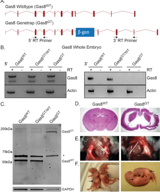
genetrap insertion indicates that the 5’end of the transcript is generated (Fig 1B, left). In
con-trast, primers located 3’to the insertion failed to detect any Gas8 mRNA (Fig 1B, right).
Western blot analysis shows a product of expected size (57kDa) in wildtype and
heterozy-gous Gas8 mice. This product is absent in homozyheterozy-gous mutants (Fig 1C). Additionally, the
Fig 1. Generation of mutant Gas8GTmice and phenotype description.(A) Schematic of the wildtype
Gas8 allele (Gas8WT) and the Gas8 genetrap allele (Gas8GT). The relative position of theβ-geo cassette is
indicated by the blue box. Arrows indicate the primers used for RT-PCR analysis. (B) RT-PCR expression analysis of Gas8 transcript in Gas8WT, Gas8GT/WT, Gas8GTwhole embryos shows the presence of the 5’end of Gas8 transcript (left panels) and absence of the 3’end (right panels). Actin served as a positive template control in all samples. Reactions with reverse transcriptase are indicated (+) and negative RT controls (-). (C) Western blot for Gas8 protein on Gas8WT, Gas8GT/WT, and Gas8GTtrachea. Wildtype Gas8 is located at 57 kDa (Gas8WT) while the genetrap allele is at approximately 230kDa (Gas8GT).*denotes a spurious band recognized by polyclonal antibody. GAPDH was used as a loading control. (D) Nissl stained coronal section of P21 Gas8WTand Gas8GTbrain. (E) Gas8GTmice displaysitus inversusas noted by the reversed direction
of the heart apex (white lines indicate heart axis) and (F) the stomach location in P2 pups (arrow).
doi:10.1371/journal.pgen.1006220.g001
Gas8::β-geo fusion protein is detected (approx. 230kDa) in heterozygous and homozygous mutants indicating that the genetrap allele is being transcribed and translated. Loss of Gas8 led to lethality at approximately postnatal day 14 (P14) with few living to P21. All mutants
pre-sented with severe hydrocephalus (Fig 1D). Gas8GTmutant mice also presented withsitus
inversusat a rate of 36% (6 of 16 mutants) in live births based on position of the heart and
stomach (Fig 1E and 1F). Both the hydrocephalic andsitus inversusphenotypes suggested a
defect in the function of motile cilia.
Loss of Gas8 does not result in defective Hh signaling
Based on a previous study reporting Gas8 as a positive effector of Hh signaling in mammals,
we anticipated Gas8GTmice would present with phenotypes related to Hh signaling defects,
especially since this mutation would lack the putative Smo binding domain (amino acids 386– 478). However, we did not observe any hedgehog-associated phenotypes in limb patterning or neural tube formation. To further test a role for Gas8 in the Hh pathway, we isolated Mouse
Embryonic Fibroblasts (MEFs) from Gas8WTand Gas8GTmutant mice and treated them with
150nM Smoothened agonist (SAG). The MEFs were then immunolabeled for Smo and
acety-lated tubulin to analyze differences in Smo trafficking into the cilium (Fig 2A). In contrast to
the outcome of the knockdown studies, there was no difference between the amounts of Smo
present in the cilia of Gas8GTmutants when compared to Gas8WTcilia (Fig 2B). These data
indicate that a least in the Gas8GTmutants, Gas8 is not an essential factor involved in
regulat-ing Smo cilia traffickregulat-ing. Similarly, none of the Gas8GTmutants exhibited defects in dorsal
ven-tral patterning of the neural tube typical of altered Hh signaling (Fig 2C).
Gas8
GTmice present with dyskinetic cilia and subtle cilia ultrastructural
defects
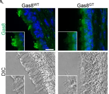
To investigate the hydrocephalus phenotype, cilia morphology, ultrastructure and motility on ependymal and tracheal cells was assessed. DIC analysis and immunofluorescence staining of
tra-chea indicate motile cilia are present on the epithelium, but the Gas8GTprotein fails to localize to
these cilia (Fig 3). We counted cilia from trachea TEMs for broken doublet rings and found that
about 9% of cilia from Gas8GTmutants showed disorganization of the arrangement of the
micro-tubule doublets (Fig 4Aarrowhead and4E). To analyze ultrastructure within the doublets, we
averaged 202 doublets of both genotypes to reduce variability due to random sectioning of the 96nm repeat of the microtubule doublet. We did not observe any major structural differences in
the inner or outer dynein arms (Fig 4B). However, high speed video and Fourier transformation
analysis revealed that cilia are largely static with only a few moving (Fig 4C and 4D,S1 Movie
andS2 Movie). Those cilia that did moved were dyskinetic, resulting in an inability of cilia to
pro-pel fluid as seen by tracking of fluorescent beads added to either brain ventricle or trachea
prepa-rations (Fig 4F and 4G). Beat frequency of cilia that remained motile in Gas8GTmutants was
modestly decreased from 17.0Hz in Gas8WTto 12.7Hz in Gas8GT(Fig 4H). Cilia length is also
affected in Gas8GTmice, with Gas8GTmotile cilia measuring 0.9μm shorter than Gas8WTmotile
cilia (Gas8WT5.3μm and Gas8GT4.4μm) (Fig 4I). Cilia orientation in Gas8GTtracheas is also
more randomized than in Gas8WTcontrols (Fig 4J). These phenotypes observed in the motile
cilia of Gas8GTmutant mice are similar to those observed in PCD patients and animal models.
GAS8 is a disease causing gene in humans
The phenotypes in the Gas8GTmutants led us to evaluate whether mutations inGAS8are
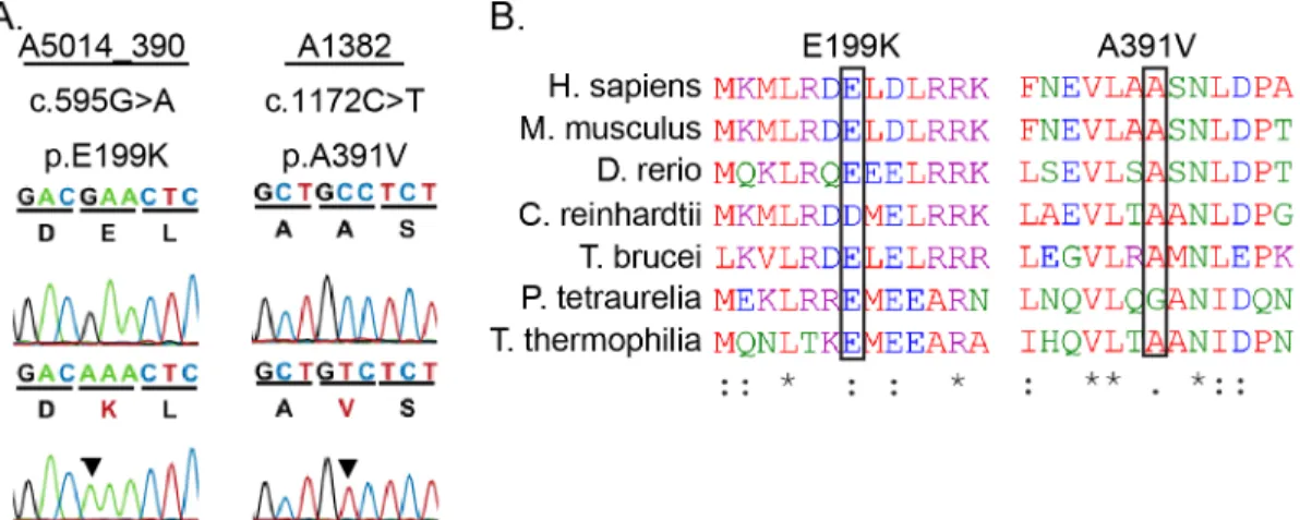
asso-ciated with PCD in humans. We identified two independent missense variants, c.595G>A
5A) [32]. The E119K patient is of Latino decent and presented with heterotaxy. Unaffected parents of the patient are heterozygotes, and an unaffected female sibling is a homozygote. This allele appears at a frequency of 11% in Latino populations (87 homozygotes and 1279 het-erozygotes in a total of 11564 alleles sequenced according to ExAC). The prevalence of this allele in the Latino community makes it unlikely to be associated with disease. The A391V patient met the diagnostic criteria for PCD. This allele is infrequent, occurring only 3 times het-erozygously and 0 times homozygously in 84864 alleles sequenced according to ExAC. Both
variants affect highly conserved regions across multiple species (Fig 5B). We utilized the
Phen-2 program to predict the pathogenicity of these alleles. The A391V allele had a Poly-Phen-2 score of 0.762 suggesting that it is a potentially damaging mutation while the E199K allele had a score of only 0.082, suggesting that this is a benign mutation. Given the low allele frequency, PolyPhen-2 score, and the confirmation of the PCD diagnosis in the patient carry-ing the A391V variant, we chose to test potential pathogenicity of this allele in mice.
To further assess the potential pathogenicity of the human allele, we created a mouse har-boring the A391V mutation via homology driven repair with CRISPR/Cas9 technology.
Sequencing confirmed the presence of the c.1172 C>T mutation resulting in an A391V amino
acid change (Fig 6A). We crossed Gas8AVmice onto the Gas8GTbackground to create
Fig 2. Gas8 mutant embryos have no overt Hh phenotypes.(A) Vehicle (Control) and drug treated (SAG) Gas8WTand Gas8GTmutant MEFs stained for Smo (green) and acetylated tubulin (purple). Scale bar is
15μm. (B) Quantification of Smo translocation into the cilium. All bars are normalized to WT CONT. (*= significantly different than WT CONT, # = Significant different than GT CONT p<0.05). (C) Gas8WTneural
tube sections and Gas8GTsections. The Pax7 and Sonic hedgehog (SHH) domains in the notochord, floor plate and neural tube all appear similar between Gas8GTand Gas8WTlittermates. Scale is 90μm.
doi:10.1371/journal.pgen.1006220.g002
compound heterozygous (Gas8GT/AV) mice. To determine the impact on motile cilia and test possible cause of the hydrocephalus, we took brains from 6 week old mice and analyzed cilia beat and the ability of motile cilia to move fluid. While there were no differences in beat
fre-quency, bead flow analysis shows a modest decrease in the ability of Gas8GT/AVcilia to move
fluid compared to Gas8GT/WTcilia (Fig 6B and 6C). Compound heterozygotes develop mild
hydrocephalus at approximately 10 weeks of age (Fig 6D) but there were no evident laterality
defects. While all the Gas8GT/AVmice analyzed (n = 6) display hydrocephalus at this age, the
severity ranged from mild (arrowhead) to moderate (arrow). The phenotype in the Gas8GT/AV
mice is not as severe as in the Gas8GTmice and hydrocephalus was not present in any (n = 2)
of the Gas8GT/WTor (n = 2) of the Gas8WT/AVmice analyzed.
We chose to generate the A391V mouse model because of the PCD symptoms of the patient but given the lack of full PCD symptoms in the E199K patient, we decided to test first whether
or not the E199K is pathogenic inChlamydomonasbefore proceeding to a potential
mamma-lian model. Alignment of GAS8 and theChlamydomonasorthologue DRC4 revealed that E199
in GAS8 aligns with D198 in DRC4 (Fig 5B). To better understand the mechanisms underlying
these defects, we generated strains expressing theChlamydomonasequivalent (D198K) of the
human E199K alleles in a null mutant background (pf2). Interestingly, transformation with DRC4-DK-GFPrescued the severe motility defects seen in thepf2null mutant, but measure-ments of forward swimming velocities revealed a subtle defect in the swimming phenotype of
the rescued strains (Fig 6E). Furthermore, the DRC4-DK-GFP protein is assembled at
wild-type levels in the flagellar axonemes ofChlamydomonas, as assayed by western blot (S1 Fig).
These observations show that the D198K DRC4 mutant protein is properly localized in the axoneme and may not correspond to a pathogenic allele.
Discussion
Defects involving cilia motility cause severe phenotypes in humans including infertility, hydro-cephalus, respiratory defects, and reversal of left-right asymmetry. Much of our understanding
Fig 3. Gas8 localizes to cilia.Gas8 immunofluorescence staining on Gas8WTand Gas8GTp21 trachea
(Scale Bar = 10μm). Gas8 is present in wild-type axonemes but absent from mutant axonemes. DIC images are shown to visualize the cilia on the tracheal epithelium.
Fig 4. Gas8GTmice present with cilia motility phenotypes.(A) TEM of Gas8WTand Gas8GTtracheal cilia
about cilia motility has come from studies in organisms such asChlamydomonas. These studies and how defects in cilia motility cause disease are now being extended into mammalian sys-tems. Recently GAS8 was implicated as a cause for Primary Ciliary Dyskinesia (PCD) as well as
a positive effector of Smoothened transport into cilia during Hh pathway activation [12,13,19].
To further evaluate the connection between Gas8 and PCD in mammals, we generated a
mouse with aβ-geo cassette inserted in intron 7 of the Gas8 gene. Insertion of theβ-geo
gene-trap cassette effectively eliminated the presence of wildtype transcript and protein in mutants
as verified by RT-PCR and western blot analysis. Though the Gas8GTmutant allele is translated
into a large fusion protein between the N-terminal portion of Gas8 andβ-geo, it does not
local-ize to motile cilia. Gas8GTmutant mice present with hydrocephalus starting at postnatal day 5
(P5) that becomes more pronounced as the mice mature and eventually leads to mortality between P14-P21. Development of hydrocephalus is associated with severe impairment of cilia motility on ependymal cells lining the ventricles of the brain.
Previous studies using image average procedures to analyze flagella ultrastructure in
Chla-mydomonasshowed that strains with mutations in PF2/DRC4, the Gas8 homolog, were associ-ated with the loss of the majority of the N-DRC complex along with a subset of the IDAs
[6,16,17,33]. In contrast to theChlamydomonasresults, the N-DRC and IDA do not appear to
be overtly affected in Gas8 mutant mice based on standard thin-section TEM analysis. How-ever, loss of Gas8 does effect microtubule organization, as indicated by a higher percentage of
showing no major structural defects (Gas8WTN = 202 doublets from 1 trachea, Gas8GTN = 202 doublets
from 1 trachea). (C) Representative kymograph measurement of ciliary waveform in Gas8WTand Gas8GT tracheal cilia. (D) Schematic depicting waveform defects observed in Gas8GTcilia. (E) Percentage of cilia with disorganized doublets in wildtype and mutant trachea (Gas8WTN = 77 cilia from 1 trachea, Gas8GT
N = 287 cilia from 1 trachea). (F and G) Fluorescent bead tracking of Gas8 mice in ependyma and trachea epithelium respectively. Bead flow is significantly impaired in Gas8GTversus Gas8WTEpendyma: Gas8WT= 147.5μm/sec, Gas8GT= 10.2μm/sec (Ependyma: Gas8WTN = 4 brains, Gas8GTN = 4 brains). Trachea: Gas8WT= 24.15μm/sec, Gas8GT= 0.62μm/sec. (Trachea: Gas8WTN = 3 trachea, Gas8GTN = 3 trachea). (H)
Tracheal cilia beat frequency (CBF) captured by DIC and analyzed using fast Fourier transform test. Gas8WT
CBF = 17.0Hz and Gas8GTCBF = 12.7Hz (Gas8WTN = 108 cilia from 4 trachea, Gas8GTN = 46 cilia from 3 trachea). (I) Cilia length is decreased in Gas8GTmutants as measured from DIC images (p<0.01, Gas8WT N = 321 cilia from 3 trachea, Gas8GTN = 343 cilia from 3 trachea). (J) Cilia orientation is significantly altered
in Gas8GTmice (p = 0.025) (Gas8WTN = 184 cilia from 1 trachea, Gas8GTN = 375 cilia from 1 trachea).*=
p<0.01,**= p<0.001
doi:10.1371/journal.pgen.1006220.g004
Fig 5. PCD patient missense mutations in highly conserved regions of Gas8.(A) Sanger Sequence trace and amino acid sequence of human mutations. (B) ClustalOmega amino acid alignment indicates the high conservation of the missense mutant residues found in patients which are indicated by black boxes.
cilia with disorganized microtubule doublets in Gas8GTmutant mice when compared to
Gas8WTmice. Altered cilia microtubules were recently also recently observed in human Gas8
patients [12,13]. Together these data suggest that defects in the mammalian N-DRC may not
always be detectable using traditional TEM averaging of cross-sections. The inability to observe ultrastructural defects in human PCD patients could be attributed to having only one N-DRC per 96nm repeat. Future studies using better imaging approaches such as cryo-electron tomog-raphy and image averaging of longitudinal sections to assess the human N-DRC will likely con-tinue to reveal structural and functional differences similar to those described for the radial
spokes by Lin,et al2014.
Most Gas8GTmutant cilia failed to move, however those that were observed moving
dis-played a modest decrease in beat frequency. The most distinguishable phenotype observed in the cilia that moved was a very rigid and short wave pattern. This pattern has also been
observed in other cilia motility mutants thought to affect the NDRC [10–12]. These changes in
waveform and the lack of overall motility result in the defective fluid flow observed in these mice. Previous data show a complex relationship between planar cell polarity (PCP) and fluid
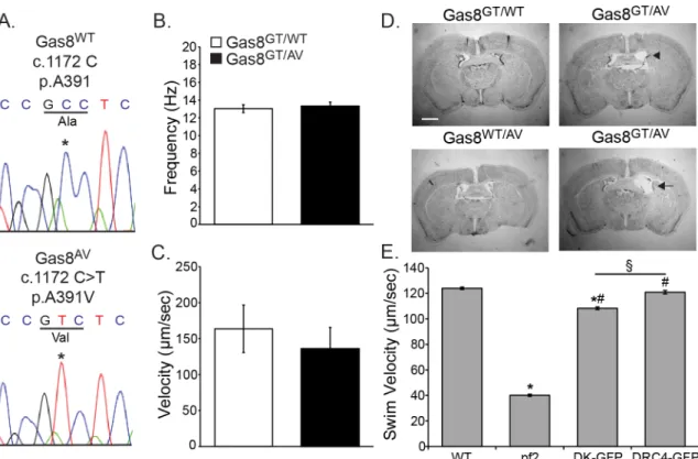
Fig 6. A391V is a potential pathogenic allele.(A) Sanger Sequence confirmation of the 1172 C>T point mutation in Gas8AVmice reproducing the A391V missense mutation found in the human patient. (B) Ciliary beat frequency analysis on
tracheal cilia of Gas8GT/WTand Gas8GT/AVmice shows no difference between controls and compound heterozygotes (n = 86
points from 3 trachea for Gas8GT/WT(13.04 Hz), n = 76 points from 3 tracheas for Gas8GT/AV(13.34 Hz)). (C) Tracking of red fluorescent latex beads added to lateral ventricles shows a trending but not significant decrease in ability of Gas8GT/AVcilia to move fluid. (n = 3 for Gas8GT/WT(163.7μm/sec), n = 2 for Gas8GT/AV(135.9μm/sec)). (D) Nissl stained brains of 10 week
old Gas8GT/WT, Gas8WT/AV, and Gas8GT/AVmice. Mild to moderate hydrocephalus is present in the Gas8GT/AVbrains. Scale
is 1mm (Arrowhead indicates mild, arrow indicates moderate) (n = 4). (E) Swim speed quantification of rescue of
DRC4-D198K construct inpf2deficientChlamydomonas.“pf2”denotespf2deficientChlamydomonas,“DK-GFP”denotes pf2deficientChlamydomonasexpressing the DRC4-D198K-GFP construct,“DRC4-GFP”denotespf2deficient
Chlamydomonasexpressing the DRC4-GFP wild-type construct.*= significant difference from WT (p<0.05), # = significant difference frompf2(p<0.05), § = significant difference between DK-GFP and DRC4-GFP (p<0.05) (n = 390 for WT (123.9μm/sec), n = 271 forpf2(40.1μm/sec), n = 180 for DK-GFP (108.3μm/sec), n = 299 for DRC4-GFP (120.9μm/sec)).
doi:10.1371/journal.pgen.1006220.g006
flow in establishing motile cilia orientation [34,35]. Gas8GTcilia show a more random
distribu-tion of cilia orientadistribu-tion than their Gas8WTcounterparts supporting the necessity of proper
fluid flow in establishing cilia orientation.
Variants in Gas8 were recently identified in human PCD patients. These mutations resulted in a similar, albeit not significant, decrease in beat frequency along with an abnormally rigid
ciliary waveform [12,13]. This motility phenotype is similar to our observation in the mutant
mice. Here we identified an additional independent missense mutation, c.1172C>T A391V, in
PCD patients as well as a variant c.595G>A E199K that appears to have minimal effect on cilia
motility. The A391V mutations lies in close proximity to the other published mutants, C309,
A334, and G357suggesting that this area is critical for GAS8 function. Similarly, the genetrap
cassette in the Gas8GTallele was inserted in close proximity (K337) to the A334mutation. The
E199K mutation also affects a highly conserved region within Gas8 that is proposed to be a
Microtubule Association Domain (GMAD) [36].
To test pathogenicity of the A391V allele, we used CRISPR/CAS9 homology driven repair (HDR) to generate a mouse line mimicking the human mutation. Mice compound
heterozy-gous for the Gas8GTand Gas8AVmutations develop age dependent, mild hydrocephalus, but
did not present with situs defects (n = 6 Gas8GT/AVmice). The phenotype was associated with a
reduced ability of ependymal cilia to move fluid. Interestingly, beat frequency was not signifi-cantly altered from that of controls, suggesting that the defect lies within a subtle waveform dif-ference or in cilia orientation. These data suggest that the A391V allele is pathogenic though more in-depth analysis of ciliary defects will be necessary to determine the precise mechanism.
Data from theChlamydomonasrescue experiments suggest that the E199K allele may have
very subtle effects on motility. The D198K rescued strain inChlamydomonasshowed a small
but statistically significant reduction in forward swim velocity of approximately 10 percent. While statistically significant, additional work is needed to determine whether such small changes might impact ciliary motility and have pathogenic consequences in different tissues and different organisms. As this variant is commonly found in Latino populations, it seems more likely that this variant is a benign polymorphism.
Gas8 was previously implicated as a modulator of the Hh pathway.In vitrodata indicated
that the C-terminal region of Gas8 binds to Smoothened (Smo) and acts at the base of primary
cilia as a regulator of Smo entry into the cilium following Hh pathway activation [19]. These
data showed that in the absence of Gas8, Smo accumulation in the cilium is abrogated and that it cannot activate the Gli transcription factors and turn on downstream genes. Based on these in vitrofindings, we expected to see Hh defects in our mutant mice. However, the Gas8 mutants survive to birth and have normal digit number and patterning as well as normal neural tube dorsal ventral patterning. Furthermore, there were no significant differences in Smo
accu-mulation in cilia between Gas8WTand Gas8GTMEFs after SAG stimulation, suggesting that in
this mutant model, Gas8 does not act as a regulator for Smo entry. The role that Gas8 plays at the base of primary cilia remains uncertain; however, we do not see any other pathologies that would suggest there is a defect in primary cilia such as cystic kidney disease.
The data presented here solidifyGAS8as a disease causing gene in humans and elucidate
the mechanisms by which loss of Gas8 causes disease. We identified new independent, homozygous missense mutations and used model systems to test the pathogenicity of the alleles. Importantly, these results suggest the A391V allele is pathogenic while the E199K var-iant is not. Our results demonstrate the importance of testing the potential pathogenicity of
human alleles in easily amenable model systems such asChlamydomonasand further reveal
Materials and Methods
Mice
The Gas8 mutant mouse line was generated using embryonic stem cell line CH0760
(BayGe-nomics) in which aβ-galactosidase neomycin resistance fusion cassette was inserted into intron
7 of Gas8. The insertion site was confirmed by genomic PCR and sequence analysis. PCR
prim-ers for genotyping were designed based on the insertion site and are as follows 5’-GGGACAA
GCAGATTCTGGTC-3’, 5’-CAGGGTTACACACAGAGAAACC-3’, and 5’-CCGCAAACT
CCTATTTCTG-3’. The Gas8GTembryonic stem cells were from the 129P2/OlaHsd genetic
background and were injected into C57BL/6 blastocysts using standard procedures. Chimeras were bred with albino C57BL/6 females and germline transmission was confirmed by coat color and subsequent PCR genotyping.
Ethics statement
All experimental procedures were approved by the Institutional Animal Care and Use Com-mittee (IACUC) regulations at the University of Alabama at Birmingham under the animal protocol number (130208061).
RT-PCR
RNA was isolated from Gas8WT, Gas8GT/WT, and Gas8GTmouse embryonic fibroblasts using
Trizol reagent according to the manufacturer’s protocol (cat# 15596–026, Thermo-Fisher
Sci-entific). cDNA was generated using Verso cDNA kit (cat# AB-1453/B, Thermo-Fisher
Scien-tific). 5’Gas8 RT-PCR was performed using the following primers spanning exons 3 and 4:
5’-GAATCGAAGAATACCACCATC-3’and 5’-CTGAGAAGATGGCTATGTAG-3’. 3’Gas8
RT-PCR was performed with primers spanning exons 9 and 10: 5’-CTGGACCCCACAGCAT
TAAC-3’and 5’-CTTGATGGTGGTATTCTTCG-3’. Actin control primers: 5’-ATGGGTCA
GAAGGACTCCTA-3’and 5’-GGTGTAAAACGCAGCTCA-3’were used in all samples.
Tissue preparation
Animals were anesthetized by a 0.1 ml per 10 g of body weight intraperitoneal injection of
2.5% tribromoethanol (cat# T48402, Sigma–Aldrich), killed by cardiac puncture, and perfused
with PBS followed by 4% paraformaldehyde (cat# 19943, Thermo-Fisher Scientific). The brains were further fixed in 4% paraformaldehyde 1h at room temperature followed by successive dehydration through 1 hour alcohol incubations at 30% and 50% and placed finally in 70% overnight. Tissues were further dehydrated through 1 hour alcohol incubations at 80%, 95%, and finally 100%. Tissues were placed in xylenes for 1 hour and then placed in a 50/50 xylenes/ paraffin mix for 1 hour at 60°C under vacuum followed by a final paraffin penetration in paraf-fin at 60°C under vacuum for 1 hour and then parafparaf-fin embedded. The brains were sectioned at
10μm and stained with Cresyl Violet stain as previously described [37].
Immunoblotting
Fresh tracheas were extracted from p21 Gas8WT, Gas8GT/WT, and Gas8GTmice. Samples were
submerged in ice cold RIPA (10mM Tris pH7.5, 150mM NaCl, 1%NP-40, 1% sodium deoxy-cholate, 0.1% SDS) mixed with one cOmplete Protease Inhibitor tablet (cat# 11 836 170 001, Roche Diagnostics) per 10mL at 300uL per 5mg of tissue. Tissues were sonicated 3x for 10 sec-onds each. After sonication, tissues were placed on a rotary agitator for 2 hours at 4°C and then spun for 20 minutes at 12,000rpm at 4°C. Supernatant was removed and protein levels were
assayed using a BioRad DC protein assay kit (cat# 5000111, Bio-Rad). Approximately 20μg per
sample was used for SDS-PAGE with a 12% Tris-Glycine gel (cat# 00252562, Thermo—Fisher Scientific). Proteins were transferred overnight to nitrocellulose. The membrane was blocked for 45 minutes in 5% milk in PBS and incubated with primary antibody in 5% milk in PBS with 0.02% Tween-20 overnight at 4°C. Primary rabbit anti-Gas8/DRC4 antibody was used at
1:20000 [6]. Primary mouse anti-GAPDH was used as a loading control at 1:1000 (cat#
ab8245, Abcam, Cambridge UK). Blots were washed 5x for 5 minutes each in 0.02% PBS-Tween-20. Secondary antibody in 5% milk in 0.02% PBS-Tween-20 was added and the blot was incubated for 1 hour at room temperature with the following secondary antibodies:
anti-mouse IRDye 800CW (cat# 827–08364, LI-COR, Lincoln NE USA) and anti-rabbit IRDye
680RD (cat# 926–68071, LI-COR). Blots were washed 5x for 5 min each in 0.02% PBS-Tween-20 and then dried. Images were taken on a LI-COR Odyssey CLx imaging system (LI-COR).
Mouse embryonic fibroblast generation
Mouse embryonic fibroblasts (MEFs) were generated from E14.5 embryos and cultured in DMEM growth medium with High Glucose, 0.05mg/ml Penicillin/Streptomycin, 2mM
L-Glu-tamine, 0.2mMβ-mercaptoethanol, and 20% Fetal Bovine Serum (FBS). Prior to
immunolabel-ing, MEFs were cultured in reduced serum medium containing 0.5% FBS for 48 hours to
induce primary cilia formation as previously described [38].
Immunofluorescence
Cells were fixed in 4% paraformaldehyde and permeabilized with 0.1% Triton X-100 in PBS with 2% donkey serum, 0.02% sodium azide and 10 mg/ml bovine serum albumin (BSA). Cells
were labeled with anti-acetylatedα-tubulin, 1:1000 (cat# T-6793, Sigma-Aldrich), anti-SmoN,
1:1000 (gift from Dr. Matthew Scott, Stanford University). Sections from E10.5 neural tubes were immunolabeled with the following antibodies from Developmental Studies Hybridoma Bank (University of Iowa, Iowa City, IA): anti-ShhN 1:1000 (5E1), anti-FoxA2 1:1000 (74.5a5), anti-Mnr2 1:1000 (81.5C10), anti-Pax7 1:1000 (Pax7), and anti-Msx1+2 1:1000 (4G1) as
previ-ously described [38]. Trachea sections were labeled with anti-Gas8/DRC4, 1:2000 [6]. All
incu-bations and washes were carried out in PBS with 2% normal donkey serum, 0.02% sodium
azide and 1% BSA. Primary antibody incubations were performed for 16–24 hours at 4°C and
secondary antibody incubations were performed for 1 hour at room temperature. Secondary antibodies all from Thermo-Fisher Scientific include the following: Alexa Fluor-594 donkey anti-mouse (cat# A21203), Alexa 488 donkey anti-mouse (cat# A21202), Alexa Fluor-594 donkey anti-rabbit (cat# A21207), and Alexa Fluor-488 donkey anti-rabbit (cat# A21206). Nuclei were visualized by Hoechst nuclear stain. Coverslips were mounted using Immu-Mount (cat# 9990402, Thermo-Fisher Scientific). Fluorescence imaging was performed using a Nikon TE-2000U inverted microscope (Melville, KY) outfitted with a PerkinElmer UltraVIEW ERS 6FE-US spinning disk laser apparatus (Shelton, CT) and a Hamamatsu C9100.
DIC images of p14 trachea prepared for IF were used for length analysis. Images were cap-tured with a 40x objective (Plan-Fluor, 1.3NA). Length was measured manually by drawing a line from the tip of the cilium to the base using Volocity v6.3.
Smoothened trafficking assay
the immunofluorescence section and imaged by spinning disk confocal. Amount of Smooth-ened per cilia volume was measured using Volocity v6.3 software.
Transmission electron microscopy
Postnatal day 14 (P14 mice were anesthetized and perfused with PBS followed by a perfusion of 2% glutaraldehyde in 0.1M cacodylate buffer pH 7.4. Tracheas were extracted and fixed overnight at 4°C in 2% glutaraldehyde in 0.1M cacodylate buffer pH 7.4. Samples were then washed thoroughly four times for 15 minutes each in 0.1M cacodylate Buffer pH 7.4. A post fix in 1% OsO4 in 0.1M cacodylate buffer pH 7.4 was performed. Samples were washed two times for 10 minutes each in 0.1 M cacodylate pH 7.0. Samples were then prepped in 1% tannic acid in 0.1M cacodylate Buffer pH 7.0; 30 minutes followed by 1% NaSO4 in 0.1M cacodylate Buffer pH 7.0; 5 minutes. Dehydrate the samples in 50%, 75%, and 95% at 4°C for 20 minutes each and finally 100% EtOH for 20 minutes; warm to RT°. Dehydrate samples totally with four washes of 100% EtOH 15 minutes each. Infiltrate the sample with Propylene Oxide for 30 min-utes. Mix the EMbed 812 according to instructions from EMS and Infiltrate with 25% Embed in propylene oxide for 30 minutes, 50% for 40 minutes, 75% overnight, 100% for four hours, 100% for 1 hour and harden at 60°C. Samples were sliced at 90nm and imaged on a Phillips CM110 Electron Microscope.
TEM averaging of doublets was performed by isolating individual doublets from cilia and importing the doublets into Photoshop CS5. Individual doublets were aligned to a single tem-plate doublet and then averaged and flattened.
TEMs were used to determine cilia orientation. Cilia orientation was determined by measur-ing the angle of central pairs by drawmeasur-ing a line across the central doublets and measurmeasur-ing the angle relative to the image. Each angle was normalized to the average (or most common angle) after setting the average angle to 0°. The frequency of angles in each image was measured and plotted.
Bead flow analysis
Brains of experimental mice were extracted, sliced in half to expose the ependymal of the lateral ventricles and placed in pre-warmed, pre-oxygenated artificial cerebrospinal fluid (125mM
NaCl, 2.5mM KCl, 1.25mM NaH2PO4, 2mM CaCl2, 1mM MgCl2, 25mM NaHCO3, 25mM
Glucose, pH 7.35). Brains were placed on a Zeiss Axioskop microscope and imaged with a 5x objective (Plan-Neofluor, 0.15NA) and a 10x objective (Fluor, 0.5NA) using a Photometrics CoolSnap HQ CCD camera at 30fps. Red fluorescent latex beads (cat# L3530-1mL,
Sigma-Aldrich) were diluted 1:100 from stock and 10μL of diluted beads was added to the ventricles.
Bead tracking analysis was performed using the MTrack2 plugin in FIJI.
High speed video microscopy
Mice trachea were dissected out into fresh PBS and cut lengthwise into strips. Trachea were kept in warm media (DMEM F/12, 20% FBS, and Pen-Strep and allowed to adapt for 20
min-utes in an environmental chamber (37°C, 45% relative humidity, and 5% CO2) before imaging
with Differential Interference Contrast (DIC). All high speed video was captured at 240fps using a modified Casio Exilim EX-ZR100 attached to a Nikon TE-200 using a 60x water objec-tive (Plan-Apo WI NA = 1.2). Videos saved as quick time files were then extracted into individ-ual frames using Virtindivid-ualDub 1.10.4 software and all analysis was performed in ImageJ.
Kymographs were created using Metamorph v6.1. A line was drawn from the tip of the cil-ium to the base and kymographs were made from the results.
Generation of Chlamydomonas lines
To make the D198K mutation inChlamydomonas, the DRC4-GFP plasmid [6] was used as
template for PCR with the primers 5’-CAGTGCTGTGAGCCTGACG and 5’-AAACCAAAG
CACCTTGAGCG to generate a 1483bp product that contains the restriction sitesBclI and
ClaI flanking the desired mutation site. The PCR product was cloned into pGEM-T-Easy (cat# A1360, Promega Corp) to generate the plasmid pf2-Y1-A. This plasmid was further digested withKpnI andSpeI to removed repetitive DNA and subcloned into pBlueScript to generate the plasmid pf2-Y1-B. The D198K mutation was introduced into pf2-Y1-B using the primers
5’-GAAGATGCTGCGAGACaAaATGGAGCTGCGGAGAAAG-3’and 5’-CTTCTACGACG
CTCTGtTtTACCTCGACGCCTCTTTC-3’and the QuickChange II kit (Agilent Technologies)
to generate the plasmid pf2-Y1-C. After sequence verification by Genewiz, the pf2-Y1-C
plas-mid was digested withKpnI andSpeI and subcloned back into pf2-Y1-A to generate the
plas-mid pf2-Y1-D. The pf2-Y1-D plasplas-mid was digested withBclI andClaI to release the 1483 bp
fragment now carrying the D198K mutation. This fragment was subcloned back into the origi-nal DRC4-GFP plasmid by Genewiz. The completed plasmid, DRC4-D198K-GFP, was
linear-ized withEcoRI for transformation into thepf2-4strain [6]. Transformants were screened as
described above. RT-PCR confirmed that the D198K mutation was expressed in the rescued strains without any other sequence modification. Forward swimming velocity was recorded
and measured as previously described [6]. For transformations with the control DRC4-GFP
plasmid, rescued colonies were recovered at a frequency of 5–15%
Generation of the Gas8
AVmouse allele
CRISPR/sgRNA target sequences were queried using the MIT CRISPR server. Three sites most proximal to the desired SNP change were selected to test nuclease efficiency. CRISPR1: 5’- CT
TCTCCACAGCAGCGTTCAGGG-3’(reverse strand), CRISPR2: 5’-GGTGCTGGCCGCCTC
CAACCTGG-3’(forward strand), CRISPR3: 5’-GACACAAGCGTTAATGCTGTGGG-3’
(re-verse strand). Pronuclear injections were performed with Cas9 mRNA (100 ng/ul), CRISPR3/ sgRNA (50 ng/ul) and ssODN (200 ng/ul). Efficiency of nuclease activity was assessed using a blastocyst assay. In brief, injected zygotes were cultured to the blastocyst stage and lysed to obtain genomic DNA. Genomic DNA was used in PCR and the amplicons (215 bp) were resolved by heteroduplex mobility assay (HMA). CRISPR3 was found to be most efficient and was used to generate the SNP edited mouse (C57Bl/6 background). Injected zygotes were cul-tured to 2-cell stage in KSOM mixed with the NHEJ inhibitor SCR7 at a final concentration of 10 mM. The 2-cell stage embryos were transferred to psuedopregnant recipient female mice, which gave birth to 13 pups. The SNP was introduced with the help of a 154 nt single stranded
oligo DNA (ssODN) HDR template. Since the PAM sequence (CCC>Pro) could not be
modi-fied without changing the amino acid, multiple silent changes were made in the protospacer (sgRNA binding) sequence (indicated by small letters in the sequence below). These changes were made to eliminate the chances of the sgRNA binding to the repaired allele. The SNP change introduced a restriction enzyme recognition enzyme site (BsmBI/Esp3I) and the silent changes introduced two new restriction enzyme recognition sites (BtgI and HaeII). HaeII sites were used to distinguish the wildtype and the modified alleles. Specific primers were also
designed that can preferentially amplify the modified allele. HDR template (ssODN) 5’-GGCC
CTGAACGCTGCTGTGGAGAAGAGAGAGGTTCAGTTCAATGAGGTGCTGGCCGTCT
complete knock-in of the edited/repair sequence, 1 pup had incorporated the silent changes but did not have the desired SNP change, and 1 pup had indels. F0 animals were bred with wildtype C57Bl/6 mice to test germline transmission of the desired alleles. All alleles were suc-cessfully transmitted through the germline, and the positive F1 animals were used to create homozygous and compound heterozygous F2 animals.
Statistical analysis
Cilia length analysis, bead flow tracking, cilia orientation, and cilia beat frequency were tested with Student’s t-test and graphed in Microsoft Excel. Smoothened trafficking assay and
Chla-mydomonas swim speeds were tested by ANOVA followed by Student’s t-test with a
Bonfer-roni correction and graphed in Microsoft Excel. All error is represented in Standard Error of Means (SEM).
Supporting Information
S1 Fig. DRC4-DK-GFP is expressed and localizes to the axoneme.Western Blot of Chlamy-domonasflagellar axonemes showing thatpf2cells transformed with DRC4-DK-GFP (DK) have proper localization of the protein product. The DRC4-GFP fusion proteins are detected by a DRC4 antibody (top panel) and a GFP antibody (bottom panel). An IC69 antibody against an outer arm dynein intermediate chain serves as a loading control for the blot (bottom panel). (TIF)
S1 Movie. Cilia in Gas8WTtrachea display normal beat frequency and waveform (Captured at 240 fps and played back at 30 fps).
(AVI)
S2 Movie. Cilia in Gas8GTtrachea display slower beat frequency and a rigid waveform (Captured at 240 fps and played back at 30 fps).
(AVI)
Acknowledgments
We would like to acknowledge Dr. Bonnie Blazer-Yost for critical reading of the manuscript and Dr. Karl Lechtreck for critical reading and experimental design. The authors would also
like to acknowledge Jason Sakizadeh (UMN) for work withChlamydomonasand Mandy J.
Croyle and Devan M. Rockwell for technical assistance in manuscript preparation as well as Holly R. Thomas for technical assistance as well as the patients and families for contribution of clinical data.
Author Contributions
Conceived and designed the experiments: WRL EBM DT BKY NFB. Performed the experi-ments: WRL EBM DT RB RCP JDP SEB SS CA MRK MAZ AKC MEP NFB. Analyzed the data: WRL EBM DT RB RCP SEB SS CA MAZ AKC RAK SMR IAD JMP FH MEP BKY NFB. Contributed reagents/materials/analysis tools: AKC RAK SMR IAD FH MEP BKY. Wrote the paper: WRL EBM AKC JMP FH MEP BKY NFB.
References
1. Drummond IA (2012) Cilia functions in development. Curr Opin Cell Biol 24: 24–30. doi:10.1016/j.ceb. 2011.12.007PMID:22226236
2. Brooks ER, Wallingford JB (2014) Multiciliated cells. Curr Biol 24: R973–982. doi:10.1016/j.cub.2014. 08.047PMID:25291643
3. Nonaka S, Yoshiba S, Watanabe D, Ikeuchi S, Goto T, et al. (2005) De novo formation of left-right asymmetry by posterior tilt of nodal cilia. PLoS Biol 3: e268. PMID:16035921
4. Hirokawa N, Tanaka Y, Okada Y (2012) Cilia, KIF3 molecular motor and nodal flow. Curr Opin Cell Biol 24: 31–39. doi:10.1016/j.ceb.2012.01.002PMID:22285930
5. Hirokawa N, Tanaka Y, Okada Y (2009) Left-right determination: involvement of molecular motor KIF3, cilia, and nodal flow. Cold Spring Harb Perspect Biol 1: a000802. doi:10.1101/cshperspect.a000802 PMID:20066075
6. Bower R, Tritschler D, Vanderwaal K, Perrone CA, Mueller J, et al. (2013) The N-DRC forms a con-served biochemical complex that maintains outer doublet alignment and limits microtubule sliding in motile axonemes. Mol Biol Cell 24: 1134–1152. doi:10.1091/mbc.E12-11-0801PMID:23427265
7. Heuser T, Raytchev M, Krell J, Porter ME, Nicastro D (2009) The dynein regulatory complex is the nexin link and a major regulatory node in cilia and flagella. J Cell Biol 187: 921–933. doi:10.1083/jcb. 200908067PMID:20008568
8. Piperno G, Mead K, LeDizet M, Moscatelli A (1994) Mutations in the "dynein regulatory complex" alter the ATP-insensitive binding sites for inner arm dyneins in Chlamydomonas axonemes. J Cell Biol 125: 1109–1117. PMID:8195292
9. Piperno G, Mead K, Shestak W (1992) The inner dynein arms I2 interact with a "dynein regulatory com-plex" in Chlamydomonas flagella. J Cell Biol 118: 1455–1463. PMID:1387875
10. Wirschell M, Olbrich H, Werner C, Tritschler D, Bower R, et al. (2013) The nexin-dynein regulatory com-plex subunit DRC1 is essential for motile cilia function in algae and humans. Nat Genet 45: 262–268. doi:10.1038/ng.2533PMID:23354437
11. Austin-Tse C, Halbritter J, Zariwala MA, Gilberti RM, Gee HY, et al. (2013) Zebrafish Ciliopathy Screen Plus Human Mutational Analysis Identifies C21orf59 and CCDC65 Defects as Causing Primary Ciliary Dyskinesia. Am J Hum Genet 93: 672–686. doi:10.1016/j.ajhg.2013.08.015PMID:24094744
12. Olbrich H, Cremers C, Loges NT, Werner C, Nielsen KG, et al. (2015) Loss-of-Function GAS8 Muta-tions Cause Primary Ciliary Dyskinesia and Disrupt the Nexin-Dynein Regulatory Complex. Am J Hum Genet 97: 546–554. doi:10.1016/j.ajhg.2015.08.012PMID:26387594
13. Jeanson L, Thomas L, Copin B, Coste A, Sermet-Gaudelus I, et al. (2016) Mutations in GAS8, A Gene Encoding A Nexin-Dynein Regulatory Complex Subunit, Cause Primary Ciliary Dyskinesia With Axone-mal Disorganization. Hum Mutat.
14. Whitmore SA, Settasatian C, Crawford J, Lower KM, McCallum B, et al. (1998) Characterization and screening for mutations of the growth arrest-specific 11 (GAS11) and C16orf3 genes at 16q24.3 in breast cancer. Genomics 52: 325–331. PMID:9790751
15. Rupp G, Porter ME (2003) A subunit of the dynein regulatory complex in Chlamydomonas is a homo-logue of a growth arrest-specific gene product. J Cell Biol 162: 47–57. PMID:12847082
16. Brokaw CJ, Kamiya R (1987) Bending patterns of Chlamydomonas flagella: IV. Mutants with defects in inner and outer dynein arms indicate differences in dynein arm function. Cell Motil Cytoskeleton 8: 68– 75. PMID:2958145
17. Lin J, Tritschler D, Song K, Barber CF, Cobb JS, et al. (2011) Building blocks of the nexin-dynein regu-latory complex in Chlamydomonas flagella. J Biol Chem 286: 29175–29191. doi:10.1074/jbc.M111. 241760PMID:21700706
18. Colantonio JR, Bekker JM, Kim SJ, Morrissey KM, Crosbie RH, et al. (2006) Expanding the role of the dynein regulatory complex to non-axonemal functions: association of GAS11 with the Golgi apparatus. Traffic 7: 538–548. PMID:16643277
19. Evron T, Philipp M, Lu J, Meloni AR, Burkhalter M, et al. (2011) Growth Arrest Specific 8 (Gas8) and G protein-coupled receptor kinase 2 (GRK2) cooperate in the control of Smoothened signaling. J Biol Chem 286: 27676–27686. doi:10.1074/jbc.M111.234666PMID:21659505
20. Corbit KC, Aanstad P, Singla V, Norman AR, Stainier DY, et al. (2005) Vertebrate Smoothened func-tions at the primary cilium. Nature 437: 1018–1021. PMID:16136078
21. Haycraft CJ, Banizs B, Aydin-Son Y, Zhang Q, Michaud EJ, et al. (2005) Gli2 and Gli3 localize to cilia and require the intraflagellar transport protein polaris for processing and function. PLoS Genet 1: e53. PMID:16254602
22. Rohatgi R, Milenkovic L, Scott MP (2007) Patched1 regulates hedgehog signaling at the primary cilium. Science 317: 372–376. PMID:17641202
24. Roy S (2009) The motile cilium in development and disease: emerging new insights. Bioessays 31: 694–699. doi:10.1002/bies.200900031PMID:19492356
25. King SM (2000) The dynein microtubule motor. Biochim Biophys Acta 1496: 60–75. PMID:10722877
26. O'Callaghan C, Rutman A, Williams GM, Hirst RA (2011) Inner dynein arm defects causing primary cili-ary dyskinesia: repeat testing required. Eur Respir J 38: 603–607. doi:10.1183/09031936.00108410 PMID:21406509
27. Sakakibara H, Takada S, King SM, Witman GB, Kamiya R (1993) A Chlamydomonas outer arm dynein mutant with a truncated beta heavy chain. J Cell Biol 122: 653–661. PMID:8335691
28. Smith EF, Yang P (2004) The radial spokes and central apparatus: mechano-chemical transducers that regulate flagellar motility. Cell Motil Cytoskeleton 57: 8–17. PMID:14648553
29. Merveille AC, Davis EE, Becker-Heck A, Legendre M, Amirav I, et al. (2011) CCDC39 is required for assembly of inner dynein arms and the dynein regulatory complex and for normal ciliary motility in humans and dogs. Nat Genet 43: 72–78. doi:10.1038/ng.726PMID:21131972
30. Becker-Heck A, Zohn IE, Okabe N, Pollock A, Lenhart KB, et al. (2011) The coiled-coil domain contain-ing protein CCDC40 is essential for motile cilia function and left-right axis formation. Nat Genet 43: 79– 84. doi:10.1038/ng.727PMID:21131974
31. Antony D, Becker-Heck A, Zariwala MA, Schmidts M, Onoufriadis A, et al. (2013) Mutations in CCDC39 and CCDC40 are the major cause of primary ciliary dyskinesia with axonemal disorganization and absent inner dynein arms. Hum Mutat 34: 462–472. doi:10.1002/humu.22261PMID:23255504
32. Halbritter J, Porath JD, Diaz KA, Braun DA, Kohl S, et al. (2013) Identification of 99 novel mutations in a worldwide cohort of 1,056 patients with a nephronophthisis-related ciliopathy. Hum Genet 132: 865– 884. doi:10.1007/s00439-013-1297-0PMID:23559409
33. Gardner LC, O'Toole E, Perrone CA, Giddings T, Porter ME (1994) Components of a "dynein regulatory complex" are located at the junction between the radial spokes and the dynein arms in Chlamydomo-nas flagella. J Cell Biol 127: 1311–1325. PMID:7962092
34. Guirao B, Meunier A, Mortaud S, Aguilar A, Corsi JM, et al. (2010) Coupling between hydrodynamic forces and planar cell polarity orients mammalian motile cilia. Nat Cell Biol 12: 341–350. doi:10.1038/ ncb2040PMID:20305650
35. Matsuo M, Shimada A, Koshida S, Saga Y, Takeda H (2013) The establishment of rotational polarity in the airway and ependymal cilia: analysis with a novel cilium motility mutant mouse. Am J Physiol Lung Cell Mol Physiol 304: L736–745. doi:10.1152/ajplung.00425.2012PMID:23525783
36. Bekker JM, Colantonio JR, Stephens AD, Clarke WT, King SJ, et al. (2007) Direct interaction of Gas11 with microtubules: implications for the dynein regulatory complex. Cell Motil Cytoskeleton 64: 461– 473. PMID:17366626
37. Bishop GA, Berbari NF, Lewis J, Mykytyn K (2007) Type III adenylyl cyclase localizes to primary cilia throughout the adult mouse brain. J Comp Neurol 505: 562–571. PMID:17924533
38. Berbari NF, Kin NW, Sharma N, Michaud EJ, Kesterson RA, et al. (2011) Mutations in Traf3ip1 reveal defects in ciliogenesis, embryonic development, and altered cell size regulation. Dev Biol 360: 66–76. doi:10.1016/j.ydbio.2011.09.001PMID:21945076