REVIEW ARTICLE
Tissue Engineering Strategies for Myocardial Regeneration:
Acellular Versus Cellular Scaffolds?
Maribella Domenech, PhD,1Lilliana Polo-Corrales, PhD,1,2 Jaime E. Ramirez-Vick, PhD,1,3 and Donald O. Freytes, PhD4,5
Heart disease remains one of the leading causes of death in industrialized nations with myocardial infarction
(MI) contributing to at least one fifth of the reported deaths. The hypoxic environment eventually leads to
cellular death and scar tissue formation. The scar tissue that forms is not mechanically functional and often
leads to myocardial remodeling and eventual heart failure. Tissue engineering and regenerative medicine
principles provide an alternative approach to restoring myocardial function by designing constructs that will
restore the mechanical function of the heart. In this review, we will describe the cellular events that take place
after an MI and describe current treatments. We will also describe how biomaterials, alone or in combination
with a cellular component, have been used to engineer suitable myocardium replacement constructs and how
new advanced culture systems will be required to achieve clinical success.
Keywords:
cardiac tissue engineering, cardiac patch, acellular scaffolds, extracellular matrix scaffolds,
myo-cardial infarction, heart repair
Introduction
H
eart disease remains one of the leading causes of death in industrialized nations with myocardial in-farction (MI) contributing to at least*20% of the reported deaths.1 An acute MI occurs when a coronary artery that feeds oxygenated blood to the right and left ventricles gets occluded, thus resulting in areas of hypoxia. The hypoxic environment, if maintained for sufficient amount of time, eventually leads to cellular death due to lack of oxygen and nutrients triggering an inflammatory response. The inflam-matory environment is responsible for the clearing of dead cells and stimulating neighboring cells to increase matrix production ultimately leading to scar tissue formation.The scar tissue that forms after an MI is unable to con-tract and, due to the high stresses present during the normal pumping action of the heart, the infarct area deforms over time leading to myocardial remodeling and reduced cardiac output. Although reperfusion and pharmacological treat-ments have shown some improvetreat-ments in patients after an MI, the scar tissue is not completely removed and cardiac output is not restored to pre-MI levels.2If significant dam-age is sustained, a heart transplant is a potential treatment option. However, heart transplantation remains limited by
low availability and the need for life immunosuppression of the transplant recipient.
Tissue engineering and regenerative medicine principles provide an alternative approach to restoring myocardial function after an MI. By combining the expertise of multiple fields, such as engineering, biology, medicine, biochemistry, and pharmacology, tissue engineers try to create suitable tissue replacements capable of restoring function and im-proving quality of life. This review will first describe the cellular events that take place after an MI with emphasis on the host tissue response dominated by inflammatory cells such as macrophages. The review then will describe how biomaterials, alone or in combination with a cellular com-ponent, have been used to engineer suitable myocardium replacement constructs. Given the complexity of the myo-cardial tissue, we will also discuss ideas for new advanced culture systems that can help assemble and test the new generation of engineered cardiac devices.
Myocardial Infarction
A heart attack or MI is caused by the stenosis and/or occlusion of a coronary artery leading to improper delivery of oxygenated blood to regions of the heart. This condition
1Department of Chemical Engineering, Universidad de Puerto Rico, Mayagu¨ez, Puerto Rico. 2
Department of Agroindustrial Engineering, Universidad de Sucre, Sucre, Colombia.
3
Department of Biomedical, Industrial & Human Factors Engineering, Wright State University, Dayton, Ohio.
4
The New York Stem Cell Foundation Research Institute, New York, New York.
5
Joint Department of Biomedical Engineering, NC State/UNC-Chapel Hill, Raleigh, North Carolina.
TISSUE ENGINEERING: Part B Volume 22, Number 6, 2016
ªMary Ann Liebert, Inc. DOI: 10.1089/ten.teb.2015.0523
is classified based on the extent of occlusion into ST-Elevation myocardial infarction (STEMI) when occlusion is completely blocked, or non-ST-Elevation myocardial in-farction (NSTEMI) when occlusion is not complete. The physiological and clinical description of MI is described in details elsewhere2; in brief, the occlusion can affect the major coronary arteries such as the left anterior descending, left circumflex, and the right coronary artery. Once the ar-tery is sufficiently occluded, oxygenation and nutrient deficit downstream of the occlusion result in the gradual death of the myocardial tissue (Fig. 1). The infarct site refers to the portion of the necrotic myocardium that is damaged or in the process of being damaged by the hypoxic conditions. Most infarcts involve the death of the full thickness of the ven-tricular wall (transmural infarction), although in some cases perfusion from neighboring vessels can help delay and/or minimize injury. At the early stages of the infarct there is a decrease in aerobic glycolysis, an increase in anaerobic glycolysis (accumulation of lactic acid), production of high-energy phosphates, and reduced contractility.2
Necrosis and apoptosis begin to occur, leading to the acti-vation of the inflammatory response through the recruitment of neutrophils and subsequently monocytes from peripheral blood. Recent studies have shown the active recruitment of circulating peripheral blood monocytes and a monocyte population resi-dent in the spleen following an MI.3In addition, studies have shown waves of pro- and anti-inflammatory monocytes
circu-lating at different times after the infarct with classically acti-vated monocytes found soon after the ischemic injury and then followed by alternatively activated monocytes.4Once recruited to the infarct, monocytes differentiate toward macrophages and begin the labor of removing cellular debris.5,6
During the initial phase of the inflammatory response after the recruitment of the monocytes, there is a strong presence of classically activated macrophages or M1 macrophages within the myocardial tissue. These are macrophages associated with the removal of pathogens and cellular debris and express proinflammatory cytokines such as IL-1b, TNF-a, and IL-6. These macrophages are present at high numbers during the first week after an infarct in mice with a gradual decrease over time.4,5,7,8Following this initial proinflammatory phase, there is a gradual shift in the type of macrophage toward more al-ternatively activated macrophages (M2), which are character-ized by the secretion of CCL-17, TIMP-1, and IL-10. These macrophages are typically associated with wound healing re-sponses and are thought to activate fibroblasts, smooth muscle, and endothelial cells.5,6,9–11
Myocardial regeneration is the process by which the in-jured myocardium is restored to its original structure and function. As described above, the normal healing process for postinfarction cardiac tissue involves the generation of a fibrous scar, which provides mechanical support but is devoid of functional cardiomyocytes. Most treatment strategies focus on improving the performance of an already damaged tissue
and not in regenerating the myocardium to restore or establish normal function. More recently, new therapeutic approaches based on progenitor cells have been developed with the goal of regenerating tissue to its normal structure and function.
Current Cardiac Treatments
Advancements in medical interventions have improved the prognosis post-MI considerably, but the incidence of heart failure is still increasing, likely as a result of the in-creasing number of patients who now survive the initial attack. Currently, the only approved treatments for end-stage heart failure post-MI are left ventricular assist devices and heart transplantation. The first is plagued by the com-plications of a chronic external assist device, which include bleeding (30%), right ventricular failure (20–30%), throm-boembolism (3–35%), primary device failure (6%-6 months, 64%-2 years), and infection (18–59%).12The second is a limited resource in which proper matching of the donor organ to the patient is a great challenge, limiting even fur-ther its use. A number of surgical approaches have been developed as preventative measures to improve patient survival and their quality of life, and which can be an option for patients excluded from cardiac transplantation lists. The most common include angioplasty, left ventricular recon-struction, and cellular cardiomyoplasty.
Ischemic tissue revascularization
There is agreement that initial treatment for STEMI is restoring blood flow to the ischemic tissue via tissue re-perfusion. The main alternatives for reperfusion can be classified into pharmacologic, surgical, or mechanical. The pharmacological breakdown of blood clots (thrombus) in stenotic coronary arteries is known as thrombolysis. The mechanical alternative to reperfusion is known as primary percutaneous surgical alternative coronary intervention or primary coronary angioplasty, where the occlusion is me-chanically expanded to allow blood flow to resume. The surgical alternative is known as coronary artery bypass graft (CABG) surgery, which when compared with angioplasty is highly invasive (requiring open heart surgery) and requires extra surgery to obtain the vein graft.
The use of primary angioplasty for the treatment of STEMI was first described as a rescue treatment in the case of failed intracoronary thrombolysis and was studied extensively as an adjunctive therapy. In general terms, the procedure consists of feeding a deflated balloon or other device (e.g., stent) on a catheter from the inguinal femoral artery or radial artery up through blood vessels until they reach the site of blockage in the heart. At the blockage, the balloon is inflated to open the artery, allowing blood to flow. Primary angioplasty has been shown to be more effective to thrombolysis for treatment of patients with acute STEMI in randomized trials.13–16The use of angioplasty requires the procedure to be performed pref-erably within 90 min of the patient presenting to the emer-gency room, which most hospitals cannot provide.
There is strong evidence that with increasing duration and severity of ischemia, more cardiac tissue damage can develop, allowing a variety of reperfusion-associated pathologies, known as reperfusion injury. This condition results in cardiac tissue damage through myocardial stunning, microvascular and endothelial injury, and irreversible cell damage, necrosis,
apoptosis, autophagy, or necroptosis.17,18 Reperfusion injury has been observed in each of the cardiac tissue revasculari-zation strategies mentioned above and under certain condi-tions can be lethal. There are various pharmacological and nonpharmacological interventions used to reduce reperfusion injury. In the case of pharmacological interventions, the use of drugs such as cyclosporine-A, metoprolol, and glucose modulators has shown some promising results, but a long list of failed examples makes them a weak alternative. In con-trast, nonpharmacological interventions have focused on limiting the infarct size as means to reduce reperfusion injury.
Left ventricular reconstruction
After MI, the formation of scar tissue leads to changes in left ventricular (LV) size, shape, structure, and physiology through a process known as myocardial remodeling.19 During this process, there is thinning of the LV walls, with the elliptical LV becoming more spherical and dilated.20A number of different surgical techniques and modifications have been developed to restore LV shape and reduce its volume to improve LV function and are collectively known as LV reconstruction.21–24 This is a specific surgical pro-cedure developed for the management of heart failure with LV remodeling caused by coronary artery disease.25Despite its success, these procedures have not found general ac-ceptance in the medical community. Possible reasons in-clude a lack of robust prospective randomized data showing the mortality benefit of this technique in patients with is-chemic cardiomyopathy and dilated ventricles that were referred for CABG. To address these concerns, the Surgical Treatment for Ischemic Heart Failure (STICH) trial was developed to evaluate the role of cardiac surgery in the treatment of patients with coronary artery disease and LV systolic dysfunction.26 A major question addressed by this study was if left ventricular reconstruction improved pa-tient outcome when combined with CABG. The results of this clinical trial showed no significant difference between performing CABG alone or when combined with LV re-construction.26 These surgical techniques, and the use of nonbioactive materials as tissue replacements, helped spark the interest in exploring innovative use of biomaterials and tissue engineering constructs.
Cellular cardiomyoplasty
Cell transplantation is an area of growing interest in clinical cardiology, as a potential means of treating pa-tients after acute MI. Cellular cardiomyoplasty is a thera-peutic strategy in which progenitor cells are used to repair regions of damaged or necrotic myocardium. The ability of transplanted progenitor cells to improve function within the failing heart has been shown in experimental animal mod-els and in some human clinical trials.27The progenitor cells involved in these new therapeutic approaches include bone marrow or adipose tissue-derived mesenchymal stem cells (MSCs), hematopoietic precursor cells, endothelial pro-genitor cells, endogenous cardiac stem cells, and skeletal muscle-derived cells.28,29
Three mechanisms have been proposed to describe how cardiomyoplasty improves myocardial function: (1) transdiffer-entiation of the administered stem cells into cardiomyocytes, endothelial cells, and smooth muscle cells,30,31 (2) fusion
between the stem cell and endogenous cardiac myocytes,32 and (3) release of paracrine factors that stimulate endo-genous cardiac repair mechanisms.33,34 Conflicting results showing a lack of transdifferentiation have put into question its role in cardiomyoplasty and motivated the search of al-ternative hypotheses like fusion and paracrine signaling.30,35 Further studies suggested that the lack of transdifferentiation shown was related to differences in experimental proce-dures, but the exact mechanism remains unknown.36 In addition, it has been consistently reported that the level of fusion between stem cells and cardiomyocytes remains
low,32,37 suggesting that additional mechanisms may be
involved. Current results support paracrine signaling as the principal mechanism for the improvement of myocardial function. In it, stem cells release cytokines and chemokines to stimulate other cells into the regenerative process. In fact, MSCs have been shown to stimulate host myocardial pre-cursor cells to amplify and differentiate into cardiomyocytes
in vivo.38
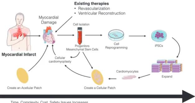
The clinical application of cellular cardiomyoplasty for the treatment of the ischemic tissues after acute MI involves tissue revascularization, isolation of autologous stem cells from the patient, and implementation through repeat cardiac catheterization or intramyocardial injection.39 A major limitation for the application of cellular cardiomyoplasty as a treatment option is stem cell retention and engraftment after intramyocardial implantation.40 A significant propor-tion of the transplanted cells leak out through the needle track that is made by the puncturing needle or enter systemic circulation.41,42 Cell retention is normally less than 10%, regardless of the delivery route within the first 24 h.43 Al-though it was initially Al-thought that cell death by apoptosis was the reason for low engraftment,44,45 it has been dem-onstrated that venous drainage and the contraction of the beating heart are the main reasons for cell loss.40,46 Short-term cell retention is necessary for subsequent long-Short-term engraftment and cardiac tissue functional improvement after acute MI. Other unresolved issues include cell delivery method and route, cell distribution, time transplantation, cell type, cell number, and viability. There are new therapeutic approaches involving engineering culture systems, the use of novel biomaterials for mechanical support of the cells and for controlled release of therapeutics, and tissue engi-neering (Fig. 2).
Engineered Cardiac Patch
Engineered cardiac patch is fabricated to mimic the native extracellular matrix (ECM) and offer mechanical support and cell delivery into the region of infarction. Its application helps to limit LV remodeling, prevent dilatation and thin-ning of the infarct zone, enhance mechanical properties of ventricle, and reduce cardiomyocyte apoptosis. In addition, it aids to retain viable transplanted stem cells, which stim-ulate the formation of vasculature, myofibroblasts, and cardiomyocytes. Hence, the optimal properties of a scaffold involve high porosity, microenvironment similar to ECM, good mechanical properties, biodegradability, and biocom-patibility. Natural polymers (i.e., collagen, fibrin, chitosan, alginate, natural ECM, peptides) and synthetic polymers (i.e., polycaprolactone [PCL], polyglycerol sebacate [PGS], and polyurethanes) are a choice of materials to fabricate
scaffolds47,48(Fig. 3). The Tables 1 and 2 summarize some characteristics of natural and synthetic polymers used for cardiac patch, respectively.
Acellular biomaterial approach to cardiac repair
Cardiac tissue has limited self-renewal capacity, which limits its ability to regenerate and repair itself after injury. Due to the challenges and limitations on the use of bioma-terials with exogenous cells, acellular injectable biomateri-als and patches have been evaluated as mechanical supports for MI. Acellular scaffolds have several advantages com-pared to cellular scaffolds such as (1) their off-the-shelf availability for immediate implantation (e.g., SynerGraft, AlloDerm, DermaMatrix), (2) their limited immune re-action,49and (3) low cost and extended shelf life.50Cardiac tissue scaffolds should exhibit elasticity matching the myocardium, host cell integration and vascularization, me-chanical stability, and nonimmunogenicity to support tissue function and regeneration. The following sections briefly discuss the advantages and limitations of major biomateri-als used as acellular constructs for myocardial repair and regeneration with emphasis given to naturally occurring biomaterials.
Collagen. Due to its abundance in connective tissue, collagen type I scaffolds are increasingly being used in tissue repair applications. Collagen type I provides a tissue-like environment for cell attachment and growth, which facilitates host cell integration. Its main attributes include biocompatibility, biodegradability, and fibrous contractile structure.51,52Collagen type I comprises about 80% of the collagen matrix in cardiac tissue,53making it the choice of preference for cardiac tissue scaffolds. Gaballa et al.54
grafted a three-dimensional (3D) collagen type I scaffold onto infarcted myocardium in rats and found that the scaf-fold induced neovessel formation and reduced LV re-modeling 3 weeks after implantation. However, solid porous collagen has a lower elastic modulus,55which can limit its mechanical integration to the cardiac tissue. Serpooshan
et al. optimized the elastic modulus of collagen type I gel
to improve myocardial contractility in the injured heart.56 Collagen type I was molded using a plastic compression technique to generate dense tissue scaffolds with a high elastic modulus. Four weeks post-MI in mice, collagen type I patches showed host cell infiltration and new blood vessel formation. Echocardiography showed significant improve-ment in cardiac function, diminished fibrosis, and inhibition of LV dilation and wall thinning.
after injury. Cardiac cell integration and function should be further evaluated to determine its long-term clinical potential.
Hyaluronic acid. Hyaluronic acid (HA), also known as hyaluronan, is a natural linear polysaccharide abundantly found in the ECM of several tissues. Its structure and ligand binding properties have been linked to angiogenesis59 and
tissue repair.60 Thus, HA has become an important com-ponent in scaffolds used for tissue repair and regeneration. In cardiac tissue, HA has shown modest results for cardiac function recovery postinfarction. Yoonet al.were one of the first groups that demonstrated the regenerative potential of HA for heart tissue. A significant decrease in both infarcted area and apoptotic index as well as an increase in local
FIG. 3. Diagrammatic representation of tissue engineering strategies using cellular and acellular scaffolds. The goal is to find the optimal configuration that restores myocardial function to preinfarct levels. Color images available online at www.liebertpub.com/teb
FIG. 2. Diagrammatic representation of the different approaches that can be used to repair infarcted myocardial tissue. An acellular patch can be used as an off-the-shelf product that can be implanted soon after myocardial infarction. Alter-natively, cells can be harvested (i.e., progenitor cells) and injected back into the patient. Another approach can be the isolation of somatic cells (i.e., blood cells), reprogrammed, expanded, differentiated, and assembled into a bioengineered cardiac tissue that can then be implanted back into the patient as an autologous patch. These approaches have different timing and expenses associated with them that can have potential impact on their clinical use. Artwork provided by Servier Medical Arts. Color images available online at www.liebertpub.com/teb
vasculature were observed in rats injected with an acrylated HA hydrogel into the epicardium of the infarcted region.61 Similar results have been observed by Abdalla et al. by evaluating the recovery of cardiac function in the infarcted heart of rats postinjection of HA gel into the peri-infarct region.62 Transthoracic echocardiography revealed a sig-nificant increase of about 18% in ejection fraction in HA-injected groups compared to the control group. Decreased collagen deposition and increased levels of VEGF were also observed supporting reduced scarring and new vasculature formation in response to HA.
The molecular weight of HA has been shown to affect its regenerative potential in the myocardium. Evaluation of different molecular weights of HA-based hydrogels (50, 130, and 170 kDa) showed that the lowest molecular weight had the most significant regeneration and function recovery of the infarcted myocardium.63The regenerative potential of HA-based hydrogel was markedly reduced in chronic models of MI, indicating that the injection time is a major determinant for cardiac repair. The compressive modulus of HA gels is also an important variable for stabilization of the infarcted myocardium. Use of hydrogels with high com-pression modulus (43 kPa) significantly reduced LV
re-modeling and improved function when compared with lower modulus hydrogel and control groups in an ovine model.64 Due to the increase in wall stress during systole, a high compression modulus may be more suitable to reduce myocardial stress distribution. Thus, two major factors, the molecular weight of HA and injection time, are main determinants for HA-mediated repair in the infarcted myo-cardium. Overall, several studies support the use of HA-based hydrogels as a promising novel therapy to reduce scarring and promote vasculature formation in the infarcted myocardium.
Alginate. Alginate is an anionic polysaccharide present in brown seaweed. It is biocompatible and has been used for food and pharmaceutical applications.65 Its capacity forin situ gelation and nonthrombogenic properties makes it at-tractive for cardiac tissue applications. In heart tissues, in-tramyocardial injections of alginate hydrogels have shown significant clinical potential for the improvement of LV function and reduced remodeling potentially by providing mechanical support to the damaged ECM. Yuet al.showed that acellular alginate hydrogels could improve cardiac function, reduce remodeling, and increase neovascularization Table1. Some Natural Polymers for Cardiac Patch
Material Stem cells
Porosity and/or
pore size In vivomodel Signaling Ref.
Collagen hMSCs — Male CDF rats a-SMA 118
Autologous stem cells 400–600mm Male C57/BL6 mouse Anti-sarcomeric actin antibody and Anti-vWF
121
hMSCs hESC-MC
— Male athymic RNU
nude rats
CD105, CD73,
Angiogenin, PDGF-B, VEGF, and CXCL1
105
Autologous mesenchymal stem cells
— Wistar rats CD44+, CD90+, CD45-,
CD34-, Desmine, and a-smooth muscle actinin
119
hESC-derived cardiovascular progenitors Autologous r-ADSCs
— Female Wistar rats
Rowett nude female rats
Tbx5 and Nkx 2.5 116
Natural ECM BM-MSCs 91.2%–1.3%;
130.5–25.3 mm
Male syngeneic Lewis rats
a-SMA, bFGF, vWF, PDGF-B, IGF-1, HGF, MEF2D, MYH6, Type I collagen, SMMHC, CD68, and IL-6
139
hMSCs — Adult mongrel dogs Sarcomerica-actinin,
Atrial natriuretic peptide Cardiotin, Subunit of the Cav 1.2, and cardiac troponin-T
137
Cardiac progenitor cells — — a-MHC, Troponin T,
Troponin C, GATA-4, nkx 2.5,a-SMA, Smooth muscle 22a, Fibroblast-specific protein 1, vWF, tie2
135
BMMCs 19.5–17.9mm — Sarcomerica-actinin,
Myosin heavy chain Cardiac troponin T, and vWF
134
Table 2. Other Polymeric Material for Cardiac Patch Stem cell Polymer Porosity/pore size Elastic modulus (MPa) In vivo model Signaling Ref. Tensile stress Tensile strain (MPa) Tensile strain at break Cardiomyocytes differentiated from hESC Poly(glycerol sebacate) — — Adult male Sprague Dawley rats — 144 cSca-1, BMMCs, and ADSCs Nanopeptides (cell–PuraMatrix ) — — Wild mice (C57Bl/6J) Anti-vWF), anti-SMA, anti-a -sarcomeric actinin anti-CD31,VEGF, bFGF, and PDGF-bb 152 BM-MSCs PPC, PU, and [P(3HB-co-4HB)] — — — CD34, CD45, CD90, CD73, CX43, and cTn T 153 Neonatal cardiomyocytes PGS/collagen — 2.06 MPa (TS) 57.87% (TSA) 83.65% (TSB) 4.24 (EM). — Cardiac-specific marker proteins a -actinin, Troponin, b -MHC, and cx43 146 BM-MSC PCL/Gelatin 83.6% – 0.8%/ 0.83 – 0.15 m m — Female Sprague Dawley rats CD31, cTnT, and Cx43 47 Cardiac progenitor cells PCL/CNT — 1 1 MPa (EM) 1.3 MPa (TS), 131% (TSB) — sca-1, CD34, GATA-4, CD44, CD29, and CD31 150 BMMSCs PGS/Fibrinogen — — Farm pigs (Sus scrofa) Yorkshire Swine a -sarcomeric actinin, troponin T, CD68, and CD31 117 hMSCs PEG/Alginate — — Male nude rats (Crl:NIH-Foxn1 rnu ) anti-vWF, anti-BRDU 154 Neonatal rat cardiac cell Fibrin — 86.0 – 3.8 (EM) 75.7 – 11.5 (UTS) Female Fisher F344
syngeneic immune-competent rats
in a chronic rodent model of ischemic cardiomyopathy.66 The angiogenic effect of alginate can be further enhanced by incorporating RGD peptides into the biopolymer, but the structural changes reduce the therapeutic effects of the hy-drogel.67Landaet al.evaluated the effect of alginate hydrogel in the recovery of cardiac function of rats post-MI.68 There was an increase in scar thickness, and reduced LV systolic and diastolic dilatation was typically observed even after injection 60 days postinfarction. These results were comparable to those achieved by neonatal cardiomyocyte transplantation.
The use of alginate hydrogels has also been shown to improve cardiac function in large animal models. Intra-coronary injection of alginate hydrogel prevented LV re-modeling and increased scar thickness in a swine model of MI.69 The alginate hydrogel was replaced by myofibro-blasts, which support local tissue restoration while limiting general myocardial remodeling. Implantation of alginate hydrogel in dogs with heart failure produced by intra-coronary microembolizations (LV ejection fraction <30%) significantly improved ventricular wall stability and func-tion.70Injection of alginate hydrogel expanded the LV wall and improved the LV systolic and diastolic functions at levels comparable to those observed in dogs in long-term therapy with beta-blockers.71These observations motivated evaluation in patients with ischemic (n=4) and nonischemic (n=2) dilated cardiomyopathy. Patients that received algi-nate implants showed improvements in LV size and func-tion as early as 3 days. Reducfunc-tions in LV volumes and an increase in ejection fractions were sustained for over 3 months. Due to their promising results, alginate hydrogels are to date the only and first injectable biomaterial in clinical trials for treating MI. However, lack of integration between alginate and cardiac cells might be the major limitation for tissue regenerative applications.
Chitosan. Chitosan is a cationic hydrophilic polysac-charide derivate from chitin commonly found in crustacean shells. It has been extensively used in biomedical applica-tions, including wound healing,72,73 drug delivery sys-tems,74 and surgical adhesives.75 The porosity of chitosan can be controlled as a function of freeze-drying,76which is important for host cell migration and tissue integration. However, chitosan alone is noncell adhesive and has a high compressive modulus, which requires its chemical modifi-cation and/or mixing with other biomaterials to obtain op-timal mechanical and physiological properties for cardiac tissue. Poket al. evaluated a multilayered scaffold formed by a gelatin–chitosan hydrogel around a self-assembled PCL core for use as a cardiac patch.77Gelatin and chitosan ratios of 50:50 and 25:75 significantly improved cell adhesion while retaining the mechanical strength of PCL. Mixtures of chitosan and collagen have also shown potential to improve cardiac function. Denget al. combined chitosan with col-lagen to increase the compressive modulus of colcol-lagen as a potential implant for stabilization of the ventricular wall.57 Ahmadiet al.investigated the effects of a collagen chitosan matrix on cardiac remodeling.78Mice received local injec-tion of collagen–chitosan matrix 2 weeks post-MI. LV ejection fraction was improved only in collagen–chitosan-treated mice over a 3-week follow-up period. Thus, com-bination of porous chitosan with lower compressive moduli and cell-adherent biomaterials may have the potential to
increase tissue integration and therefore increase the me-chanical stability of the ventricle postinfarction.
Fibrin. Fibrin-based scaffolds are biopolymer gels formed from fibrinogen, a glycoprotein that contains two Arginine–Glycine–Aspartic acid (RGD) sequences in each amino acid chain and is converted by thrombin into fibrin during blood clot formation simulating the last step of the blood coagulation cascade.79,80 Fibrin polymerizes in situ
upon the combination of fibrinogen and thrombin. Fibrin glue has been tested as an injectable scaffold for cardiac tissue repair. Christman et al. examined the effects of in-jectable fibrin glue as a scaffold and wall support in the ischemic myocardium in rats. Fibrin glue alone or with skeletal myoblasts was injected into the LV 1–2 days after left coronary artery occlusion. Five weeks after injection, fibrin glue alone or with cells preserved infarct wall thick-ness, reduced infarct size, increased blood flow, and im-proved cardiac ejection function.81,82 Huang et al.
performed a comparative study to determine the therapeutic potential of fibrin, collagen type I, and Matrigel as injectable biomaterials for MI repair. Injection of each individual biomaterial into the infarct zone significantly increased vascularization compared to the saline solution control group at 5 weeks post-treatment in rats. The angiogenic potential was similar for all three polymers probably due the shared common binding sites for avb3 integrin, which is associated to angiogenesis. The major disadvantage of fibrin gels is poor mechanical properties, not able to support the stresses generated during myocardial contraction.83 There-fore, fibrin should be combined with other high-compression moduli components such as chitosan or collagen to increase mechanical strength and reduce degradation rates.
approach its normal thickness, and finally resulting in im-proved regional mechanical function.86,87Moreover, natural matrices from small intestine submucosa (SIS)88 and por-cine sternum89 used in rats models have showed to be a good alternative to promote angiogenesis, enhance cardiac function, and decrease apoptosis, through the recruitment of c-kit+cells, myofibroblasts, and macrophages after MI. Tan
et al. used a decellularized SIS patch with MSCs on a MI
rabbit model90and showed significant improvements in LV function, wall thickness, and vasculature. Decellularized ventricular and pericardial matrices are two additional op-tions for MI repair.91–93 Singelyn et al. have shown that decellularized porcine ventricular tissue can be solubilized and self-assembled in situ upon injection into myocardial tissue. Smooth muscle cells and endothelial cells were able to infiltrate the decellularized matrix both in vitro and
in vivo with a significant increase in blood vessel density.
These studies were performed in the healthy rat myocar-dium, which have better recovery rates than in MI.
Synthetic materials. Synthetic materials have been widely used in tissue engineering applications due to im-proved mechanical properties, material uniformity, and low risk of infection compared to natural biomaterials. Synthetic polymers can be modified with high precision to meet tissue-specific properties such as appropriate degradation rates, porosity, and mechanical strength. Several synthetic polymers have been evaluated for cardiac tissue implants, including poly(ethylene glycol)(PEG), polyvinyl alcohol, poly(caprolactone)(PCL), polypropylene, polyester, and poly(N-isopropylacrylamide) (PNIPAM).94,95Meshes made of polyester and poly(propylene) have been successfully used as LV restraints to prevent LV remodeling and dilation in animal models and human patients.96–100 Injection of a thermosensitive hydrogel containing PCL and PNIPAM into the myocardium 4 days postinfarction in rabbits was found effective to prevent ventricular wall thinning and reduce systolic and diastolic dilatation after 30 days of treatment.95 Similarly, PEG hydrogels have prevented LV remodeling and dilation, but lack vascularization unless codelivered with cells.101While the mechanical properties and stability of synthetic polymers are superior to natural polymers, cell integration is a major limitation. Blends of synthetic and natural polymers with or without growth factors are often preferred to support cell migration and tissue replacement of the implant.102,103
Engineered cellular constructs for cardiac repair
Although acellular scaffolds have many advantages over cellular scaffolds, the use of cells has also been shown to improve healing and tissue regeneration. In many cases, the addition of a cellular component showed improvement over the material alone.104 The use of an engineered cellular construct is a therapeutic strategy to regenerate the myo-cardium lost after an MI by supporting the function of the myocardium via a mechanically suitable material and the surviving cells using an appropriate exogenous repair cell. The cardiac patch is a 3D carrier for cell delivery fabricated
in vitro and implanted over the infarcted tissue47with the
goals of improving repair cell retention and engraftment, limiting LV remodeling, preventing LV dilatation and
thinning, enhancing the mechanical properties of ventricle, and reducing cardiomyocyte apoptosis.48In addition, it can also provide the means to stimulate angiogenesis, release of cytokines, and myocardial perfusion.48,105 All these prop-erties depend on the choice of scaffold material and repair cells that will ultimately couple with the native cells.
One of the first patches developed for delivering cells to an MI site consisted of a cardiomyocyte-enriched extract from fetal rat ventricular muscle dispersed in a commercially available gelatin scaffold (i.e., Gelfoam, Merck, Co.).106 Although this cardiac patch showed improved cell survival and retention, it was not able to demonstrate any improve-ment in cardiac function. In contrast, a similar type of patch based on alginate and fetal rat cardiomyocytes showed im-provement on both cell survival and cardiac function in a rat MI model.107 This variability in results is common and needs to be considered. These are mainly due to differences in degree of injury to the heart due to the infarct procedure used.108 Additional studies in the development of cardiac patches have shown a limited use of cardiomyocytes. This is because these cell types are terminally differentiated, thus limiting their proliferative capacity.109
As an alternative, many researchers are studying a recently detected small population cardiac progenitor cells (CPCs) negative for blood lineage markers (Lin-) and positive for stem cell surface markers (i.e., c-kit, Sca-1, and MDR-1), which have the potential for myocardial regeneration.110 A potential treatment option could be to attract this cell popu-lation to the infarct site and provide them with the appropriate environment to stimulate myocardial regeneration. This en-vironment should promote not only the migration of CPCs but also of any other cell types that might promote myocar-dial regeneration (e.g., macrophages, MSCs).
It is important to first differentiate between the term re-pair, which defines the natural healing process that replaces the damaged tissue with a scar, and regeneration (our ther-apeutic objective), in which the damaged tissue rebuilds itself to its normal structure and function.111 The cardiac repair process involves an inflammatory response, where macrophages remove dead cardiomyocytes and secrete an-giogenic and profibrotic cytokines, chemokines, and prote-ases.112This is followed by fibrosis, in which macrophages and myofibroblasts work together in reinforcing the ven-tricular wall through the secretion of connective tissue, eventually forming the scar tissue. This structure is so dense that it prevents the migration of CPCs, essential for cardiac regeneration, making fibrosis an important hurdle in mam-mals’ capacity to regenerate myocardium after MI.113 In addition, it has been found that acute inflammation is nec-essary to activate the regenerative response in the neonatal mouse heart.114
Another type of progenitor cells that have been tested for myocardial regeneration are MSCs loaded into scaffolds made of natural and synthetic polymers.115 Collagen, fi-brin, PGS, PCL, nanopeptides, polyurethane (PU), and 3-hydroxybutyrate-co-4hydroxybutyrate [P(3HB-co-4HB)] are examples of these scaffold biomaterials. The use of these patches has shown increased vascularization and improve-ment in cardiac function. They are also an improveimprove-ment over cellular cardiomyoplasty, which is limited because in this technique cell survival involves enzymatic cell dissociation before injection and because of the poor vascularization in
the infarct site.116 Cardiac patches could overcome these limitations by retaining viable transplanted stem cells in a scaffold that would facilitate cell migration and cell adhe-sion toward the infarcted area.48 In addition, these trans-planted cells could stimulate formation of vasculature and cardiomyocytes. As with most of the cell-bearing scaffolds, optimal properties of these include high porosity, high surface area to volume ratio, microenvironment similar to natural ECM, good mechanical properties, biodegradability, and biocompatibility.47,117 These parameters can be mod-ulated with the choice of materials used and the synthesis conditions chosen for its fabrication.
Collagen. Collagen matrices loaded with MSCs have proven to be a good alternative biomaterial for cardiac patches. Simpsonet al.used collagen matrices seeded with human MSCs (hMSCs) to treat MI in the male CDF rat model.118 The results showed that the application of this patch promoted myocardial regeneration and induced the expression ofa-SMA marker, which was associated with an increase in myofibroblasts within the infarct region. Like-wise, Maureira et al. developed a cardiac patch based on collagen and autologous MSCs, which was implanted in an MI rat model.119The results of this study showed reverse remodeling of the infarcted area, improvement of perfusion, a reduced infarction, an increase of ventricular wall thickness, enhanced angiogenesis, and formation of myofibroblast-like tissue.
Similarly, the effectiveness of collagen seeded with adi-pose tissue-derived stem cell epicardial patches to preserve LV function, decrease fibrosis, and increase vessels in the infarct area was confirmed in reported studies.116 Further-more, studies have evaluated the response of collagen ma-trices seeded with hMSCs and human ESC-derived mesenchymal cells (hESC-MC) to repair the infarcted myocardium of athymic nude rats.105 This in vitro study showed similar responses with regard to stem cell potency, viability, and cell proliferation for both cell types, while the results from thein vivostudy demonstrated maintenance of the diastolic function and attenuated adverse LV remodel-ing. Interestingly, even with the observed differences in response to secreted paracrine factors, local angiogenic ef-fects were seen in both cell types. Shiet al.demonstrated that collagen scaffolds covalently conjugated with the Sca-1 antibody (present in adult murine hematopoietic stem cells120) can attract native autologous stem cells to en-courage cardiomyocyte regeneration after MI.121In another study, collagen I patches seeded with human bone marrow CD133+cells were used on cryoinjured rat hearts to assess theirin vitroand in vivocardiomyocyte differentiation po-tential.122Results showed a capacity to induce angiogenesis, but not to induce cell differentiation. In a similar type of study, a collagen type I patch seeded with human umbilical cord blood mononuclear cells was used in MI mouse model to assess their cardiomyocyte differentiation potential.123 These patches showed improvements in vascularization and cardiac function, as measured by an increase in ejection fraction. The same group also conducted the Myocardial Assistance by Grafting a New Bioartificial Upgraded Myocardium (MAGNUM) phase I clinical trial using the same autologous cell-seeded patches.124 Ten patients pre-senting LV postischemic myocardial scars that were
un-dergoing CABG had the seeded patches fixed onto the ischemic tissue. Results showed that the patch increased the LV wall with viable tissue and helped normalize cardiac wall stress in injured regions, improving diastolic function.
Fibrin. Fibrin is a hydrogel that contains adhesion mol-ecules and has been shown to be biocompatible. However, fibrin-based scaffolds must be stabilized with other materi-als to compensate for their rapid reabsorption.125,126Studies demonstrate the feasibility of applying injectable fibrin gels into the infarcted myocardium.127,128 Recently, Bagoet al.
observed cardiomyogenic differentiation of adipose tissue-derived progenitor cells using fibrin scaffolds in a mouse model.129 This system promoted endothelial lineage, in-creased vessel density, improved cardiac function, and di-minished scar size after MI. Wendel et al.reported infarct size reduction, elimination of LV wall thinning, vasculari-zation, and the restoration of cardiac function in rats 4 weeks after transplantation of the cardiac patch, which consisted of fibrin scaffolds seeded with neonatal rat cardiac cells.48 Geusset al.showed PEGylated fibrin gels as an alternative to promote proliferation and cardiomyogenic differentia-tion.126 However, by comparing two-dimensional (2D) versus 3D cell cultures, contractile activity was maintained when cells were cultured on layers of PEGylated fibrin and significantly reduced when cultured as aggregates. The au-thors argued that the lack of contractility was related to low cardiomyocyte proliferation and not to the influence of mechanical properties (i.e., elastic modulus).
To develop cardiac patches that more closely mimic ECM, current tissue engineering approaches to myocardial regeneration are implementing the use of therapeutic genes to enhance paracrine action. An example, insulin-like growth factor-1 (IGF-1), which is a hormone that has been shown to enhance cardiac differentiation, reduces apoptosis and enhances neovascularization.79,130,131Recently, Liet al.
published the first study showing cardiac repair in a large animal model using fibrin patch seeded with IGF-1-modified MSCs.79The results of this study were interesting because, despite the absence of cardiomyogenic differentiation
in vivo, it promoted cardiac repair through other pathways.
Similarly, Ye and colleagues used three cell types: human iPSC (hiPSC)-derived cardiomyocytes, endothelial cells, and smooth muscle cells in combination with fibrin patches seeded with IGF-1-encapsulated microspheres to treat MI in a porcine model.132The results demonstrated cardiac repair without developing ventricular arrhythmias. In addition, Xiong et al. seeded fibrin patches with hESC-derived en-dothelial and smooth muscle cells and implanted on a por-cine MI model.133Results after 7 days showed a significant improvement in LV ejection fraction, which was maintained for over 4 weeks.
ECM scaffolds. An innovative alternative that has pro-ven very effective is the use of natural matrices as scaffolds.
Wanget al.demonstrated that a cardiac patch consisting of
Results showed a significant improvement in cardiac func-tion and vascularizafunc-tion. French et al. indicated that CPCs seeded on naturally derived cardiac ECM show enhanced adhesion, maturation, proliferation, and survival compared with a collagen matrix.135 Singelyn et al. used porcine
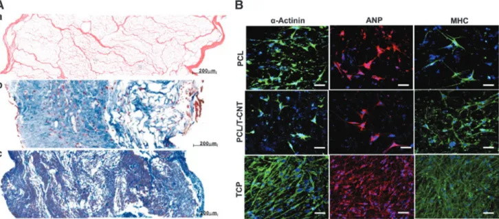
myocardial tissue as an injectable myocardial matrix into rat myocardium.91The results demonstrated that this matrix not only allowed the formation of nanofibrous structures con-taining glycosaminoglycans but also the migration of en-dothelial and smooth muscle cells. The feasibility of this FIG. 4. (A)Decellularized porcine myocardium can support cardiomyogenic and angiogenic differentiation.(a)Large pores evenly distributed across the 2 mm thick acellular scaffold (H&E).(b)Edge to edge view of thorough recellularization at 2 weeks in which cells were found to infiltrate and distribute within the myocardial scaffold.(c)Tissue remodeling was observed in the 4-week recellularization tissue construct; cells were still observed, and cell density was found higher than of the 2 week’s construct. Reprinted with permission from Wanget al.134(B)Immunofluorescence images showing the expression of cardiomyocyte markers,a-actinin, ANP, and MHC in CPCs that have been induced to differentiate 21 days after seeding onto PCL or PCL–TCNT meshes, or TCP. Scale bar, 50mm. Reprinted with permission from Wickham et al.150 ANP, atrial natriuretic peptide; CPC, cardiac progenitor cell; MHC, myosin heavy chain; TCP, tissue culture plastic. Color images available online at www.liebertpub.com/teb
FIG. 5. Summary of engineered approaches to cardiac tissue regeneration. Extracellular matrix materials such as collagen, alginate, hyaluronic acid, and chitosan are used alone or combined as a cardiac patch to provide mechanical strength and stabilize the ventricular wall (acellular). Addition of cells to extracellular matrix constructs (cellular) or engineered matrices improves cardiac function and vascularization compared to acellular models. The question mark (?) implies it remains to be determined in future studies. Color images available online at www.liebertpub.com/teb
approach was demonstrated in a large animal model through a transendocardial catheter injection.136
Extracellular matrices derived from porcine UBM can also serve as an inductive scaffold for myocardial repair. Potapovaet al.found formation of myocytes and improved mechanical function in canine heart using UBM seeded with partially differentiated hMSCs.137 Shahet al. also probed the ability of this natural matrix to support and promote the growth of mature cardiomyocytes.138In addition, Weiet al.
used decellularized bovine pericardial tissue as scaffolds seeded with multilayered MSCs on a male syngeneic Lewis rats MI model.139The advantage of this study was the de-velopment of vasculogenesis, angiogenesis, and differenti-ation of MSCs toward myofibroblast, although no mature cardiomyocytes were found within the patch.
Poly (glycerol sebacate). In 2002, Langer and coworkers reported the synthesis of PGS, a biodegradable elastomer composed of glycerol and sebacic acid with desirable che-mical and physical properties for tissue engineering appli-cations.140 In addition, this polyester is considered a good material for cardiac patches because of its biodegradability, excellent mechanical properties, and low cost.141–143Chen
et al. used PGS patches to deliver differentiated
cardio-myocytes from hESCs to treat MI in adult male Sprague Dawley rats.144The results showed that this system not only maintained active cardiomyocytes beating for an extended time but it also supported cells duringin vivoimplantation. In addition, the PGS/fibrinogen cardiac patch has demon-strated excellent mechanical properties, good cell–scaffold interactions, and capacity to stimulate the expression of cardiac-specific markers (i.e.,a-actinin, troponin, ß-myosin heavy chain, and connexin 43).145In addition, PGS/fibrinogen/ VEGF scaffolds loaded with MCSs have been shown to promote both the expression of cardiac and endothelial cell markers while preventing negative ventricular remodeling
in vivoin a porcine MI model.117Likewise, it was
demon-strated that PGS/collagen patches can provide a good local microenvironment similar to the natural interactions exist-ing between cells and the native ECM, stimulatexist-ing cardio-genic differentiation of MSCs.146
Polycaprolactone. PCL is a FDA-approved biodegrad-able synthetic polymer used as a scaffold material for car-diac tissue regeneration due to its mechanical properties and biodegradability. Nevertheless, PCL must be combined with other polymers (e.g., dextran, vinylalcohol, and poly-ethylene oxide) to improve its hydrophobicity through the presence of ester and keto groups in its structure, which inhibit cell binding.147 Therefore, electrospun nanofibrous scaffolds fabricated from this material can simulate the ECM microenvironment, allowing diffusion of nutrients, cell adhesion, cell growth, migration, proliferation, and differentiation.148 Studies have reported that the use of a PCL/oligomer hydrogel can simulate ECM.149 The incor-poration of thiophene-conjugated carbon nanotubes into PCL scaffolds enhances the mechanical properties, cell ad-hesion, proliferation, and differentiation of CPCs (Fig. 4B).150 Similarly, in vivo studies in rats have demonstrated that PCL/gelatin scaffolds loaded with MSCs can restrain LV remodeling, improve cardiac function, and promote both an-giogenesis and cardiomyogenesis in the infarcted area.47In
addition, scaffolds of the PCL derivative poly(glycolide-co-caprolactone) were seeded with bone marrow mononuclear cells and fixed in a rat MI model.151Results after 4 weeks showed significant improvements in ejection fraction, ven-tricular diameter, and hemodynamics.
Others. Research in the development of cardiac patches has also explored other polymeric materials. For instance, a commercial biological matrix (cell-PuraMatrix) formed of self-assembled peptides can serve as a biological scaffold to evaluate the feasibility of different types of cells involved in myocardial repair in mice [(i.e., clonal stem cell antigen-1-positive cardiac progenitors (cSca-1), bone marrow mono-nuclear cells, skeletal myoblasts, and adipose tissue-derived MSCs)]. Results indicate that cSca-1 is better in preventing cardiac remodeling and systolic dysfunction compared to the other cell types.152 In vitro and in vivo studies have demonstrated that [P(3HB-co-4HB)] and PU can support stem cell growth,153 while a cardiac patch consisting of hMSCs encapsulated in alginate and seeded in polyethylene glycol hydrogels stimulates healing after a heart attack.154In addition, Miyagiet al.developed a hybrid scaffold using a gelatin scaffold (i.e., Gelfoam, Merck, Co.) coated with PCL (for reinforcement) and seeded with bone marrow MSCs and the cytokine stem cell factor and stromal cell-derived factor-1alpha.155Results in a small animal ventriculotomy model showed a significant improvement in cardiac func-tion when compared to controls (i.e., animals and unmodi-fied gelatin sponge). This improvement was observed in all groups with the scaffold alone, with or without cytokines or cells.
Advanced Culture Systems
Appropriate cell function and tissue integration depend on cell–cell and ECM interactions. In the myocardium, elec-trical stimuli are propagated through interconnected cardi-omyocytes highlighting the importance of gap junctions and cell polarization for appropriate function. Intercellular communication between cardiomyocytes is essential for the coordinated and synchronized contraction of the cardiac muscle. This communication occurs through gap junctions, which are membrane channels formed by the connexin family proteins which allow the propagation of rapid an-isotropic impulses. These connexins have been shown to play a crucial role in determining impulse conduction and the heart morphogenesis, as well as in several cardiomy-opathies, such as myocardial ischemia.156In vitro, cell po-larization and cell–cell interconnectivity can be achieved by modulating cell adhesion to the culture surface. In this section, we discussed current engineered techniques used to develop optimal ECM architecture and improve cardiac cell–cell adhesion and function.
Electrospinning nanofibers
technique for cardiac tissue regeneration applications. Zong
et al.used electrospinning to fabricate biodegradable
non-woven poly(lactide)- and poly(glycolide)-based (PLGA) scaffolds.157In these polymeric fibers, primary cardiomyo-cytes developed mature contractile machinery (sarcomeres) and showed electrical activity using voltage-sensitive dyes over 7 days. Similar cardiomyocyte contractility and sur-vival have been observed with other biodegradable poly-mers used in electrospinning, such as polyurethane,158 poly(lactic-co-glycolic) acid (PLGA),159 and PCL.160 However, thesein vitrostudies were carried out in the ab-sence of fibroblasts, which can modulate cardiomyocyte function and structure161 and thereby impact long-term survival and tissue integration. Hussain’s group addressed the fibroblast gap in 3D cardiac tissue constructs by fabri-cating a 3D chitosan nanofiber scaffold using an electro-spinning technique to maintain cardiomyocyte function with fibroblast cocultures.162A mat of chitosan fibers (*150mm thick) was coated with fibronectin to enhance cell adhesion of neonatal cardiomyocytes isolated from rats in coculture with 3t3-J2 fibroblasts. Cardiomyocyte cocultures main-tained cell polarization and expressed high levels of Con-exin43, a gap junction protein required to propagate electrical stimuli, over 3 weeks. Image analysis of intra-cellular calcium ion staining showed that cardiomyocytes maintained an elongated morphology and beating frequency of 17–3 contractions per minute only in 3D cocultures, but not in 2D or 3D monocultures highlighting the importance of fibroblasts for long-term function in cardiac tissue con-structs. Overall electrospinning techniques have shown great potential for heart tissue scaffold. However,in vivo studies are needed to evaluate heart function and recovery over time following MI.
Engineered cell sheets
Cells sheets have improved cell survival compared to single cells injected in tissues163and have been used clini-cally for the repair of several tissues such as eye cornea,164 cartilage,165 and heart.166 Cell sheets are formed by de-tachment of 2D cell monolayers using surface detachable polymers or magnetic particles. Okano and colleagues de-velop a method to harvest cell sheets by lowering the culture temperature, using the temperature-responsive polymer poly(N-isopropylacrylamide) (PIPAAm).167 When cova-lently grafted on the surface of culture dishes, cells adhere
via serum and cell membrane proteins, due to PIPAAm hydrophobicity at the standard culture temperature of 37C. Below its critical temperature of 32C, PIPAAm becomes very hydrophilic and protein nonadhesive; thus, at lower temperatures, cell sheets detach without damaging cell–cell junctions or ECM proteins underneath the cell sheets. Using this method, Okano and colleagues fabricated a 3D sheet of pulsatile neonatal rat cardiomyocytes.167 The multilayer cardiac construct was macroscopically observed to pulse spontaneously and showed a diffuse pattern of Connexin 43 gap junctions. Long-term survival was observed up to 1 year, and the cell sheet construct showed well-differentiated sarcomeres and abundant mitochondria.168 Usually thick cell sheets (>300mm) are preferred for tissue transplant, but limited due to lack of sufficient vascularization. To over-come this limitation, Okano’s research group explored the
possibility of adding endothelial cells to the 3D contractile constructs.169Cell sheets were stacked andtransplantedinto the dorsal subcutaneous tissue of rats. One week after transplantation, fluorescence-labeled endothelial cells in the stack formed continuous tubular blood vessel networks in the construct and integrated with the underlying host tissues. The myocardial endothelial tissue grafts showed improved vascularization compared to myocardial cell sheets that can potentially overcome the limits of mass transport to create functional integrated tissues. However, these studies were carried out in subcutaneous tissue rather than in damaged heart tissue; thus, results may not fully reflect their potential for myocardial repair.
Sawa’s group evaluated the use of myoblast sheets for treatment of MI. Two weeks after MI, cell sheets were directly implanted over the scar area and heart function was monitored. Myoblast sheet MI-treated group demon-strated uniform repair in the anterior wall and improved function probably due to remodeling of the geometry of the LV chamber. In addition, myoblast sheets showed higher secretion of proangiogenic factors HGF and VEGF com-pared to single cell injection and controls.168 In another study, clinical implantation of autologous myoblast sheets in a patient who had been supported with a LV assist system for dilated cardiomyopathy showed significant improvement.170
Another strategy termed magnetite force-based tissue engineering has been successfully used to generate cell sheets for tissue engineering applications.171,172 This method uses a magnetic field to generate sheets of mag-netized cells. Liposomes are loaded with magnetite to display a positive charge that electrostatically fuses with the cell membrane. Cells are grouped to form sheets by placing a magnet at the bottom of a cell culture well. Ishii
et al.used this technique to implant a multilayered sheet of
adipose-derived regenerative cells (ADRC) in the infarcted myocardium.173 After 28 days of implantation, echocar-diographic measurements revealed that the decreases in LVFS and LVEF following MI had significantly improved in ADRC sheets compared to collagen gel and ADRC controls. One of the main advantages of this cell sheet technique is the improvement in angiogenesis more ef-fectively than direct injection of cell suspensions, which can explain the observed significant decrease in cardiac fibrosis and increased myocardial capillaries, compared with the control groups. One major drawback in this study is that ADRC sheets were placed on the surface on the infarcted myocardium after 1 min of coronary occlusion, which could have contributed to the decrease of adverse remodeling as necrotic tissue is minimal. Cell death can occur as quickly as 20 min after coronary artery occlusion, which is within the time frame of MI, and can negatively impact the regenerative potential of the tissue. Thus, ad-ditional studies with various times of sheet implantation are required to determine the potential of these cell sheets for cardiac regeneration.
Multicellular constructs and advanced culture systems
As our understanding of the architecture and composition of myocardial tissue increases, it becomes apparent that a single cell type-based tissue engineering approach may not
be sufficient to repair the myocardium. Two culture and triculture systems might be required to provide the appro-priate combination able to improve cell coupling with the host’s cells and ultimately improve function. This will also require a new 3D culture system to recapitulate the ultra-structural composition and orientation of the myocardium while allowing for proper diffusion of nutrients and cell survival. The only cell sources that can reproducibly provide functional cardiomyocytesin vitroare pluripotent stem cells (ESCs and iPSCs). Although these cells provide functional myocytes, when assembled into 3D tissues an optimal ratio of supporting cells (such as fibroblast) is needed for the formation of myocardial-like tissue.174As shown in previ-ous sections, the infiltration of these supporting cells is a requirement for a viable scaffold. For instance, the combi-nation of cardiomyocytes with endothelial cells and mes-enchymal cells has been shown to improve contractility
in vitro, further supporting the role of multiple cells during
the formation of engineered tissues.175 Another level of complexity that will need to be added is the incorporation of biophysical stimulation. This has been reviewed else-where,176,177but will be needed to improve the function and maturity of cardiac constructsin vitro.
Another interesting approach has been to decellularize whole organs to provide the best 3D environment for cells to attach and grow in their appropriate in vivo compart-ments.178,179The work by Wanget al.is a good example, in which a 2-mm section of porcine myocardium was reseeded with differentiated bone marrow mononuclear cells.134This scaffold maintained the cardiomyocyte-like phenotype of the seeded cells and showed angiogenesis potential and re-covery of the native tissue’s mechanical properties after remodeling. Knowing the extent of recapitulation of the native structure or the correct combination of cells needed to provide physiologically relevant bioengineered tissues still remains to be determined. It is clear that although some cardiomyocyte 2D culture systems have proved adequate for gathering cardiac physiological data in applications such as drug discovery,180these are not adequate for tissue regen-eration applications, where the in vivo tissue complexity needs to be recapitulated. Future culture systems should advance toward 3D cultures providing appropriate bio-physical conditions and oxygen tension to support native-like bioengineered cardiac tissues for implantation.
In addition to creating tissues in the laboratory, it is also important to test such constructs under physiologically rel-evant conditions. The traditional approach has been to test cardiac patches in animal models (often in small animal models). This approach provides information regarding survival of the cardiac repair cells, but remains a poor model when testing human-derived cells given the physiological differences between rodents and humans and the need for immune-compromised animals. The development ofin vitro
culture systems that use relevant cells during the host tissue response such as macrophages to test bioengineered cardiac tissues has been proposed.9–11 As explained in previous sections, after an MI there is a dynamic response driven by different subtypes of macrophage (proinflammatory vs. prohealing), which play a major role during the remodeling process. However, the interactions between inflammatory cells and engineered cardiac patches have been widely ig-nored duringin vitroscreenings. A recent study by Pallotta
et al. showed that there is potential cross talk between
the macrophage subtype and cardiomyocytes derived from human ESCs further supporting the need to understand these interactions.181 Advanced culture systems that incorporate the host tissue response and repair cells will allow for the manufacture and screening of bioengineered cardiac tis-sues that can be more readily translated to clinical use. The main advantages and challenges of the different approaches to cardiac tissue regeneration are summarized in Figure 5.
Conclusions and Future Directions
Engineering approaches toward the repair of myocardial tissue have shown promise in the past, but full restoration of myocardial function remains elusive. As our understanding of the heart’s physiology and function grows, tissue engi-neering principles can be applied toward the design of an engineered cardiac patch that ultimately restores function to the heart and prevents myocardial wall remodeling. Among the potential approaches are (1) acellular scaffolds that provide biological activity and biophysical support to the heart and (2) cellular scaffolds that provide the minimum combination of cells and biomaterial composition needed for increased biological activity and biophysical support. It is also clear that advanced culture systems (i.e., triculture systems) will be needed to create more advance engineered cardiac tissues and to create better screening tools to test bioengineered cardiac patchesin vitro.
Acknowledgments
This work was supported by NIH-NCI 1K01 CA188167, NYSTEM C026721A Empire State Stem Cell Scholars: Fellow-to-Faculty Award, Oak Foundation, and NYSCF. The authors would also like to thank Dr. Isabella Pallotta for her careful evaluation and help during the preparation of this review.
Disclosure Statement
No competing financial interests exist.
References
1. Go, A.S., Mozaffarian, D., Roger, V.L., Benjamin, E.J., Berry, J.D., Blaha, M.J., Dai, S., Ford, E.S., Fox, C.S., Franco, S., Fullerton, H.J., Gillespie, C., Hailpern, S.M., Heit, J.A., Howard, V.J., Huffman, M.D., Judd, S.E., Kissela, B.M., Kittner, S.J., Lackland, D.T., Lichtman, J.H., Lisabeth, L.D., Mackey, R.H., Magid, D.J., Marcus, G.M., Marelli, A., Matchar, D.B., McGuire, D.K., Mohler, E.R., 3rd, Moy, C.S., Mussolino, M.E., Neumar, R.W., Nichol, G., Pandey, D.K., Paynter, N.P., Reeves, M.J., Sorlie, P.D., Stein, J., Towfighi, A., Turan, T.N., Virani, S.S., Wong, N.D., Woo, D., Turner, M.B., American Heart Association Statistics, C., and Stroke Statistics, S. Heart disease and stroke statistics—2014 update: a report from the American Heart Association. Circulation 129,
e28, 2014.
2. Robbins, S.L., Kumar, V., and Cotran, R.S. Robbins and Cotran Pathologic Basis of Disease. 8th ed. Philadelphia, PA: Saunders/Elsevier, 2010.
Chudnovskiy, A., Waterman, P., Aikawa, E., Mempel, T.R., Libby, P., Weissleder, R., and Pittet, M.J. Identifi-cation of splenic reservoir monocytes and their deploy-ment to inflammatory sites. Science325,612, 2009. 4. Nahrendorf, M., Swirski, F.K., Aikawa, E., Stangenberg,
L., Wurdinger, T., Figueiredo, J.L., Libby, P., Weissleder, R., and Pittet, M.J. The healing myocardium sequentially mobilizes two monocyte subsets with divergent and complementary functions. J Exp Med204,3037, 2007. 5. Nahrendorf, M., and Swirski, F.K. Monocyte and
macro-phage heterogeneity in the heart. Circ Res 112, 1624, 2013.
6. Swirski, F.K., and Nahrendorf, M. Macrophage-stem cell crosstalk after myocardial infarction. J Am Coll Cardiol
62, 1902, 2013.
7. Frangogiannis, N.G. The immune system and cardiac re-pair. Pharmacol Res58,88, 2008.
8. Frangogiannis, N.G. The inflammatory response in myo-cardial injury, repair, and remodelling. Nat Rev Cardiol
11, 255, 2014.
9. Freytes, D.O., Kang, J.W., Marcos-Campos, I., and Vunjak-Novakovic, G. Macrophages modulate the via-bility and growth of human mesenchymal stem cells. J Cell Biochem114,220, 2013.
10. Freytes, D.O., Santambrogio, L., and Vunjak-Novakovic, G. Optimizing dynamic interactions between a cardiac patch and inflammatory host cells. Cells Tissues Organs
195,171, 2012.
11. Spiller, K.L., Freytes, D.O., andVunjak-Novakovic, G. Macrophages modulate engineered human tissues for en-hanced vascularization and healing. Ann Biomed Eng43,
616, 2015.
12. Gordon, R.J., Quagliarello, B., and Lowy, F.D. Ven-tricular assist device-related infections. Lancet Infect Dis
6, 426, 2006.
13. Danchin, N., Coste, P., Ferrieres, J., Steg, P.G., Cottin, Y., Blanchard, D., Belle, L., Ritz, B., Kirkorian, G., Angioi, M., Sans, P., Charbonnier, B., Eltchaninoff, H., Gueret, P., Khalife, K., Asseman, P., Puel, J., Goldstein, P., Cambou, J.P., Simon, T., and Investigators, F.-M. Comparison of thrombolysis followed by broad use of percutaneous coronary intervention with primary percutaneous coronary intervention for ST-segment-elevation acute myocardial infarction: data from the french registry on acute ST-elevation myocardial infarction (FAST-MI). Circulation
118,268, 2008.
14. Aversano, T., Aversano, L.T., Passamani, E., Knatterud, G.L., Terrin, M.L., Williams, D.O., Forman, S.A., and Atlantic Cardiovascular Patient Outcomes Research, T. Thrombolytic therapy vs primary percutaneous coronary intervention for myocardial infarction in patients pre-senting to hospitals without on-site cardiac surgery: a randomized controlled trial. JAMA287,1943, 2002. 15. Grines, C.L., Browne, K.F., Marco, J., Rothbaum, D.,
Stone, G.W., O’Keefe, J., Overlie, P., Donohue, B., Chelliah, N., Timmis, G.C., et al. A comparison of im-mediate angioplasty with thrombolytic therapy for acute myocardial infarction. The Primary Angioplasty in Myo-cardial Infarction Study Group. N Engl J Med 328,673, 1993.
16. de Boer, M.J., Ottervanger, J.P., van ’t Hof, A.W., Hoorntje, J.C., Suryapranata, H., Zijlstra, F., and Zwolle Myocardial Infarction Study, G. Reperfusion therapy in elderly patients with acute myocardial infarction: a
ran-domized comparison of primary angioplasty and throm-bolytic therapy. J Am Coll Cardiol39,1723, 2002. 17. Verma, S., Fedak, P.W., Weisel, R.D., Butany, J., Rao, V.,
Maitland, A., Li, R.K., Dhillon, B., and Yau, T.M. Fun-damentals of reperfusion injury for the clinical cardi-ologist. Circulation105,2332, 2002.
18. Ibanez, B., Heusch, G., Ovize, M., andVan de Werf, F. Evolving therapies for myocardial ischemia/reperfusion injury. J Am Coll Cardiol65,1454, 2015.
19. Gaudron, P., Eilles, C., Ertl, G., and Kochsiek, K. Com-pensatory and noncomCom-pensatory left ventricular dilatation after myocardial infarction: time course and hemody-namic consequences at rest and during exercise. Am Heart J123,377, 1992.
20. Di Donato, M., Sabatier, M., Dor, V., Toso, A., Maioli, M., and Fantini, F. Akinetic versus dyskinetic post-infarction scar: relation to surgical outcome in patients undergoing endoventricular circular patch plasty repair. J Am Coll Cardiol29,1569, 1997.
21. Cooley, D.A., Collins, H.A., Morris, G.C., Jr., and Chapman, D.W. Ventricular aneurysm after myocardial infarction; surgical excision with use of temporary car-diopulmonary bypass. J Am Med Assoc167, 557, 1958. 22. Dor, V., Saab, M., Coste, P., Kornaszewska, M., and
Montiglio, F. Left ventricular aneurysm: a new surgical approach. Thorac Cardiovasc Surg37,11, 1989.
23. Mickleborough, L.L., Carson, S., andIvanov, J. Repair of dyskinetic or akinetic left ventricular aneurysm: results obtained with a modified linear closure. J Thorac Cardio-vasc Surg121,675, 2001.
24. Stoney, W.S., Alford, W.C., Jr., Burrus, G.R., and Thomas, C.S., Jr. Repair of anteroseptal ventricular an-eurysm. Ann Thorac Surg15,394, 1973.
25. Athanasuleas, C.L., Stanley, A.W., Jr., and Buckberg, G.D. Restoration of contractile function in the enlarged left ventricle by exclusion of remodeled akinetic anterior segment: surgical strategy, myocardial protection, and angiographic results. J Card Surg13,418, 1998.
26. Jones, R.H., Velazquez, E.J., Michler, R.E., Sopko, G., Oh, J.K., O’Connor, C.M., Hill, J.A., Menicanti, L., Sadowski, Z., Desvigne-Nickens, P., Rouleau, J.L., Lee, K.L., and Investigators, S.H. Coronary bypass surgery with or without surgical ventricular reconstruction. N Engl J Med360,1705, 2009.
27. Murry, C.E., Field, L.J., and Menasche, P. Cell-based cardiac repair: reflections at the 10-year point. Circulation
112,3174, 2005.
28. Wang, J.S., Shum-Tim, D., Galipeau, J., Chedrawy, E., Eliopoulos, N., and Chiu, R.C. Marrow stromal cells for cellular cardiomyoplasty: feasibility and potential clinical advantages. J Thorac Cardiovasc Surg120,999, 2000.
29. Hutcheson, K.A., Atkins, B.Z., Hueman, M.T., Hopkins, M.B., Glower, D.D., and Taylor, D.A. Comparison of benefits on myocardial performance of cellular cardio-myoplasty with skeletal myoblasts and fibroblasts. Cell Transplant9,359, 2000.
30. Makino, S., Fukuda, K., Miyoshi, S., Konishi, F., Koda-ma, H., Pan, J., Sano, M., Takahashi, T., Hori, S., Abe, H., Hata, J., Umezawa, A., and Ogawa, S. Cardiomyocytes can be generated from marrow stromal cells in vitro. J Clin Invest103,697, 1999.
31. Mangi, A.A., Noiseux, N., Kong, D., He, H., Rezvani, M., Ingwall, J.S., and Dzau, V.J. Mesenchymal stem cells