Human Cytomegalovirus Stimulates the Synthesis of Select
Akt-Dependent Antiapoptotic Proteins during Viral Entry To Promote
Survival of Infected Monocytes
Megan A. Peppenelli,aKyle C. Arend,bOlesea Cojohari,aNathaniel J. Moorman,b Gary C. Chana Department of Microbiology and Immunology, SUNY Upstate Medical University, Syracuse, New York, USAa
; Department of Microbiology and Immunology, Lineberger Comprehensive Cancer Center, University of North Carolina at Chapel Hill, Chapel Hill, North Carolina, USAb
ABSTRACT
Primary peripheral blood monocytes are responsible for the hematogenous dissemination of human cytomegalovirus (HCMV) following a primary infection. To facilitate viral spread, we have previously shown HCMV to extend the short 48-h life span of monocytes. Mechanistically, HCMV upregulated two specific cellular antiapoptotic proteins, myeloid leukemia sequence 1 (Mcl-1) and heat shock protein 27 (HSP27), to block the two proteolytic cleavages necessary for the formation of fully active caspase 3 and the subsequent initiation of apoptosis. We now show that HCMV more robustly upregulated Mcl-1 than normal myeloid growth factors and that Mcl-1 was the only myeloid survival factor to rapidly induce HSP27 prior to the 48-h cell fate checkpoint. We determined that HCMV glycoproteins gB and gH signal through the cellular epidermal growth factor receptor (EGFR) and␣v3 integrin, respectively, during viral entry in order to drive the increase of Mcl-1 and HSP27 in an Akt-depen-dent manner. Although Akt is known to regulate protein stability and transcription, we found that gB- and gH-initiated signal-ing preferentially and cooperatively stimulated the synthesis of Mcl-1 and HSP27 through mTOR-mediated translation. Overall, these data suggest that the unique signaling network generated during the viral entry process stimulates the upregulation of se-lect antiapoptotic proteins allowing for the differentiation of short-lived monocytes into long-lived macrophages, a key step in the viral dissemination strategy.
IMPORTANCE
Human cytomegalovirus (HCMV) infection is endemic within the human population. Although primary infection is generally asymptomatic in immunocompetent individuals, HCMV is a significant cause of morbidity and mortality in the immunocom-promised. The multiorgan inflammatory diseases associated with symptomatic HCMV infection are a direct consequence of the monocyte-mediated systemic spread of the virus. In order for peripheral blood monocytes to facilitate viral dissemination, HCMV subverts the short 48-h life span of monocytes by inducing the expression of cellular antiapoptotic proteins Mcl-1 and HSP27. Here, we demonstrate that the rapid and simultaneous upregulation of Mcl-1 and HSP27 is a distinctive feature of HCMV-induced monocyte survival. Moreover, we decipher the signaling pathways activated during viral entry needed for the robust synthesis of Mcl-1 and HSP27. Identifying the virus-specific mechanisms used to upregulate select cellular factors re-quired for the survival of HCMV-infected monocytes is important to the development of new classes of anti-HCMV drugs.
H
uman cytomegalovirus (HCMV), a betaherpesvirus, is highly prevalent throughout the United States, with 50 to 80% of the population being seropositive (1). HCMV infection is generally asymptomatic in immunocompetent individuals although infec-tion has been linked to chronic inflammatory diseases, such as atherosclerosis and acute infection mononucleosis (2,3). In con-trast, HCMV infection causes significant morbidity and mortality in immunocompromised individuals, including AIDS patients, neonates, and transplant recipients (4–6). In these patients, HCMV diseases are diverse with respect to the organ site, includ-ing eyes (retinitis), brain (encephalopathy), central nervous sys-tem (polyradiculopathy), lungs (pneumonitis), and gastrointesti-nal tract (gastroenteritis), which can lead to systemic organ failure (7,8). The systemic pathogenesis established by HCMV is depen-dent on the spread of the virus from the initial point of infection to peripheral organs.Monocytes are believed to be the primary cell type responsible for the systemic dissemination of HCMV during both symptom-atic and asymptomsymptom-atic infections (9–11). However, monocytes have a limited life span of 48 h following release from the bone
marrow, after which these cells either undergo apoptosis or differ-entiate into long-lived macrophages in the presence of a differen-tiation stimulus (12,13). The cell fate decision at this 48-h viability gate has been attributed to the amount of activated caspase 3 pres-ent in the cell (14–17). Myeloid differentiation factors granulocyte macrophage colony-stimulating factor (GM-CSF) and macro-phage colony-stimulating factor (M-CSF) block the activation of caspase 3 prior to the 48-h viability checkpoint (14,17). Similarly, we found that HCMV infection inhibited the activation of caspase
Received12 November 2015Accepted30 December 2015
Accepted manuscript posted online6 January 2016
CitationPeppenelli MA, Arend KC, Cojohari O, Moorman NJ, Chan GC. 2016. Human cytomegalovirus stimulates the synthesis of select Akt-dependent antiapoptotic proteins during viral entry to promote survival of infected monocytes. J Virol 90:3138 –3147.doi:10.1128/JVI.02879-15.
Editor:K. Frueh
Address correspondence to Gary C. Chan, [email protected].
3 for the first 48 h postinfection (hpi), facilitating an antiapoptotic state and promoting monocyte-to-macrophage differentiation (14, 18, 19). The survival of infected short-lived monocytes is crucial for viral spread since, unlike monocytes, macrophages are permissive to viral replication (20–23). However, while both HCMV and myeloid growth factors block the activation of caspase 3, the mechanisms of inhibition appear to be distinct. Caspase 3 activation is dependent on two proteolytic cleavage steps; we found that HCMV inhibits both cleavages, whereas GM-CSF and M-CSF inhibit only the first cleavage (14, 17), suggesting that HCMV utilizes a virus-specific mechanism to prevent caspase 3-mediated apoptosis of infected monocytes and allows viral spread.
We identified two cellular caspase 3 regulatory proteins, my-eloid cell leukemia 1 (Mcl-1) and heat shock protein 27 (HSP27), that were rapidly induced following HCMV infection of mono-cytes (14,17,18). Mcl-1, similar to other antiapoptotic members of the Bcl-2 family of proteins, blocks the release of cytochromec from mitochondria and is crucial for the survival of myeloid cells during myelopoiesis (24, 25). Mcl-1 expression is rapidly lost upon release of monocytes into circulation and thus acts as a bio-logical clock, ensuring the short life span of these inflammatory blood sentinels (18,26). HSP27 blocks apoptosis by directly bind-ing to procaspase 3 to inhibit the second proteolytic cleavage re-quired for the formation of fully activated caspase 3 (27). In con-trast to Mcl-1, monocytes express low basal levels of HSP27, which is induced late following M-CSF treatment once differentiation into macrophages is complete (27). We found HCMV to have the unique ability to rapidly and simultaneously increase both Mcl-1 and HSP27 expression to mediate an early survival state of in-fected monocytes (14,18), whereby Mcl-1 acts as the first line of defense and HSP27 functions as a final safeguard against the caspase 3-mediated apoptosis. However, the mechanism by which HCMV concurrently induces both Mcl-1 and HSP27 remains to be elucidated.
The lack of viral lytic gene expression in HCMV-infected monocytes suggests that HCMV may be regulating cellular signal-ing pathways dursignal-ing viral entry to rapidly induce Mcl-1 and HSP27 expression. In support of this, infection with UV-inacti-vated HCMV or treatment with soluble glycoproteins stimulates prosurvival events, similar to live virus (18,20,28,29). Indeed, we found that inhibition of phosphatidylinositol 3-kinase (PI3K) during viral entry blocked the HCMV-mediated antiapoptotic state and inhibition of caspase 3, demonstrating that the PI3K signaling pathway is vital for the survival of infected monocytes (18). Accordingly, we found that PI3K regulated the rapid induc-tion of Mcl-1 but not other Bcl-2 family members (18,30). Tran-scriptome studies also showed increased HSP27 expression to be regulated in a PI3K-dependent manner (30). However, despite also rapidly activating PI3K, M-CSF treatment does not increase expression of Mcl-1 and HSP27 until 5 and 7 days posttreatment, respectively, suggesting that a unique regulation of the PI3K path-way by HCMV during viral entry may promote the expression of select antiapoptotic proteins (27,31). Akt (the downstream target of PI3K) substrate specificity is regulated by the phosphorylation ratio of two phosphorylation sites, indicating that upstream sig-naling events are key determinants to the functional outcome of PI3K signaling (32, 33). HCMV directly stimulates monocyte PI3K/Akt signaling as a result of HCMV glycoprotein gB binding to the cellular epidermal growth factor receptor (EGFR) (19,34,
35). We have also shown that unidirectional cross talk to EGFR from gH engagement of␣v3 integrin also contributes to PI3K/ Akt signal strength, which is in contrast to bidirectional cross talk that occurs between EGFR and␣v3 during viral entry into fibro-blasts (19,36). However, the mechanism by which this virus-spe-cific signalsome generated during viral entry into monocytes in-duces Mcl-1 and HSP27 expression in order to stimulate the survival of infected cells is unclear.
In this study, we examine the mechanism used by HCMV to increase the expression of Mcl-1 and HSP27 to allow short-lived monocytes to survive to become long-lived persistently infected macrophages. We found that while normal myeloid growth fac-tors activate the PI3K/Akt signaling cascade, only HCMV infec-tion stimulated the rapid and robust upregulainfec-tion of both Mcl-1 and HSP27 in a PI3K/Akt-dependent fashion. We showed that the unique signaling network induced during HCMV entry increases both the transcription and translation of Mcl-1 and HSP27. More specifically, activation of both the EGFR and␣v3 signaling path-ways contributed to the increased translation of Mcl-1 and HSP27 following HCMV infection. Taken together, our data indicate that gB-to-EGFR and gH-to-integrin signaling pathways work coop-eratively to drive the selective synthesis of Mcl-1 and HSP27 in order to bypass the 48-h viability gate and promote viral dissem-ination with the infected host.
MATERIALS AND METHODS
Viral preparation and infection.HCMV (Towne/E strain; passages 39 and 40) was cultured in human embryonic lung (HEL) fibroblasts. Virus was purified on a 0.5 M sucrose cushion, resuspended in RPMI 1640 medium (Cellgro, Mediatech, Herndon, VA), and used to infect mono-cytes at a multiplicity of infection (MOI) of 5 for each experiment. We have previously shown that infection with an MOI of 5 results in 100% of monocytes being infected with HCMV at 4 h postinfection (hpi) (20). Monocytes were mock infected using equivalent volumes of RPMI 1640 medium alone. For glycoprotein blocking studies, purified virus was pre-treated with an anti-gB antibody (US Biological, Salem, MA) or an anti-gH blocking antibody (Thermo Scientific Pierce, Rockford, IL) for 1 h prior to challenge of monocytes.
Human peripheral blood monocyte isolation.Isolation of human peripheral blood monocytes was performed as previously described (20). Briefly, blood was drawn by venipuncture and centrifuged through a Fi-coll Histopaque 1077 gradient (Sigma-Aldrich, St. Louis, MO) at 200⫻g
for 30 min at room temperature (RT). Mononuclear cells were collected and washed six times with phosphate-buffered saline (PBS) to remove platelets at 200⫻gfor 10 min at RT. Monocytes were then layered on top of a 45% and 52.5% isosmotic Percoll gradient and centrifuged for 30 min at 400⫻g at RT yielding a⬎90% monocyte population. Cells were washed three times with saline at 200⫻gfor 10 min at RT to remove residual Percoll and suspended in RPMI 1640 medium (Cellgro, Manas-sas, VA) supplemented with 1% human serum (Sigma-Aldrich, St. Louis, MO), unless otherwise stated. University Institutional Review Board and Health Insurance Portability and Accountability Act guidelines for the use of human subjects were followed for all experimental protocols in our study.
for 10 min. Equal amounts of total cellular protein from each sample were separated using SDS-PAGE, followed by immunoblotting. Blots were blocked in a 5% bovine serum albumin (BSA)–Tris-buffered saline (TBS)–Tween 20 solution, followed by incubation overnight at 4°C with an anti-HSP27 antibody, an anti-Mcl-1 antibody, an anti-phospho-gly-cogen synthase kinase 3(p-GSK3) antibody, or an anti--actin anti-body (Santa Cruz Biotechnology, Santa Cruz, CA). Blots were then incu-bated with diluted horseradish peroxidase (HRP)-conjugated secondary antibodies for 10 min at RT and developed using ECL Plus (Amersham Biosciences, Piscataway, NJ) according to the manufacturer’s protocol.
Real-time PCR analysis.Monocyte (2⫻107cells) total RNA was
harvested using an E.Z.N.A. Total RNA kit I (Omega, Norcross, GA). Quantitative reverse transcription-PCR (qRT-PCR) analysis was per-formed with the following PCR mix: total RNA (100 ng), 2⫻one-step SYBR green reaction mix (iTaq DNA polymerase, buffers, and 6-carboxy-X-rhodamine [ROX] normalization dyes) (Bio-Rad, Hercules, CA), iScript reverse transcriptase (Bio-Rad, Hercules, CA), and primers (Inte-grated DNA Technologies, Coralville, IA). The following forward and reverse primers were used: Mcl-1, 5=-CTGATTGTTCTGCTCCCTCTAC-3= (for-ward) and 5=-GTCACAGCACCCATGGTATTA-3=(reverse); HSP27, 5=-TG CTTCACGCGGAAATACA-3=(forward) and 5=-TTTACTTGGCGGCAGT CTC-3=(reverse); 18S, 5=-GTAACCCGTTGAACCCCATT-3=(forward) and 5=-CCATCCAATCGGTAGTAGCG-3=(reverse). Relative quantitation was done to analyze changes in the presence of Mcl-1 or HSP27 relative to 18S using the algorithm 2⫺⌬⌬CT(whereCTis threshold cycle). Statistical
signifi-cance between experimental means (Pvalue) was determined using one-way analysis of variance (ANOVA). Results are representative of three indepen-dent experiments from different donors, unless otherwise stated in the figure legends.
Cytochromecrelease assay.Procedural details were previously de-scribed (37). Briefly, monocytes were mock or HCMV infected or GM-CSF or M-GM-CSF treated for 24 h. Cells were then lysed in mitochondrion isolation buffer (250 mM sucrose, 10 mM Tris-HCl [pH 7.4], 0.1 mM EGTA) and passed once through a 27-gauge needle or a Dounce homog-enizer. After samples were centrifuged at 600⫻gfor 10 min, the resulting supernatant was centrifuged at 10,000⫻gfor 10 min to obtain mitochon-dria. Mitochondria were then resuspended in experimental buffer (125 mM KCl, 10 mM Tris-morpholinepropanesulfonic acid [MOPS], pH 7.4, 5 mM glutamate, 2.5 mM malate, 1 mM KPO4, and 10M EGTA-Tris, pH 7.4) to a concentration of 0.3 to 0.5 mg/ml protein and exposed to 100
M concentrations of synthetic peptides constituting the BH3 domains of BH3-only proteins Noxa A, Noxa B, Bmf, and Bid (GenScript, Piscataway, NJ) for 40 min at RT. After treatment with BH3 peptides, mitochondria were separated, and the cytochromecconcentration was measured in the mitochondrial and supernatant fractions by enzyme-linked immunosor-bent assay (ELISA) (eBioscience, San Diego, CA).
Analysis of polysome-associated RNAs.Monocytes (10⫻107cells)
were treated with 0.1 mg/ml cycloheximide (CHX) (inhibitor of transla-tion) at 37°C for 10 min prior to harvest. The cells were then washed with PBS containing CHX at 4°C and pelleted by centrifugation at 4°C. Pellets were resuspended in polysome lysis buffer (20 mM Tris-HCl [pH 7.4], 140 mM KCl, 5 mM MgCl2, Triton X-100, 10 mM dithiothreitol [DTT], and
0.1 mg/ml CHX) and passed through a 27-gauge needle five times. Resid-ual insoluble debris and mitochondria were pelleted by centrifugation for 10 min at 15,000⫻gin a microcentrifuge. The clarified lysate was then layered onto a linear 10 to 50% sucrose gradient and spun at 32,000 rpm for 2 h without braking in a Sorvall SW-41 rotor. The locations of ribo-somal subunits, monosomes, and polysomes in the gradient were deter-mined by continuous monitoring of the absorbance at an optical density of 254 nm (OD254) during gradient fractionation using a Brandel gradient
fractionator system coupled to a spectrophotometer (ISCO). We rou-tinely monitored the efficiency of fractionation by performing Western blotting for the nuclear protein lamin A/C and the cytoplasmic protein tubulin (38). Total RNA was extracted from an equal volume of each gradient fraction using TRIzol, and contaminating DNA was removed
with DNase (Ambion, Austin, TX). The RNA was converted to single-stranded cDNA using a first-strand cDNA synthesis kit (Life Technolo-gies, Grand Island, NY), and equal volumes of cDNA were analyzed by qRT-PCR using the primers and probe sets described above. The copy number of Mcl-1 or HSP27 transcript in each gradient fraction was deter-mined by comparing the threshold values for each sample to a standard curve generated with an Mcl-1 or HSP27 expression vector. The transla-tion efficiency of each gene was determined by comparing the amount of mRNA present in polysome-containing fractions to the sum total of the mRNA present in all fractions of the gradient combined.
RESULTS
HCMV specifically upregulates Mcl-1 and HSP27 in order to promote an early antiapoptotic state within infected mono-cytes.Similar to normal myeloid growth factors GM-CSF and M-CSF, HCMV prevents the intrinsic programming of mono-cytes to undergo apoptosis by inhibiting the activation of caspase 3 during the first 48 h of infection (14,17). However, unlike GM-CSF and M-GM-CSF that simply block the first of two proteolytic cleavage steps required for the full activation of caspase 3, HCMV infection induces Mcl-1 and HSP27 to block both cleavages, sug-gesting a critical role of these two proteins in the early HCMV-induced prosurvival state (14,17). Indeed, M-CSF treatment does not increase expression of Mcl-1 or HSP27 until 5 to 7 days post-treatment (27, 31), indicating that the simultaneous and rapid induction of both antiapoptotic regulators may be unique to HCMV infection. In accordance with Mcl-1 and HSP27 acting as late survival proteins during M-CSF-induced differentiation, we found Mcl-1 and HSP27 expression to be increased by 24 hpi in HCMV-infected but not M-CSF-treated monocytes (Fig. 1Aand
B). In GM-CSF-treated monocytes, HSP27 was not induced while Mcl-1 was increased to a lesser extent than with HCMV infection (Fig. 1A andB), further supporting a vital role for Mcl-1 and HSP27 in mediating survival following infection of monocytes.
Mcl-1 prevents the release of cytochromecvia inhibition of Bax and Bak, which are responsible for the formation of pores within the outer mitochondrial membrane (24). To confirm the biological activity of Mcl-1, mitochondria were isolated from HCMV-infected, GM-CSF-treated, or M-CSF-treated cells and incubated with Noxa A or Noxa B peptide (binds and inhibits Mcl-1), Bid peptide (directly activates Bax and Bak), or Bmf pep-tide (binds and inhibits all antiapoptotic Bcl-2 family members) (39). As expected, Bid and Bmf treatment induced cytochromec release regardless of the survival treatment (Fig. 1C). In accord with our Mcl-1 expression profile, mitochondria from HCMV-infected and GM-CSF-treated, but not M-CSF-treated, cells re-leased cytochrome c upon exposure to Noxa A and Noxa B peptides, indicating that Mcl-1 is responsible for regulating mito-chondrial membrane potential. These data demonstrate that HCMV rapidly selects for Mcl-1 as the central Bcl-2 regulator of cytochromecrelease following infection. Together with our pre-vious functional studies demonstrating that, unlike GM-CSF and M-CSF, HCMV blocks the second proteolytic cleavage step of caspase 3 in an HSP27-dependent manner (14), these results sug-gest that a virus-specific mechanism is responsible for selective and rapid upregulation of both Mcl-1 and HSP27 in order to drive the survival of infected monocytes past the 48-h viability gate.
glyco-proteins and UV-inactivated virus particles to stimulate a prosur-vival state (18,20,28,29), and the PI3K/Akt-dependent inhibition of caspase 3 (18) indicate that the activation of the PI3K/Akt path-way during viral entry is crucial for the early survival of infected monocytes. In support of these observations, inhibition of PI3K or Akt with selective small-molecule inhibitors blocked the
induc-tion of Mcl-1 and HSP27 following HCMV infecinduc-tion (Fig. 2Aand
B). HCMV gB and gH bind to EGFR and␣v3, respectively, to initiate PI3K/Akt signaling (19,34–36, 40), suggesting that gB-and gH-initiated signaling may cooperatively regulate Mcl-1 gB-and HSP27 expression following HCMV infection of monocytes. In-deed, HCMV particles coated with neutralizing gB or gH antibod-ies was unable to induce Mcl-1 and HSP27 (Fig. 2C). It should be noted that these glycoprotein-neutralizing antibodies have been validated to bind and block signaling from their respective target glycoproteins without affecting either the receptor/ligand interac-tions of the other glycoproteins or the ability of HCMV to bind to the cell surface (36,40). Accordingly, inhibition either of EGFR signaling using an anti-EGFR antibody or AG1478 (AG; an EGFR kinase-specific inhibitor) or of integrin signaling using an anti-␣v3 antibody or PP2 (an inhibitor of Src; a kinase downstream of integrins) abrogated HCMV-induced Mcl-1 and HSP27 expres-sion (Fig. 2C). These data indicate that gB/EGFR and gH/␣v3 work in concert to orchestrate the rapid upregulation of Mcl-1 and HSP27 and thus the early antiapoptotic state of HCMV-in-fected monocytes.
HCMV gB- and gH-initiated signaling promotes the tran-scription of Mcl-1 and HSP27.How the HCMV-induced signal-some regulates the levels of Mcl-1 and HSP27 is unclear as PI3K/ Akt signaling can control protein expression through protein stability, transcription, and/or translation. Mcl-1 is a unique member of the Bcl-2 family with an extremely short half-life of 45 min to 4 h (41,42). Mcl-1’s stability is regulated by Mcl-1 ubiqui-tin ligase E3 (Mule), which is solely dedicated to binding and targeting Mcl-1 for polyubiquitination and proteasome-depen-dent degradation (42,43). Binding of Mule to Mcl-1 is facilitated by glycogen synthesis kinase 3 (GSK3) phosphorylation of Mcl-1. GSK3activity is blocked by Akt-dependent phosphoryla-tion on serine 9 (44,45). Yet, despite the rapid increase in Akt activity following HCMV infection, GSK3was not additionally phosphorylated at 15 min postinfection (p.i.) (Fig. 3A), suggesting that HCMV-activated Akt does not regulate Mcl-1 protein stabil-ity. Indeed, HCMV-infected monocytes treated with CHX at 24
FIG 1HCMV infection exhibits enhanced ability to upregulate Mcl-1 and HSP27. Peripheral blood monocytes were mock infected, HCMV infected, GM-CSF treated, or M-CSF treated for 24 h. (A and B) Mcl-1 and HSP27 expression was detected by immunoblotting. Membranes were then reprobed for-actin as a loading control. Data are representative of 3 to 6 independent blood donors. (C) Mitochondria were isolated and resuspended in 100M synthetic peptides constituting the BH3 domains of Noxa A, Noxa B, Bmf, and Bid. After treatment with BH3 peptides, mitochondria were separated, and the cytochromecconcentration was measured in the mitochondrial and superna-tant fractions by ELISA. Data represent the means and standard deviations from 3 to 5 independent blood donors. *,P⬍0.001.
hpi resulted in a rapid loss of Mcl-1 protein to levels comparable to those in mock-infected cells after 1 h of CHX treatment (Fig. 3B). In contrast, HCMV-induced expression of HSP27, which is a rel-atively stable protein (46), was unaffected by CHX treatment. Taken together, these data support an alternative PI3K/Akt-de-pendent mechanism to stimulate Mcl-1 and HSP27 expression following HCMV infection.
HCMV rapidly regulates the transcriptional machinery to in-crease the monocyte antiapoptotic transcriptome leading to the differentiation and survival of infected monocytes (28,30,47,48). In line with HCMV regulating survival via transcriptional regula-tion, we found Mcl-1 and HSP27 mRNAs to be elevated at 24 hpi
(Fig. 4A). Moreover, the levels of Mcl-1 and HSP27 transcripts were unaffected by the presence of the transcriptional inhibitor actinomycin D (ActD), indicating that HCMV infection directly increases transcription rather than mRNA stability (Fig. 4A). In agreement with protein expression, increased transcription of Mcl-1 and HSP27 was dependent on PI3K/Akt signaling, as dem-onstrated by the loss of mRNA expression in the presence of PI3K and Akt inhibitors (Fig. 4BandC). Signaling initiated by both gB and gH was required for maximum expression as neutralization of either glycoprotein blocked the ability of HCMV to stimulate transcription of Mcl-1 and HSP27 (Fig. 4D). Accordingly, tran-scription was also dependent on EGFR and Src activities (Fig. 4E).
FIG 3HCMV infection does not increase Mcl-1 and HSP27 protein stability. (A) Peripheral blood monocytes were mock or HCMV infected for 15 min. Phosphorylation of GSK3at serine 9 and total GSK3were determined by immunoblotting. Relative levels of p-GSK3and total GSK3were determined by densitometry. (B) Primary monocytes were mock or HCMV infected for 24 h (T0) and further treated for 1 h, 2 h, 3 h, or 4 h with CHX. Whole-cell lysates were
extracted, and Mcl-1 and HSP27 protein expression was examined by immunoblotting. Membranes were then reprobed for-actin as a loading control. Data are representative of three independent blood donors.
However, EGFR appears to play a slightly larger role in mediating HCMV-induced transcription as the loss of EGFR activity had a more inhibitory effect on transcription than the loss of Src activity (Fig. 4E). In contrast to the protein expression studies where HCMV induced a more robust upregulation of Mcl-1 and HSP27 than myeloid growth factors, Mcl-1 and HSP27 mRNAs were in-creased similarly by HCMV, GM-CSF, and M-CSF (Fig. 4F), in-dicating that increased transcription is not the only mechanism regulating protein expression. Nonetheless, since we have previ-ously shown gH-initiated signaling to cross talk with EGFR (19), these data suggest that EGFR activation through both direct gB binding and indirect gH cross talk, as well as integrin activation through gH, is required for the increase in Mcl-1 and HSP27 mRNA levels.
HCMV increases translation of Mcl-1 and HSP27 in an EGFR- and Src-dependent manner.A direct downstream sub-strate of the PI3K/Akt pathway is the mammalian target of rapa-mycin (mTOR) kinase, a known regulator of translation and cell survival (49–51). We found the rapid activation of mTOR, via phosphorylation at serine 2448, was observed only following HCMV infection (Fig. 5A), suggesting that the differential regula-tion of Mcl-1 and HSP27 protein expression between HCMV and myeloid growth factors may be due to differences in the rates of mTOR-mediated translation. Inhibition of mTOR with rapamy-cin significantly limited the ability of HCMV to increase Mcl-1 and HSP27 protein expression (Fig. 5B). To directly examine the role of translation in controlling Mcl-1 and HSP27 expression, we measured the effect of HCMV infection on the association of the Mcl-1 and HSP27 mRNAs with ribosomes. Cytoplasmic lysates were fractionated on linear sucrose gradients to resolve ribosomal subunits (40S and 60S), monosomes (80S), and polysome-con-taining fractions (Fig. 5C). The abundance of each mRNA in the fractions containing polysomes was determined by qRT-PCR. We found 50% of HSP27 mRNAs in polysome-containing fractions
from HCMV-infected monocytes. In contrast, only 5% of the HSP27 mRNA was associated with polysomes in uninfected cells (Fig. 5D). Similarly, 80% of Mcl-1 transcripts were associated with polysomes in HCMV-infected monocytes while only 20% were associated with polysomes in uninfected cells (Fig. 5D). In con-trast, GM-CSF and M-CSF treatment did not increase the associ-ation of either Mcl-1 or HSP27 mRNAs with polysomes, indicat-ing that the rapid loadindicat-ing of ribosomes onto these transcripts is unique to the HCMV-induced survival process (Fig. 5D).
Next, we examined the involvement of the HCMV-induced signaling network in regulating translation. Treatment with AG completely blocked the phosphorylation of mTOR, suggesting that EGFR activation contributes to increased translation follow-ing HCMV infection (Fig. 6A). However, integrin signaling also contributes to mTOR activation by HCMV in monocytes as PP2 treatment led to a partial reduction of mTOR phosphorylation. The abundance of polysomes was reduced in the presence of the EGFR and Src inhibitors as determined by integrating the area under the curve of the polysome-containing fractions (Fig. 6Band
C). Although these results suggest a role of EGFR and Src signaling in regulating Mcl-1 and HSP27 translation, we were unable to directly assess the levels of these mRNAs actively translating in the absence of these signaling pathways as AG or PP2 treatment pre-vents Mcl-1 or HSP27 transcription (Fig. 4E) and likely their load-ing into ribosomes, resultload-ing in insufficient levels to detect upon fractionation (18, 40). Nonetheless, these data suggest that the disparity between the capacities of HCMV and myeloid growth factors to induce Mcl-1 and HSP27 is due to the unique ability of the HCMV-induced signalsome to rapidly stimulate mTOR-me-diated translation.
DISCUSSION
Differentiation of infected monocytes is critical to HCMV dissem-ination and the establishment of chronic infection in peripheral
FIG 5HCMV stimulates mTOR activity to promote Mcl-1 and HSP27 translation. For the experiments shown in panels A, C, and D, monocytes were mock infected, HCMV infected, GM-CSF treated, or M-CSF treated for 24 h. For the experiment shown in panel B, monocytes were pretreated for 1 h with rapamycin and then mock or HCMV infected for 24 h. (A and B) Phosphorylation of mTOR at serine 2448 was determined via immunoblotting, and membranes were reprobed for-actin as a loading control. (C) Cytoplasmic lysates from treated cells were fractionated over linear 10 to 50% sucrose gradients with continuous monitoring of the UV absorbance at the OD254. (D) qRT-PCR was performed to determine the levels of Mcl-1 and HSP27 mRNAs within the individual fractions.
organ sites since, in contrast to monocytes, macrophages are per-missive for viral replication (20–23). Monocytes have a biologi-cally programmed short life span of 48 h in the absence of a sur-vival stimulus (12,13). We have previously shown that HCMV infection promptly inhibits caspase 3 activation to allow infected monocytes to successfully navigate the 48-h viability gate (14,18). In the absence of viral antiapoptotic proteins during the critical first 48-h time period of infection (18), we determined that HCMV rapidly upregulated Mcl-1 and HSP27 to block the two proteolytic cleavage steps necessary for the full activation of caspase 3 (14). In contrast, normal myeloid growth factors simply block the first cleavage of caspase 3 to promote monocyte-to-macrophage survival (14,17), indicating that the rapid induction of Mcl-1 and HSP27 may be unique to HCMV infection. In the current study, we provide evidence that an HCMV-specific signal-some generated during viral entry is responsible for the selective increase in Mcl-1 and HSP27 following infection of monocytes.
Studies have demonstrated that increased Mcl-1 and HSP27 activity plays a vital role during the later stages of the myeloid maturation process induced by myeloid growth factors. Accord-ingly, a more rapid and robust increase in Mcl-1 and HSP27 was
observed following HCMV infection than with GCSF and M-CSF treatments. The reason for this difference in the kinetics of Mcl-1 and HSP27 upregulation by HCMV and myeloid growth factors is unclear. However, we speculate that although both HCMV and growth factors must subvert the intrinsic biological programming of monocytes to activate caspase 3, HCMV must also abrogate the antiviral proapoptotic signaling initiated by host cells in response to infection (52). In support of this, transcrip-tome studies suggest that HCMV infection causes a surge in the intracellular proapoptotic protein milieu of monocytes (47). Con-sequently, HCMV likely evolved a more stringent strategy to en-sure that caspase 3 is not cleaved into the fully active form. To-gether with our previous data showing that HCMV-induced Mcl-1 and HSP27 work in concert to block caspase 3-mediated apoptosis (14,18), our data here demonstrate that the rapid and robust upregulation of these antiapoptotic proteins is unique to HCMV infection and required for the survival of infected mono-cytes.
The specific induction of Mcl-1 and HSP27 indicates an evo-lutionary advantage of these caspase 3 regulators to mediate the survival of HCMV-infected monocytes. Mcl-1, unlike other highly stable Bcl-2 family members (53), has a short half-life that allows for short-term inducible effects on cell viability and the rapid control of death signals. Our data hint at the presence of a basal stable and an HCMV-induced unstable pool of Mcl-1 fol-lowing infection as treatment with CHX returned Mcl-1 expres-sion to the same level as that in uninfected cells. Because we have shown that intermediate levels of caspase 3 activation after the 48-h viability gate are required for myeloid differentiation to oc-cur (14), the induction of an unstable pool of Mcl-1 by HCMV likely halts early caspase 3 activation while allowing for a temporal activation of caspase 3 once infected monocytes have bypassed the 48-h checkpoint. The reason for selecting HSP27 activity over inhibitor of apoptosis proteins (IAPs), which also block the sec-ond cleavage of caspase 3 (54–56), during this crucial cell fate decision period is likely a result of the antiviral activity possessed by the multifunctional IAPs (57). Indeed, HSP27 has not been shown to possess any antiviral activity, and several viruses exploit the nonapoptotic functions of HSP27 to enhance viral replication (58–60). Thus, the unique properties of Mcl-1 and HSP27 appear to be ideal for balancing the acquisition of an antiapoptotic state with the progression of the myeloid differentiation process of HCMV-infected monocytes.
The establishment of an antiapoptotic state within infected monocytes requires the activation of the PI3K/Akt signaling path-way during viral entry (18,19,48). HCMV gB and gH binding to EGFR and␣v3, respectively, cosignals the activation of PI3K/Akt signaling following infection of monocytes (19,34–36,40). Ac-cordingly, we found that signaling initiated by both gB and gH was required for maximum Mcl-1 and HSP27 expression. However, GM-CSF and M-CSF, which also rapidly activate PI3K/Akt signal-ing, were unable to induce robust Mcl-1 and HSP27 expression, suggesting that the HCMV-induced signalsome may be regulating Akt in a distinct manner to allow the production of select antiapo-ptotic proteins. Akt substrate specificity is regulated by the phos-phorylation ratio between two residues at serine 473 (S473) and threonine 308 (T308) (32,33). Because the p-S473/p-T308 ratio following HCMV infection is different from that found after treat-ment with myeloid growth factors (data not shown), we suggest that HCMV-activated Akt possesses unique substrate specificity
FIG 6HCMV-induced translation of Mcl-1 and HSP27 is predominantly EGFR dependent. (A and B) Monocytes were pretreated with AG or PP2 for 1 h. Cells were then mock or HCMV infected for 24 h in the presence of either inhibitor. Levels of p-mTOR and-actin were determined by densitometry (A). Sample treatments were normalized to the levels in HCMV-infected monocytes. Cytoplasmic lysates from treated cells were fractionated over lin-ear 10 to 50% sucrose gradients with continuous monitoring of the UV absor-bance at the OD254(B). (C) To determine the total abundance of polysomes,
and thus downstream effects. In support of this is the observation that GSK3was not phosphorylated while mTOR was phosphor-ylated following HCMV infection. How HCMV alters Akt phos-phorylation patterns and thus substrate specificity is under investigation. Nonetheless, our data suggest that the targeted up-regulation of a specific subset of Akt-dependent proteins by HCMV mediates the survival of infected monocytes.
The differential ability of the HCMV-induced signalsome to upregulate Mcl-1 and HSP27 appears to occur at the level of translation as only HCMV promoted the rapid loading of ribo-somes onto Mcl-1 and HSP27 mRNAs while both HCMV and myeloid growth factors stimulated transcription of Mcl-1 and HSP27. Our data further suggest that EGFR and␣v3 cosig-naling contributes to enhanced translation following HCMV infection although EGFR appears to be the master regulator as inhibition of EGFR completely abrogated HCMV-induced mTOR phosphorylation and translation. Even though PI3K/ Akt/mTOR is a principle pathway regulating translation (61– 63), we found that only HCMV, and not growth factors, rapidly activated mTOR, suggesting that the HCMV-specific signaling network distinctly regulates Akt activity in order to rapidly target mTOR phosphorylation. How the HCMV-induced sig-nalsome alters Akt activity to possess unique substrate speci-ficity is unclear. The concurrent targeting of Akt by EGFR ki-nase (EGFRK) and Src may induce a unique Akt activation profile as both have been shown to directly regulate Akt/ mTOR-dependent translation (64, 65). Alternatively, gH/Src signaling may also directly control mTOR-mediated transla-tion as Src has also been shown to regulate mTOR in an Akt-independent manner (66). However, our data suggest that cross talk from the gH/␣v3/Src pathway may modify EGFR activity, resulting in an atypical activation of Akt. In support of this is the observation that blocking EGFR activity completely abrogated HCMV-induced transcription and translation, whereas a partial reduction was observed upon Src inhibition. Moreover, we have previously found that unidirectional cross talk from gH/␣v3/Src signaling to EGFR occurs following HCMV infection of monocytes and that HCMV induces a chronic versus a transient EGF-mediated activation of EGFR in monocytes (19); LaMarca et al. found that HCMV stimulates a distinct phosphorylation profile on the cytoplasmic tail of EGFR compared to that of EGF (the cognate ligand of EGFR) (67). Regardless, our study demonstrates that the simultaneous activation of EGFR and␣v3 during HCMV entry into mono-cytes leads to a rapid and robust activation of mTOR, allowing for the early translation of Mcl-1 and HSP27.
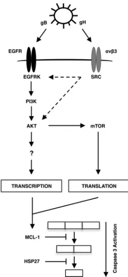
HCMV entry induces a signaling network necessary for the survival of infected monocytes, a necessary step in the hematoge-nous dissemination process. Our work here begins to decipher the molecular mechanisms by which HCMV selectively increases the expression of Mcl-1 and HSP27, which cooperatively function to block the activation of caspase 3 within HCMV-infected mono-cytes. We found that gB/EGFR and gH/integrin cooperatively function to increase Mcl-1 and HSP27 expression (Fig. 7). Fur-thermore, EGFR activation from cross talk from the gH/␣v3/Src cascade may also contribute to the upregulation of Mcl-1 and HSP27. We also demonstrate that although Akt is known to pro-mote protein stability, HCMV-activated Akt preferentially regu-lates the synthesis of antiapoptotic proteins though an increase in transcription and an increase in mTOR-mediated translation.
Overall, we suggest that HCMV rapidly induces the survival of newly infected monocytes through a unique signalsome generated during the viral entry process. Specifically, our data indicate that gB- and gH-initiated signaling stimulates the expression of select antiapoptotic proteins, Mcl-1 and HSP27, in order to block caspase 3-mediated apoptosis and allow monocyte-to-macro-phage differentiation. Deciphering the distinct mechanisms by which HCMV induces monocyte survival may allow the develop-ment of novel pharmaceutical drugs that directly target HCMV-infected monocytes.
ACKNOWLEDGMENTS
We thank Christine Burrer for maintenance of virus stocks and technical support.
We have no conflicting financial interests.
FIG 7A model for the selective upregulation of Mcl-1 and HSP27 during HCMV infection of monocytes. HCMV gB binding to EGFR and gH binding to␣v3 trigger the PI3K/Akt pathway. In addition, cross talk from the gH/
FUNDING INFORMATION
Sinsheimer Scholar Award provided funding to Gary C. Chan. North Carolina University Cancer Research Fund provided funding to Nathan-iel John Moorman. HHS | NIH | National Institute of Allergy and Infec-tious Diseases (NIAID) provided funding to Gary C. Chan under grant number R56AI110803-01. HHS | NIH | National Institute of Allergy and Infectious Diseases (NIAID) provided funding to Nathaniel John Moor-man under grant number R01AI03311. American Heart Association (AHA) provided funding to Gary C. Chan.
The funders had no role in study design, data collection and interpreta-tion, or the decision to submit the work for publication.
REFERENCES
1.Staras SA, Dollard SC, Radford KW, Flanders WD, Pass RF, Cannon MJ.2006. Seroprevalence of cytomegalovirus infection in the United States, 1988 –1994. Clin Infect Dis43:1143–1151.http://dx.doi.org/10 .1086/508173.
2.Streblow DN, Dumortier J, Moses AV, Orloff SL, Nelson JA. 2008. Mechanisms of cytomegalovirus-accelerated vascular disease: induction of paracrine factors that promote angiogenesis and wound healing. Curr Top Microbiol Immunol325:397– 415.
3.Jordan MC, Rousseau W, Stewart JA, Noble GR, Chin TD. 1973. Spontaneous cytomegalovirus mononucleosis. Clinical and laboratory observations in nine cases. Ann Intern Med79:153–160.
4.Ho M.1977. Virus infections after transplantation in man. Brief review. Arch Virol55:1–24.http://dx.doi.org/10.1007/BF01314475.
5.Masur H, Whitcup SM, Cartwright C, Polis M, Nussenblatt R.1996. Advances in the management of AIDS-related cytomegalovirus retinitis. Ann Intern Med125:126 –136.http://dx.doi.org/10.7326/0003-4819-125 -2-199607150-00009.
6.Stagno S, Pass RF, Dworsky ME, Henderson RE, Moore EG, Walton PD, Alford CA.1982. Congenital cytomegalovirus infection: the relative importance of primary and recurrent maternal infection. N Engl J Med
306:945–949.http://dx.doi.org/10.1056/NEJM198204223061601. 7.Emery VC.2001. Investigation of CMV disease in immunocompromised
patients. J Clin Pathol54:84 – 88.http://dx.doi.org/10.1136/jcp.54.2.84. 8.Ljungman P, Engelhard D, Link H, Biron P, Brandt L, Brunet S,
Cordonnier C, Debusscher L, de Laurenzi A, Kolb HJ, Messina C, Newland AC, Prentice HG, Richard C, Ruutu T, Tilg H, Verdonck L.
1992. Treatment of interstitial pneumonitis due to cytomegalovirus with ganciclovir and intravenous immune globulin: experience of European Bone Marrow Transplant Group. Clin Infect Dis14:831– 835.http://dx .doi.org/10.1093/clinids/14.4.831.
9.Manez R, Kusne S, Rinaldo C, Aguado JM, St George K, Grossi P, Frye B, Fung JJ, Ehrlich GD. 1996. Time to detection of cytomegalovirus (CMV) DNA in blood leukocytes is a predictor for the development of CMV disease in seronegative recipients of allografts from CMV-seropositive donors following liver transplantation. J Infect Dis173:1072– 1076.http://dx.doi.org/10.1093/infdis/173.5.1072.
10. Taylor-Wiedeman J, Sissons JG, Borysiewicz LK, Sinclair JH. 1991. Monocytes are a major site of persistence of human cytomegalovirus in peripheral blood mononuclear cells. J Gen Virol72:2059 –2064.http://dx .doi.org/10.1099/0022-1317-72-9-2059.
11. Taylor-Wiedeman J, Sissons P, Sinclair J.1994. Induction of endoge-nous human cytomegalovirus gene expression after differentiation of monocytes from healthy carriers. J Virol68:1597–1604.
12. Gonzalez-Mejia ME, Doseff AI.2009. Regulation of monocytes and mac-rophages cell fate. Front Biosci14:2413–2431.
13. Whitelaw DM.1972. Observations on human monocyte kinetics after pulse labeling. Cell Tissue Kinet5:311–317.
14. Chan G, Nogalski MT, Yurochko AD.2012. Human cytomegalovirus stimulates monocyte-to-macrophage differentiation via the temporal reg-ulation of caspase 3. J Virol86:10714 –10723.http://dx.doi.org/10.1128 /JVI.07129-11.
15. Sordet O, Rebe C, Plenchette S, Zermati Y, Hermine O, Vainchenker W, Garrido C, Solary E, Dubrez-Daloz L.2002. Specific involvement of caspases in the differentiation of monocytes into macrophages. Blood100:
4446 – 4453.http://dx.doi.org/10.1182/blood-2002-06-1778.
16. Droin N, Cathelin S, Jacquel A, Guery L, Garrido C, Fontenay M, Hermine O, Solary E.2008. A role for caspases in the differentiation of
erythroid cells and macrophages. Biochimie90:416 – 422.http://dx.doi .org/10.1016/j.biochi.2007.08.007.
17. Jacquel A, Benikhlef N, Paggetti J, Lalaoui N, Guery L, Dufour EK, Ciudad M, Racoeur C, Micheau O, Delva L, Droin N, Solary E.2009. Colony-stimulating factor-1-induced oscillations in phosphatidylinosi-tol-3 kinase/AKT are required for caspase activation in monocytes under-going differentiation into macrophages. Blood114:3633–3641.http://dx .doi.org/10.1182/blood-2009-03-208843.
18. Chan G, Nogalski MT, Bentz GL, Smith MS, Parmater A, Yurochko AD.2010. PI3K-dependent upregulation of Mcl-1 by human cytomega-lovirus is mediated by epidermal growth factor receptor and inhibits apoptosis in short-lived monocytes. J Immunol184:3213–3222.http://dx .doi.org/10.4049/jimmunol.0903025.
19. Chan G, Nogalski MT, Stevenson EV, Yurochko AD.2012. Human cytomegalovirus induction of a unique signalsome during viral entry into monocytes mediates distinct functional changes: a strategy for viral dissemination. J Leukoc Biol92:743–752. http://dx.doi.org/10 .1189/jlb.0112040.
20. Smith MS, Bentz GL, Alexander JS, Yurochko AD.2004. Human cyto-megalovirus induces monocyte differentiation and migration as a strategy for dissemination and persistence. J Virol78:4444 – 4453.http://dx.doi .org/10.1128/JVI.78.9.4444-4453.2004.
21. Fish KN, Depto AS, Moses AV, Britt W, Nelson JA. 1995. Growth kinetics of human cytomegalovirus are altered in monocyte-derived mac-rophages. J Virol69:3737–3743.
22. Ibanez CE, Schrier R, Ghazal P, Wiley C, Nelson JA.1991. Human cytomegalovirus productively infects primary differentiated macro-phages. J Virol65:6581– 6588.
23. Sinclair J, Sissons P.1996. Latent and persistent infections of monocytes and macrophages. Intervirology39:293–301.
24. Michels J, Johnson PW, Packham G.2005. Mcl-1. Int J Biochem Cell Biol
37:267–271.http://dx.doi.org/10.1016/j.biocel.2004.04.007.
25. Opferman JT, Iwasaki H, Ong CC, Suh H, Mizuno S, Akashi K, Korsmeyer SJ.2005. Obligate role of anti-apoptotic Mcl-1 in the survival of hematopoietic stem cells. Science307:1101–1104.http://dx.doi.org/10 .1126/science.1106114.
26. Liu H, Perlman H, Pagliari LJ, Pope RM.2001. Constitutively activated Akt-1 is vital for the survival of human monocyte-differentiated macro-phages. Role of Mcl-1, independent of nuclear factor (NF)-B, Bad, or caspase activation. J Exp Med194:113–126.
27. Voss OH, Batra S, Kolattukudy SJ, Gonzalez-Mejia ME, Smith JB, Doseff AI.2007. Binding of caspase-3 prodomain to heat shock pro-tein 27 regulates monocyte apoptosis by inhibiting caspase-3 proteo-lytic activation. J Biol Chem282:25088 –25099.http://dx.doi.org/10 .1074/jbc.M701740200.
28. Yurochko AD, Huang ES.1999. Human cytomegalovirus binding to human monocytes induces immunoregulatory gene expression. J Immu-nol162:4806 – 4816.
29. Yurochko AD, Hwang ES, Rasmussen L, Keay S, Pereira L, Huang ES.
1997. The human cytomegalovirus UL55 (gB) and UL75 (gH) glycopro-tein ligands initiate the rapid activation of Sp1 and NF-kB during infec-tion. J Virol71:5051–5059.
30. Chan G, Bivins-Smith ER, Smith MS, Yurochko AD.2008. Transcrip-tome analysis of NF-B- and phosphatidylinositol 3-kinase-regulated genes in human cytomegalovirus-infected monocytes. J Virol82:1040 – 1046.http://dx.doi.org/10.1128/JVI.00864-07.
31. Busca A, Saxena M, Kumar A.2012. Critical role for antiapoptotic Bcl-xL and Mcl-1 in human macrophage survival and cellular IAP1/2 (cIAP1/2) in resistance to HIV-Vpr-induced apoptosis. J Biol Chem287:15118 – 15133.http://dx.doi.org/10.1074/jbc.M111.312660.
32. Hart JR, Vogt PK.2011. Phosphorylation of AKT: a mutational analysis. Oncotarget2:467– 476.http://dx.doi.org/10.18632/oncotarget.293. 33. Yung HW, Charnock-Jones DS, Burton GJ.2011. Regulation of AKT
phosphorylation at Ser473 and Thr308 by endoplasmic reticulum stress modulates substrate specificity in a severity dependent manner. PLoS One
6:e17894.http://dx.doi.org/10.1371/journal.pone.0017894.
34. Chan G, Nogalski MT, Yurochko AD. 2009. Activation of EGFR on monocytes is required for human cytomegalovirus entry and mediates cellular motility. Proc Natl Acad Sci U S A106:22369 –22374.http://dx.doi .org/10.1073/pnas.0908787106.
cytomegalovirus. Nature 424:456 – 461. http://dx.doi.org/10.1038 /nature01818.
36. Wang X, Huang DY, Huong SM, Huang ES.2005. Integrin ab3 is a coreceptor for human cytomegalovirus. Nat Med11:515–521.http://dx .doi.org/10.1038/nm1236.
37. Cojohari O, Burrer CM, Peppenelli MA, Abulwerdi FA, Nikolovska-Coleska Z, Chan GC.2015. BH3 profiling reveals selectivity by herpesvi-ruses for specific Bcl-2 proteins to mediate survival of latently infected cells. J Virol89:5739 –5746.http://dx.doi.org/10.1128/JVI.00236-15. 38. Lenarcic EM, Ziehr B, De Leon G, Mitchell D, Moorman NJ.2014.
Differential role for host translation factors in host and viral protein syn-thesis during human cytomegalovirus infection. J Virol88:1473–1483.
http://dx.doi.org/10.1128/JVI.02321-13.
39. Certo M, Del Gaizo Moore V, Nishino M, Wei G, Korsmeyer S, Armstrong SA, Letai A.2006. Mitochondria primed by death signals determine cellular addiction to antiapoptotic BCL-2 family members. Cancer Cell9:351–365.http://dx.doi.org/10.1016/j.ccr.2006.03.027. 40. Nogalski MT, Chan G, Stevenson EV, Gray S, Yurochko AD. 2011.
Human cytomegalovirus-regulated paxillin in monocytes links cellular pathogenic motility to the process of viral entry. J Virol85:1360 –1369.
http://dx.doi.org/10.1128/JVI.02090-10.
41. Akgul C, Moulding DA, White MR, Edwards SW.2000. In vivo locali-sation and stability of human Mcl-1 using green fluorescent protein (GFP) fusion proteins. FEBS Lett478:72–76.http://dx.doi.org/10.1016/S0014 -5793(00)01809-3.
42. Mojsa B, Lassot I, Desagher S.2014. Mcl-1 ubiquitination: unique reg-ulation of an essential survival protein. Cells3:418 – 437.http://dx.doi.org /10.3390/cells3020418.
43. Zhong Q, Gao W, Du F, Wang X.2005. Mule/ARF-BP1, a BH3-only E3 ubiquitin ligase, catalyzes the polyubiquitination of Mcl-1 and regulates apoptosis. Cell 121:1085–1095. http://dx.doi.org/10.1016/j.cell.2005.06 .009.
44. Maurer U, Charvet C, Wagman AS, Dejardin E, Green DR. 2006. Glycogen synthase kinase-3 regulates mitochondrial outer membrane permeabilization and apoptosis by destabilization of MCL-1. Mol Cell
21:749 –760.http://dx.doi.org/10.1016/j.molcel.2006.02.009.
45. Nandy D, Asmann YW, Mukhopadhyay D, Basu A. 2010. Role of AKT-glycogen synthase kinase axis in monocyte activation in human be-ings with and without type 2 diabetes. J Cell Mol Med14:1396 –1407.
http://dx.doi.org/10.1111/j.1582-4934.2009.00900.x.
46. Chang WH, Tien CL, Chen TJ, Nukina N, Hsieh M.2009. Decreased protein synthesis of Hsp27 associated with cellular toxicity in a cell model of Machado-Joseph disease. Neurosci Lett454:152–156.http://dx.doi.org /10.1016/j.neulet.2009.03.004.
47. Chan G, Bivins-Smith ER, Smith MS, Smith PM, Yurochko AD.2008. Transcriptome analysis reveals human cytomegalovirus reprograms monocyte differentiation toward an M1 macrophage. J Immunol181:
698 –711.http://dx.doi.org/10.4049/jimmunol.181.1.698.
48. Chan G, Bivins-Smith ER, Smith MS, Yurochko AD.2009. NF-B and phosphatidylinositol 3-kinase activity mediates the HCMV-induced atyp-ical M1/M2 polarization of monocytes. Virus Res144:329 –333.http://dx .doi.org/10.1016/j.virusres.2009.04.026.
49. Chiang GG, Abraham RT.2005. Phosphorylation of mammalian target of rapamycin (mTOR) at Ser-2448 is mediated by p70S6 kinase. J Biol Chem280:25485–25490.http://dx.doi.org/10.1074/jbc.M501707200. 50. Mamane Y, Petroulakis E, LeBacquer O, Sonenberg N.2006. mTOR,
translation initiation and cancer. Oncogene25:6416 – 6422.http://dx.doi .org/10.1038/sj.onc.1209888.
51. Edinger AL, Thompson CB.2002. Akt maintains cell size and survival by increasing mTOR-dependent nutrient uptake. Mol Biol Cell13:2276 – 2288.http://dx.doi.org/10.1091/mbc.01-12-0584.
52. Barber GN.2001. Host defense, viruses and apoptosis. Cell Death Differ
8:113–126.http://dx.doi.org/10.1038/sj.cdd.4400823.
53. Rooswinkel RW, van de Kooij B, de Vries E, Paauwe M, Braster R, Verheij M, Borst J.2014. Antiapoptotic potency of Bcl-2 proteins pri-marily relies on their stability, not binding selectivity. Blood123:2806 – 2815.http://dx.doi.org/10.1182/blood-2013-08-519470.
54. Deveraux QL, Reed JC.1999. IAP family proteins—suppressors of apop-tosis. Genes Dev13:239 –252.http://dx.doi.org/10.1101/gad.13.3.239. 55. Shi Y.2002. Mechanisms of caspase activation and inhibition during
apoptosis. Mol Cell9:459 – 470.http://dx.doi.org/10.1016/S1097-2765 (02)00482-3.
56. Shi Y.2004. Caspase activation, inhibition, and reactivation: a mechanistic view. Protein Sci13:1979 –1987.http://dx.doi.org/10.1110/ps.04789804. 57. Lopez J, Meier P.2010. To fight or die—inhibitor of apoptosis proteins at
the crossroad of innate immunity and death. Curr Opin Cell Biol22:872– 881.http://dx.doi.org/10.1016/j.ceb.2010.08.025.
58. Mathew SS, Della Selva MP, Burch AD.2009. Modification and reorga-nization of the cytoprotective cellular chaperone Hsp27 during herpes simplex virus type 1 infection. J Virol83:9304 –9312.http://dx.doi.org/10 .1128/JVI.01826-08.
59. Singh D, McCann KL, Imani F.2007. MAPK and heat shock protein 27 activation are associated with respiratory syncytial virus induction of hu-man bronchial epithelial monolayer disruption. Am J Physiol Lung Cell Mol Physiol 293:L436 –L445. http://dx.doi.org/10.1152/ajplung.00097 .2007.
60. Suomalainen M, Nakano MY, Boucke K, Keller S, Greber UF.2001. Adenovirus-activated PKA and p38/MAPK pathways boost microtubule-mediated nuclear targeting of virus. EMBO J20:1310 –1319.http://dx.doi .org/10.1093/emboj/20.6.1310.
61. Shahbazian D, Roux PP, Mieulet V, Cohen MS, Raught B, Taunton J, Hershey JW, Blenis J, Pende M, Sonenberg N.2006. The mTOR/PI3K and MAPK pathways converge on eIF4B to control its phosphorylation and activity. EMBO J25:2781–2791.http://dx.doi.org/10.1038/sj.emboj .7601166.
62. Ruggero D, Sonenberg N.2005. The Akt of translational control. Onco-gene24:7426 –7434.http://dx.doi.org/10.1038/sj.onc.1209098.
63. Laplante M, Sabatini DM.2012. mTOR signaling in growth control and disease. Cell149:274 –293.http://dx.doi.org/10.1016/j.cell.2012.03.017. 64. Diehl N, Schaal H.2013. Make yourself at home: viral hijacking of the
PI3K/Akt signaling pathway. Viruses5:3192–3212.http://dx.doi.org/10 .3390/v5123192.
65. Soung YH, Korneeva N, Kim TH, Chung J.2013. The role of c-Src in integrin (␣64) dependent translational control. BMC Cell Biol14:49.
http://dx.doi.org/10.1186/1471-2121-14-49.
66. Vojtechova M, Tureckova J, Kucerova D, Sloncova E, Vachtenheim J, Tuhackova Z.2008. Regulation of mTORC1 signaling by Src kinase ac-tivity is Akt1-independent in RSV-transformed cells. Neoplasia10:99 – 107.http://dx.doi.org/10.1593/neo.07905.