Arabidopsis RAD51, RAD51C and XRCC3
proteins form a complex and facilitate RAD51
localization on chromosomes for meiotic
recombination
Hang Su1☯, Zhihao Cheng1,2☯, Jiyue Huang1, Juan Lin1, Gregory P. Copenhaver3,4, Hong Ma1,5, Yingxiang Wang1
*
1 State Key Laboratory of Genetic Engineering and Collaborative Innovation Center of Genetics and
Development, Ministry of Education Key Laboratory of Biodiversity Sciences and Ecological Engineering, Institute of Plant Biology, School of Life Sciences, Fudan University, Shanghai, China, 2 Institute of Banana and Plantain, Chinese Academy of Tropic Agriculture Science, Haikou, Hainan province, China,
3 Department of Biology and the Integrative Program for Biological and Genome Sciences, University
of North Carolina at Chapel Hill, Chapel Hill, North Carolina, United States of America, 4 Lineberger Comprehensive Cancer Center, University of North Carolina School of Medicine, Chapel Hill, North Carolina, United States of America, 5 Center for Evolutionary Biology, Institutes of Biomedical Sciences, School of Life Sciences, Fudan University, Shanghai, China
☯These authors contributed equally to this work. *[email protected]
Abstract
Meiotic recombination is required for proper homologous chromosome segregation in plants and other eukaryotes. The eukaryotic RAD51 gene family has seven ancient paralogs with important roles in mitotic and meiotic recombination. Mutations in mammalian RAD51 homologs RAD51C and XRCC3 lead to embryonic lethality. In the model plant Arabidopsis thaliana, RAD51C and XRCC3 homologs are not essential for vegetative development but are each required for somatic and meiotic recombination, but the mechanism of RAD51C and XRCC3 in meiotic recombination is unclear. The non-lethal Arabidopsis rad51c and xrcc3 null mutants provide an opportunity to study their meiotic functions. Here, we show that AtRAD51C and AtXRCC3 are components of the RAD51-dependent meiotic recombi-nation pathway and required for normal AtRAD51 localization on meiotic chromosomes. In addition, AtRAD51C interacts with both AtRAD51 and AtXRCC3 in vitro and in vivo, sug-gesting that these proteins form a complex (es). Comparison of AtRAD51 foci in meiocytes from atrad51, atrad51c, and atxrcc3 single, double and triple heterozygous mutants further supports an interaction between AtRAD51C and AtXRCC3 that enhances AtRAD51 locali-zation. Moreover, atrad51c-/+atxrcc3-/+double and atrad51-/+atrad51c-/+atxrcc3-/+triple heterozygous mutants have defects in meiotic recombination, suggesting the role of the AtRAD51C-AtXRCC3 complex in meiotic recombination is in part AtRAD51-dependent. Together, our results support a model in which direct interactions between the RAD51C-XRCC3 complex and RAD51 facilitate RAD51 localization on meiotic chromosomes and RAD51-dependent meiotic recombination. Finally, we hypothesize that maintenance of
a1111111111 a1111111111 a1111111111 a1111111111 a1111111111 OPEN ACCESS
Citation: Su H, Cheng Z, Huang J, Lin J,
Copenhaver GP, Ma H, et al. (2017) Arabidopsis RAD51, RAD51C and XRCC3 proteins form a complex and facilitate RAD51 localization on chromosomes for meiotic recombination. PLoS Genet 13(5): e1006827.https://doi.org/10.1371/ journal.pgen.1006827
Editor: Wojciech P. Pawlowski, Cornell University,
UNITED STATES
Received: January 17, 2017
Accepted: May 17, 2017
Published: May 31, 2017
Copyright:©2017 Su et al. This is an open access article distributed under the terms of theCreative Commons Attribution License, which permits unrestricted use, distribution, and reproduction in any medium, provided the original author and source are credited.
Data Availability Statement: All relevant data are
within the paper and its Supporting Information files.
Funding: This work was supported by grants from
RAD51 function facilitated by the RAD51C-XRCC3 complex could be highly conserved in eukaryotes.
Author summary
Meiotic recombination and sister chromatid cohesion are important for maintaining the association between homologous chromosomes and ensuring their accurate segregation. Meiotic recombination starts with a set of programmed DNA double-strand breaks (DSBs), catalyzed by the SPO11 endonuclease. Processing of DSB ends produces 30 single-stranded DNA tails, which form nucleoprotein filaments with RAD51 and DMC1, homo-logs of the prokaryotic RecA protein. The eukaryoticRAD51gene family has seven ancient paralogs, in addition to RAD51 and DMC1, the other five members in mammals form two complexes: RAD51B-RAD51C-RAD51D- XRCC2 (BCDX2) and RAD51C-XRCC3 (CX3). To date, the molecular mechanism of CX3 in animal meiosis remains largely unknown due to the essential roles of these two proteins in embryo development. In Ara-bidopsis, RAD51C and XRCC3 are required for meiosis and fertility, but their specific mechanisms are unclear. Here we present strong evidence thatArabidopsisRAD51 forms a protein complex with AtRAD51C-AtXRCC3in vivo. Our data also support the previous hypothesis that CX3 promotes RAD51-denpendet meiotic recombination by affecting its localization on chromosomes. Given that the RAD51, RAD51C and XRCC3 proteins are highly conserved in plants and vertebrates, the mechanism we present here could be important for the regulation of meiotic recombination in both plants and vertebrate animals.
Introduction
Homologous recombination (HR) is important for repairing DNA damage and maintaining genomic stability. Meiotic HR and sister chromatid cohesion are required for maintaining physical associations between homologous chromosomes (homologs) and ensuring their accu-rate segregation. Meiotic HR is initiated by programmed DNA double-strand breaks (DSBs) that are catalyzed by SPO11, a topoisomerase-like protein [1]. The resulting DSB ends are pro-cessed by the MRE11- RAD50-NBS1 (MRN) protein complexes to generate 30single-stranded DNA (ssDNA) tails [2,3], which are subsequently protected by replication protein A (RPA) [4]. Functional homologs of theE.coliRecA protein, RAD51 and DMC1 [5,6] bind to the 3’ tails to form nucleoprotein filaments with the help of several proteins identified in multiple species, includingSaccharomyces cerevisiae(Rad52 [7], Rad54 [8], Tid1/Rhd54 [9], Rad55-Rad57 [10], Swi5-Sfr1 [11] and PCSS complex [12]),Arabidopsis thaliana(RAD51C [13], XRCC3 [14], MND1-HOP2[15] and ATR/ATRIP [16]), and mammals (Mnd1-Hop2 [17] and Brca2-Dss1 [18]). The nucleoprotein filaments facilitate single-end invasion of a non-sister chromatid, resulting in the formation of a recombination intermediate called a D-loop, which is then processed to ultimately produce either crossovers (COs) or non-crossovers (NCOs) [19].
In vertebrate animals and plants, theRAD51gene family is highly conserved with seven members:DMC1,RAD51,RAD51B,RAD51C,RAD51D,XRCC2andXRCC3[20–23], which share Walker A and Walker B motifs with over 37.5% similarity [24]. In mice, mutations in any of the paralogs, exceptDMC1, lead to embryonic lethality following spontaneous DNA damage or errors [25–29]. In the model plantArabidopsis thaliana, all seven genes are decision to publish, or preparation of the
manuscript.
Competing interests: The authors have declared
dispensable for vegetative growth [13,14,24,30–33]. However,AtRAD51,AtRAD51Cand
AtXRCC3are required for somatic and meiotic recombination, as well as plant fertility.
Muta-tions in any of these three genes result in a meiotic chromosome fragmentation phenotype [13,14,24,30–32]. Moreover,AtDMC1is specifically required for meiotic homolog pairing and recombination [34,35]. In contrast toatrad51,atrad51candatxrcc3mutants,atdmc1mutants do not suffer meiotic chromosome fragmentation; instead their DSBs are thought to be repaired using sister chromatids as templates [34,35]. The three other paralogs,AtRAD51B,
AtRAD51DandAtXRCC2, seem to be unnecessary for meiotic DSB repair, because the triple
mutant has normal chromosome morphology and fertility [33]. Except for slight differences in synapsis, the chromosome morphology using light microscopy for DAPI-stained chromo-somes and fertility phenotypes ofatrad51candatxrcc3mutants are similar to those ofatrad51, suggesting that their functions are related, but further analyses are needed to understand their mechanistic roles in meiotic DSB repair.
Biochemical studies in human cells demonstrate that RAD51 paralogs associate with one another in two distinct complexes: RAD51B-RAD51C-RAD51D-XRCC2 (BCDX2) and RAD51C-XRCC3 (CX3) [36,37]. The CX3 complex stabilizes RAD51 binding to ssDNA [36–
39]in vitro, thus promoting single-end invasion. Moreover, RAD51C and XRCC3 also help
mediate Holliday junction (HJ) resolutionin vitro[40], suggesting a later role in meiotic recombination. A yeast two-hybrid assay demonstrated that theArabidopsisRAD51 paralogs also interact with each other [41], supporting the idea that RAD51 paralogs function by forma-tion of distinct protein complexes in both animals and plants. However, whether the RAD51 paralogs associate with each otherin plantahas not been tested.
In this study, we report thatArabidopsishomologs of RAD51, RAD51C and XRCC3 show highly similar meiotic chromosome morphological defects using immune-localization for key markers. We also provide evidence that AtRAD51C and AtXRCC3 are required for AtRAD51 localization on chromosomes. Bothin vitroandin vivodata demonstrate that AtRAD51C interacts with AtRAD51 and AtXRCC3. Furthermore, observation of AtRAD51 foci inatrad51,
atrad51candatxrcc3single, double and triple heterozygotes reveals that AtRAD51C and
AtXRCC3 both are involved in AtRAD51 loading. Triple heterozygotes also experience non-homolog chromosome associations and have reduced CO frequencies. Together, these results demonstrate that AtRAD51C, AtXRCC3 and AtRAD51 form a complexin plantaand are required for AtRAD51 loading on chromosomes.
Results
AtRAD51, AtRAD51C and AtXRCC3 have non-redundant roles in
meiotic recombination
Previous studies have found thatAtRAD51,AtRAD51CandAtXRCC3are required for meiotic DSB repair and plant fertility and mutation of individual genes cause indistinguishable chro-mosome entanglement and fragmentation phenotypes [13,14,31,32]. The similarity of the phe-notypes suggests that these genes might function in the same genetic pathway or process. To test this hypothesis, we generated double mutants betweenatrad51-3(SAIL_873_C08) [42],
atrad51c(SALK_021960) [13], andatxrcc3(SALK_045564) [14] and found that the
chromo-some morphologies ofatrad51 atrad51c(48 cells),atrad51 atxrcc3(65 cells), andatrad51c
atxrcc3(54 cells) double mutants showed no obvious differences compared with each of the
single mutants (S1 Fig). The lack of an additive phenotype in the double mutants further sup-ports the hypothesis that they act together in the same biological process.
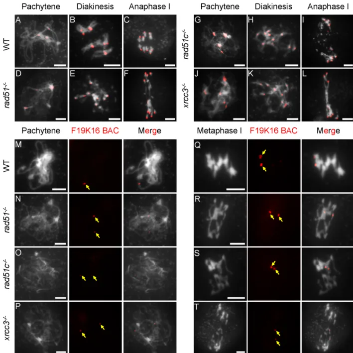
atxrcc3(81cells) and a bacterial artificial chromosome (BAC-F19K16) probe that targets a telo-mere proximal region on chromosome 1 foratrad51(31 cells);atrad51c(45 cells) andatxrcc3
(22 cells) [43]. Wild-type (WT) meiocytes had three to five centromere signals at pachytene, indicative of paired homologous centromeres in a cluster (Fig 1A). Although the three mutants had no typical pachytene chromosomes, they all displayed similar centromere clusters or num-bers of signals at a stage similar to that of WT, suggesting thatAtRAD51,AtRAD51Cand
AtXRCC3are not required for early centromere pairing or clustering (Fig 1D, 1G and 1J). At
diakinesis and metaphase I, WT meiocytes had five bivalents, each with two paired centromere signals corresponding to the associated homologs (Fig 1B). In contrast, the three mutants each had 10 centromere signals located on abnormally associated chromosomes (multivalents-with more than two chromosomes) (Fig 1E, 1H and 1K), indicating a failure to maintain homolog association, at least at the centromere regions. We next examined homolog pairing on the chromosome arms using the telomere-proximal BAC probe. Unlike the single focus observed on WT pachytene chromosomes, indicative of fully synapsed homologs, meiocytes from each of the three mutants showed two separate signals, indicating a failure to pair properly (Fig
1M–1P). We also performed ASY1 and ZYP1 immuno-localization in WT and mutants. No
obvious difference of ASY1 signals at zygotene was found between WT and mutants (S2 Fig). However, unlike WT with linear ZYP1 distribution on pachytene chromosomes, ZYP1 was completely disappeared inrad51, while some punctate or discontinuous ZYP1 signals were observed inxrcc3andrad51c(S2 Fig). Together, these results demonstrate thatAtRAD51,
AtRAD51CandAtXRCC3are not required for recombination-independent centromere
clus-tering, but are necessary for homolog pairing, consistent with previous findings obtained using FISH experiment [44]. The similarities of the mutant phenotypes further support the idea that they act in the same process.
Arabidopsis RAD51C and XRCC3 are required for normal localization of
RAD51 on chromosomes
Loading of RAD51 on ssDNA is aided by several proteins, including Rad52 [45], Rad55-57 (Rad51 paralogs) [46] and Sfr1-Swi5 [11] in yeast, the Brca2-Dss1 complex in mammalian cells [47], and also by AtBRCA2 inArabidopsis[48]. The similarity of meiotic defects in
Arabi-dopsis rad51,rad51candxrcc3mutants suggests the RAD51 paralogs RAD51C and XRCC3
may function in meiotic recombination by affecting RAD51 function. To test this hypothesis, we performed an immunofluorescence assay using an AtRAD51 antibody [49]. InArabidopsis, formation of DSBs is thought to occur at leptotene [50]. At a similar stage, we found that WT plants had 187.7±24.5 AtRAD51 foci per meiocyte (n = 14), but the number of foci was greatly reduced inatrad51c(36.1±9.7, n = 17; P = 1.5E-13) andatxrcc3(33.7±10.3, n = 34; P = 5.7E-13) mutant meiocytes (Fig 2A, 2C, 2D and 2Q). In contrast, a parallel experiment did not detect any AtRAD51 foci inatrad51mutant meiocytes at zygotene (Fig 2B). A similar pattern was also observed using pachytene meiocytes (Fig 2E–2H). These results provide evidence that
ArabidopsisRAD51C and XRCC3 are required for formation of wild type level of RAD51 foci
on meiotic chromosomes. This is consistent with the previous findings for Rad51 paralogs in yeast [46]. Nevertheless, the reduction of AtRAD51 foci inatrad51candatxrcc3homozygous mutants does not preclude the possibility that normal level DSBs are formed in these mutants. To test whether DSB frequency is altered inatrad51candatxrcc3mutants, we examined the distribution of a DSB marker, phosphorylated histone H2AX (γ-H2AX) [51]. At zygotene, after DSBs have been formed, no significant differences in the number ofγ-H2AX foci were detected between WT (189.3±26.5, n = 39),atrad51(176.7±15.5, n = 19; P = 0.062),atrad51c
and 2R). InArabidopsis, most meiotic DSBs are thought to be repaired during zygotene-pachy-tene. We found thatγ-H2AX foci were obviously reduced in WT (56.9±15.2, n = 55) pachytene meiocytes compared those ofatrad51(132.1±15.4, n = 13; P = 1.5E-11),atrad51c(120.6±16.6, n = 14; P = 7.2E-11) andatxrcc3(122.2±18.8, n = 18; P = 1.7E-11) mutants (Fig 2M–2P and Fig 1. Fluorescence in situ hybridization (FISH) analysis of chromosome interactions in atrad51, atrad51c and atxrcc3 mutants.
(A-L) Chromosome morphologies of wild type (WT), atrad51, atrad51c and atxrcc3 mutants at pachytene, diakinesis and anaphase I. Centromeres appear as red dots. (M-P) WT pachytene chromosomes showing a single signal from the BAC-F19K16 probe and atrad51,
atrad51c, and atxrcc3 chromosomes showing two signals from the same probe. (Q-S) Equal segregation of BAC-F19K16 signals at
metaphase I in WT and unequal segregation in atrad51 and atrad51c mutants. (T) BAC-F19K16 signals located on atxrcc3 chromosome fragments at metaphase I. For FISH analysis of chromosomes using the BAC-F19K16 probe, the left panels show the chromosome morphology following staining with 6-diamidino-2-phenylindole (DAPI), the middle panels show the BAC-F19K16 signals (red dots) and the right panels merge the DAPI staining and BAC-F19K16 signals. Scale bar: 5μm.
2S). The presence of normal numbers of zygoteneγ-H2AX foci and reduced AtRAD51 foci suggests that AtRAD51C and AtXRCC3 are not required for meiotic DSB formation, but are necessary for AtRAD51 loading.
Arabidopsis RAD51C interacts with RAD51 and XRCC3 in vitro and in
vivo
In yeast, the Rad51 paralogs Rad55 and Rad57 form a heterodimeric complex to stimulate RAD51 activity [10]. Vertebrate Rad51 paralogs interact with one another to form two distinct complexes: BCDX2 and CX3 [52]. Like vertebrates,Arabidopsishas seven RAD51 paralogs, and previous yeast two-hybrid assays have shown that XRCC3 interacts with both RAD51C and RAD51 [41]. However, whether these proteins interactin plantahas not been investigated.
As an initial test for potential interactions we used a yeast two-hybrid assay (Y2H) and found that AtXRCC3 interacts with both AtRAD51 and AtRAD51C (Fig 3A), consistent with the previously identified interactions in Y2H system [41]. The interaction between AtRAD51C and AtXRCC3 was further supported by a pull-down assay using recombinant fusion protein of glutathione S-transferase (GST) with AtRAD51C and an AtXRCC3-His tag fusion protein (Fig 3B). In addition to the previously identified interactions, we also found that GST-A-tRAD51 interacts with AGST-A-tRAD51C-His (Fig 3B). To explore whether these associations also occurredin planta, we examined the interactions using bimolecular fluorescence complemen-tation (BiFC) in tobacco (Nicotiana benthamiana) cells. Strong nuclear signals, indicating interaction, were observed for AtRAD51C with either AtRAD51 or AtXRCC3 (Fig 3C). These results provide the first direct evidence that plant RAD51 paralogs RAD51C and XRCC3 inter-act directly with RAD51in vitroandin planta.
Male meiotic defects are observed in atrad51c
-/+atxrcc3
-/+double and
atrad51
-/+atrad51c
-/+atxrcc3
-/+triple heterozygous mutants
A recent study identified a weakatrad51allele,atrad51-2[42], with a T-DNA insertion in the 30-untranslated region (UTR) that results in reduced AtRAD51 protein levels. This mutant had mild chromosome fragmentation and partial synapsis, as well as some bivalent formation with homologs and non-homologs [42]. In contrast, theatrad51-1null mutant had severe chromosome fragmentation and formed multivalents during meiotic prophase I [31]. These findings suggest that reducing AtRAD51 level might be a strategy for investigating its meiotic function. Alternatively, analysis of double heterozygous mutants in genes encoding compo-nents of a complex can reveal phenotypic defects, even though the corresponding single heterozygotes are phenotypically normal [53,54]. We hypothesized that double/triple hetero-zygotes ofatrad51,atrad51candatxrcc3might reduce, but not abolish, their interactions in a complex and reveal informative meiotic phenotypes
respectively. (B, F) No signal was detected in the atrad51 mutant at similar stages. (C, D, G, H) The numbers of AtRAD51 foci were significantly reduced in the atrad51c and atxrcc3 mutants compared with WT at zygotene and pachytene, respectively. (I, M) Localization ofγ-H2AX on wild-type (WT) chromosomes at zygotene and pachytene stages, respectively. (J-L) No significant differences were detected between
atrad51, atrad51c and atxrcc3 mutants at zygotene. (N-P) Moreγ-H2AX foci were detected at pachytene in
atrad51, atrad51c and atxrcc3 mutants compared to WT. (Q) The number of RAD51 foci on chromosomes
from wild type and mutants at zygotene. (R-S) The number ofγ-H2AX foci on chromosomes from wild type and mutants at zygotene and pachytene. Left panels show the chromosome morphology following staining with 6-diamidino-2-phenylindole (DAPI), middle panels show AtRAD51 foci (red dots), and right panels merge the DAPI-stained images with the AtRAD51 foci images. Scale bar: 5μm.**p<0.01 (two-tailed Student’s t-test).
Fig 3. AtRAD51C interacts with AtRAD51 and AtXRCC3 in vitro and in vivo. (A) Yeast two-hybrid assay showing AtXRCC3 interaction
with AtRAD51 and AtRAD51C. The known DYT1-DYT1 interaction is used as a positive control [82]. AD refers to the activation domain, BD refers to the DNA-binding domain, SD-2 refers to SD-Leu-Trp and SD-4 refers to SD-Leu-Trp-His-Ade+X-gal. Blue indicates a positive interaction. (B) Pull-down assay showing that AtRAD51C interacts with AtRAD51 and AtXRCC3. (C) BiFC assay in tobacco cells showing strong nuclear signals for AtRAD51-AtRAD51C and AtRAD51C-AtXRCC3 interactions. Left panels are yellow fluorescent protein (YFP) signals, middle panels are nuclei stained with 6-diamidino-2-phenylindole (DAPI), and right panels merge the DAPI-stained images with the YFP signals and bright field images.
To test this hypothesis, we generatedatrad51-/+,atrad51c-/+andatxrcc3-/+double and triple heterozygous mutants and compared their meiotic phenotypes with WT. Analysis of meiotic chromosome morphology after DAPI staining showed thatatrad51-/+,atrad51c-/+and
atxrcc3-/+single heterozygote meiocytes andatrad51-/+atrad51c-/+andatrad51-/+atxrcc3-/+
double heterozygotes had similar phenotypes compared to WT (Fig 4A–4L;S3 Fig). In addi-tion, meiocytes fromatrad51c-/+atxrcc3-/+double heterozygotes had chromosome morphol-ogy similar to WT at pachytene (Fig 4M), but at diakinesis, WT formed five bivalents, whereas 32.8% (20 of 61, n = 61) of theatrad51-/+atxrcc3-/+double heterozygote meiocytes had non-homologous chromosome associations (Fig 4N). The cell appears to be able to resolve these
Fig 4. Centromere-fluorescence in situ hybridization analysis of chromosome morphology in double and triple heterozygous mutants. (A-L) Wild type (WT), atrad51-/+atrad51c-/+and atrad51-/+atxrcc3-/+
mutant chromosome morphologies at pachytene, diakinesis, metaphase I and metaphase II. Compared with WT, atrad51-/+atrad51c-/+and atrad51-/+atxrcc3-/+mutants had similar chromosome morphologies. (M)
atrad51c-/+atxrcc3-/+chromosome morphology at pachytene was similar to WT, but showed 32.8% (n = 61)
non-homolog association at diakinesis (N). (O, P) atrad51c-/+atxrcc3-/+chromosome morphology at
metaphase I and II, respectively. Chromosome morphology of atrad51-/+atrad51c-/+atxrcc3-/+at pachytene (Q) and diakinesis (R), metaphase I (S) and metaphase II (T). The atrad51-/+atrad51c-/+atxrcc3-/+triple
chromosome morphology showed more severe defects than atrad51c-/+atxrcc3-/+. Red dots indicate
centromere signals. Yellow arrows show non-homologous chromosome associations (N, R, S). The red numbers in N, R, S refer to the number of abnormal cells out of all cells observed. Scale bar: 5μm.
associations since equal division of chromosomes was observed at anaphase I and II (Fig 4O and 4P). Meiocytes fromatrad51-/+atrad51c-/+atxrcc3-/+triple heterozygotes had a more severe non-homolog association phenotype (47.8% at diakinesis, 22 of 46, n = 46,Fig 4R) and had unequal chromosome segregation at metaphase II (16.7%, 2 of 12, n = 12,Fig 4T). No chromosome fragments were observed in the triple heterozygote, suggesting it is still capable of DSB repair. The results also suggest that RAD51C and XRCC3 are functionally more related to each other than either is to RAD51. Previous studies showed that T-DNA translocation can cause a similar pattern of chromosome association using light microscopy because the translo-cated chromosome can associated with two normal chromosomes [55,56]. We have verified the T-DNA insertion site by sequencing the junction with flanking genomic DNAs and the results indicated that these mutations are not associated with translocations.
Repair of meiotic DSBs is delayed in atrad51
-/+, atrad51c
-/+and
atxrcc3
-/+single, double and triple heterozygous mutants
To test whether meiotic DSB repair is delayed in the heterozygotes, we performed immunos-taining experiments using aγH2AX antibody. As mentioned above, WT meiocytes had 189.3
±26.5 (n = 39) and 56.9±15.2 (n = 55)γH2AX foci at zygotene and pachytene, respectively (Fig
5A, 5I, 5Q and 5RandTable 1). All single, double and triple heterozygotes had no obvious dif-ferences in the number ofγH2AX foci at zygotene, but had significantly more foci at pachytene (Fig 5A–5R,S1 Table). Moreover, the double and triple heterozygotes had more foci at pachy-tene than the single heterozygotes. There are significantly fewer foci inatrad51-/+(78.1±19.4, n = 17),atrad51c-/+(83.3±10.8, n = 12) andatxrcc3-/+(82.0±25.9, n = 24) (S1 Table) compared to the double mutantsatrad51-/+atrad51c-/+(96.3±15.4, n = 30),atrad51-/+atxrcc3-/+(100.4
±14.8, n = 21) andatrad51c-/+atxrcc3-/+(105.2±24.1, n = 15), which in turn have significantly
fewer foci (S1 Table) than the tripleatrad51-/+atrad51c-/+atxrcc3-/+(113.3±14.8, n = 46) (Fig 5J–5P and 5RandTable 1). These data suggest that DSB formation is normal in the heterozy-gotes, but there is a defect in the progression of DSB repair, and that AtRAD51, AtRAD51C and AtXRCC3 function in this process.
Because AtRAD51C and AtXRCC3 are required for normal AtRAD51 localization, we next examined AtRAD51 localization in heterozygous mutant meiocytes. As described above, WT meiocytes have 187.7±24.5 (n = 14) AtRAD51 foci at zygotene and 51.2±14.0 (n = 65) foci at pachytene (seeFig 6A and 6Ifor examples andTable 1). In contrast, single, double and triple heterozygous mutant meiocytes have significantly fewer AtRAD51 foci at zygotene (p<0.05;
Fig 6A–6H and 6Q). At pachytene, the three single mutant heterozygotes show no obvious
differences in the number of AtRAD51 foci compared with WT (Fig 6I–6L and 6R), but the double and triple heterozygotes exhibited reduced AtRAD51 foci (p<0.05;Fig 6M–6P and 6R). These findings are consistent with the earlier results, suggesting that AtRAD51C and AtXRCC3 play related roles in AtRAD51 loading on chromosomes, likely in a protein complex.
CO number is reduced in atrad51
-/+atrad51c
-/+atxrcc3
-/+triple
heterozygotes
differences were observed in the single heterozygotes:atrad51-/+with 9.6±0.7 (n = 20; P = 0.072) per meiocyte,atrad51c-/+with 9.6±0.7 (n = 21; P = 0.052) per meiocyte andatxrcc3-/+
with 9.6±0.8 (n = 24; P = 0.066) per meiocyte. Theatrad51-/+atrad51c-/+,atrad51-/+atxrcc3-/+
andatrad51c-/+atxrcc3-/+double heterozygotes showed a slight, but statistically significant, reduction of chiasmata with 8.4±1.2 (n = 14; P = 6.0E-05), 8.0±0.8 (n = 10; P = 2.9E-06) and 7.1±1.0 (n = 34; P = 1.2E-20) per meiocyte, respectively (Fig 7A–7D and 7K). Theatrad51-/+
atrad51c-/+atxrcc3-/+triple heterozygous mutant also had a significant reduction, with only
6.9±1.0 (n = 15; P = 2.6E-10) chiasmata per meiocyte formed (Fig 7E and 7K). Furthermore, the chiasmata numbers per meiocyte ofatrad51c-/+atxrcc3-/+double heterozygote (7.1; P values = 2.0E-03 and 1.1E-02, respectively) and the triple heterozygote (6.9, P values = 1.7E-03 and 8.8E-1.7E-03, respectively) were significantly lower than those of the other two double heterozygotes.
Arabidopsisforms two types of COs: interference-sensitive Type I COs that require ZMM
proteins like MSH4 and MLH1 [57–59], and interference-insensitive class II COs that are MUS81-dependent [60,61]. To assess the impact of RAD51 and its paralogs on Type I COs, we used an AtMLH1 antibody to visualize AtMLH1 foci, in WT,atrad51-/+atrad51c-/+,atrad51-/+ atxrcc3-/+,atrad51c-/+atxrcc3-/+andatrad51-/+atrad51c-/+atxrcc3-/+meiocytes at diakinesis [59]. On average, WT meiocytes had 9.0±1.2 foci (n = 61,Fig 7F), whereas at similar stages,
atrad51-/+atrad51c-/+,atrad51-/+atxrcc3-/+,atrad51c-/+atxrcc3-/+andatrad51-/+atrad51c-/+
atxrcc3-/+mutants had 7.9±1.4 (n = 40; P = 5.8E-05), 7.7±1.6 (n = 25; P = 5.4E-04), 6.4±1.3
(n = 39; P = 5.9E-16) and 5.9±1.0 (n = 16; P = 5.5E-12) foci, respectively (Fig 7G–7J and 7L). The reduction of AtMLH1 foci in the mutants is consistent with the observed reduction in chi-asmata, and supports the idea that Type-I COs are reduced in the mutants.
Although the CO number was obviously reduced by ~30% in theatrad51-/+atrad51c-/+
atxrcc3-/+triple heterozygote, no univalents were observed, consistent with a mechanism that
ensures at least one CO per chromosome [62]. If the COs were distributed among the 5
Arabi-dopsisbivalents randomly, they would follow the Poisson function P (k COs per bivalent) =
(λke-λ)/k! whereλis the mean number of COs per bivalent. Using this function, from the chromosomes showed no obvious differences among the eight genotypes examined. (I-P) Theγ-H2AX distribution in pachytene chromosomes from single, double and triple heterozygous mutants. Left panels show the chromosome morphology following staining with 6-diamidino-2-phenylindole (DAPI), middle panels showγ-H2AX foci (red dots), and right panels merge the DAPI-stained images with theγ-H2AX foci images. (Q-R) The number ofγ-H2AX foci in chromosomes from the eight genotypes at zygotene and pachytene in A-P. Scale bar: 5μm.*p<0.05,**p<0.01 (two-tailed Student’s t-test).
https://doi.org/10.1371/journal.pgen.1006827.g005
Table 1. Numbers ofγ-H2AX and AtRAD51 foci in single, double and triple heterozygous mutants at zygotene and pachytene.
AtRAD51 γ-H2AX
Genetype Zygotene Pachytene Zygotene Pachytene
WT 187.7±24.5 (n = 14) 51.2±14.0 (n = 65) 189.3±26.5 (n = 39) 56.9±15.2 (n = 55)
atrad51-/+ 143.3±20.2 (n = 16) 49.8±13.2 (n = 25) 199.5±20.6 (n = 22) 78.1±19.4 (n = 17)
atrad51c-/+ 143.8±32.7 (n = 22) 57.3±10.4 (n = 12) 191.0±28.3 (n = 30) 83.3±10.8 (n = 12)
atxrcc3-/+ 142.2±29.7 (n = 19) 49.3±14.7 (n = 29) 201.9±35.4 (n = 24) 82.0±25.9 (n = 24)
atrad51-/+atrad51c-/+ 116.4±23.1 (n = 12) 34.4±10.2 (n = 29) 202.9±20.2 (n = 19) 96.3±15.4 (n = 30)
atrad51-/+atxrcc3-/+ 108.1±8.9 (n = 14) 35.5±9.8 (n = 35) 187.4±29.0 (n = 21) 100.4±14.8 (n = 21)
atrad51c-/+atxrcc3-/+ 106.9±8.2 (n = 10) 32.2±9.0 (n = 33) 192.2±24.8 (n = 30) 105.2±24.1 (n = 15)
atrad51-/+atrad51c-/+atxrcc3-/+ 99.5±13.0 (n = 18) 30.7±8.1 (n = 61) 199.5±16.3 (n = 35) 113.3±14.8 (n = 46)
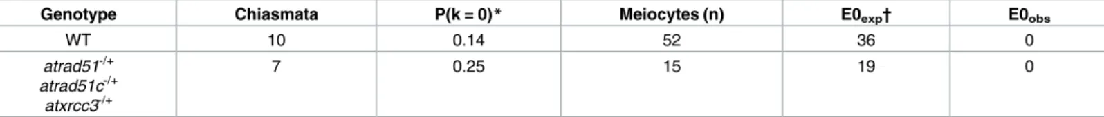
analyses of 52 WT and 15atrad51-/+atrad51c-/+atxrcc3-/+triple heterozygote meiocytes, we would expect to find 36 and 19 univalents in WT and the triple mutant, respectively, but none were observed (Table 2).
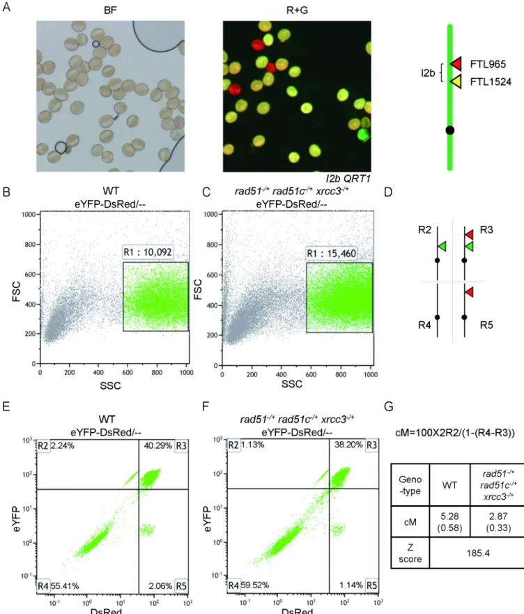
To further quantify the remaining COs inatrad51-/+atrad51c-/+atxrcc3-/+, we used a flow cytometry-based assay that measures the segregation of transgenes encoding fluorescent marker proteins expressed using a pollen-specific LAT52 promoter (FTL markers) [63,64]. The number of viable pollen grains is dramatically reduced inatrad51-/+atrad51c-/+atxrcc3-/+, but it was still feasible to measure CO frequencies using this assay. We crossedatrad51-/+
atrad51c-/+atxrcc3-/+with lineI2b, which carries two FTL markers (YFP and DsRed) on
chro-mosome 2 (Fig 8A). Pollen grains which express both fluorescent proteins have not experi-enced a crossover between the markers, while those that express only one or the other have. The relative abundance of these two classes can be used to calculate the genetic distance between the two markers [65]. We scored 10,092 WT pollen grains and 15,460 pollen grains from the triple heterozygote (Fig 8B and 8C). TheI2bmap distance was 5.28±0.58 cM in WT and 2.87±0.33 cM in the triple heterozygote (Fig 8D–8G). The genetic distance between the two fluorescent markers was significantly reduced inatrad51-/+atrad51c-/+atxrcc3-/+(Z score = 185.4, P value<<0.01) (Fig 8G), consistent with the reduction in chiasmata described above.
Discussion
Formation of protein complexes between RAD51 and its paralogs is
highly conserved
RAD51 family members are conserved across species, from yeast to humans [20]. The bud-ding yeastS.cerevisiaehas four RAD51 paralogs (Rad51, Dmc1, Rad55 and Rad57) [10], whereas humans have seven paralogs (RAD51, DMC1, RAD51B, RAD51C, RAD51D, XRCC2, XRCC3) [20]. In yeast, Rad55 interacts with Rad57 to form a stable heterodimer [10]. Simi-larly, in humans, two complexes are formed by the RAD51 paralogs: the BCDX2 and the CX3 complexes [36,37,39]. Moreover, a recent study inCaenorhabditis elegansshowed that the RAD51 paralogs, RFS-1 and RIP-1, also exist as a heterodimer and interact with RAD51 [66].
ArabidopsisXRCC3 has been shown to interact with both RAD51 and RAD51C using a
yeast two-hybrid assay [41]. We confirmed the yeast two-hybrid result (Fig 3A) and demon-strated that the AtRAD51C-AtXRCC3 interaction occursin plantaby using pull-down and BiFC assays (Fig 3B and 3C). It is noteworthy that both pull-down and BiFC assays support an interaction between AtRAD51C with AtRAD51 and AtXRCC3. Our results strongly sup-port the idea that AtRAD51C is a central factor in complex formation, and is associated with AtRAD51 and AtXRCC3. These findings are consistent with previous results in human cells that show AtRAD51C associates with two protein complexes [36,37,39]. Our study is also the first time to show that RAD51 paralogs form a protein complex with RAD51 in plants, sup-porting the hypothesis that formation of RAD51-paralogs associated protein complexes is highly conserved across eukaryotes, including yeast, humans and plants.
zygotene chromosomes from atrad51-/+, atrad51c-/+, atxrcc3-/+, atrad51-/+atrad51c-/+, atrad51-/+atxrcc3-/+,
atrad51c-/+atxrcc3-/+and atrad51-/+atrad51c-/+atxrcc3-/+heterozygous mutant meiocytes showed reductions compared to wild type (WT). (I-L) The location and (R) number of AtRAD51 foci on pachytene chromosomes in the three single heterozygotes showed no obvious differences compared with WT. (M-P) The location and (R) number of AtRAD51 foci on pachytene chromosomes in the double and triple heterozygous mutants were significantly reduced relative to WT. Left panels show the chromosome morphology following staining with 6-diamidino-2-phenylindole (DAPI), middle panels show AtRAD51 foci (red dots), and right panels merge the DAPI-stained images with the AtRAD51 foci images. Scale bar: 5μm.**p<0.01 (two-tailed Student’s t-test).
Fig 7. Number of chiasmata and AtMLH1 foci in double and triple heterozygous mutants. (A-E)
Centromere-fluorescence in situ hybridization showing wild type (WT) and atrad51-/+atrad51c-/+, atrad51-/+atxrcc3-/+, atrad51c-/+
atxrcc3-/+and atrad51-/+atrad51c-/+atxrcc3-/+double and triple heterozygous mutant chromosome morphologies at
diakinesis. Red dots indicate centromere signals. (F-J) Immunostaining of AtMLH1 foci (red dots) on WT, atrad51-/+
atrad51c-/+, atrad51-/+atxrcc3-/+, atrad51c-/+atxrcc3-/+and atrad51-/+atrad51c-/+atxrcc3-/+diakinesis chromosomes. (K) Number of chiasmata in WT, double and triple mutant chromosomes. (L) Number of AtMLH1 foci in WT, double and triple mutants. Scale bar: 5μm.*p<0.05,**p<0.01 (one-tailed Student’s t-test).
Formation of a RAD51 paralog complex is required to facilitate RAD51 in
HR
Previous studies in yeast showed that RAD51 paralogs are unable to form filaments with ssDNA and do not have a direct role in homology search or single strand invasion [10,23]. Nevertheless, studies in different organisms have reported that RAD51 paralogs play impor-tant roles in promoting RAD51 function in both mitotic and meiotic HR [46,67–69]. For example, the yeast Rad55-Rad57 complex has a role in RAD51-dependent HR [10,46]. Similar roles have been found for theC.elegansheterodimer of RAD51 paralogs RFS-1/RIP-1 [66] and the human CX3 complex [36,37,39].
Due to the lack of direct biochemical data, the role of RAD51 paralogs in meiotic HRin
plantais unclear. Studies in the monocot model plant,Oryza sativa(rice), showed that the
RAD51 paralogs OsRAD51C and OsXRCC3 are required for meiotic DSB repair and muta-tions in either result in sterility, chromosome entanglement and fragmentation [70,71]. These results are consistent with similar findings inArabidopsis[13,14,32]. Immunostaining showed that OsXRCC3 is required for OsRAD51C localization on chromosomes [70], suggesting the existence of a potential OsRAD51C-OsXRCC3 complex in rice. Additionally, the single-end processing proteins OsCOM1 and OsDMC1 no longer associate with DSB sites in rice
osxrcc3, which suggests that OsXRCC3, and by extension OsRAD51C, might function
up-stream of OsRAD51 [70,72]. Nevertheless, the relationship between OsRAD51 and its paralogs OsRAD51C and OsXRCC3 remains unclear, because a RAD51 antibody is currently unavail-able in rice. In the present study, we showed thatArabidopsisRAD51 foci were obviously reduced inatrad51candatxrcc3mutants, consistent to the discovery in rice. Together, these studies, in both rice andArabidopsis, strengthen the idea that AtRAD51 depends on its para-logs for normal function and that this relationship is highly conserved in eukaryotes.
RAD51 paralogs have a role in meiotic CO formation
Previous studies showed that RAD51 paralogs have a later role in processing meiotic re-combination intermediates [40]. Direct evidence to support the RAD51C-XRCC3 complex having a role in the later meiotic recombination process come from the observation that the RAD51C-XRCC3 complex is associated with HJ resolvase activity. Moreover, RAD51C- and XRCC3-defective hamster cells have reduced resolvase activity and HJ progression [40,73]. Similarly, theArabidopsisRAD51 paralogs AtRAD51B and AtXRCC2 were also reported to affect meiotic recombination in terms of CO number [74]. However, mutations in these para-logs show an increase in meiotic recombination frequency [74], suggesting that they have roles in meiotic CO formation. In the present study, we found thatatrad51c atxrcc3double hetero-zygous mutant and theatrad51 atrad51c atxrcc3triple heterozygous mutant have significantly fewer COs (Fig 7D, 7E and 7I–7L), compared with WT. Given that the reduced number of AtRAD51 foci observed in the double and triple heterozygous mutants, we propose that a
Table 2. Observed and expected nonexchange chromosomes.
Genotype Chiasmata P(k = 0)* Meiocytes (n) E0exp† E0obs
WT 10 0.14 52 36 0
atrad51-/+ atrad51c-/+
atxrcc3-/+
7 0.25 15 19 0
*P(k) = (λke-λ)/k!
† E0exp= P(k = 0)*n*5 (number of bivalents per meiocyte)
Fig 8. Meiotic crossover frequency of I2b in WT and triple heterozygote via flow cytometry. (A) Pollen grains from plants that are
diminished capacity to form wild type level of RAD51 foci results in fewer COs in the mutants. The previous finding further supports this idea that a weakeratrad51allele had fewer chromo-some fragments and chromo-some univalents, and also formed bivalents between homologs and non-homologs [42]. Therefore, we speculate that AtRAD51 could function in two manners, both dependent on the AtRAD51 paralogs AtRAD51C and AtXRCC3. Most AtRAD51 foci are required for DNA repair using either homologs or sister chromatids as templates without CO formation, while a small number of AtRAD51 foci might play a role in normal CO formation dependent also on AtDMC1. Therefore, the AtRAD51C-AtXRCC3 is critical for ensuring wild type number of AtRAD51 foci and COs and facilitating proper homolog recombination and association.
A model for the role of RAD51 paralogs in meiotic recombination
Based on our results and previous studies, we propose a model for how AtRAD51C and AtXRCC3 function in conjunction with AtRAD51 in meiotic HR (Fig 9). Meiotic recombination is initiated by programmed DSBs that are catalyzed by AtSPO11-1 and other proteins. The bro-ken ends are further processed by the MRX protein complex to produce ssDNA tails [2,75–77]. In WT, interaction between the AtRAD51C-AtXRCC3 complex and AtRAD51 is proposed to alter the latter’s configuration and facilitates its binding with the ssDNA tails, thus resulting in single end invasion. Consequently, repair of the DSBs yields either COs or NCOs. In the hetero-zygous mutants, the reduced AtRAD51 level is likely insufficient for supporting the AtDMC1 function, consistent with previous studies in bothArabidopsisand yeast showing that normal
DMC1function in meiosis requiresRAD51[6,78]. Thus, with reduced amounts of RAD51 pro-teins, single end invasion is possibly more promiscuous and targets both homologous and non-homologous templates, resulting in multivalent formation. This aspect of the model is supported by the observation that the triple heterozygous mutant and the weakatrad51mutant had non-homologous associations and reduced COs. In the homozygous mutants, when AtRAD51 is either completely absent or reduced below a threshold, most or all DSBs are unrepaired, leading to severe chromosome fragmentation and chromosome entanglements. Further investigations are needed to establish the precise AtRAD51 thresholds and how the AtRAD51 paralogs main-tain the necessary level of AtRAD51 during the single-end invasion process.
In summary, meiotic DSB repair is essential for sexual reproduction in eukaryotes includ-ing buddinclud-ing yeast, animals and flowerinclud-ing plants. RAD51 paralogs facilitate the establishment of RAD51 at DSBs and mediate and single end invasion. These functions are also highly con-served in eukaryotes. We propose that facilitation of normal RAD51 function by its paralogs, such as RAD51C and XRCC3, may be a general mechanism for meiotic DSB repair.
Materials and methods
Plant material and genotyping
The mutantsatrad51-3(SAIL_873_C08) [42],atrad51c(SALK_021960) [13],atxrcc3
(SALK_045564) [14] used in this study were shown previously to be null mutants in the Side scatter (SSC)/ forward scatter (FSC) plot of eYFP-DsRed/-- pollen grains (R1 gate) in triple heterozygote with the number of pollen grains counted. Events in the R1 gate are represented as green points. (D) The R2 gate is comprised of yellow-only pollen grains; the R3 gate is comprised of the yellow+red pollen grains; the R4 gate R4 is comprised of non-fluorescent pollen grains; the R5 gate is comprised of red-only pollen grains. (E) DsRed / eYFP plot of eYFP-DsRed/-- pollen grains in WT with R2-R5 gates with the percentage of events in each quadrant is shown in each gate. (F) DsRed / eYFP plot of eYFP-DsRed/-- pollen grains in triple heterozygote with R2-R5 gates with the percentage of events in each quadrant is shown in each gate. (G) Formula to calculate the genetic distance of an interval flanked by the two fluorescent markers with results in WT and triple heterozygote. Standard deviations are shown in parentheses. Z score is 185.4: P value<<0.01.
Fig 9. Model of the role of RAD51C- and XRCC3-mediated meiotic recombination (A) The number of RAD51 foci observed in different
Columbia (Col-0) background.atrad51-/+atrad51c-/+andatrad51-/-atrad51c-/-mutants were crossed byatrad51-/+(male parent) andatrad51c-/+(female parent),atrad51-/+xrcc3-/+and
atrad51-/-xrcc3-/-mutants were crossed byatrad51-/+(male parent) andxrcc3-/+(female parent),
atrad51c-/+xrcc3-/+andatrad51c-/-xrcc3-/-mutants were crossed byatrad51c-/+(male parent)
andxrcc3-/+(female parent). Triple heterozygous mutants were crossed byatrad51c-/+(male
parent) andatrad51-/+atxrcc3-/+(female parent). Plants were grown at 21˚C with 16 h light and 8 h dark. Mutant genotypes were confirmed by PCR using the primers described inS2 Table.
Phenotype analysis
A minimum of 10 plants were characterized for each mutant. Chromosome spreads were stained with DAPI and centromere FISH, and immuno-localization experiments were carried out as described previously [79]. Rabbit polyclonal AtRAD51 andγ-H2AX antibodies were used at 1:200 fold dilutions and Alexa Fluor 488 Goat Anti-Rat IgG (H+L) secondary antibody (A-21428, Invitrogen, Carlsbad, CA, USA) was used at a 1:1000 fold dilution [80]. Chiasmata distribution statistics were performed following the protocol of Sanchezet al. [81]. BAC DNA extraction (F19K16) and probe labeling were described previously [43]. Images of chromo-some spreads were obtained using an Axio Imager A2 microscope (Zeiss, Heidelberg, Ger-many) equipped with a digital camera (Canon, Tokyo, Japan), and processed using Photoshop CS (Adobe Systems, Mountain View, CA). Images were initially captured in black & white and, if necessary, globally false-colored post-capture for visual contrast. AtRAD51 andγ -H2AX foci in WT and mutant lines were counted and statistically analyzed using ImageTool version 3.0 software (University of Texas Health Science Center, San Antonio, USA).
In mutants that lacked synapsis, we distinguished zygotene from pachytene chromosomes by their relative condensation, with pachytene being more condensed than zygotene
chromosomes.
Constructs
To construct the vectors for yeast two-hybrid, pull-down and BiFC assays, full-length
AtRAD51,AtRAD51CandAtXRCC3cDNA were PCR-amplified using Phanta Super-Fidelity
DNA polymerase (Vazyme Biotech Co., Ltd, China) and appropriate primers (S2 Table). For the Y2H assay, full-lengthAtRAD51andAtXRCC3cDNA were purified and ligated into pGADT7 pGBKT7 byNdeI andBamHI double-enzyme digestion, and full-length
AtRAD51CcDNA was purified and ligated into pGADT7 and pGBKT7 byNdeI andEcoRI
double-enzyme digestion.
For the BiFC assay, full-lengthAtRAD51andAtXRCC3cDNA was purified and ligated into pXY103, pXY104, pXY105 and pXY106 byBamHI andSalI double-enzyme digestion, and full-lengthAtRAD51CcDNA was purified and ligated into pXY103, pXY104, pXY105 and pXY106 byXbaI andSalI double-enzyme digestion.
For the pull down assay, full-lengthAtRAD51andAtXRCC3cDNA was purified and ligated into pET32a and pGEX-6P-1 byBamHI andSalI double-enzyme digestion and full-length ends are processed by the MRE11- RAD50-NBS1 (MRN) protein complexes to generate 30ssDNA tails [2,3]. (D) In wild type, with normal
RAD51 function, interactions between the RAD51C-XRCC3 complex and RAD51 may alter the configuration of RAD51 proteins and facilitate its binding to the single-strand DNA tails (E), and formation of nucleoprotein filaments (F). (G) The RAD51-mediated single end invasion results in formation of a recombination intermediate called a D-loop. (H) In the heterozygous mutant backgrounds, insufficient levels of RAD51C and XRCC3 lead to reduced RAD51 binding, leading to homolog invasion when RAD51 binds (G) or non-homolog invasion (J) in the absence of RAD51. (I-J) In the homozygous mutants, due to the absence, or severe reduction of RAD51, the single-ended DNA may form joint molecules with homologs, non-homologs or sister chromatids. The weight of the arrows indicates the proportional balance of the various intermediate products.
AtRAD51CcDNA was purified and ligated into pET32a and pGEX-6P-1 byEcoRI andSalI double-enzyme digestion. All constructs were verified by DNA sequencing.
Yeast two-hybrid assay
Plasmid vectors were transformed into the Y2H gold yeast strain (pGBKT7 constructs) or the Y187 yeast strain (pGADT7 constructs) using the LiAc/PEG method. Transformants were mated on YPDA medium for 48 h, and selected on SD/–Trp–Leu plates for 36 h. Transfor-mants were then selected on SD/–His–Ade–Trp–Leu with X-α-Gal and AbA plates to test for positive interactions [82].
Pull-down assay
AtRAD51, AtRAD51C and AtXRCC3 were expressed inE.coliusing the pGEX6P-1 and pET32a plasmids. The tagged proteins were mixed and incubated for 2 h at 4˚C, then pulled down by GST beads for 1 h at 4˚C. The protein mixture was confirmed by western blotting with a GST antibody (AG768, Beyotime Co. Ltd, China) or a His-tag antibody (AH367, Beyo-time Co. Ltd, China) at 1:100 dilutions, followed by application of an horseradish peroxidase (HRP) goat anti-mouse IgG (H+L) secondary antibody (A0216, Beyotime Co. Ltd, China) at a 1:2000 dilution.
BiFC assay
BiFC plasmids (pXY103/104/105/106-RAD51, pXY103/104/105/106-RAD51C, pXY103/104/ 105/106-XRCC3 and pXY103/104/105/106) were transformed intoAgrobacteriumGV3101 cells. Transformants were harvested once the OD600reached 2.0, and resuspended in MES/
MgCl2/acetosyringone solution to a final OD600of 1.0. Cell suspensions were mixed in 1:1
ratios of various combinations, and youngNicotiana benthamianaleaves were infiltrated. Leaves were excised and visualized using a LSM-710 confocal microscope (Zeiss) following 36 h incubation [83].
Flow cytometry
Open flowers from WT plants oratrad51-/+atrad51c-/+atxrcc3-/+plants that were hemizygous for the fluorescent-tagged line (FTL) interval I2b and eitherQRT+/+orqrt-/+were collected [64]. The flowers (50 or more) were mixed with 1 mL PBS buffer (10 mM CaCl2, 1 mM KCl, 2
mM MES, 5% w/v sucrose, pH 6.5) supplemented with 0.01% Triton X-100 in a 1.5-mL micro-centrifuge tube. The mixture was vortexed at maximum speed for 2–3 min and the solution fil-tered through a 70-μm Falcon1cell strainer (352350, Corning Life Sciences, Tewksbury, MA, USA) at 450×gfor 2 min at 4˚C. The flow-through was resuspended in a fresh tube with 1 mL PBS buffer at 4˚C. Flow cytometry analysis was performed using a Gallios flow cytometer (Beckman Coulter, Inc.). Statistical analysis was performed using Kaluza Analysis 1.3 software (Beckman Coulter, Inc.) using the two-color analysis methods described previously [65,84].
Statistical methods
Supporting information
S1 Fig. Chromosome morphologies inatrad51-/-atrad51c-/-,atrad51-/-atxrcc3-/-and atrad51c-/-atxrcc3-/-double homozygous mutants. (A-T) Wild-type (WT),atrad51 -/-atrad51c-/-,atrad51-/-atxrcc3-/-andatrad51c-/-atxrcc3-/-mutant chromosome morphologies at pachytene, diakinesis, anaphase I, anaphase II and tetrad formation. In comparison with single homozygotes,atrad51-/-atrad51c-/-,atrad51-/-atxrcc3-/-andatrad51c-/-atxrcc3-/-mutants had similar chromosome phenotypes. Scale bar: 5μm.
(PDF)
S2 Fig. Immunofluorescence of ASY1 at zygotene and ZYP1 at pachytene in wild type, atrad51, atrad51c and atxrcc3 mutants. (A) Localization of ASY1 on wild-type (WT) chro-mosomes at zygotene. (B-D) Immunofluorescence of ASY1 at zygotene inatrad51,atrad51c
andatxrcc3mutants. (E) Localization of ZYP1 on wild-type (WT) chromosomes at pachytene.
(F-H) Localization of ZYP1 at pachytene inatrad51,atrad51candatxrcc3mutants. Left panels show the chromosome morphology following staining with 6-diamidino-2-phenylindole (DAPI), middle panels show ASY1 signal (red line) or ZYP1 signal (red point/line), and right panels merge the DAPI-stained images with the ASY1/ZYP1 signal images. Scale bar: 5μm. (PDF)
S3 Fig. Fluorescencein situ hybridization analysis of chromosome behavior in atrad51-/+, atrad51c-/+andatxrcc3-/+single heterozygous mutants. (A-P) Wild type (WT),atrad51-/+,
atrad51c-/+andatxrcc3-/+mutant chromosome morphologies and centromere signals (shown
as red dots) at pachytene, diakinesis, metaphase I and tetrad formation. In comparison with
WT,atrad51-/+,atrad51c-/+andatxrcc3-/+mutants had similar chromosome phenotypes. Scale
bar: 5μm. (PDF)
S4 Fig. Immunostaining of DMC1 signals in single, double and triple heterozygous mutant chromosomes at zygotene and pachytene stages. The distribution of DMC1 among the eight
genotypes examined shows no obvious differences on zygotene (A-H) and pachytene (I-P) chromosomes. Left panels show the chromosome morphology following staining with 6-diami-dino-2-phenylindole (DAPI), middle panels show DMC1 foci (red dots), and right panels merge the DAPI-stained images with the DMC1 foci images. (Q-R) The number of DMC1 foci in chromosomes from the eight genotypes at zygotene and pachytene in A-P. Scale bar: 5μm. (PDF)
S1 Table. P-values of numbers ofγ-H2AX foci for all comparisons at zygotene and pachy-tene.
(PDF)
S2 Table. Primers used in this study.
(XLSX)
Acknowledgments
We greatly appreciate the kind gift of AtMLH1 antibody from Prof. Raphae¨l Mercier at INRA, Centre de Versailles-Grignon (France). We also thank theArabidopsisBiological Resource Center at Ohio State University for theArabidopsismutant seeds.
Author Contributions
Data curation: HS ZC.
Formal analysis: HS ZC GPC.
Funding acquisition: YW HM.
Investigation: YW HM HS ZC.
Project administration: YW HM JL.
Resources: YW HM.
Software: GPC JH.
Supervision: YW HM GPC.
Validation: HM GPC.
Visualization: YW HM HS ZC GPC JH.
Writing – original draft: YW HS ZC.
Writing – review & editing: YW HS ZC HM GPC.
References
1. Keeney S, Giroux CN, Kleckner N (1997) Meiosis-specific DNA double-strand breaks are catalyzed by Spo11, a member of a widely conserved protein family. Cell 88: 375–384. PMID:9039264
2. Amiard S, Charbonnel C, Allain E, Depeiges A, White CI, et al. (2010) Distinct roles of the ATR kinase and the Mre11-Rad50-Nbs1 complex in the maintenance of chromosomal stability in Arabidopsis. Plant Cell 22: 3020–3033.https://doi.org/10.1105/tpc.110.078527PMID:20876831
3. Paull TT, Gellert M (1999) Nbs1 potentiates ATP-driven DNA unwinding and endonuclease cleavage by the Mre11/Rad50 complex. Genes Dev 13: 1276–1288. PMID:10346816
4. Gasior SL, Wong AK, Kora Y, Shinohara A, Bishop DK (1998) Rad52 associates with RPA and func-tions with rad55 and rad57 to assemble meiotic recombination complexes. Genes Dev 12: 2208–2221. PMID:9679065
5. Muller B, Koller T, Stasiak A (1990) Characterization of the DNA binding activity of stable RecA-DNA complexes. Interaction between the two DNA binding sites within RecA helical filaments. J Mol Biol 212: 97–112.https://doi.org/10.1016/0022-2836(90)90307-8PMID:2319601
6. Cloud V, Chan YL, Grubb J, Budke B, Bishop DK (2012) Rad51 is an accessory factor for Dmc1-medi-ated joint molecule formation during meiosis. Science 337: 1222–1225.https://doi.org/10.1126/ science.1219379PMID:22955832
7. Sung P (1997) Function of yeast Rad52 protein as a mediator between replication protein A and the Rad51 recombinase. J Biol Chem 272: 28194–28197. PMID:9353267
8. Petukhova G, Stratton S, Sung P (1998) Catalysis of homologous DNA pairing by yeast Rad51 and Rad54 proteins. Nature 393: 91–94.https://doi.org/10.1038/30037PMID:9590697
9. Shinohara M, Gasior SL, Bishop DK, Shinohara A (2000) Tid1/Rdh54 promotes colocalization of rad51 and dmc1 during meiotic recombination. Proc Natl Acad Sci U S A 97: 10814–10819. PMID:11005857 10. Sung P (1997) Yeast Rad55 and Rad57 proteins form a heterodimer that functions with replication
pro-tein A to promote DNA strand exchange by Rad51 recombinase. Genes Dev 11: 1111–1121. PMID:
9159392
11. Haruta N, Kurokawa Y, Murayama Y, Akamatsu Y, Unzai S, et al. (2006) The Swi5-Sfr1 complex stimu-lates Rhp51/Rad51- and Dmc1-mediated DNA strand exchange in vitro. Nat Struct Mol Biol 13: 823– 830.https://doi.org/10.1038/nsmb1136PMID:16921379
12. Sasanuma H, Tawaramoto MS, Lao JP, Hosaka H, Sanda E, et al. (2013) A new protein complex pro-moting the assembly of Rad51 filaments. Nat Commun 4: 1676.https://doi.org/10.1038/ncomms2678
PMID:23575680
14. Bleuyard JY, White CI (2004) The Arabidopsis homologue of Xrcc3 plays an essential role in meiosis. EMBO J 23: 439–449.https://doi.org/10.1038/sj.emboj.7600055PMID:14726957
15. Vignard J, Siwiec T, Chelysheva L, Vrielynck N, Gonord F, et al. (2007) The interplay of RecA-related proteins and the MND1-HOP2 complex during meiosis in Arabidopsis thaliana. PLoS Genet 3: 1894– 1906.https://doi.org/10.1371/journal.pgen.0030176PMID:17937504
16. Kurzbauer M-T, Uanschou C, Chen D, Schlo¨gelhofer P (2012) The recombinases DMC1 and RAD51 are functionally and spatially separated during meiosis in Arabidopsis. The Plant Cell 24: 2058–2070.
https://doi.org/10.1105/tpc.112.098459PMID:22589466
17. Petukhova GV, Pezza RJ, Vanevski F, Ploquin M, Masson JY, et al. (2005) The Hop2 and Mnd1 pro-teins act in concert with Rad51 and Dmc1 in meiotic recombination. Nat Struct Mol Biol 12: 449–453.
https://doi.org/10.1038/nsmb923PMID:15834424
18. Yang H, Jeffrey PD, Miller J, Kinnucan E, Sun Y, et al. (2002) BRCA2 function in DNA binding and recombination from a BRCA2-DSS1-ssDNA structure. Science 297: 1837–1848.https://doi.org/10. 1126/science.297.5588.1837PMID:12228710
19. Hunter N, Kleckner N (2001) The single-end invasion: an asymmetric intermediate at the double-strand break to double-holliday junction transition of meiotic recombination. Cell 106: 59–70. PMID:11461702 20. Lin Z, Kong H, Nei M, Ma H (2006) Origins and evolution of the recA/RAD51 gene family: evidence for
ancient gene duplication and endosymbiotic gene transfer. Proc Natl Acad Sci U S A 103: 10328– 10333.https://doi.org/10.1073/pnas.0604232103PMID:16798872
21. Suwaki N, Klare K, Tarsounas M (2011) RAD51 paralogs: roles in DNA damage signalling, recombina-tional repair and tumorigenesis. Semin Cell Dev Biol 22: 898–905.https://doi.org/10.1016/j.semcdb. 2011.07.019PMID:21821141
22. Bleuyard JY, Gallego ME, White CI (2006) Recent advances in understanding of the DNA double-strand break repair machinery of plants. DNA Repair (Amst) 5: 1–12.
23. Karpenshif Y, Bernstein KA (2012) From yeast to mammals: recent advances in genetic control of homologous recombination. DNA Repair (Amst) 11: 781–788.
24. Bleuyard JY, Gallego ME, Savigny F, White CI (2005) Differing requirements for the Arabidopsis Rad51 paralogs in meiosis and DNA repair. Plant J 41: 533–545.https://doi.org/10.1111/j.1365-313X.2004. 02318.xPMID:15686518
25. Pittman DL, Schimenti JC (2000) Midgestation lethality in mice deficient for the RecA-related gene, Rad51d/Rad51l3. Genesis 26: 167–173. PMID:10705376
26. Kuznetsov SG, Haines DC, Martin BK, Sharan SK (2009) Loss of Rad51c leads to embryonic lethality and modulation of Trp53-dependent tumorigenesis in mice. Cancer Res 69: 863–872.https://doi.org/ 10.1158/0008-5472.CAN-08-3057PMID:19155299
27. Shu Z, Smith S, Wang L, Rice MC, Kmiec EB (1999) Disruption of muREC2/RAD51L1 in mice results in early embryonic lethality which can Be partially rescued in a p53(-/-) background. Mol Cell Biol 19: 8686–8693. PMID:10567591
28. Lim DS, Hasty P (1996) A mutation in mouse rad51 results in an early embryonic lethal that is sup-pressed by a mutation in p53. Mol Cell Biol 16: 7133–7143. PMID:8943369
29. Deans B, Griffin CS, Maconochie M, Thacker J (2000) Xrcc2 is required for genetic stability, embryonic neurogenesis and viability in mice. EMBO J 19: 6675–6685.https://doi.org/10.1093/emboj/19.24.6675
PMID:11118202
30. Couteau Florence, Belzile Francis, Horlow Christine, Grandjean Olivier, Vezon Daniel, et al. (1999) Random Chromosome Segregation without Meiotic Arrest in Both Male and Female eiocytes of a dmc1 Mutant of Arabidopsis. The Plant Cell 11: 1623–1634. PMID:10488231
31. Li W, Chen C, Markmann-Mulisch U, Timofejeva L, Schmelzer E, et al. (2004) The Arabidopsis AtRAD51 gene is dispensable for vegetative development but required for meiosis. Proc Natl Acad Sci U S A 101: 10596–10601.https://doi.org/10.1073/pnas.0404110101PMID:15249667
32. Abe K, Osakabe K, Nakayama S, Endo M, Tagiri A, et al. (2005) Arabidopsis RAD51C gene is important for homologous recombination in meiosis and mitosis. Plant Physiol 139: 896–908.https://doi.org/10. 1104/pp.105.065243PMID:16169964
33. Wang Y, Xiao R, Wang H, Cheng Z, Li W, et al. (2014) The Arabidopsis RAD51 paralogs RAD51B, RAD51D and XRCC2 play partially redundant roles in somatic DNA repair and gene regulation. New Phytol 201: 292–304.https://doi.org/10.1111/nph.12498PMID:24102485
35. Couteau F, Belzile F, Horlow C, Grandjean O, Vezon D, et al. (1999) Random chromosome segregation without meiotic arrest in both male and female meiocytes of a dmc1 mutant of Arabidopsis. Plant Cell 11: 1623–1634. PMID:10488231
36. Wiese C, Collins DW, Albala JS, Thompson LH, Kronenberg A, et al. (2002) Interactions involving the Rad51 paralogs Rad51C and XRCC3 in human cells. Nucleic Acids Res 30: 1001–1008. PMID:
11842112
37. Masson JY, Stasiak AZ, Stasiak A, Benson FE, West SC (2001) Complex formation by the human RAD51C and XRCC3 recombination repair proteins. Proc Natl Acad Sci U S A 98: 8440–8446.https:// doi.org/10.1073/pnas.111005698PMID:11459987
38. Bishop DK, Ear U, Bhattacharyya A, Calderone C, Beckett M, et al. (1998) Xrcc3 is required for assem-bly of Rad51 complexes in vivo. Journal of Biological Chemistry 273: 21482–21488. PMID:9705276 39. Liu N, Schild D, Thelen MP, Thompson LH (2002) Involvement of Rad51C in two distinct protein
com-plexes of Rad51 paralogs in human cells. Nucleic Acids Res 30: 1009–1015. PMID:11842113 40. Liu Y, Masson JY, Shah R, O’Regan P, West SC (2004) RAD51C is required for Holliday junction
pro-cessing in mammalian cells. Science 303: 243–246.https://doi.org/10.1126/science.1093037PMID:
14716019
41. Osakabe K, Yoshioka T, Ichikawa H, Toki S (2002) Molecular cloning and characterization of RAD51-like genes from Arabidopsis thaliana. Plant Mol Biol 50: 71–81. PMID:12139010
42. Pradillo M, Lo´pez E, Linacero R, Romero C, Cuñado N, et al. (2012) Together yes, but not coupled: new insights into the roles of RAD51 and DMC1 in plant meiotic recombination. The Plant Journal 69: 921– 933.https://doi.org/10.1111/j.1365-313X.2011.04845.xPMID:22066484
43. Wang Y, Cheng Z, Huang J, Shi Q, Hong Y, et al. (2012) The DNA replication factor RFC1 is required for interference-sensitive meiotic crossovers in Arabidopsis thaliana. PLoS Genet 8: e1003039.https:// doi.org/10.1371/journal.pgen.1003039PMID:23144629
44. Da Ines O, Abe K, Goubely C, Gallego ME, White CI (2012) Differing requirements for RAD51 and DMC1 in meiotic pairing of centromeres and chromosome arms in Arabidopsis thaliana. PLoS Genet 8: e1002636.https://doi.org/10.1371/journal.pgen.1002636PMID:22532804
45. Shinohara A, Ogawa T (1998) Stimulation by Rad52 of yeast Rad51-mediated recombination. Nature 391: 404–407.https://doi.org/10.1038/34943PMID:9450759
46. Liu J, Renault L, Veaute X, Fabre F, Stahlberg H, et al. (2011) Rad51 paralogues Rad55-Rad57 balance the antirecombinase Srs2 in Rad51 filament formation. Nature 479: 245–248.https://doi.org/10.1038/ nature10522PMID:22020281
47. Jensen RB, Carreira A, Kowalczykowski SC (2010) Purified human BRCA2 stimulates RAD51-medi-ated recombination. Nature 467: 678–683.https://doi.org/10.1038/nature09399PMID:20729832 48. Seeliger K, Dukowic-Schulze S, Wurz-Wildersinn R, Pacher M, Puchta H (2012) BRCA2 is a mediator
of RAD51- and DMC1-facilitated homologous recombination in Arabidopsis thaliana. New Phytol 193: 364–375.https://doi.org/10.1111/j.1469-8137.2011.03947.xPMID:22077663
49. Li X, Qian W, Zhao Y, Wang C, Shen J, et al. (2012) Antisilencing role of the RNA-directed DNA methyl-ation pathway and a histone acetyltransferase in Arabidopsis. Proc Natl Acad Sci U S A 109: 11425– 11430.https://doi.org/10.1073/pnas.1208557109PMID:22733760
50. Mercier R, Mezard C, Jenczewski E, Macaisne N, Grelon M (2015) The molecular biology of meiosis in plants. Annu Rev Plant Biol 66: 297–327.https://doi.org/10.1146/annurev-arplant-050213-035923
PMID:25494464
51. Lowndes NF, Toh GW (2005) DNA repair: the importance of phosphorylating histone H2AX. Curr Biol 15: R99–R102.https://doi.org/10.1016/j.cub.2005.01.029PMID:15694301
52. Masson JY, Tarsounas MC, Stasiak AZ, Stasiak A, Shah R, et al. (2001) Identification and purification of two distinct complexes containing the five RAD51 paralogs. Genes Dev 15: 3296–3307.https://doi. org/10.1101/gad.947001PMID:11751635
53. Huffaker TC, Hoyt MA, Botstein D (1987) Genetic analysis of the yeast cytoskeleton. Annual review of genetics 21: 259–284.https://doi.org/10.1146/annurev.ge.21.120187.001355PMID:3327466 54. Phizicky EM, Fields S (1995) Protein-protein interactions: methods for detection and analysis. Microbiol
Rev 59: 94–123. PMID:7708014
55. Crismani W, Mercier R (2013) Identifying meiotic mutants in Arabidopsis thaliana. Plant Meiosis: Meth-ods and Protocols: 227–234.
57. Higgins JD, Armstrong SJ, Franklin FC, Jones GH (2004) The Arabidopsis MutS homolog AtMSH4 functions at an early step in recombination: evidence for two classes of recombination in Arabidopsis. Genes Dev 18: 2557–2570.https://doi.org/10.1101/gad.317504PMID:15489296
58. Jackson N, Sanchez-Moran E, Buckling E, Armstrong SJ, Jones GH, et al. (2006) Reduced meiotic crossovers and delayed prophase I progression in AtMLH3-deficient Arabidopsis. EMBO J 25: 1315– 1323.https://doi.org/10.1038/sj.emboj.7600992PMID:16467846
59. Chelysheva L, Grandont L, Vrielynck N, le Guin S, Mercier R, et al. (2010) An easy protocol for studying chromatin and recombination protein dynamics during Arabidopsis thaliana meiosis: immunodetection of cohesins, histones and MLH1. Cytogenet Genome Res 129: 143–153.https://doi.org/10.1159/ 000314096PMID:20628250
60. Berchowitz LE, Copenhaver GP (2010) Genetic interference: don’t stand so close to me. Curr Geno-mics 11: 91–102.https://doi.org/10.2174/138920210790886835PMID:20885817
61. Berchowitz LE, Francis KE, Bey AL, Copenhaver GP (2007) The role of AtMUS81 in interference-insen-sitive crossovers in A. thaliana. PLoS Genet 3: e132.https://doi.org/10.1371/journal.pgen.0030132
PMID:17696612
62. Jones GH, Franklin FC (2006) Meiotic crossing-over: obligation and interference. Cell 126: 246–248.
https://doi.org/10.1016/j.cell.2006.07.010PMID:16873056
63. Francis KE, Lam SY, Harrison BD, Bey AL, Berchowitz LE, et al. (2007) Pollen tetrad-based visual assay for meiotic recombination in Arabidopsis. Proc Natl Acad Sci U S A 104: 3913–3918.https://doi. org/10.1073/pnas.0608936104PMID:17360452
64. Berchowitz LE, Copenhaver GP (2008) Fluorescent Arabidopsis tetrads: a visual assay for quickly developing large crossover and crossover interference data sets. Nat Protoc 3: 41–50.https://doi.org/ 10.1038/nprot.2007.491PMID:18193020
65. Yelina NE, Ziolkowski PA, Miller N, Zhao X, Kelly KA, et al. (2013) High-throughput analysis of meiotic crossover frequency and interference via flow cytometry of fluorescent pollen in Arabidopsis thaliana. Nat Protoc 8: 2119–2134.https://doi.org/10.1038/nprot.2013.131PMID:24113785
66. Taylor MR, Spirek M, Chaurasiya KR, Ward JD, Carzaniga R, et al. (2015) Rad51 Paralogs Remodel Pre-synaptic Rad51 Filaments to Stimulate Homologous Recombination. Cell 162: 271–286.https:// doi.org/10.1016/j.cell.2015.06.015PMID:26186187
67. Liu N, Lamerdin JE, Tebbs RS, Schild D, Tucker JD, et al. (1998) XRCC2 and XRCC3, new human Rad51-family members, promote chromosome stability and protect against DNA cross-links and other damages. Mol Cell 1: 783–793. PMID:9660962
68. Pierce AJ, Johnson RD, Thompson LH, Jasin M (1999) XRCC3 promotes homology-directed repair of DNA damage in mammalian cells. Genes Dev 13: 2633–2638. PMID:10541549
69. Lio YC, Schild D, Brenneman MA, Redpath JL, Chen DJ (2004) Human Rad51C deficiency destabilizes XRCC3, impairs recombination, and radiosensitizes S/G2-phase cells. J Biol Chem 279: 42313– 42320.https://doi.org/10.1074/jbc.M405212200PMID:15292210
70. Zhang B, Wang M, Tang D, Li Y, Xu M, et al. (2015) XRCC3 is essential for proper double-strand break repair and homologous recombination in rice meiosis. J Exp Bot 66: 5713–5725.https://doi.org/10. 1093/jxb/erv253PMID:26034131
71. Tang D, Miao C, Li Y, Wang H, Liu X, et al. (2014) OsRAD51C is essential for double-strand break repair in rice meiosis. Front Plant Sci 5: 167.https://doi.org/10.3389/fpls.2014.00167PMID:24847337 72. Wang H, Hu Q, Tang D, Liu X, Du G, et al. (2016) OsDMC1 Is Not Required for Homologous Pairing in
Rice Meiosis. Plant Physiol 171: 230–241.https://doi.org/10.1104/pp.16.00167PMID:26960731 73. Liu Y, Tarsounas M, O’Regan P, West SC (2007) Role of RAD51C and XRCC3 in genetic
recombina-tion and DNA repair. J Biol Chem 282: 1973–1979.https://doi.org/10.1074/jbc.M609066200PMID:
17114795
74. Da Ines O, Degroote F, Amiard S, Goubely C, Gallego ME, et al. (2013) Effects of XRCC2 and RAD51B mutations on somatic and meiotic recombination in Arabidopsis thaliana. Plant J 74: 959–970.https:// doi.org/10.1111/tpj.12182PMID:23521529
75. grelon M, Vezon D, Gendort G, Pelleiter G (2001) AtSPO11-1 is necessary for eficient meiotic recombi-nation in plant. The EMBO journal 20: 589–600.https://doi.org/10.1093/emboj/20.3.589PMID:
11157765
76. Hamant O, Ma H, Cande WZ (2006) Genetics of meiotic prophase I in plants. Annu Rev Plant Biol 57: 267–302.https://doi.org/10.1146/annurev.arplant.57.032905.105255PMID:16669763
78. Da Ines O, Degroote F, Goubely C, Amiard S, Gallego ME, et al. (2013) Meiotic recombination in Arabi-dopsis is catalysed by DMC1, with RAD51 playing a supporting role. PLoS Genet 9: e1003787.https:// doi.org/10.1371/journal.pgen.1003787PMID:24086145
79. Wang Y, Cheng Z, Lu P, Timofejeva L, Ma H (2014) Molecular cell biology of male meiotic chromo-somes and isolation of male meiocytes in Arabidopsis thaliana. Methods Mol Biol 1110: 217–230.
https://doi.org/10.1007/978-1-4614-9408-9_10PMID:24395259
80. Huang J, Cheng Z, Wang C, Hong Y, Su H, et al. (2015) Formation of interference-sensitive meiotic cross-overs requires sufficient DNA leading-strand elongation. Proc Natl Acad Sci U S A 112: 12534– 12539.https://doi.org/10.1073/pnas.1507165112PMID:26392549
81. Sanchez Moran E, Armstrong SJ, Santos JL, Franklin FC, Jones GH (2001) Chiasma formation in Ara-bidopsis thaliana accession Wassileskija and in two meiotic mutants. Chromosome Res 9: 121–128. PMID:11321367
82. Cui J, You C, Zhu E, Huang Q, Ma H, et al. (2016) Feedback Regulation of DYT1 by Interactions with Downstream bHLH Factors Promotes DYT1 Nuclear Localization and Anther Development. Plant Cell 28: 1078–1093.https://doi.org/10.1105/tpc.15.00986PMID:27113773
83. Zhu E, You C, Wang S, Cui J, Niu B, et al. (2015) The DYT1-interacting proteins bHLH010, bHLH089 and bHLH091 are redundantly required for Arabidopsis anther development and transcriptome. Plant J 83: 976–990.https://doi.org/10.1111/tpj.12942PMID:26216374