The handle
http://hdl.handle.net/1887/26502
holds various files of this Leiden University
dissertation.
Author
: Willems, Lianne Irene
Direct and two-step activity-based profiling of
proteases and glycosidases
PROEFSCHRIFT
ter verkrijging van
de graad van Doctor aan de Universiteit Leiden, op gezag van Rector Magnificus prof. mr. dr. C. J. J. M. Stolker,
volgens het besluit van het College voor Promoties te verdedigen op dinsdag 24 juni 2014
klokke 13:45 uur
door
Lianne Irene Willems
Overige leden Prof. dr. J. M. F. G. Aerts Prof. dr. J. Brouwer Prof. dr. D. J. Vocadlo Dr. M. Verdoes Prof. dr. J. Lugtenburg
Chapter 1
General introduction 9
Chapter 2
Bioorthogonal chemistry 25
Chapter 3
Two-step labeling of endogenous enzymatic activities by Diels-Alder ligation 39
Chapter 4
Triple bioorthogonal ligation strategy for simultaneous labeling of multiple enzymatic activities
57
Chapter 5
Chapter 6
A potent and selective activity-based probe for human retaining α-galactosidases 103
Chapter 7
Activity-based profiling of human retaining β-galactosidases 117
Chapter 8
Summary and future prospects 129
Samenvatting 157
List of publications 163
6
α-gal αGal A αGal B Ahx APT ATP β-gal Bn Boc Bodipy BOP bs BSA Bz c/m calcd. cDNA Cos-7 COSY d δ D DBU α-D-galactopyranoside α-galactosidase A α-galactosidase B 6-aminohexanoic acid attached proton test adenosine triphosphate β-D-galactopyranoside benzyl tert-butoxycarbonyl boron-dipyrromethene benzotriazol-1-yloxy-tris-(dimethylamino)phosphonium hexafluorophosphate broad singlet
bovine serum albumin benzoyl
chloroform/methanol calculated
complementary DNA African green monkey kidney fibroblast cell line
correlation spectroscopy doublet chemical shift Aspartic acid 1,8-diazabicycloundec-7-ene DFT DiPEA DMAP DMEM DMF DMSO DNA dt dtd DTNB DTT E EDC EEDQ El-4 eq. Et FBS FCS Fmoc G GBA1
density functional theory N,N-diisopropylethylamine 4-(N,N-dimethylamino)pyridine Dulbecco’s modified Eagle’s medium
N,N-dimethylformamide dimethylsulfoxide deoxyribonucleic acid double triplet double triple doublet
5,5’-dithiobis-(2-nitrobenzoic acid) dithiothreitol Glutamic acid 1-ethyl-3-(3-dimethylamino-propyl)-carbodiimide 2-ethoxy-1-ethoxycarbonyl-1,2-dihydroquinoline
murine lymphoid cell line molar equivalents ethyl
fetal bovine serum fetal calf serum
(9H -fluoren-9-yl)methoxy-carbonyl
Glycine
7 HCTU HEK-293T HPLC hr(s) HRMS HRP HSQC Hz IC50 J LC/MS Leu Lys M m/z mCPBA Me MES min mRNA MS NAGA NCBI NMP NMR 2-OG PAGE PBS ganglioside O -(6-chlorobenzotriazol-1-yl)-N,N,N',N'-tetramethyluronium hexafluorophosphate
human embryonic kidney cell line high-performance liquid
chromatography hour(s)
high resolution mass spectrometry horseradish peroxidase
heteronuclear single quantum coherence
Hertz
half maximal inhibitory concentration coupling constant liquid chromatography/mass spectrometry Leucine Lysine multiplet
mass to charge ratio
meta-chloroperoxybenzoic acid methyl
2-(N-morpholino)ethanesulfonic acid
minutes
messenger ribonucleic acid mass spectrometry N-acetylgalactosamine
National Center for Biotechnology Information
N-methyl-2-pyrrolidone nuclear magnetic resonance 2-oxoglutarate
polyacrylamide gel electrophoresis phosphate-buffered saline PetEt ppm PVDF q RAW Rf Rt rt s SD SDS Su t TBAF TBDMS TBS TBS-t(+) TBTA tBu TCEP td tdd TEV TFA THF TIS TLC TMS Tr Tris Ts Tyr UV WT petroleum ether parts per million polyvinylidene difluoride quartet
mouse leukaemic monocyte macrophage cell line retention factor retention time room temperature singlet
standard deviation sodium dodecyl sulfate succinimidyl
triplet
tetrabutyl ammonium fluoride tert-butyldimethylsilyl Tris-buffered saline TBS with 0.1% Tween-20 N,N,N-tris((1-benzyl-1H -1,2,3-triazol-4-yl)methyl)amine tert-butyl
tris(2-carboxyethyl)phosphine triple doublet
triple double doublet Tobacco Etch virus trifluoroacetic acid tetrahydrofuran triisopropylsilane
1
General introduction
10
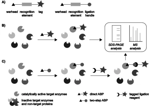
proteolytically cleaved in order to become catalytically active.7,8 Therefore, methods to quantify mRNA or protein levels would provide only limited information on the amount of active cysteine proteases, a parameter of great physiological relevance for these enzymes. To enable the profiling of enzymatic activity, ABPP approaches make use of chemical probes termed activity-based probes (ABPs) that are directed to the active site of an enzyme or enzyme family. These probes are designed to be recognized by a specific target protein and react in a mechanism-based manner with an active site residue, thereby labeling only the catalytically active form of the enzyme. The binding of the probe to the enzyme should be covalent and irreversible in order to facilitate the analysis of labeled proteins by for instance SDS-PAGE or mass spectrometry. A large number of ABPs that target many different enzyme classes has been developed over the past decades, enabling the selective labeling of enzymatic activity in complex biological samples such as cell lysates, cell cultures and sometimes even living organisms.1-3 Besides the monitoring of a specific enzymatic activity, ABPP strategies are also used to identify and characterize (unknown) protein functions, to study up- and down-regulation of enzymatic activity in various disease states, to discover and evaluate putative new enzyme inhibitors and to identify the protein targets of covalently binding natural products.1-6
11 structure of the recognition element and the reactivity of the warhead can be tuned to provide selective targeting of a specific enzyme or multiple members of an enzyme class. The third common feature of all ABPs is a means to visualize, isolate, identify and/or quantify the labeled proteins by for example SDS-PAGE, microscopy or mass spectrometry analysis. This functionality can either be a reporter group that enables the direct visualization and/or isolation of target enzymes (Figure 1.1B) or a reactive moiety that can be used to introduce the tag in a later stage (Figure 1.1C). Based on the nature of this third element ABPs are classified as either direct or two-step ABPs, respectively. Commonly used reporter entities for ABPP studies include fluorescent tags that enable visualization of the labeled proteins on gel and by fluorescence microscopy, affinity tags such as biotin for enrichment of labeled proteins and subsequent mass spectrometry analysis or detection by Western blotting, or a combination of these. Quenched fluorophores that only become fluorescent after binding of the ABP to a target enzyme can strongly reduce background labeling and thereby provide significant advantages for (in vivo) imaging applications.9 The three main elements of ABPs may be separated by additional linkers that can be used to minimize steric hindrance or modulate solubility and membrane-permeability of the probe.
12
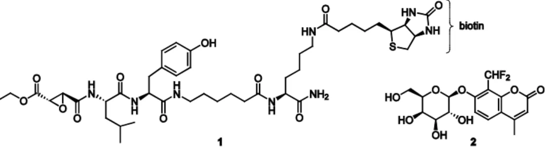
site of the enzyme. An example is the labeling of cysteine proteases using the biotinylated peptide epoxysuccinate DCG-04 (1) (Figure 1.2), which was shown to react specifically with a number of cysteine proteases of the cathepsin family in cell extracts.18 The epoxide moiety of the ABP, which functions as the warhead, is attacked by the catalytically active cysteine residue in the active site, resulting in opening of the epoxide ring and the formation of a stable covalent bond between the enzyme and the probe (Figure 1.3A).19 In cases where no covalent enzyme-substrate intermediate is formed, activity-based profiling can be achieved by applying suicide substrates that act via an indirect binding mechanism. For example, fluorogenic ABP 2 (Figure 1.2) was developed to report on the activity of β-galactosidases by reacting as shown in Figure 1.3B.20 This probe contains a latent quinone methide precursor which is released upon hydrolysis by a target enzyme. The highly reactive species that is formed can subsequently react with a nearby nucleophilic amino acid residue, yielding an immobilized fluorescent label. Although suicide substrates bind in a mechanism-based manner, the reactive species that is released may react with a nucleophile distant from the active site or even another component in the sample, so that this type of ABP does not specifically label the protein of interest. In this thesis the focus is on the first class of ABPs which directly modify a catalytically active amino acid residue. It should be kept in mind that the use of these probes results in the subsequent inhibition of catalytic activity and may thereby potentially disturb the system at hand, especially when applied to living organisms.
13
Figure 1.3. Examples of two mechanisms by which ABPs can bind to their target enzymes. A) Direct alkylation of the catalytically active amino acid residue: labeling of cysteine proteases by ABP 1. B) Targeting with suicide substrates: labeling of inverting β-galactosidases by fluorogenic ABP 2. ‘Nuc’: nucleophile.
1.2 Two-step activity-based probes
14
was first illustrated by the labeling of enzymatic activity in cell extracts with azide-functionalized sulfonate 3 followed by reaction with rhodamine-alkyne 4 and copper(I) as the catalyst (Figure 1.4C).24 A second example of a bioorthogonal ligation reaction is the Staudinger-Bertozzi ligation, in which an azide is again used as a ligation handle. In this case two-step labeling is achieved via selective reaction with a phosphine reagent (Figure 1.4B).25 This strategy has been applied to profile the activity of the proteasome β-subunits by treating cultured cells with azide-modified proteasome inhibitor 5 (Figure 1.4C) and labeling the target proteins after cell lysis with biotin-phosphine 6.26 A further description of the various bioorthogonal ligation techniques and their application in two-step ABPP is given in Chapter 2.
15
1.3 Probe design and selected examples
As a starting point for the design of novel ABPs, the structure of a natural substrate of the enzyme of interest can be used, which is then equipped with a tag and modified in such a way that reaction with the enzyme results in covalent and irreversible binding. Knowledge of the catalytic mechanism and/or reactivity towards particular electrophiles is essential. In many cases known mechanism-based inhibitors, in turn frequently based on natural substrates, are used as a scaffold to which a reporter entity is attached for analysis of the target proteins. In this respect, nature often provides a source of inspiration. For example, the structure of the potent biotin-tagged proteasome ABP 7 was derived from the naturally occurring irreversible proteasome inhibitor epoxomicin by extension of the N-terminus of the peptide epoxyketone with a biotinylated linker (Figure 1.5).27,28 The proteasome contains three catalytically active β-subunits that have different substrate preferences but each employ an N-terminal threonine residue as the active site nucleophile, a distinctive feature that is only found in a few other proteases. This particular aspect of the proteasome’s catalytic mechanism is exploited by the inhibitor to achieve selective targeting of this enzyme. The binding mechanism involves reaction of both the hydroxyl group and the primary amine of a catalytic threonine residue with the ketone and the epoxide of the inhibitor warhead, respectively, to form a stable morpholine ring (Figure 1.6A).29
16
Figure 1.6. Binding mechanisms of various classes of ABPs. Inhibition of the proteasome by epoxomicin derivatives (A), retaining β-glucosidases by cyclophellitol analogues (B) and deubiquitinating enzymes by ubiquitin-vinyl sulfone (C). Ub75: Ubiquitin lacking its C-terminal glycine residue.
Another example of ABP development guided by a mechanism-based inhibitor is based on the retaining β-glucosidase inhibitor cyclophellitol, which is itself a remarkable example of rational design coinciding with the isolation of the natural product.30-32 This mechanism-based inhibitor has served as a basis for the synthesis of direct and two-step ABPs (8 and 9, Figure 1.5) that selectively label the human enzyme glucocerebrosidase in cell extracts and cultured cells.33 Interestingly, the introduction of the hydrophobic fluorescent tag in probe 9 proved to strongly enhance its affinity as compared to the parent compound cyclophellitol. Irreversible binding of these probes is thought to occur by attack of the nucleophilic glutamic acid residue in the active site of retaining β-glucosidases on the epoxide moiety of the ABPs (Figure 1.6B).34 A second glutamic acid residue, which is essential for substrate hydrolysis by acting as a general acid/base, was shown to be also requisite for binding of the epoxide probes.33 In a later study, an aziridine-based ABP (10) was revealed to inhibit retaining β-glucosidases with enhanced potency and enabled in vivo profiling of enzymatic activity.35 However, labeling also occurred in the absence of the general acid/base residue in the active site, presumably due to the high reactivity of the aziridine moiety. As a result the probe is capable of labeling enzymes that lack catalytic activity on their natural substrate and therefore it may be considered not to be a ‘true’ ABP.
17 epoxysuccinate warhead, was modified by elaboration of the peptide portion to allow incorporation of an affinity tag.18
Besides inhibitor-inspired probe design, the structure of a substrate and its binding mechanism to the protein of interest can also serve directly as a basis for novel ABPs. This strategy was illustrated by the synthesis of ABPs based on the small (76 amino acids) protein ubiquitin to target deubiquitinating enzymes, a class of cysteine proteases that recognize a range of substrates modified with one or more ubiquitin units which are linked via their C -terminus to a lysine residue of another ubiquitin or to the protein substrate. The substitution of the C-terminal glycine residue of recombinant ubiquitin with a vinyl sulfone warhead and subsequent radioiodination led to an ABP that labels a number of deubiquitinating enzymes in cell extracts (Figure 1.6C).37 In later studies, other thiol-reactive electrophiles were attached to epitope-tagged ubiquitin to modify the reactivity of the probes towards specific deubiquitinating and ubiquitin-conjugating enzymes.38,39 These probes also exemplify the uncommon use of protein-derived ABPs rather than small molecule chemical probes, with a major drawback being that their large size excludes in situ and in vivo applications.
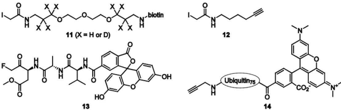
18
Figure 1.7. Structures of biotinylated (11) and alkyne-functionalized (12) iodoacetamide reagents and fluorescently labeled ABPs that target caspases (13) and deubiquitinating enzymes (14).
with low reactivity by using varying concentrations of the iodoacetamide reagent. While the cysteine residues with low reactivity are labeled in a concentration-dependent manner, labeling of the most reactive cysteine residues is equally efficient when a substoichiometric amount or an excess of the reagent is used. This strategy enables the annotation of highly reactive cysteines regardless of their natural abundance and can be used to identify new residues with putative catalytic activity.
In contrast to the iodoacetamide reagents, fluoromethylketone inhibitors and their derivatives, such as the fluorescein-functionalized ABP 13 (Figure 1.7),42 have a very low intrinsic reactivity towards cysteine residues because the fluoride is a very poor leaving group and the carbon-fluorine bond is much more stable than the carbon-iodine bond. In this case, selective activity-based labeling of a specific class of cysteine proteases, the caspases, was achieved by incorporation of a peptide into the ABP that specifically binds these enzymes and thereby brings the warhead in close proximity to the active site cysteine residue so that a reaction takes place.
19 contrast, the functionalization of larger peptide fragments of a natural caspase-1 substrate with analkyne warhead yielded selective caspase-1 ABPs.
1.4 Aim and outline of this thesis
Most of the ABPs that have been developed to date target hydrolytic enzymes, in particular proteases and esterases, that employ an amino acid residue (serine, threonine, cysteine) as the active site nucleophile. Novel ABPP strategies are increasingly more directed towards the development of ABPs that target other enzyme families, often with different catalytic mechanisms or low expression levels. At the same time the targeting of enzyme activities with tight substrate specificity and the interest in in vivo applications of ABPP necessitates the use of ABPs that are modified with a small ligation handle instead of a reporter group. Relatively few bioorthogonal ligation methods, however, have been applied to the two-step profiling of enzymatic activity. The first part of this thesis therefore aims to develop new bioorthogonal ligation methods for use in ABPP. In addition, the complexity of biological processes and the involvement of many proteins and other biomolecules in cellular processes and disease development often requires the study of multiple targets simultaneously, and therefore attention is also focused on tandem labeling strategies in which multiple ligation reactions are performed concurrently in a single experiment.
Chapter 2 gives an overview of the various bioorthogonal ligation methods that have been described in literature, with an emphasis on the application of bioorthogonal chemistry in ABPP. Frequently used ligation reactions include copper(I)-catalyzed as well as strain-promoted [2+3] cycloadditions of an azide with an alkyne, Staudinger-Bertozzi ligation of an azide with a phosphine, Diels-Alder reactions between electron-rich dienes and an electron-deficient dienophiles and inverse-electron-demand Diels-Alder reactions of tetrazine with strained alkenes (Figure 1.8).21
20
Figure 1.8. Reagents for commonly used bioorthogonal ligation reactions.
presented in which the Diels-Alder ligation is used together with the Staudinger-Bertozzi ligation for the labeling of multiple enzymatic activities in the same sample.
The inherent reactivity of maleimide towards cysteine residues poses severe limitations on its use in bioorthogonal labeling experiments. In order to improve the selectivity of the ligation reaction, an alternative two-step ABPP strategy that is based on the inverse-electron-demand Diels-Alder reaction of tetrazine with norbornene (Figure 1.8D) is investigated in Chapter 4. Labeling of proteasome activity is accomplished by using a norbornene-functionalized proteasome ABP and a fluorescently labeled or biotin-tagged tetrazine reagent. This approach enables the selective labeling of active proteasome β-subunits in vitro and in situ. Moreover, a triple ligation strategy involving tetrazine ligation, Staudinger-Bertozzi ligation and copper(I)-catalyzed click reaction is developed for the simultaneous labeling of three different proteolytic activities in a single experiment.
21 proteins on gel.45,46 In a later study another type of glycosidase ABPs was developed based on the irreversible retaining β-glucosidase inhibitor cyclophellitol (see Figure 1.5).33,35 In a comparative study the cyclophellitol derivatives proved to be superior to 2-deoxy-2-fluoroglucosides in terms of potency and labeling efficiency on two retaining β-glucosidases.46 The second part of this thesis describes the development of differently configured isomers of the cyclophellitol-based ABPs for the labeling of two mechanistically related glycosidases, retaining α- and β-galactosidases, for which at present no covalent and irreversible probes have been reported in literature.
Chapter 5 details the synthesis of galactopyranose-configured probes that contain an electrophilic aziridine or epoxide moiety in either an α- or a β-configuration47 with the aim to achieve activity-based profiling of retaining α- and β-galactosidases, respectively (Figure 1.9). Both direct ABPs functionalized with a fluorescent Bodipy tag or a biotin tag are synthesized as well as probes that are derivatized with an azide for two-step labeling experiments. The biological evaluation of these probes is presented in the following two chapters. In Chapter 6 the inhibitory potency of the α-configured epoxide- and aziridine-based probes (15 and 16, Figure 1.9) is evaluated on the recombinant human enzyme α-galactosidase A. The most potent compounds, the aziridines ABPs, are then used to visualize the active enzyme on gel. In addition, it is demonstrated that the fluorescently labeled aziridine ABP enables the labeling of endogenous retaining α-galactosidase activity in cell extracts. Chapter 7 describes the evaluation of the β-configured epoxide-based probes (17, Figure 1.9) for their ability to inhibit and label the recombinant enzyme galactocerebrosidase, a human retaining β-galactosidase. Furthermore, it is revealed that the small structural difference between the β-galactopyranose probe scaffold developed in this thesis and the previously reported β-glucopyranose-configured isomers (e.g. ABPs 8 and 9, Figure 1.5), namely only the stereochemistry of a single hydroxy substituent, has resulted in a dramatic change in selectivity. Consequently, the two differently configured ABPs allow the selective labeling of retaining β-galactosidases and retaining β-glucosidases, respectively, without any observed cross-reactivity.
22
9. Blum, G.; Von Degenfeld, G.; Merchant, M. J.; Blau, H. M.; Bogyo, M. Nat. Chem. Biol. 2007, 3, 668.
10. Geurink, P. P.; Florea, B. I.; Li, N.; Witte, M. D.; Verasdonck, J.; Kuo, C.-L.; van der Marel, G. A.; Overkleeft, H. S. Angew. Chem. Int. Ed. 2010, 49, 6802.
11. Speers A. E.; Cravatt, B. F. J. Am. Chem. Soc. 2005, 127, 10018.
12. Verhelst, S. H. L.; Fonovic, M.; Bogyo, M. Angew. Chem. Int. Ed. 2007, 46, 1284. 13. Dirksen, A.; Yegneswaran, S.; Dawson, P. E. Angew. Chem. Int. Ed. 2010, 49, 2023. 14. Yang, Y.; Hahne, H.; Kuster, B.; Verhelst, S. H. L. Mol. Cell. Proteomics 2013, 12, 237. 15. Gartner, C. A.; Elias, J. E.; Bakalarski, C. E.; Gygi, S. P. J. Proteome Res.2007, 6, 1482.
16. Hillaert, U.; Verdoes, M.; Florea, B. I.; Saragliadis, A.; Habets, K. L.; Kuiper, J.; van Calenbergh, S.; Ossendorp, F.; van der Marel, G. A.; Driessen, C.; Overkleeft, H. S. Angew. Chem. Int. Ed. 2009, 48, 1629.
17. Edgington, L. E.; Berger, A. B.; Blum, G.; Albrow, V. E.; Paulick, M. G.; Lineberry N.; Bogyo, M. Nat. Med. 2009, 15, 967.
18. Greenbaum, D.; Medzihradszky, K. F.; Burlingame, A.; Bogyo, M. Chem. Biol. 2000, 7, 569.
19. Matsumoto, K.; Mizoue, K.; Kitamura, K.; Tse, W.-C.; Huber, C. P.; Ishida, T. Biopolymers1999, 51, 99.
20. Kwan, D. H.; Chen, H.-M.; Ratananikom, K.; Hancock, S. M.; Watanabe, Y.; Kongsaeree, P. T.; Samuels, A. L.; Withers, S. G. Angew. Chem. Int. Ed. 2011, 50, 300.
21. For reviews see: a) Sletten, E. M.; Bertozzi, C. R. Acc. Chem. Res. 2011, 44, 666; b) Devaraj, N. K.; Weissleder, R. Acc. Chem. Res. 2011, 44, 816; c) Ngo, J. T.; Tirell, D. A. Acc. Chem. Res. 2011, 44, 677; d) Hao, Z.; Hong, S.; Chen, X.; Chen, P. R. Acc. Chem. Res.2011, 44, 742; e) Heal, W. P.; Tate, E. W. Org. Biomol. Chem. 2010, 8, 731; f) Willems, L. I.; van der Linden, W. A.; Li, N.; Li, K.-Y.; Liu, N.; Hoogendoorn, S.; van der Marel, G. A.; Florea, B. I.; Overkleeft, H. S. Acc. Chem. Res. 2011, 44, 718; g) Ramil, C. P.; Lin, Q. Chem. Commun. 2013, 49, 11007; h) Lang, K.; Chin, J. W. ACS Chem. Biol. 2014, 9, 16.
22. Rostovtsev, V. V. ; Green, L. G.; Fokin, V. V.; Sharpless, K. B. Angew. Chem. Int. Ed.2002, 41, 2596.
23. Wang, Q.; Chan, T. R.; Hilgraf, R.; Fokin, V. V.; Sharpless, K. B.; Finn, M. G. J. Am. Chem. Soc.2003, 125, 3192. 24. Speers, A. E.; Adam, G. C.; Cravatt, B. F. J. Am. Chem. Soc. 2003, 125, 4686.
25. Saxon, E.; Bertozzi, C. R. Science 2000, 287, 2007.
26. Ovaa, H.; van Swieten, P. F.; Kessler, B. M.; Leeuwenburgh, M. A.; Fiebiger, E.; van den Nieuwendijk, A. M. C. H.; Galardy, P. J.; van der Marel, G. A.; Ploegh, H. L.; Overkleeft, H. S. Angew. Chem. Int. Ed. 2003, 42, 3626. 27. Hanada, M.; Sugawara, K.; Kaneta, K.; Toda, S.; Nishiyama, Y.; Tomita, K.; Yamamoto, H.; Konishi, M.; Oki, T.
J. Antibiot. 1992, 45, 1746.
28. Sin, N.; Kim, K. B.; Elofsson, M.; Meng, L.; Auth, H.; Kwok, B. H. B.; Crews, C. M. Bioorg. Med. Chem. Lett. 1999, 9, 2283.
29. Groll, M.; Kim, K. B.; Kairies, N.; Huber, R.; Crews, C. M. J. Am. Chem. Soc. 2000, 122, 1237. 30. Caron, G.; Withers, S. G. Biochem. Biophys. Res. Commun. 1989, 163, 495.
23 32. Atsumi, S.; Umezawa, K.; Iinuma, H.; Naganawa, H.; Nakamura, H.; Iitaka, Y.; Takeuchi, T. J. Antibiot. 1990, 43,
49.
33. Witte, M. D.; Kallemeijn, W. W.; Aten, J.; Li, K.-Y.; Strijland, A.; Donker-Koopman, W. E.; van den Nieuwendijk, A. M. C. H.; Bleijlevens, B.; Kramer, G.; Florea, B. I.; Hooibrink, B.; Hollak, C. E. M.; Ottenhoff, R.; Boot, R. G.; van der Marel, G. A.; Overkleeft, H. S.; Aerts, J. M. F. G. Nat. Chem. Biol. 2010, 6, 907.
34. Gloster, T. M.; Madsen, R.; Davies, G. J. Org. Biomol. Chem. 2007, 5, 444.
35. Kallemeijn, W. W.; Li, K.-Y.; Witte, M. D.; Marques, A. R. A.; Aten, J.; Scheij, S.; Jiang, J.; Willems, L. I.; Voorn-Brouwer, T. M.; van Roomen, C. P. A. A.; Ottenhoff, R.; Boot, R. G.; van den Elst, H.; Walvoort, M. T. C.; Florea, B. I.; Codée, J. D. C.; van der Marel, G. A.; Aerts, J. M. F. G.; Overkleeft, H. S. Angew. Chem. Int. Ed. 2012, 51, 12529.
36. Barrett, A. J.; Kembhavi, A. A.; Brown, M. A.; Kirschke, H.; Knight, C. G.; Tamai. M.; Hanada, K. Biochem. J. 1982, 201, 189.
37. Borodovsky, A.; Kessler, B. M.; Casagrande, R.; Overkleeft, H. S.; Wilkinson, K. D.; Ploegh, H. L. EMBO J. 2001, 20, 5187.
38. Borodovsky, A.; Ovaa, H.; Kolli, N.; Gan-Erdene, T.; Wilkinson, K. D.; Ploegh, H. L.; Kessler, B. M. Chem. Biol.
2002, 9, 1149.
39. Love, K.; Pandya, R.; Spooner, E.; Ploegh, H. ACS Chem. Biol. 2009, 4, 275.
40. Gygi, S. P.; Rist, B.; Gerber, S. A.; Turecek, F.; Gelb, M. H.; Aebersold, R. Nat. Biotechnol. 1999, 17, 994. 41. Weerapana, E.; Wang, C.; Simon, G. M.; Richter, F.; Khare, S.; Dillon, M. B.; Bachovchin, D. A.; Mowen, K.; Baker,
D.; Cravatt, B. F. Nature 2010, 468, 790.
42. Bedner, E.; Smolewski, P.; Amstad, P.; Darzynkiewicz, Z. Exp. Cell Res. 2000, 259, 308.
43. Ekkebus, R.; van Kasteren, S. I.; Kulathu, Y.; Scholten, A.; Berlin, I.; Geurink, P. P.; De Jong, A.; Goerdayal, S.; Neefjes, J.; Heck, A. J.; Komander, D.; Ovaa, H. J. Am. Chem. Soc. 2013, 135, 2867.
44. Martell, J.; Weerapana, E. Molecules 2014, 19, 1378.
45. Vocadlo, D. J.; Bertozzi, C. R. Angew. Chem. Int. Ed. 2004, 43, 5338.
46. Witte, M. D.; Walvoort, M. T. C.; Li, K.-Y; Kallemeijn, W. W.; Donker-Koopman, W. E.; Boot, R. G.; Aerts, J. M. F. G.; Codée, J. D. C.; van der Marel, G. A.; Overkleeft, H. S. ChemBioChem2011, 12, 1263.
2
Bioorthogonal chemistry
2.1 Introduction
26
criteria of bioorthogonality.
2.2 Bioorthogonal chemistry: scope and limitations
27
Figure 2.1. Schematic representations of commonly used bioorthogonal ligation reactions. A) Ketone-hydrazide condensation. B,C) Reactions using an azide as one of the reagents: copper(I)-catalyzed Huisgen [3+2] azide-alkyne cycloaddition, strain-promoted azide-alkyne cycloaddition and Staudinger-Bertozzi ligation of an azide with a phosphine. D) Light-induced reaction of tetrazole with an alkene. E) Diels-Alder [4+2] cycloaddition of a conjugated diene with a dienophile. F) Inverse-electron-demand Diels-Alder reaction of tetrazine with a strained alkene.
28
29
Figure 2.2. Quenched Bodipy-tetrazine (1) that becomes strongly fluorescent after reaction with a dienophile (2). In synthetic organic chemistry the utility of chemical transformations is judged by their efficiency, so that at least the most elaborate or expensive reaction partner is transformed in (near) quantitative fashion, as well as their selectivity, so that unwanted side reactions are limited. Yield, the nature of (side) products and reaction kinetics are also parameters that should be considered when evaluating the merits of a bioorthogonal ligation. Whereas the kinetics of newly developed ligation reactions are frequently analyzed using isolated reagents, the efficiency of a particular ligation reaction is likely to be influenced by the nature of the biological sample, for example a cell extract or an intracellular environment, and the experimental conditions. A detailed study in which the copper(I)-catalyzed click reaction, strain-promoted click reaction and Staudinger-Bertozzi ligation were directly compared for the labeling of proteins in cell extracts and glycoproteins on live cell surfaces revealed that, as expected, the former reaction is superior in terms of efficiency but its use is restricted to in vitro experiments.9 The strain-promoted azide-alkyne cycloaddition and Staudinger ligation performed similarly well, with the efficiency of the latter reaction depending on the specific structure of the azide.
Staudinger-30
Figure 2.3. Staudinger-Bertozzi ligation in two-step labeling of proteasome activity. A) Structures of azide-functionalized fluorescent proteasome ABP 3 and biotin-phosphine 4. B) Strategy to evaluate the efficiency of the Staudinger-Bertozzi ligation using ABP 3 and phosphine 4. Ligation with the phosphine reagent results in a concentration-dependent gel-shift of the fluorescently labeled proteasome subunits. SDS-PAGE: sodium dodecyl sulfate polyacrylamide gel electrophoresis.
Bertozzi ligation, as compared to the corresponding lower-running bands. The ligation reaction was shown to reach completion, provided that a large excess of biotin-phosphine 4 was added relative to ABP 3. The poor yield in terms of conversion of reagent 4 is most likely due to the inherent instability of the trivalent phosphine, which has been shown to be susceptible to aerobic and metabolic oxidation.40
31
Figure 2.4. Reagents for two-step labeling of proteasome activity via strain-promoted click reaction. Structures of azide-functionalized fluorescent proteasome ABP 5 and biotin-functionalized cyclooctynes 6-8.
that most of the azide-independent labeling by cyclooctynes occurs via thiol-yne addition with cysteine residues.43 Consequently, while the use of functionalized cyclooctynes is an effective strategy for addressing cell surface azides, alkylation of free thiols appears to be requisite to limit background labeling when strain-promoted click chemistry is applied to complex biological samples such as cell extracts.
32
tetrazines display reactivity towards specific cyclooctynes,47,48 so that the proper reagents need to be carefully selected in order to minimize cross-reactivity. Related dual labeling strategies were used for cell surface glycan imaging in which ligation with tetrazine was accomplished by making use of cyclopropene or terminal alkene ligation handles instead of trans-cyclooctene.49-51 In addition, it has been demonstrated that properly substituted cyclopropenes enable orthogonal reactions with either tetrazines or nitrile imines52 and that the reaction of tetrazine with isonitriles is orthogonal to the azide-dibenzocyclooctyne cycloaddition,53 although in both cases no tandem bioorthogonal labeling experiments were performed. A triple ligation strategy that involves the copper(I)-catalyzed click reaction, the Staudinger-Bertozzi ligation and the inverse-electron-demand Diels-Alder reaction of tetrazine with norbornene was shown to enable the simultaneous labeling of three different enzymatic activities in cell extracts (Figure 2.6). Despite the fact that tetrazines revealed to be poorly compatible with click chemistry, the successive performance of these ligation reactions in the same sample proved to result in efficient and selective labeling of the intended target enzymes (see Chapter 4).54
33
Figure 2.6. Schematic representation of a triple ligation strategy involving copper(I)-catalyzed click reaction, Staudinger-Bertozzi ligation and tetrazine ligation for simultaneous labeling of three enzymatic activities.
2.3 Bioorthogonal chemistry in ABPP
34
which two-step labeling strategies appear to be indispensable is that of the exo-glycosidases, which typically have a sterically confined active site so that modification of a substrate (analogue) with a large tag is usually not tolerated. This limitation is likely responsible for the fact that few ABPs have been developed for this class of enzymes. The first ABPs that were reported to target exo-glycosidases are based on 2-deoxy-2- fluoroglycosides, mechanism-based retaining exo-glycosidase inhibitors,59 in which the primary hydroxyl is substituted with an azide to enable two-step labeling of target enzymes. For instance, retaining β-galactosidase inhibitor 11 served as a basis for the development of the two-step ABP 12 (Figure 2.8).60 Even though this small modification resulted in some loss of affinity, the probe retained sufficient affinity to enable labeling of target enzymes via Staudinger-Bertozzi ligation with a tagged phosphine reagent. In an alternative approach, ABPs for retaining β-glucosidases were designed by making use of the potent irreversible retaining β-glucosidase inhibitor cyclophellitol (13)61,62 which contains an epoxide moiety with which it covalently binds its target enzymes. Substitution of the primary hydroxyl with an azide led to an equally potent two-step ABP (14).63 This probe can be used to label recombinant retaining β-glucosidases via copper(I)-catalyzed azide-alkyne cycloaddition.64 Suprisingly, however, a direct ABP that was derived from 14 by installment of a Bodipy fluorophore (15) proved to label the human enzyme glucocerebrosidase with much higher potency than the azide-functionalized ABP.63 It was suggested that a hydrophobic pocket near the active site of the enzyme is responsible for tight binding of the hydrophobic fluorescent tag, thereby enhancing the affinity of the probe.
35
Figure 2.8.Structures of retaining β-galactosidase inhibitor 11 and azide-functionalized analogue 12, retaining β -glucosidase inhibitor cyclophellitol (13) and derivatives functionalized with an azide (14) or Bodipy fluorophore (15).
2.4 Cleavable linkers in ABPP
36
Figure 2.9. A) Structures of commonly used cleavable linker systems for the enrichment of proteins from biological samples. Cleavage sites are indicated by dashed lines. B) Mechanism of hydrazine-mediated cleavage of levulinoyl-based linker 21.
References
1. Part of this chapter has been published in: Willems, L. I.; Van der Linden, W. A.; Li, N.; Li, K.-Y.; Liu, N.; Hoogendoorn, S.; van der Marel, G. A.; Florea, B. I.; Overkleeft, H. S. Acc. Chem. Res. 2011, 44, 718.
2. Mahal, L. K.; Yarema, K. J.; Bertozzi, C. R. Science1997, 276, 1125. 3. Saxon, E.; Bertozzi, C. R. Science 2000, 287, 2007.
4. Huisgen, R. 1,3-Dipolar Cycloaddition Chemistry; Padwa, A., Ed.; Wiley-Interscience: New York 1984; Chapter 1, pp 1–176.
5. Tornøe, C. W.; Christensen, C.; Meldal, M. J. Org. Chem. 2002, 67, 3057.
6. Rostovtsev, V. V. ; Green, L. G.; Fokin, V. V.; Sharpless, K. B. Angew. Chem. Int. Ed. 2002, 41, 2596.
7. Wang, Q.; Chan, T. R.; Hilgraf, R.; Fokin, V. V.; Sharpless, K. B.; Finn, M. G. J. Am. Chem. Soc.2003, 125, 3192. 8. Speers, A. E.; Adam, G. C.; Cravatt, B. F. J. Am. Chem. Soc. 2003, 125, 4686.
9. Agard, N. J.; Baskin, J. M.; Prescher, J. A.; Lo, A.; Bertozzi, C. R. ACS Chem. Biol. 2006, 1, 644. 10. Agard, N. J.; Prescher, J. A.; Bertozzi, C. R. J. Am. Chem. Soc. 2004, 126, 15046.
11. Baskin, J. M.; Prescher, J. A.; Laughlin, S. T.; Agard, N. J.; Chang, P. V.; Miller, I. A.; Lo, A.; Codelli, J. A.; Bertozzi, C. R. Proc. Natl. Acad. Sci. USA 2007, 104, 16793.
37 14. Jewett, J. C.; Sletten, E. M.; Bertozzi, C. R. J. Am. Chem. Soc. 2010, 132, 3688.
15. Chang, P. V.; Prescher, J. A.; Sletten, E. M.; Baskin, J. M.; Miller, I. A.; Agard, N. J.; Lo, A.; Bertozzi, C. R. Proc. Natl. Acad. Sci. USA 2010, 107, 1821.
16. Debets, M. F.; van Berkel, S. S.; Schoffelen, S.; Rutjes, F. P. J. T.; van Hest, J. C. M.; van Delft, F. L. Chem. Commun. 2010, 46, 97.
17. Kuzmin, A.; Poloukhtine, A.; Wolfert, M. A.; Popik, V. V. Bioconjugate Chem. 2010, 21, 2076.
18. Dommerholt, J.; Schmidt, S.; Temming, R.; Hendriks, L. J. A.; Rutjes, F. P. J. T.; van Hest, J. C. M.; Lefeber, D. J.; Friedl, P.; van Delft, F. L. Angew. Chem, Int. Ed. 2010, 49, 9422.
19. van den Bosch, S. M.; Rossin, R.; Renart Verkerk, P.; Ten Hoeve, W.; Janssen, H. M.; Lub, J.; Robillard, M. S. Nucl. Med. Biol. 2013, 40, 415.
20. Debets, M. F.; van der Doelen, C. W. J.; Rutjes, F. P. J. T.; van Delft, F. L. ChemBioChem2010, 11, 1168. 21. Schultz, M. K.; Parameswarappa, S. G.; Pigge, F. C. Org. Lett. 2010, 12, 2398.
22. McKay, C. S.; Blake, J. A.; Cheng, J.; Danielson, D. C.; Pezacki, J. P. Chem. Commun. 2011, 47, 10040. 23. Song, W.; Wang, Y.; Qu, J.; Madden, M. M.; Lin, Q. Angew. Chem. Int. Ed. 2008, 47, 2832.
24. Song, W.; Wang, Y.; Qu, J.; Lin, Q. J. Am. Chem. Soc. 2008, 130, 9654.
25. Willems, L. I.; Verdoes, M.; Florea, B. I.; van der Marel, G. A.; Overkleeft, H. S. ChemBioChem 2010, 11, 1769. 26. Devaray, N. K.; Weissleder, R.; Hilderbrand, S. A. Bioconjug. Chem. 2008, 19, 2297.
27. Yang, J.; Seckute, J.; Cole, C. M.; Devaraj, N. K. Angew. Chem. Int. Ed. 2012, 51, 7476.
28. Patterson, D. M.; Nazarova, L. A.; Xie, B.; Kamber, D. N.; Prescher, J. A. J. Am. Chem. Soc. 2012, 134, 18638. 29. Devaray, N. K.; Upadhyay, R.; Haun, J. B.; Hilderbrand, S. A.; Weissleder, R. Angew. Chem. Int. Ed. 2009, 48,
7013.
30. Rossin, R.; Renart Verkerk, P.; van den Bosch, S. M.; Vulders, R. C. M.; Verel, I.; Lub, J.; Robillard, M. S. Angew. Chem. Int. Ed. 2010, 49, 3375.
31. Rossin, R.; van den Bosch, S. M.; Ten Hoeve, W.; Carvelli, M.; Versteegen, R. M.; Lub, J.; Robillard, M. S. Bioconjugate Chem. 2013, 24, 1210.
32. Neves, A. A.; Stöckmann, H.; Wainman, Y. A.; Kuo, J. C.-H.; Fawcett, S.; Leeper, F. J.; Brindle, K. M. Bioconjugate Chem. 2013, 24, 934.
33. Devaray, N. K.; Hilderbrand, S.; Upadhyay, R.; Mazitschek, R.; Weissleder, R. Angew. Chem. Int. Ed. 2010, 49, 2869.
34. Blackman, M. L.; Royzen, M.; Fox, J. M. J. A. Chem. Soc. 2008, 130, 13518.
35. Taylor, M. T.; Blackman, M. L.; Dmitrenko, O.; Fox, J. M. J. Am. Chem. Soc. 2011, 133, 9646. 36. Karver, M. R.; Weissleder, R.; Hilderbrand, S. A. Bioconjugate Chem. 2011, 22, 2263.
37. Stairs, S.; Neves, A. A.; Stöckmann, H.; Wainman, Y. A.; Ireland-Zecchini, H.; Brindle, K. M.; Leeper, F. J.
ChemBioChem 2013, 14, 1063.
38. Carlson, J. C. T.; Meimetis, L. G.; Hilderbrand, S. C.; Weissleder, R. Angew. Chem. Int. Ed. 2013, 52, 6917. 39. Verdoes, M.; Florea, B. I.; Hillaert, U.; Willems, L. I.; van der Linden, W. A.; Sae-Heng, M.; Filippov, D. V.;
Kisselev, A. F.; van der Marel, G. A.; Overkleeft, H. S. ChemBioChem2008, 9, 1735.
40. Lin, F. L.; Hoyt, H. M.; van Halbeek, H.; Bergman, R. G.; Bertozzi, C. R. J. Am. Chem. Soc. 2005, 127, 2686. 41. van der Linden, W. A.; Li, N.; Hoogendoorn, S.; Ruben, M.; Verdoes, M.; Guo, J.; Boons, G-J.; van der Marel,
G. A.; Florea, B. I.; Overkleeft, H. S. Bioorg. Med. Chem. 2012, 20, 662.
42. Berry, A. F.; Heal, W. P.; Tarafder, A. K.; Tolmachova, T.; Baron, R. A.; Seabra, M. C.; Tate, E. W. Chembiochem
2010, 11, 771.
43. van Geel, R.; Pruijn, G. J. M.; van Delft, F. L.; Boelens, W. C. Bioconjugate Chem. 2012, 23, 392.
38
53. Stairs, S.; Neves, A. A.; Stöckmann, H.; Wainman, Y. A.; Ireland-Zecchini, H.; Brindle, K. M.; Leeper, F. J.
ChemBioChem 2013, 14, 1063.
54. Willems, L. I.; Li, N.; Florea, B. I.; Ruben, M.; van der Marel, G. A.; Overkleeft, H. S. Angew. Chem. Int. Ed. 2012, 51, 4431.
55. Speers, A. E.; Adam, G. C.; Cravatt, B. F. J. Am. Chem. Soc. 2003, 125, 4686.
56. Ovaa, H.; van Swieten, P. F.; Kessler, B. M.; Leeuwenburgh, M. A.; Fiebiger, E.; van den Nieuwendijk, A. M. C. H.; Galardy, P. J.; van der Marel, G. A.; Ploegh, H. L.; Overkleeft, H. S. Angew. Chem. Int. Ed. 2003, 42, 3626. 57. Britton, M.; Lucas, M. M.; Downey, S. L.; Screen, M.; Pletnev, A. A.; Verdoes, M.; Tokhunts, R. A.; Amir, O.;
Goddard, A. L.; Pelphrey, P. M.; Wright, D. L.; Overkleeft, H. S.; Kisselev, A. F. Chem. Biol. 2009, 16, 1278. 58. Verdoes, M.; Willems, L. I.; van der Linden, W. A.; Duivenvoorden, B. A.; van der Marel, G. A.; Florea, B. I.;
Kisselev, A. F.; Overkleeft, H. S. Org. Biomol. Chem. 2010, 8, 2719. 59. Withers, S. G.; Rupitz, K.; Street, I. P. J. Biol. Chem.1988, 263, 7929. 60. Vocadlo, D. J.; Bertozzi, C. R. Angew. Chem. Int. Ed. 2004, 43, 5338.
61. Tatsuta, K.; Niwata, Y.; Umezawa, K.; Toshima, K.; Nakata, M. Tetrahedron Lett. 1990, 31, 1171.
62. Atsumi, S.; Umezawa, K.; Iinuma, H.; Naganawa, H.; Nakamura, H.; Iitaka, Y.; Takeuchi, T. J. Antibiot. 1990, 43, 49.
63. Witte, M. D.; Kallemeijn, W. W.; Aten, J.; Li, K.-Y.; Strijland, A.; Donker-Koopman, W. E.; van den Nieuwendijk, A. M. C. H.; Bleijlevens, B.; Kramer, G.; Florea, B. I.; Hooibrink, B.; Hollak, C. E. M.; Ottenhoff, R.; Boot, R. G.; van der Marel, G. A.; Overkleeft, H. S.; Aerts, J. M. F. G. Nat. Chem. Biol. 2010, 6, 907.
64. Witte, M. D.; Walvoort, M. T. C.; Li, K.-Y; Kallemeijn, W. W.; Donker-Koopman, W. E.; Boot, R. G.; Aerts, J. M. F. G.; Codée, J. D. C.; van der Marel, G. A.; Overkleeft, H. S. ChemBioChem2011, 12, 1263.
65. Shimkus, M.; Levy, J.; Herman, T. Proc. Natl. Acad. Sci. USA 1985, 82, 2593. 66. Gartner, C. A.; Elias, J. E.; Bakalarski, C. E.; Gygi, S. P. J. Proteome Res.2007, 6, 1482. 67. Verhelst, S. H. L.; Fonovic, M.; Bogyo, M. Angew. Chem. Int. Ed. 2007, 46, 1284. 68. Dirksen, A.; Yegneswaran, S.; Dawson, P. E. Angew. Chem. Int. Ed. 2010, 49, 2023.
69. Olejnik, J.; Sonar, S.; Krzymañska-Olejnik, E.; Rothschild, K. J. Proc. Natl. Acad. Sci. USA 1995, 92, 7590. 70. Szychowski, J.; Mahdavi, A; Hodas, J. J.; Bagert, J. D.;, Ngo, J. T.; Landgraf, P.; Dieterich, D. C.; Schuman, E. M.;
Tirrell, D. A. J. Am. Chem. Soc. 2010, 132, 18351.
71. Geurink, P. P.; Florea, B. I.; Li, N.; Witte, M. D.; Verasdonck, J.; Kuo, C.-L.; van der Marel, G. A.; Overkleeft, H. S. Angew. Chem. Int. Ed. 2010, 49, 6802.
72. Speers A. E.; Cravatt, B. F. J. Am. Chem. Soc. 2005, 127, 10018.
3
Two-step labeling of endogenous enzymatic
activities by Diels-Alder ligation
Lianne I. Willems, Martijn Verdoes, Bogdan I. Florea, Gijsbert A. van der Marel, Herman S. Overkleeft, ChemBioChem 2010, 11, 1769‐1781.
3.1 Introduction
40
proteins. The use of an azide-functionalized ABP and subsequent reaction with a biotin-phosphine reagent enabled labeling of active proteasome subunits and also facilitated the direct identification of a novel subunit-specific inhibitor.12,13
41
Figure 3.1. The Diels-Alder cycloaddition in activity-based protein profiling. Schematic representations of a two-step labeling strategy based on the Diels-Alder ligation (A) and the simultaneous labeling of two different catalytic activities in the same sample by successive Staudinger-Bertozzi ligation and Diels-Alder ligation (B). ABP: activity-based probe.
The validity of the Diels-Alder based ABPP strategy is evaluated by using the 20S proteasome as a model protein. This multi-subunit protein complex contains three catalytically active subunits (β1, β2 and β5) that each have a different substrate preference and can be targeted by either broad-spectrum or subunit-specific ABPs.13,31-34 In order to enable two-step labeling of the catalytically active proteasome β-subunits by Diels-Alder ligation, a panel of four diene-modified ABPs (1a-d) was designed as well as a maleimide-functionalized fluorescent Bodipy tag (3) (Figure 3.2). The ABPs are based on the potent broad-spectrum proteasome inhibitor epoxomicin,35,36 extended at the N‐terminus with either a linear (1b, 1c) or a cyclic (1a, 1d) conjugated diene. The maleimide moiety is a very reactive dienophile which has been used successfully for Diels-Alder based ligation procedures on recombinant proteins.26,27 Besides the labeling of proteasome activity, the Diels-Alder approach is also applied to the labeling of another class of enzymes, lysosomal cysteine proteases of the cathepsin family. For this purpose a diene-functionalized derivative of the cathepsin ABP DCG-0437 (2b) was designed.
42
Figure 3.2. Structures of target compounds: diene-functionalized proteasome ABPs 1a-d, cathepsin probe 2b and Bodipy-tagged maleimide 3.
3.2 Results and discussion
The synthesis of diene-derivatized proteasome ABPs 1a-d and Bodipy-tagged maleimide reagent 3 has been described elsewhere.38 The ability of the four epoxomicin derivatives 1a-d to inhibit the activity of the 20S proteasome was establishe1a-d by performing a competition experiment against the fluorescent activity-based proteasome probe MV15132 in El-4 cell extracts (Figure 3.3). El-4 cells contain next to the constitutive proteasome also the immunoproteasome, in which the β1, β2 andβ5 subunits are replaced byβ1i, β2i and β5i subunits having slightly different substrate preferences.39-41 All six catalytically active subunits are targeted by MV151. Exposure of El-4 cell extracts to increasing concentrations of 1a-d followed by labeling of residual proteasome activity with MV151 revealed that all probes are able to block the fluorescent labeling of the six proteasome β-subunits with
43 similar potency. This indicates that the epoxomicin derivatives, like epoxomicin itself, are covalent and irreversible inhibitors of the catalytically active proteasome β-subunits. Complete inhibition was observed at inhibitor concentrations of 0.5-1 μM and higher.
As a first evaluation of the applicability of the synthesized probes in Diels-Alder ligation procedures, the Diels-Alder reaction of diene-derivatized probe 1c with 1 equivalent of tagged maleimide 3 was performed in the absence of proteins. The reaction was monitored by LC/MS analysis at various time points (Figure 3.4), which demonstrated the formation of the predicted Diels-Alder adduct over time. Although significant product formation wasobserved after 24 hrs, the reaction did not reach complete conversion in 3 days. Considering the potential degradation of proteins in cell extracts when left for an extended period of time and in view of practical convenience, it was decided to use an overnight incubation for the ensuing Diels-Alder two-step labeling experiments in vitro with an excess of the dienophile to further promote product formation.
The feasibility of the Diels-Alder ligation strategy for two-step profiling of enzymatic activity was demonstrated next. Cell extracts were exposed for 1 hr to ABP 1c using a concentration at which it completely blocks the active proteasome β-subunits (1 μM) and subsequently exposed to varying concentrations of Bodipy-maleimide 3 (1 - 50 μM) overnight (Figure 3.5A). Since maleimide 3 is a Michael acceptor that reacts readily with (cysteine) thiol groups and may thereby cause a substantial amount of non-specific background labeling, the proteins were denaturated and cysteine residues were masked with Ellman’s reagent (5,5’-dithiobis-(2-nitrobenzoic acid), DTNB) after incubation with 1c and prior to addition of 3. In this protocol, a chloroform/methanol precipitation step is necessary to remove remaining thiol-containing reagents before the ligation reaction, which
44
Figure 3.5. Labeling of proteasome activity in cell extracts by Diels-Alder ligation. A) Labeling in EL-4 cell extracts by exposure to 1 μM of ABP 1c for 1 hr, denaturation, masking of cysteine residues and reaction with 1 - 50 µM of Bodipy-maleimide 3 overnight. B) Optimization of cysteine capping conditions by varying DTNB concentration (using 25 µM of 3). Gels are 12.5% SDS-PAGE with fluorescent readout followed by coomassie brilliant blue staining as a loading control. ‘ctrl’: control sample labeled with 1 µM MV151. ‘ep’: 100 μM epoxomicin added to incubation with
1c. ‘M’: protein marker.
simultaneously provides the opportunity to redissolve the sample in a buffer optimal for Diels-Alder ligation (pH 6).26,27 A second precipitation step was used to quench the reaction and remove the excess of maleimide-tag 3. Analysis of the labeled proteins on gel (Figure 3.5A) reveals, despite a considerable degree of background fluorescence, the specific fluorescent labeling of a number of bands which correspond to the proteasome β-subunits as labeled by MV151. Background fluorescence was not affected by the presence or absence of 1c, confirming that it results from non-specific reactions of the maleimide-functionalized tag (3) rather than the inhibitor. In line with this observation, the concentration of cysteine capping reagent (DTNB) proved to be of crucial importance for the amount of background labeling, with an optimum around 50 mM (Figure 3.5B). The observed reduction in specific labeling when using a higher DTNB concentration is possibly due to an insufficient removal of the excess of reagent by a single precipitation step, causing it to interfere with the subsequent ligation reaction.
45
Figure 3.6. Diels-Alder based proteasome labeling in cell extracts using ABPs 1a-d. EL-4 cell lysates were exposed to 1 μM of 1a-d in the presence or absence of 100 μM epoxomicin for 1 hr, followed by denaturation, masking of cysteine residues and labeling with 25 μM Bodipy-maleimide 3 overnight. Labeled proteins were resolved by 12.5% SDS-PAGE with fluorescent readout followed by coomassie brilliant blue staining. ‘M’: protein marker.
indeed reflect labeling of the catalytically active proteasome β-subunits. The ligation reaction proceeded most efficiently with the non-cyclic dienes in 1c and especially 1b. The lower efficiency of furyl-derivative 1a is somewhat surprising as this diene is fixed in the cis -configuration which these reactants need to adopt for the Diels-Alder cycloaddition.
Using the most reactive heptadienoyl-functionalized ABP 1b, efforts were then made to further improve the efficiency of the Diels-Alder ligation by varying the concentration of dienophile (Figure 3.7A) and the reaction time of the ligation step (Figure 3.7B). Maleimide concentrations between 10 and 25 μM appeared to give the best results in terms of signal intensity and ratio of specific to background fluorescence. Both non-specific and specific labeling were also dependent on the reaction time, giving the best results when Diels-Alder ligation was performed with 25 µM of maleimide 3 for 4 - 20 hrs. After overnight incubation the signal intensity of proteasome labeling was similar to that obtained by direct labeling with fluorescent ABP MV151. Hence, a reaction time of 20 hrs and a dienophile concentration of 25 μM were selected for the following labeling experiments.
46
Figure 3.7. Optimization of Diels-Alder labeling procedure in EL-4 cell extracts by exposure to 1 μM of ABP 1b for 1 hr, denaturation, masking of cysteine residues and reaction with A) 1 - 50 µM of reagent 3 overnight, or B) 25 µM 3
for 1 - 20 hrs. Gels are 12.5% SDS-PAGE with fluorescent readout followed by coomassie brilliant blue staining. ‘ctrl’: control sample labeled with 1 µM MV151. ‘ep’: 100 μM epoxomicin added to incubation with 1b. ‘M’: protein marker.
subjected to the Diels-Alder labeling procedure with maleimide 3 (Figure 3.8B). Analysis of the fluorescent labeling on gel revealed the specific labeling of several bands that are not present in lysates from untreated cells and correspond to the proteasome β-subunits labeledby MV151. These results demonstrate that the Diels-Alder method can be applied to monitor enzymatic activity in living cells by in situ targeting with a diene-functionalized ABP and post-lysis labeling with a tagged maleimide reagent.
47 Having established the applicability of the Diels-Alder strategy for two-step labeling of proteasome activity, attention was focused on using the approach to label a class of less abundant enzymes, cysteine proteases of the papain family (cathepsins). For this purpose a heptadienoyl-functionalized derivative of the irreversible cathepsin inhibitor DCG-0437 was synthesized (2b, Scheme 3.1). Standard solid-phase peptide chemistry was employed to obtain resin-bound peptide 4. The epoxysuccinate warhead 9 was synthesized from D-(-)-diethyl tartrate (7) by using a slightly modified literature procedure.42 Briefly, the cis-diol was converted to a racemic mixture of trans-β-chloro alcohols which was then treated with DBU for epoxide formation (8), after which selective saponification of one of the ethyl esters afforded epoxysuccinate 9. Next, deprotection of the N‐terminal amine in peptide 4 followed by condensation with 9 and cleavage from the resin with concomitant deprotection provided ethyl ester 5 as well as a side product in which the ester was hydrolyzed (6). The obtained mixture of peptides was not readily separable due to the presence of a free amine in both of the compounds and was therefore used as such for the final step, reaction of the lysine ε-amine in peptides 5 and 6 with succinimidyl ester 10.38 The two products could then easily be separated to provide diene-derivatized DCG-04 analogue 2b.
Scheme 3.1 Synthesis of diene-functionalized DCG-04 derivative 2b
48
activity, two bands are visible only in ABP-treated samples which thus represent proteins that are specifically labeled by means of the two-step Diels-Alder ligation procedure. These bands correspond to the proteins that are labeled by N3-Bodipy(TMR)-DCG-04. Moreover, the fluorescent labeling was blocked by adding an excess of DCG-04, which confirms the specific labeling of catalytically active cathepsins. On the basis of the apparent molecular weights and a previously reported DCG-04 labeling profile in RAW cell extracts,44 the labeled proteases are tentatively assigned cathepsin Z and cathepsin B.
49 In a final experiment the possibility was investigated to monitor different proteolytic activities in a single sample by combining Diels-Alder two-step labeling with the Staudinger-Bertozzi ligation, another widely used bioorthogonal ligation reaction. For this purpose, the azide-derivatized proteasome probe 1145 was employed, which selectively targets the β1 proteasome subunits and enables labeling via Staudinger-Bertozzi ligation with biotin-tagged phosphine 1246 (Figure 3.10). At the same time the β2 and β5 subunits can then be labeled by Diels-Alder ligation. The dual labeling procedure was performed in HEK cell extracts, which contain only the three constitutive active proteasome subunits β1, β2 and β5. Cell extracts were first exposed to azide-modified ABP 11 using a concentration at which it completely blocks β1 activity, and then to diene-equipped probe 1b to target the non-occupied subunits β2 and β5. Next, Staudinger-Bertozzi ligation was effected by treatment with biotin-phosphine 12, after which denaturation, cysteine masking and Diels-Alder reaction with fluorescent maleimide 3 were performed as before. As shown in Figure 3.10, ligation with maleimide 3 in samples treated with both 1b and 11 resulted in the exclusive fluorescent labeling of the diene-modified subunits β2 and β5, while Staudinger ligation with phosphine 12 gave selective labeling of the β1 subunit on Western blot. Importantly, no cross-reactivity was observed and the labeling efficiency obtained by combining both ligation methods in the same sample was comparable to that of the separate ligation reactions. This experiment shows that the Diels-Alder ligation and Staudinger-Bertozzi ligation are mutually orthogonal and can be combined in the same sample to monitor different enzymatic activities.
50
with maleimide 3 for proteasome labeling in vitro. The Diels-Alder approach is fully orthogonal with respect to the Staudinger-Bertozzi ligation and both reactions can be performed in the same sample, providing the possibility to modify two different biomolecules, for example two enzymatic activities or metabolites, with different labels to study them simultaneously. Limitations of the Diels-Alder procedure are the need to mask cysteine residues prior to dienophile addition which precludes in vivo applications, and significant background labeling by the maleimide reagent which might hamper the detection of proteins with low abundance or endogenous activity. The broad use of the Diels-Alder reaction as a bioorthogonal ligation method therefore requires a dienophile that is unreactive towards cysteine residues and other functionalities present in biological samples. During the completion of the labeling experiments described in this chapter, several related studies were reported on the in situ and in vivo imaging of small molecules by inverse-electron-demand Diels-Alder reaction.48,49 The use of tetrazine as a diene and a strained alkene as a dienophile afforded very fast and selective labeling reactions and allowed the ligations to be performed in living cells. The application of a tetrazine ligation strategy for the profiling of endogenous enzymatic activities is described in Chapter 4.
Experimental procedures
A. Synthesis General
51 system (Thermo Finnigan) equipped with a C18 column (Gemini, 4.6 mm x 50 mm, 5μm particle size, Phenomenex). The applied buffers were A: H2O, B: ACN and C: 1 % aqueous TFA. Reported gradients represent the percentage of buffer B in buffer A with 10% buffer C. HRMS analysis was performed on an LTQ Orbitrap (Thermo Finnigan) mass spectrometer equipped with an electronspray ion source in positive mode (source voltage 3.5 kV, sheath gas flow 10 mL min−1, capillary temperature 250 °C) with resolution R = 60000 at m/z 400 (mass range m/z = 150 - 2000) and dioctylphtalate (m/z = 391.28428) as a "lock mass". The high resolution mass spectrometer was calibrated prior to measurements with a calibration mixture (Thermo Finnigan). 1H- and 13C-APT-NMR spectra were recorded on a Jeol JNM-FX-200 (200/50) or Bruker AV-400 (400/100 MHz). Chemical shifts are given in ppm (δ) relative to the solvent peak or to tetramethylsilane as internal standard. Coupling constants (J) are given in Hz. All presented 13 C-APT spectra are proton decoupled. Peak assignments are based on 2D 1H-COSY and 13C-HSQC NMR experiments. Compounds 1a-d, 3 and 10 were described previously.38
(2S,3S)-diethyl oxirane-2,3-dicarboxylate (8)
D-(-)-diethyl tartrate (7) (29 g, 0.14 mol) was cooled to 0 °C, before a solution of 33% HBr in acetic acid (120 mL) was added dropwise over 45 min. After complete addition, the reaction mixture was stirred at 0 °C for 15 min and then at room temperature overnight. Next, the mixture was poored onto crushed ice/H2O (300 mL) and extracted with Et2O (3x). The combined organics were washed with H2O (3x), dried over anhydrous MgSO4, filtered and concentrated in vacuo. Remaining solvents were concentrated in the presence of toluene. The crude oil was dissolved in EtOH and acetyl chloride (5.1 mL, 70 mmol, 0.5 eq.) was added. The reaction mixture was stirred under reflux for 3.5 hrs, before being concentrated at a temperature of 30 °C. The remaining yellowish oil was dissolved in Et2O (175 mL), cooled to 0 °C and put under argon atmosphere. A solution of DBU (21 mL, 0.14 mol, 1.0 eq.) in Et2O (90 mL) was added dropwise over 100 min. The reaction mixture was then stirred at 0 °C for 1 hr, more DBU (2.1 mL, 14 mmol, 0.1 eq.) was added and the reaction mixture was stirred for an additional 2 hrs, before being quenched with H2O. The mixture was washed with 1M KHSO4 solution and H2O and the organic layer was dried over anhydrous MgSO4, filtered and concentrated in vacuo. Purification by column chromatography (PetEt → 15% EtOAc in PetEt) yielded title compound 8 (15 g, 79 mmol, 56% over 3 steps). 1H NMR (400 MHz, CDCl
3): δ (ppm) 4.32-4.22 (m, 4H), 3.66 (s, 2H), 1.32 (t, J = 7.15, 7.15 Hz, 6H). 13C NMR (100 MHz, CDCl
3): δ (ppm) 166.69, 62.14, 51.96, 13.96.
(2S,3S)-3-(ethoxycarbonyl)oxirane-2-carboxylic acid (9)
A solution of compound 8 (14 g, 76 mmol) in absolute EtOH (200 mL) was cooled to 0 °C and a solution of KOH (5.0 g, 76 mmol, 1.0 eq.) in absolute EtOH (100 mL) was added dropwise over 20 min. Next, the reaction mixture was stirred at 0 °C for 3 hrs and then at room temperature for 2 hrs, before being concentrated in vacuo. H2O (200 mL) was added to the residue and the basic aqueous mixture was washed with DCM (1x 30 mL). The aqueous layer was then acidified with concentrated HCl (7.0 mL), NaCl (60 g) was added and the mixture was extracted with EtOAc (4x 200 mL). The combined organics were dried over anhydrous MgSO4, filtered and concentrated in vacuo to give title compound 9 (11 g, 66 mmol, 86%). 1H NMR (400 MHz, CDCl
3): δ (ppm) 6.24 (bs, 1H), 4.33-4.20 (m, 2H), 3.74-3.61 (m, 2H), 1.32 (t, J = 7.10, 7.10 Hz, 3H). 13C NMR (100 MHz, CDCl
3): δ (ppm) 168.54, 166.92, 61.95, 51.49, 51.42, 13.38.
MBHA-Rink amide-Lys(Boc)-Ahx-Tyr(tBu)-Leu(Fmoc) (4)
52
(2S,3S)-3-(ethoxycarbonyl)oxirane-2-carboxyl-Leu-Tyr-Ahx-Lys.TFA (5)
Resin-bound compound 4 (~0.90 mmol) was deprotected with 20% piperidine in NMP for 30 min. The resin was washed with NMP (2x) and DCM (3x) before being subjected to a condensation cycle with free acid 9 (0.36 g, 2.3 mmol, 2.5 eq.) in the presence of BOP (1.0 g, 2.3 mmol, 2.5 eq.) and DiPEA (0.45 mL, 2.7 mmol, 3.0 eq.) in NMP; the reaction mixture was shaken overnight, followed by washing with NMP (3x) and DCM (3x). The condensation cycle was repeated after which the Kaiser test indicated complete coupling. Cleavage from the resin was then accomplished by treatment with TFA/TIS/H2O (95/2.5/2.5, v/v/v) for 2 hrs at room temperature. After filtration and concentration in vacuo in the presence of toluene, the residue was recrystallized first from acetone/MeOH/EtOAc and then from MeOH/Et2O, yielding a 2:1 mixture of fully deprotected ester 5 and free acid 6 (total yield 0.73 g, 0.93 mmol, quant.) according to NMR analysis. LC/MS analysis: Rt 5.0 min (linear gradient 10 → 90% B in 15 min), m/z 677.4 [M+H]+, 1353.2 [2M+H]+. 1H NMR (400 MHz, MeOD): δ (ppm) 6.98 (d, J = 7.87 Hz, 2H), 6.66 (d, J = 7.76 Hz, 2H), 4.44 (t, J = 7.23, 7.23 Hz, 1H), 4.37 (dd, J = 7.59, 5.82 Hz, 1H), 4.30 (dd, J = 7.96, 5.15 Hz, 1H), 4.26-4.16 (m, 1H), 3.79-3.61 (m, 1H), 3.61-3.46 (m, 1H), 3.17-3.07 (m, 2H), 3.06-2.98 (m, 2H), 2.98-2.91 (m, 2H), 2.91-2.73 (m, 2H), 2.20 (t, J = 6.87, 6.87 Hz, 2H), 1.66-1.63 (m, 2H), 1.59-1.31 (m, 9H), 1.26 (t, J = 6.99, 6.99 Hz, 3H), 1.23-1.13 (m, 2H), 0.88 (d, J = 5.77 Hz, 3H), 0.84 (d, J = 5.75 Hz, 3H).
(2S,3S)-3-(ethoxycarbonyl)oxirane-2-carboxyl-Leu-Tyr-Ahx-((N)-(E)-hepta-4,6-dienoyl)Lys-H2N (2b)
A mixture of 5 and 6 (0.31 g, 0.39 mmol, non-hydrolyzed/hydrolyzed 2/1) was dissolved in DCE/DMF under argon atmosphere and made basic (pH 8.5) using DiPEA (0.13 mL, 0.78 mmol, 2.0 eq.), before a solution of OSu-ester 10
(0.23 g, 1.0 mmol, 2.6 eq.) in DCE/DMF was added. After stirring overnight at room temperature under argon atmosphere, the reaction mixture was concentrated in vacuo. The residue was taken up in MeOH/acetone and the soluble fraction was purified by column chromatography (CHCl3 → 10% MeOH in CHCl3), yielding diene-modified title compound 2b (0.16 g, 0.20 mmol, 76% from non-hydrolyzed starting material 7). 1H NMR (400 MHz, dmso-d6): δ (ppm) 8.54 (d, J = 8.19 Hz, 1H), 8.11 (d, J = 8.24 Hz, 1H), 7.82-7.74 (m, 2H), 7.30 (s, 1H), 6.96 (d, J = 8.40 Hz, 2H), 6.61 (d, J = 8.32 Hz, 2H), 6.28 (td, J = 17.03, 10.26, 10.26 Hz, 1H), 6.04 (dd, J = 15.16, 10.53 Hz, 1H), 5.74-5.64 (m, 1H), 5.02 (dd, J = 50.10, 13.54 Hz, 2H), 4.37-4.28 (m, 2H), 4.23-4.08 (m, 3H), 3.73-3.69 (m, 1H), 3.62-3.57 (m, 1H), 3.07-2.88 (m, 4H), 2.82 (dd, J = 13.69, 5.57 Hz, 1H), 2.67 (dd, J = 13.64, 8.85 Hz, 1H), 2.26 (dd, J = 14.23, 7.06 Hz, 2H), 2.17-2.05 (m, 4H), 1.59 (td, J = 9.47, 6.89, 6.89 Hz, 1H), 1.54-1.26 (m, 12H), 1.23 (t, J = 7.10, 7.10 Hz, 3H), 1.20-1.12 (m, 2H), 0.83 (dd, J = 14.79, 6.46 Hz, 6H).
Diels-Alder reaction of maleimide-tag 3 with diene-functionalized probe 1c
![Figure 1.4. A) Copper(I)-catalyzed azide-alkyne [2+3] cycloaddition. B) Staudinger-Bertozzi ligation](https://thumb-us.123doks.com/thumbv2/123dok_us/8173051.2166781/15.722.76.637.553.864/figure-copper-catalyzed-alkyne-cycloaddition-staudinger-bertozzi-ligation.webp)