Original Research Article
A study of hearing improvement gained after tympanoplasty using
various methods in cases of CSOM
Faiz Muqtadir, Rahul S.*
INTRODUCTION
Among the several sensory perceptions nature has endowed upon most of the living creatures the importance of sense of hearing, cannot be over emphasized. A person deprived of hearing (or suffering from hearing loss) feels terribly handicapped, isolated, and ineffective in communication with others. It should be noted that this ability is susceptible to pathology that causes hearing impairment may also end up causing hearing disability.1
Chronic otitis media (COM) is a chronic inflammatory disease of the middle ear and mastoid that often results in partial or total loss of the tympanic membrane (TM) and
ossicles, leading to conductive hearing loss that can range in severity up to 60 dB. COM is a common condition seen in patients attending the otolaryngology clinic and is an important public health problem with substantial economic and societal costs, affecting 0.5–30% of the community. A conservative estimate of the no. of people in the world suffering from COM is over 20 million.1 Among the various causes of ear diseases, COM is a major global cause of hearing impairment and may have serious long term effects on language, auditory, cognitive development and educational progress. As per WHO, the
prevalence of COM in Indian population is
approximately 2% which is comparatively higher than that found in developed countries like that of USA and UK where the prevalence is <1%.2 Majority of the
ABSTRACT
Background: Chronic otitis media (COM), is a common ailment with which patients present to the ENT
OPD. In patients with COM, the active infection needs to be controlled following which the definitive line of management is surgery. Tympanoplasty is the surgery performed with the goals of establishing an intact tympanic membrane, eradicating middle ear disease, creating an air-containing middle ear space and restoring the hearing by building a secure connection between the ear drum and the cochlea.
Methods: 50 patients presenting with CSOM in ENT OPD at Medical College. Preoperative audiometry was done
before tympanoplasty, followed by post-operative audiometry after 3 months.
Results: The mean air-bone gap closure was 12.06 dB; with type I tympanoplasty with cortical mastoidectomy giving
a maximum mean improvement of 16db. Minimum mean improvement of 1db was seen in type IV with modified radical mastoidectomy.
Conclusions: Type I tympanoplasty with cortical mastoidectomy is a far superior method of surgical treatment of CSOM than all other methods.
Keywords: Tympanoplasty, CSOM, Mastoidectomy
Department of ENT, ESIC Medical College, Gulbaraga, Karnataka, India
Received: 19 September 2017
Revised: 08 October 2017
Accepted: 09 October 2017
*Correspondence: Dr. Rahul S.,
E-mail: [email protected]
Copyright: © the author(s), publisher and licensee Medip Academy. This is an open-access article distributed under
the terms of the Creative Commons Attribution Non-Commercial License, which permits unrestricted non-commercial use, distribution, and reproduction in any medium, provided the original work is properly cited.
patients with COM do well with antimicrobial therapy but despite this there is a subset of the patients who
develop serious intratemporal and intracranial
complications from this otherwise self-limiting disease; and the mortality rate of these remains substantial ranging from 10–31%. The diagnosis of COM needs to be made earlier in order to prevent its long-term effects especially on hearing impairment.
With the advent of antibiotics, use of operative microscope by ENT surgeons, there has been a tremendous improvement in control and eradication of ear diseases. Several therapeutic practices and regimens have evolved to treat COM, but the surgical management –tympanoplasty– has remained the ultimate choice.3,4
To determine the degree of success in restoration of hearing (or lack of it) following tympanoplasty, several evaluation techniques are in vogue, both pre and post operatively. The autologous Temporalis Muscle Fascia and conchal cartilage was used.2 The patients were
audiologically assessed preoperatively and
post-operatively for the hearing status, with focus on audiometric evaluation of the ABG (air–bone gap).
It emerges from our analysis that a sizable number of patients show improvement postoperatively. This study attempts to document and analyse different methods of
tympanoplasty in COM and their effectiveness
postoperatively.
METHODS
50 Patients presenting with CSOM in ENT OPD at Medical College, Bangalore between October 2010 and September 2011 formed the study subjects
Percentage, mean and standard deviation are the statistical tests used in this study
Pre-op preparation
Preparation of the patients, shaving of hair of post auricular region 3 cm inside the hair line done.
Xylocaine test dose given– 0.1 ml of 2% xylocaine intradermally given, vital parameters were recorded and Informed consent was taken. Pre-op dose of antibiotic was given.
Position of the patient– supine with the face turned to one side the ear to be operated is up.
Anesthesia– local or general. Local infiltration is done with 2% lignocaine with 1:2,00,000 adrenaline. Incision may be endomeatal, endaural or postaural.
Harvesting of the temporalis fascia graft done. Other grafting material like tragal perichondrium or fascialata
Tympanic membrane is visualized. Freshening of the perforation margins done using curved pick. Curetting of the undersurface of tympanic membrane done. 6O`clock and 12O`clock incision was taken about 5 mm away fr om the annulus. The tympanomeatal flap is elevated and middle ear is inspected, status of ossicles noted.
Round window reflex is visualized and continuity of ossicular status confirmed. Graft placement is done.
Repositioning of the tympanomeatal flap is done. Gelfoam soaked with betadine is placed in the external canal. Periosteum, subcutaneous tissue and skin are sutured and mastoid dressing is done. Patient is put on antibiotics and analgesics. Suture removal is done after 1 week. Patient is followed up postoperatively at regular interval.
To eradicate disease from both the mastoid and middle ear cavity tympanoplasty can be combined with mastoidectomy. Cortical mastoidectomy is exenteration of all accessible mastoid air cells preserving the posterior meatal wall. Modified radical mastoidectomy is eradication of disease of the attic and mastoid, both of which are exteriorized into the external auditory canal by removal of posterior meatal and lateral attic wall.
Surgical procedures
A. Type I tympanoplasty
B. Type I tympanoplasty with cortical mastoidectomy
C. Type II tympanoplasty with cortical mastoidectomy
D. Type III tymapnoplasty with cortical mastoidectomy
E. Type III tympanoplasty with modified radical
mastoidectomy
F. Type IV tympanoplasty with modified radical
mastoidectomy.
For all above procedures post aural approach with William Wildes incision was used. All the patients underwent tympanoplasty. TM grafting was done using TMF employing underlay technique.
A. Type I Tympanoplasty done for patients with CSOM
of tubo tympanic variety (PPS) with central perforation.
B. Cortical mastoidectomy with type I tymapnoplasty
repositioned. External auditory canal was packed with medicated gel foam and gauze pack. Post aural incision was closed in layers using 3-0 vicryl. Post aural dressing was done and mastoid bandage was applied.
C. Cortical mastoidectomy with type III tympanoplasty The surgical approach to the middle ear is same as I AND II. After inspecting the middle ear, depending upon the type of ossicular destruction type III tympanoplasty was done using TORP or PORP, for conservation of hearing. The most common defect was erosion of the lenticular process of Incus. Sometimes destruction of the Stapes superstructure was also seen. The TMF was used for grafting TM by underlay technique.
D. Modified radical mastoidectomy with type III
tympanoplasty with the same approach as described previously, the mastoid antrum and air cell system, Aditus Ad Antrum, attic and middle ear are converted into a single cavity. The facial bridge is lowered and also the facial ridge, thus exteriorizing the cavity. Tympanoplasty was carried out at the same sitting. The TMF was grafted by underlay technique.
Post op management
1. Mastoid dressing was done for all patients and kept up to seven days
2. IV antibiotic was given for seven days
3. Sutures were removed on the seventh day.
4. Patients were reviewed once a week.
5. Aural pack was removed on the tenth day, graft visualized, topical ear drops were advised. Patients were followed up to three months.
6. All patients underwent post-operative PTA to assess
the improvement in hearing.
RESULTS
26 right ears and 24 left ears were operated in our study, based on the more severe hearing loss as measured by pre-operative PTA (Table 1).
Table 1: Laterality of surgery.
Operated ear Number
Right 26
Left 24
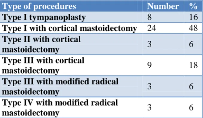
Almost half the patients underwent type I with CM. 8 and 9 cases had type I tympanoplasty and type III with CM respectively. The other procedures were divided equally among the rest of the patients (Table 2).
Table 2: Procedures performed.
Type of procedures Number %
Type I tympanoplasty 8 16
Type I with cortical mastoidectomy 24 48
Type II with cortical
mastoidectomy 3 6
Type III with cortical
mastoidectomy 9 18
Type III with modified radical
mastoidectomy 3 6
Type IV with modified radical
mastoidectomy 3 6
Table 3: Hearing improvement gained.
Improvement (db) Grade No. of cases
<5 None 16
5-10 Minimal 3
10-15 Satisfactory 11
>15 Good 20
20 patients showed a good hearing improvement with a maximum gain of 30 db. 16 patients showed no improvement, while hearing improvement between 5db to 15 db was seen in 14 patients (Table 3).
Table 4: Comparison of post op ABG. Post-op ABG Number of cases Percentage (%)
>20 13 26
<20 37 74
Table 5: Correlation of hearing improvement with procedure.
Procedure None Minimal Satisfactory Good
Type I tympanoplasty 3 2 1 2
Type I with cortical mastoidectomy 3 0 7 14
Type II with cortical mastoidectomy 1 0 1 1
Type III with cortical mastoidectomy 4 1 1 3
Type III with modified radical mastoidectomy 2 0 1 0
Type IV with modified radical mastoidectomy 3 0 0 0
At 3 months post-operative status, almost 75% patients showed an ABG of less than 20 dB (Table 4).
A graph is plotted to compare the degree of improvement in hearing loss, measured by analyzing Air-Bone Gap by pure tone audiometry (Table 5).
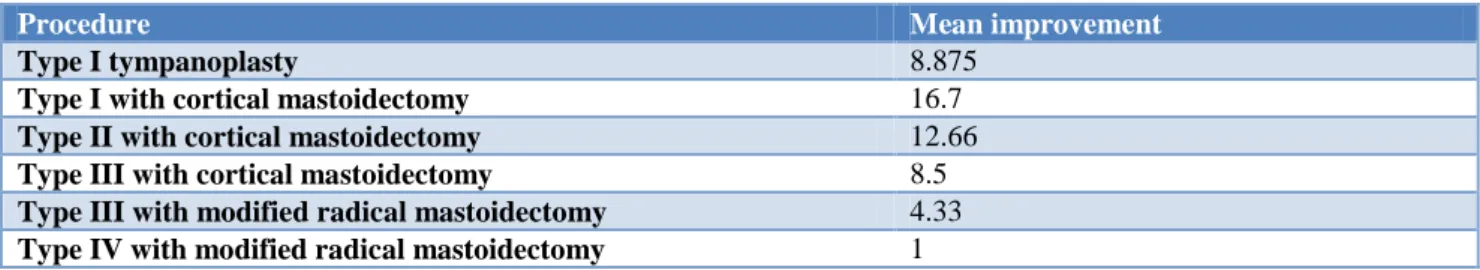
Table 6: Comparison of mean hearing improvement.
Procedure Mean improvement
Type I tympanoplasty 8.875
Type I with cortical mastoidectomy 16.7
Type II with cortical mastoidectomy 12.66
Type III with cortical mastoidectomy 8.5
Type III with modified radical mastoidectomy 4.33
Type IV with modified radical mastoidectomy 1
The mean hearing improvement obtained is plotted against each type of tympanoplasty procedures done and compared (Table 6).
DISCUSSION
Many authors like Carr and Uyar believe that the perforation location plays a more important role in surgery success than perforation size.5,6 Perforations in the anterior quadrant of the TM represent a worse surgical access in order to reach the anterior border and they are also less vascularized owing to which they are considered an important success factor for surgery.7 Hallik et al in their long term results of tympanic membrane repair found that the anterior perforations healed more poorly.8 In our study, 32 patients had a central perforation, 13 cases had an attic perforation while only 5 showed a marginal perforation.
20 patients in our study had CSOM of the right ear and left ear each and only 10 suffered from bilateral CSOM. Out of these, 26 right ears and 24 left ears were operated.
24 patients underwent type I tympanoplasty with cortical mastoidectomy, followed by 9 patients who had type III with CM and 8 underwent type I tympanoplasty. Three patients each underwent type II with CM, type III with
MRM and type IV with MRM.9
The improvement gained in the patients by measurement of the post op ABG using pure tone audiometry done after 3 months of ear surgery revealed that in our study 20 patients showed a good hearing improvement with a maximum gain of 30 dB. 16 patients showed no improvement (<5 dB); while a hearing improvement between 5 db to 15 db was seen in 14 patients.
Postoperative hearing outcomes were considered
successful, if the postoperative air-bone gap was within 20 dB.10 In this study, 37 patients showed ABG within 20
dB 3 months post operatively. Only 13 patients had ABG
persisting over 20 dB. The success rates vary in the literature, from 75 to 98%.
The correlation in the improvement gained in the various types of tympanoplasty, maximum improvement was seen in patients undergoing type I with cortical
seen in all types of tympanoplasty, signifying that many factors are responsible for the success of the surgical procedure.9 In our study, graft material was not a significant factor as TMF was used for all the cases. Multiple surgeons (including residents) performed the various procedures which could have led to differing results based on their surgical skills. It should be noted that these surgeries are complicated procedures and their success also depends on quality of post-operative care.11
Type I with cortical mastoidectomy showed the best mean improvement of 16.7 dB, followed by type II with CM which had a mean improvement of 12.66 dB.
Procedures combined with modified radical
mastoidectomy showed a very poor mean improvement in our study; only 1 dB for type IV with MRM and 4.33 dB with type III with MRM. Type I tympanoplasty and type III with CM showed equal mean improvement with 8.5 dB each.
CONCLUSION
The success of surgery is determined in terms of air-bone gap closure. Our study shows that mean AB gap closure is greatest for type I with cortical mastoidectomy; followed by type II with CM, type I and then type III with CM.
Tympanoplasty in which modified radical mastoidectomy was a part of the procedure gave poor hearing improvement as compared to other procedures.
It is essential to optimize conditions prior to surgery in order to ensure the best result post-surgery. In this study, significant improvement was noted in the subjective symptom of hearing loss following the surgery.
Funding: No funding sources Conflict of interest: None declared
Ethical approval: The study was approved by the Institutional Ethics Committee
REFERENCES
1. Merchant SN, Mckenna MJ, Rosowski JJ. Current
2. Adkins WY. Composite autograft for
Tympanoplasty & tympanomastoid surgery.
Laryngoscope. 1990;100:244-7.
3. Committee on conservation of hearing of the
American Academy of Ophthalmology and
Otolaryngology. Standard classification for surgery
of chronic ear disease. Arch Otolaryngol.
1965;81:204-5.
4. Austin DF. Ossicular reconstruction. Arch
Otolaryngol. 1971;94:525-35.
5. Carr MM, Poje CP, Nagy ML, Pizzuto MP, Brodski
LS. Success rates in paediatric tympanoplasty. J Otolaryngol. 2001;30(4):199-202.
6. Uyar Y, Keles B, Koc S, Ozturk K, Arbag H.
Tympanoplasty in paediatric patients. Int J Pediatr Otorhinolryngol. 2006;70(10):1805-9.
7. Vartiainen E, Nuutinen J. Success and pitfalls in myringoplasty: follow up study of 404 cases. Am J Otol. 1993;14(3):301-5.
8. Halik JJ, Smyth GD. Long term results of tympanic
membrane repair. Otolaryngol Head Neck Surg. 1988;98(2):162-9.
9. Fukuchi I, Cerchiari DP, Garcia E, Rezende CEB, Rapoport PB. Tympanoplasty: surgical results and a comparison of the factors that may interfere in their success. Braz J Otorhinolaryngol. 2006;72(2):267-71.
10. Gersdorff M, Garin P, Decat M, Juantegui M.
Myringoplasty: long-term results in adults and children. Am J Otol. 1995;16(4):215-8.
11. Costa SS, Sousa LCA. Otite média crônica
não-colesteatomatosa. In: Campos CAH, Costa HOO. Tratado de Otorrinolaringologia. São Paulo: Roca. 2002: 72-92.
Cite this article as: Muqtadir F, Rahul S.A study of