Management of Laparoscopic Surgery in Intraoperative
Subcutaneous Emphysema
RTH. Soepraptomo
Department of Anesthesiology and Intensive Therapy, Dr. Moewardi Hospital, Surakarta
ABSTRACT
Background: Over the past 50 years, laparos-copy has evolved from a limited gynecological surgical procedure that is only used for diagnosis and tubal ligation to become the main surgical tool used for many gynecological indications. Many studies have shown that laparoscopy is safer, more affordable, and has a shorter recovery time than laparotomy. This study aimed to report the management of laparoscopic surgery in intra-operative subcutaneous emphysema.
Case Report: The subject of the study was a 37-year-old woman with adenomyosis and chocolate cyst of sinistra with a surgery plan for resection of adenomyosis and per laparoscopic cystectomy with the physical status of the ASA II Plan GAET. The surgery was carried out on October 15, 2018, with surgery for 5 hours. At the intraoperative, subcutaneous emphysema was found from the diaphragm to the thorax. In this condition, the peritoneal insufflation was reduced to 10-12 mmHg. The head down was returned to a
posi-tion that made it easier for the patient's condiposi-tion to reduce the occurrence of decreased lung comp-liance. The patient's postoperative condition was good, the hemodynamic was stable, her breath was spontaneous, and the saturation was 99% with oxygen supplementation via Nasal Kanul 3 lpm. The subcutaneous emphysema gradually disappeared 24 hours postoperatively.
Conclusion: Subcutaneous emphysema may occur in the laparoscopic procedures. The close monitoring during surgery and the expertise of the anesthesiologist in diagnosing and interven-ing is crucial in controllinterven-ing this condition.
Keywords:subcutaneous emphysema; laparos-copy; intraoperative intervention
Correspondence:
Rth. Supraptomo. Department of Anesthesiology and Intensive Therapy, Dr. Moewardi Hospital, Surakarta, Central Java.
Cite this as:
Soepraptomo RTH (2020). Management of Laparoscopic Surgery in Intraoperative Subcutaneous Emphysema. Indones J Med. 05(03): 206-213. https://doi.org/10.26911/theijmed.2020.05.03.05.
Indonesian Journal of Medicine is licensed under a Creative Commons Attribution-Non Commercial-Share Alike 4.0 International License.
BACKGROUND
Over the past 50 years, laparoscopy has evol-ved from a limited gynecological surgical pro-cedure that is only used for diagnosis and tu-bal ligation to become the main surgical tool used for many gynecological indications. Nowadays, laparoscopy is one of the most common surgical procedures performed in the world (Ferreira et al., 2018; Onoh et al., 2018).
Almost all gynecological procedures, such as ectopic pregnancy removal, endomet-riosis treatment, ovarian cystectomy, and hysterectomy use laparoscopic procedures.
Many studies have shown that laparoscopy is safer, more affordable, and has a shorter re-covery time than laparotomy. The benefits of the laparoscopic approach to other procedu-res, including myomectomy, sacral colpo-pexy, staging, and treatment of gynecological cancers increasingly spread (Srivastava and Niranjan, 2010; Hayden and Cowman, 2011; Bajwa and Kulshrestha, 2016; Ferreira et al., 2018; Onoh) et al., 2018).
the use of pneumoperitoneum air to stop in-tra-abdominal hemorrhage, he introduced a cystoscope to the stomach to see the effect of increased pressure on the abdominal organs (Kim et al., 2013; Tiwari et al., 2013).
In the early 20th century, the diagnostic laparoscopy was performed by several gene-ral surgeons as a substitute for diagnostic la-parotomy. However, it had a substantial complication level. From the 1920s to the 1930s, the proponents of the procedure con-tinued to develop better laparoscopic equip-ment, such as pyramidal trocar for port and lens recognition with a view angle wider than 90° provided by the cystoscope. During this period, Dr. Janos Veress, a Hungarian inter-nist, developed a spring-loaded needle with an inner stylet that automatically turned a sharp tip into a round-tip. The Veress needle is still using nowadays to create a pneumope-ritoneum (Kim et al., 2013; Tiwari et al., 2013).
The use of laparoscopy has revolutio-nized the field of surgery by reducing morbi-dity with early recovery. Laparoscopic proce-dures have traditionally been performed un-der the general anesthesia (GA) due to respi-ratory changes caused by pneumoperitone-um, which are an integral part of laparoscopy (Srivastava and Niranjan, 2010; Hayden and Cowman, 2011; Bajwa and Kulshrestha, 20-16).
The precise ventilation control under controlled conditions at GA has proven to be ideal for the procedure. However, the use of regional anesthesia (RA) recently has emerg-ed as an alternative way for laparoscopy. Va-rious reports in the literature showed the sa-fety of using spinal-epidural, spinal, and epi-dural combination anesthesia in the laparos-copic procedures (Lennox et al., 2002; Zun-dert et al., 2006). The benefits of using RA were in the prevention of airway manipula-tion, the patients who woke up and breathe directly at the intraoperative, a minimal
possibility to have nausea and vomiting, the effective postoperative analgesia, and the early ambulation and recovery (Gharaibeh, 1998; Ng and Smith, 2004). However, RA could be associated with several side effects such as the need for higher sensory levels, more severe hypotension, shoulder pain due to diaphragmatic irritation, and respiratory embarrassment caused by pneumoperito-neum. Further study needs to be conducted to establish the excellence of RA over GA for its global use in different patient populations (Sharma et al., 2007; Srivastava and Niran-jan, 2010; Hayden and Cowman, 2011; Bajwa and Kulshrestha, 2016).
The gradual removal process of laparos-copy to put more complicated surgical proce-dures created the modification of existing anesthetic techniques. Various effects of pneumoperitoneum induction, an integral part of laparoscopy, can cause respiratory and cardiovascular changes. Therefore, it should be managed using GA. Since the im-plementation of laparoscopy conducted in va-rious pediatric operations, better anesthetic techniques are needed to allow early recovery and ambulation. The evolution of anesthetic drugs on a scale that is scientifically and cli-nically relevant has led to the innovations and ideas for newer but safer techniques. The development in anesthesiology has been made in many fields besides scales that clinically relevant (Srivastava and Niranjan, 2010; Hayden and Cowman, 2011; Bajwa and Kulshrestha, 2016).
Recently, RA has been documented to have equally good results in laparoscopic sur-gery. This review has been carried out to compare the advantages and disadvantages of using RA vs GA in laparoscopic surgery (Lennox et al., 2002; Zundert et al., 2006).
On October 13, 2018, a 37-year-old woman with the main complaint of left abdominal pain came. The patient said that she felt pain in her left abdomen for a year. Nausea and vomiting (-). Coughs and colds (-). She had menstruation once a month. The patient had a history of hypertension and diabetes melli-tus since 2017. Diabetes mellimelli-tus was cont-rolled. The insulin was 10 IU in the morning and 8 IU at night.
The patient's condition is fully cons-cious, with GCS E4V5M6. The patient had a blood pressure of 130/80 mmHg, pulse 88 x/min, respiration rate 16x/min, a body tem-perature of 36.4oC, and 100% oxygen satura-tion with 3 lpm nasal cannula oxygen. The patient had a bodyweight of 75 kg, a height of 154 cm, and a Body Mass Index of 31.65.
The airway was free, nasal patency (+/+), septal deviation (-/-), open mouth >3 fingers, the mallampati was 3, the teeth were in the normal limits, the neck motion was free, TMD5 cm. Breathing: the thorax was normochest, symmetrical, right chest deve-lopment=left, retraction (-/-), breathing mus-cles (-/-), sonor, vesicular base sound (+/+), additional sounds (-/-), breathing frequency in 20x/minute. Circulation: the heart sounds of I-II was regular, noisy (-). Blood pressure was 130/80mmHg, pulse was 88 x/minute, regular rhythm, enough content, CRT <2 se-conds, acral coldness (-/-). Disability: GCS E4V5M6, isochoric pupils with a diameter of 3mm/3mm, light reflexes (+/+).
RESULTS
Based on the thorax examination, there was a sonor percussion throughout the lung field, auscultation sounds a basic sound: vesicular (+/+), additional sounds: RBH /-), RBK (-/-), wheezing (-/-). On a single BJ1-2 cardio-vascular examination, the rhythm was regu-lar, murmurs (-). On the examination of the palpable abdomen, the result was supple, tenderness (-), the abdominal percussion was
tympanic, the auscultation of abdominal bowel sounds was within normal limit. The akral was warm, the edema was not found in all extremities.
Based on the result of the laboratory tests, Hb 13.6, Ht 39, AL 9.1, AT 398, AE 4.56, PT 13.7, APTT 32.5, INR 1,090, GDS 101, Cr 0.5, Ur 26, Na 136, K 4.2, Cl 109, Hbs-Ag NR. ECG The result showed sinus rhythm, HR 92 bpm, and normoaxis. Based on the re-sult of chest X-Ray, there were no abnormali-ties in the pulmo impression, the cor was in-valid. Based on the result of the USG, the VU was quite filled, the shape and the size was dbn, the hypoechoic mass was 4.55x4.52x 3.05cm. It showed that it sustained the cho-colate cyst.
Based on the patient's condition, the diagnosis of anesthesia was as follows: a 37-year-old woman with adenomyosis+chocola-te cyst of sinistra of pro-resection adenomyo-sis+per laparoscopic cystectomy with a physi-cal status of ASA II Plan GAET. Furthermore, the patient was prepared for surgery. The preparation were the family was given an ex-planation of the plan, the procedure of anes-thesia and surgery, the possibility of things that might occur during the intervention, the alternative intervention to face the risk of surgery, and the preparation of drugs and tools.
After preparing the pre-surgery, the surgery was begun. The patient entered the operating room on October 15, 2018, at 08.00, the ECG lead was conducted, oxygen saturation, blood pressure, and body tempe-rature measurements. Based on the result, the vital signs of blood pressure was 138/95 mmHg, the heart rate was 84 x/minute, the breathing frequency was 18 x/minute, the temperature was 36.3oC, the O2 saturation was 100%, the EKG was sinus rhythm.
with 2 mg of midazolam followed by giving Fentanyl 150 mcg (~2mcg/kg) slowly after 5 minutes. The patient was given 70 mg of Pro-pofol (~1mg/kg) after 3 minutes. After the airway was completely controlled, the muscle relaxant was given. It used 35 mg of
Atra-crium (0.5 mg/kg). For the anesthesia main-tenance, it used sevoflurane as much as 1.5 - 2 vol% and N250% in the administration of intraoperative analgesics as much as 0.5-1 mcg/kgbb/hour, and atracurium at 10-10 mcg/kg/min.
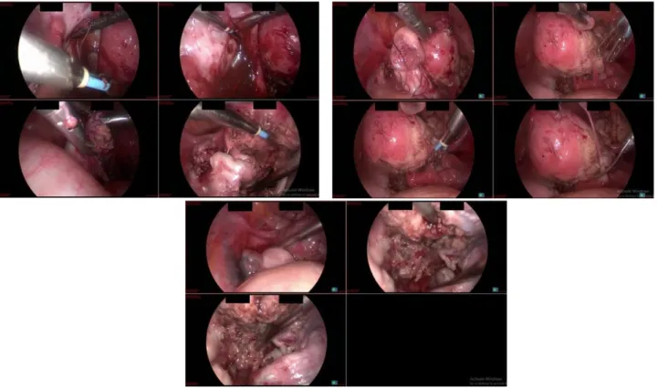
Figure 1. The figures of resection of the adenomyosis and perlaparoscopic cystectomy
Durante operation range of TD was 100-120 mmHg of systolic, 60-80 mmHg of diastolic, 64-135 mmHg of MAP, 78-88 x/min of pa-tient pulse, and 99-100% of O2 saturation. The ETCO2 of the durante operation in the first hour of operation ranged from 30-35 mmHg, using Respiration Control VCV TV as much as 350mL, RR 14x/min, I:E=1:2, PEEP=6. The head down was applied to the patient as much as 5' and the Intraabdominal Pressure Insufflation using CO2 up to 12 mmHg. 2 hours later, the operator asked to increase the CO2 insufflation pressure to 15-20 mmHg and head down that was quite ext-reme as 30'. In the first 1 minute, the ETCO2
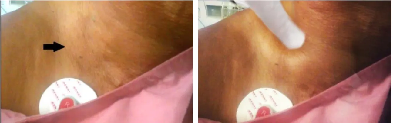
Figure 2. The subcutaneous emphysema reached the neck circumference
The subcutaneous emphysema in the thorax expanded to the 4th hour with the condition of ETCO2 has decreased to 35 mmHg from 10.20. It expanded towards the neck circum-ference. At the 5th hour, ETCO2 decreased quite well, ranging between (30-35 mmHg). The subcutaneous emphysema in the neck
circumference gradually decreased and it only lived in the thorax. After surgery, the position was neutralized. The abdominal in-sufflation was turned off. The ETCO2 re-turned to the normal range of 30-35 mmHg, followed by a fairly good reduction in the emphysema crepitus.
Figure 3. The subcutaneous emphysema in the neck circumference was reduced
The patient's postoperative condition was good, the hemodynamic was stable, her breath was spontaneous, and the saturation was 99% with oxygen supplementation via Nasal Kanul 3 lpm. The patient was observed in the Recovery Room for 2 hours. During the observation, there was no nausea and vomit-ing. The intravenous analgesic paracetamol as much as 1 gram/8 hours was given after surgery. Then, the patient was moved to the treatment room.
DISCUSSION
The use of laparoscopic techniques has been very advanced. It could be prescribed for many procedures. Nowadays, laparoscopy is the most common surgical procedure for outpatients. Laparoscopic surgery is pre-ferred because it is minimally invasive. It is also associated with a faster wound healing process. However, the laparoscopic proce-dure might be difficult to do to a patient with a poor condition due to these physiological changes. Anesthesia must be conducted care-fully and adjusted to the type of surgery and the patient's condition (Gerges et al., 2006; Upadhyay, 2019).
During laparoscopic surgery, there was an unexpected condition: Subcutaneous Em-physema. It might occur due to these con-ditions. Therefore, the patient's decline beco-mes an extreme head-down (Trendelenburg position). Besides, the CO2 insulation was quite extreme up to 15-20 mmHg. Another additional factor was also caused by a large amount of adipose tissue (BMI≥32), thus making it difficult for operators to visualize the operating field.
Close monitoring during durante opera-tion and anesthesiologist's expertise in diag-nosing and intervening in respiration was crucial in controlling this condition. Therefo-re, it did not increase its complications (Najia and Mandeep, 2018).
During pneumoperitoneum, ventilation control was adjusted to maintain PETCO2 as much as 30-35 mmHg. It required no more than 15-25% of the increase in one minute ventilation, unless the subcutaneous emphy-sema occurred. The increased breath fre-quency was preferred more than the increas-ed tidal volume in patients with Chronic Obs-tructive Pulmonary Disease (COPD) and in patients with spontaneous pneumothorax or subcutaneous emphysema. It aimed to avoid increasing alveolar inflation and reducing the risk of pneumothorax (Dora, 2002; Means et al., 2004; Carey et al., 2019).
The intra-abdominal pressure must be monitored. It must also be maintained as low as possible to reduce hemodynamic and res-piration changes. Besides, it should not exce-ed 20 mmHg. Relatively, it should be main-tained below 12 mmHg. Increased intra-ab-dominal pressure could be avoided by main-taining the depth of anesthesia. Due to the tendency for an increase in vagus tone di-rectly during laparoscopy, the Atropine must be provided for injection if needed (Gharai-beh, 1998; Hirose et al., 2018; Aseni et al., 2019).
AUTHOR CONTRIBUTION RTh. Supraptomo and Parada Jiwanggana played a role in conceptualizing the study, de-termining the methodology, collecting the da-ta, and writing the scripts.
CONSENT FOR PUBLICATION The parents of the patient provided written informed consents to publish clinical details along with any identifying images. A copy of written consent is available for review by editor-in-chief of this journal.
CONFLICT OF INTEREST This study did not have any conflict of inte-rest.
FUNDING AND SPONSORSHIP This study used a personal fund from the re-searcher.
ACKNOWLEDGEMENT
We give the best gratitude to the Faculty of Medicine, Universitas Sebelas Maret, Sura-karta and Dr. Moewardi Hospital, SuraSura-karta, who helped carry out this study.
Aseni P, Orsenigo S, Storti E, Pulici M, Arlati S (2019). Current concepts of periope-rative monitoring in high-risk surgical patients: a review. Patient Saf Surg 13-(32). https://doi.org/10.1186/s13037-019-0213-5.
Bajwa SJS, Kulshrestha A (2016). Anaesthe-sia for laparoscopic surgery: General vs regional anaesthesia. J Minim Access Surg. 12(1): 4–9. https://doi.org/10.41-03%2F0972-9941.169952.
Carey BM, Jones CN, Fawcett WJ (2019). Anaesthesia for minimally invasive ab-dominal and pelvic surgery. BJA Edu. 19(8): 254–260. https://doi.org/10.10-16/j.bjae.2019.04.001.
Dora H (2002). Anesthesia for minimally in-vasive surgery: Laparoscopy, thoracos-copy, hysteroscopy. Anesthesia & Anal-gesia. 94(1): 237. doi: 10.1213/000005-39-200201000-00054.
Ferreira H, van Belle Y, Tanos V, Rabischong B, Grimbizis G, Sardo ADS, Campo R (2018). Simulation and training of gyn-aecological skills. Facts Views Vis Ob-gyn. 10(1): 21–27. https://www.ncbi.nl-m.nih.gov/pubmed/30510664.
Gerges FJ, Kanazi GE, Jabbour-Khoury SI (2006). Anesthesia for laparoscopy: A review. J Clin Anesth. 18(1): 67-78. https://doi.org/10.1016/j.jclinane.2005 .01.013.
Gharaibeh H (1998). Anaesthetic manage-ment of laparoscopic surgery. E Me-diterr Health J, 4(1): 185–188. https://- apps.who.int/iris/bitstream/handle/- 10665/117983/emhj_1998_4_1_185_-188.pdf.
Hayden P, Cowman S (2011). Anaesthesia for laparoscopic surgery. Continuing Edu-cation in Anaesthesia Critical Care & Pain. 11(5): 177–180. https://doi.org/-10.1093/bjaceaccp/mkr027.
Hirose M, Kobayashi Y, Nakamoto S, Ueki R, Kariya N, Tatara T (2018).
Develop-ment of a hemodynamic model using routine monitoring parameters for no-ciceptive responses evaluation during surgery under general anesthesia. Me-dical Science Monitor : International Medical Journal of Experimental and Clinical Research. 24: 3324-3331. http-s://doi.org/10.12659/MSM.907484. Kim BS, Joo SH, Joh JH, Yi JW (2013).
La-paroscopic cholecystectomy in patients with anesthetic problems. World J Gas-troenterol. 19(29): 4832–4835. https:-//dx.doi.org/10.3748%2Fwjg.v19.i29.4 832.
Lennox PH, Vaghadia H, Henderson C, Martin L, Mitchell GW (2002). Small-DoDOrse Selective Spinal Anesthesia for Short-Duration Outpatient Laparos-copy: Recovery Characteristics Compa-red with Desflurane Anesthesia. Anes-thesia & Analgesia, 94(2): 346–350. https://doi.org/10.1213/00000539-20-0202000-00021
Means LJ, Green MC, Bilal R (2004). Anes-thesia for minimally invasive surgery. Semin Pediatr Surg. 13(3): 181-7. htt-ps://doi.org/10.1053/j.sempedsurg.20 04.04.006.
Najia H, Mandeep S (2018). Anesthesiology: Clinical case reviews. Anesthesia & Analgesia. 127(2): e7-e8. doi: 10.1213/-ANE.0000000000003437
Ng A, Smith G (2004). Intraperitoneal admi-nistration of analgesia: is this practice of any utility?. Br J Anaesth, 89(4): 534–7. https://doi.org/10.1093/bja/ae-f219
Sharma S, Jayaraman L, Sethi N, Sood J (2007). Anaesthetic management for laparoscopic cholecystectomy in two patients with biopsy proven polymyo-sitis. Indian J Anaesth. 51(1): 43-6. Re-trieved from http://www.ijaweb.org-/text.asp?2007/51/1/43/61114.
Srivastava A, Niranjan A (2010). Secrets of safe laparoscopic surgery: Anaesthetic and surgical considerations. J Minim Access Surg. 6(4): 91–94. https://.doi.-org/10.4103%2F0972-9941.72593 Tiwari S, Chauhan A, Chaterjee P, Alam MT
(2013). Laparoscopic cholecystectomy under spinal anaesthesia: A prospecti-ve, randomised study. J Minim Access
Surg. 9(2): 65–71. https://doi.org/10.-4103%2F0972-9941.110965.
Upadhyay S (2019). Perioperative considera-tions in laparoscopic surgery during pregnancy: A mini review and updates. EC Anaesthesia. 5(11): 07-15. https://- www.ecronicon.com/ecan/pdf/ECAN-05-00186.pdf.