Volume 16, Issue 1, Winter 2017
Editorial in Charge
Hossein Pakdaman, M.D.
Professor of Neurology, Shahid Beheshti
University of Medical Sciences,
Tehran, Iran
Editor-in-Chief
Shahriar Nafissi, M.D.
Associate Professor of Neurology,
Neurology Department, Tehran University of
Medical Sciences, Tehran, Iran
Deputy Editor
Farzad Fatehi, M.D.
Assistant Professor of Neurology, Neurology Department, Tehran University of
Medical Sciences, Tehran, Iran
Section Editors
Headache: Mansooreh Togha
, M.D.
,
Tehran
University of Medical Sciences
, Tehran, Iran
Multiple
Sclerosis:
Mohammad
Ali
Sahraian
, M.D., Neurology Department,
Tehran
University of Medical Sciences
, Tehran, Iran
Stroke: Afshin Borhanin Haghighi
, M.D.,
Shiraz University of Medical Sciences, Shiraz,
Iran
Movement Disorders: Mohammad Rohani
,
M.D., Iran University of Medical Sciences,
Tehran, Iran
Associate Editors
Shahin
Akhondzadeh,
Pharm.D.,
Ph.D,
Tehran University of Medical Sciences,
Tehran, Iran
Majid Ghafarpour,
M.D., Tehran University of
Medical Sciences, Tehran, Iran
Massoud Nabavi,
M.D., Shahed University of
Medical Sciences, Tehran, Iran
Scientific Assistant Editor
Ali Amini-Harandi
, M.D., Shahid Beheshti University of Medical Sciences, Tehran, Iran
Editorial Board
Shahram Attarian
, M.D., Centre de Référence
des Maladies Neuromusculaires et de la SLA,
France
Mahmoud R. Azarpazhooh
, M.D., Mashhad
University of Medical Sciences, Mashhad, Iran
Keivan Basiri
, M.D., Isfahan University of
Medical Sciences, Isfahan, Iran
Ahmad R. Dehpour
, Pharm.D., Ph.D., Tehran
University of Medical Sciences, Tehran, Iran
Masoud Etemadifar
, M.D., Isfahan University
of Medical Sciences, Isfahan, Iran
Kavian
Ghandehari
,
M.D.,
Mashhad
University of Medical Sciences, Mashhad, Iran
Kurosh Gharagozli
, M.D., Shahid Beheshti
University of Medical Sciences, Tehran, Iran
Mohammad H. Harirchian
, M.D., Tehran
University of Medical Sciences, Tehran, Iran
Payam Kabiri
, M.D., Ph.D., Tehran University
of Medical Sciences, Tehran, Iran
Hossein Kalani
, M.D., Shahid Beheshti
University of Medical Sciences, Tehran, Iran
Jamshid Lotfi
, M.D., Tehran University of
Medical Sciences, Tehran, Iran
Alireza Minagar
, M.D., Louisiana State University
Health Sciences Center, USA
Ali Moghtaderi
, M.D., Zahedan University of
Medical Sciences, Zahedan, Iran
Mahmood Motamedi
, M.D., Tehran University
of Medical Sciences, Tehran, Iran
Alireza Nikseresht
, M.D., Shiraz University of
Medical Sciences, Shiraz, Iran
Abdolmohamad M. Rostami
, M.D., Thomas
Jefferson University Hospitals, USA
Mohammad
Saadatnia
,
M.D.,
Isfahan
University of Medical Sciences, Isfahan, Iran
Mohammad K. Salajegheh
, M.D., Brigham and
Women's Hospital and Harvard Medical School,
USA
Gholam A. Shahidi
, M.D., Tehran University
of Medical Sciences, Tehran, Iran
Vahid
Shaygannejad
,
M.D.,
Isfahan
University of Medical Sciences, Isfahan, Iran
Akbar Soltanzadeh
, M.D., Tehran University
of Medical Sciences, Tehran, Iran
Amir A. Zamani
, M.D., Brigham and Women's
Hospital and Harvard Medical School, USA
Babak Zamani
, M.D., Tehran University of
Medical Sciences, Tehran, Iran
Secretary:
Samaneh Bahraminejad, BSc
Email: [email protected]
http://ijnl.tums.ac.ir
Copy Edit, Layout Edit, Proof Reading, Design, Print and Online Support: FaRa Publishing House (Farzanegan Radandish)
http://farapub.com Email: [email protected]
Tel/fax: +98 31 32224335, +98 31 32224382
Indexed in
PubMed,
PubMed Central,
Academic Keys,
Cite Factor (Directory Indexing of International Research Journals),
Directory of Open Access Journals (DOAJ),
Directory of Research Journal Indexing (DRJI),
Ebsco,
Electronic Journals Library,
Google Scholar,
InfoBase Index,
Islamic World Science Citation Center (ISC),
LocatorPlus,
Scientific Information Database (SID),
Ulrichsweb Global Serials Directory,
Universal Impact Factor,
WorldCat
The submission file is in Microsoft Word document file format.
The Iranian Journal of Neurology is dedicated to the Iranian Neurological Association. The journal is a peer- reviewed journal published quarterly and publishes neurological experiences in basic or clinical fields in English Language. The Iranian Journal of Neurology aims to publish manuscripts of a high scientific quality representing original clinical, diagnostic or experimental works or observations in neurological sciences. Papers in English are welcomed, particularly those which bring novel information and researches in clinical or basic fields from the neurological disorders. All received manuscripts coving the scope of the journal will be evaluated by properly competent referees.
Submissions should be accompanied by a cover letter including a declaration by the first author on behalf of the others to the effect that
(1) The paper has not been published to date (except for abstracts of conference materials).
(2) The paper has not been accepted for publication elsewhere.
(3) All persons listed as the authors have read it and approved it for publication. The cover letters should be submitted in section "Comments for the Editor".
Articles must be written in accurate scientific English appropriate for publication. The articles are subject to review and editing; however, the authors are responsible for the correctness the manuscript's English language. The articles must be submitted only online: ijnl.tums.ac.ir
The Editorial Board reserves the right to reject a paper without seeking reviewers’ opinion provide the content or the form of the paper does not meet minimum acceptance criteria or if the subject of the paper is beyond the aims and scope of the journal.
Everyone listed as the author of a paper is responsible for the reliability and completeness of data presented in the paper.
Do not submit papers that copy fully or partially previously published papers.
Indicate that this submission is ready to be considered by this journal by checking off the following:
The submission has not been previously published, nor is it before another journal for consideration (or an explanation has been provided in Comments to the Editor).
Where available, URLs for the references have been provided.
The text is double-spaced; uses an Arial 12-point font; and all illustrations, figures, and tables are placed within the text at the appropriate points, rather than at the end.
The text adheres to the stylistic and bibliographic requirements outlined in the Author Guidelines, which is found in About the Journal.
If the Editorial Board is not notified in advance and the paper is found to have been copied during editorial work, the paper shall be rejected.
We expect that all studies reported in the journal conform to the requirements of the Declaration of Helsinki (1989). Information on the consent of a relevant ethics committee to perform the trial and the informed consent of the patients to participate in the trial should be given in the Material and methods section of each paper in which diagnostic or therapeutic intervention does not follow from the standard procedure. Authors of case reports must not disclose personal data of patients described.
The journal publishes: Original Article Review Article Case Report
Short Communication Clinical Notes Editorial Letters to Editor Neurological Images Neurological Videos Iranian Neurological Events Clinical Quiz
Details
Original and review papers: The maximum length of
original and review papers (including tables and figures materials) is 3000 words.
Case reports: Case reports will be accepted only as Letter to the Editor.
Short communications: The maximum word number of
short communications should be below 1200 words with maximum one table or figure and 10 references. The manuscript should be structured including introduction, materials and methods, results, discussion, and conclusion with a structured abstracts as original articles.
neurological images or videos are welcome. They should be maximally 400 words with legends without abstract and unstructured. The videos should be uploaded as supplementary files.
Letter to the Editor: May concern short scientific reports and comments. The maximum number of words should be below 800 words with maximum 5 references, no abstract, no table or figure, and unstructured.
Clinical notes: Refer to important interesting observations which are imperative for reminders in clinical practice. The maximum number is 1000 words with maximum 5 references, 1 table and 1 figure with no abstract.
Iranian neurological events: Include the brief description of major regional events (congresses or seminar) implemented in Iran.
Manuscripts should be submitted in 12 points, Arial font, with double line spacing and sufficient margins of 2.5 cm.
The text should not be formatted.
Each section of the paper should begin on a new page
Page 1: Title Page
Page 2: Abstract and Key Words
Page 3 and subsequent pages: manuscript body including Introduction, Materials and Methods, Results, Discussion, Conclusion, References, Tables, Figures 1. Title page:
Title page should contain paper title, full names of authors, authors’ place of work, full name and address of the corresponding author (including e-mail address and telephone number), given in that order.
2. Abstract page:
The length of the abstract should be at least 200 and not more than 250 words for original papers and not more than 150 words for review papers and case reports. Abstracts of original papers should be structured to include the background, methods, results and conclusion.
Below the abstract authors should provide between three and six keywords conforming to Medical Subject Headings (Index Medicus).
3. Page three and subsequent pages of the original paper
and short communication should include the text arranged in the following order (for other manuscript type, see above):
1. Introduction: The introduction should be as concise as possible and introduce the context of the paper to the reader; the paper should clearly state the research hypothesis and the objective of the study.
2.Materials and Methods: Description of the studied
population or material should be detailed and include all information necessary to assess the reliability of results obtained in the study and/or allow the experiment to be repeated by other researchers; the section related to statistical analysis should have information on applied statistical tests and programs.
3. Results: Present results directly related to the topic of the paper only; tables and/or figures are recommended.
4.Discussion
5. Conclusions: These should be brief, follow directly from results presented above and correspond to the aim of the paper outlined in the introduction.
6.Acknowledgements: Should comprise information
on sources of funding (grant numbers); acknowledgements should concern those who made a significant contribution to the paper, but who did not meet the criteria to be listed as authors.
7.References: References should be listed in the order quoted in the paper. Please cite source and major papers that offer interested readers an opportunity to obtain more detailed information. Avoid citing review papers and conference reports, if they are not the only materials on a given topic.
In the paper references should be given in superscripts with no space between the comma and the consecutive number.
Authors are advised to carefully verify citation details. Give names of first six authors; if there are more authors, add “et al.“. Use Index Medicus abbreviations for journal titles. Then mention the volume and the issue of the journal.
The recommended style for journal references is as follows:
[Reference number][Authors]. [Article title]. [Journal Name] [Year of publication]; [volume](issue): [Pages range].
For Journal Example:
1. Janghorbani M, Amini M, Willett WC, Mehdi Gouya M, Delavari A, Alikhani S, et al. First nationwide survey of prevalence of overweight, underweight, and abdominal obesity in Iranian adults. Obesity (Silver Spring) 2007; 15(11): 2797-808.
For Books Example:
2. Ropper AH, Brown RJ. Adams and Victors principles of neurology. 8th ed. New York, NY: McGraw Hill Professional; 2005. p. 271.
Tables: Each table should be placed on a separate page. Tables should be numbered with Arabic numerals in the order in which they appear in the text. Authors should indicate the position of tables in the paper. Titles and headings of tables should be given in English. Information given in tables should not be repeated in the body of the text. Explanations concerning tables, e.g. full names of abbreviations should be given in footers below tables and should be consecutively marked: “*”,“**”,“***” etc.
Figures: Figures and photographs should be numbered
with Arabic numerals and attached as separate printouts (in the electronic version, as separate files). Figures should be saved in one of the following formats: .jpg.
delivered, so they must be prepared carefully. Please indicate where they should be placed in the text.
Abbreviations should be always clarified when used for the first time in the text (including the abstract). Abbreviations should not be used in paper titles, unless in exceptional circumstances.
Review process: All papers submitted for publication in the journal are assessed by two independent reviewers with the mutual anonymity rule as to the names of reviewers and authors observed.
Plagiarism policy: According to the plagiarism policy of Iranian Journal of Neurology, plagiarism is defined as a paper which replicates another publication with as a minimum 25% resemblance and devoid of citation.
In any time the evidence of plagiarism is detected, the manuscript will be withdrawn and the author will be sanctioned from publishing papers permanently.
Table of Contents
Original Article(s)
Comparison of the effects of low dose interferon and high dose interferon on reduction of
the number and size of plaques in patients with Multiple Sclerosis: A historical cohort
Payam Khomand, Ghobad Moradi, Behrooz Ahsan, Setareh Abtahi ... 1-6
Awareness toward stroke in a population-based sample of Iranian adults
Mozaffar
Hosseininezhad,
Hannan
Ebrahimi,
Seyed
Mohammad
Seyedsaadat,
Babak Bakhshayesh, Motahareh Asadi, Amir Reza Ghayeghran ... 7-14
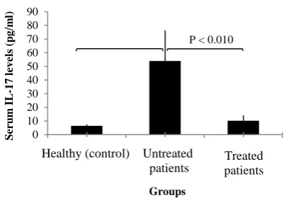
Circulating concentrations of interleukin (IL)-17 in patients with multiple sclerosis:
Evaluation of the effects of gender, treatment, disease patterns and IL-23 receptor
gene polymorphisms
Seyed Ali Ghaffari, Maryam Nemati, Hossain Hajghani, Hossainali Ebrahimi,
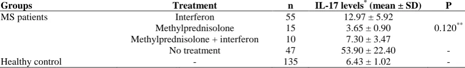
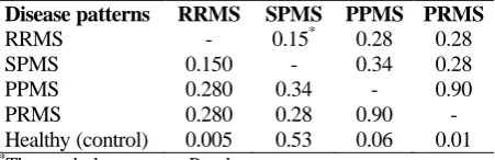
Abdolkarim Sheikhi, Abdollah Jafarzadeh ... 15-25
Knowledge, attitude, and practices among Iranian neurologists toward evidence-based
medicine
Kaveh Shafiei, Fatemeh Sedaghati ... 26-29
Polymorphisms at activated protein C cleavage sites of factor V: Are they important in
the absence of factor V Leiden?
Ehsan Kheradmand, Shaghayegh Haghjooy-Javanmard, Leila Dehghani, Mohammad Saadatnia . 30-33
Review Article
Multiple sclerosis-A disease on a dramatically rising trend in Iran: Review of
possible reasons
Mohammad Ali Sahraian, Mohammad Sahebkar, Rouhullah Dehghani, Milad Derakhshan-Jazari,
Vahid Kazami-Moghaddam, Ebrahim Kouchaki ... 34-40
Letter to Editor(s)
The first attack of multiple sclerosis presented immediately after voluntary and
intensive weight loss: A case series
Sama Bitarafan, Kiana Amani, Mohammad Ali Sahraian, Payam Sarraf, Danesh Soltani,
Abdorreza Naser Moghadasi, Mohammad Hossein Harirchian ... ………41-42
The effects of intensive language therapy in aphasic patients
Ahmad Reza Khatoonabadi, Shohreh Kaviani, Noureddin Nakhostin-Ansari, Mahsa Saadati,
Ehsan Shahverdi ... 43-44
Clinical Note(s)
Psoriasis, bulbar involvement, and diarrhea in late myoclonic epilepsy with ragged-red
fibers-syndrome due to the m.8344A > G tRNA (Lys) mutation
Josef Finsterer, Gabor Geza Kovac ... 45-49
Endovascular management of chronic internal carotid occlusion with Penumbra system
Masoud Mehrpour ... 50-52
Neurological Image/Video
Fahr disease: Idiopathic basal ganglia calcification
including relapsing-remitting form of MS (RRMS), primary progressive MS, secondary progressive MS, and isolated clinical syndrome.2
The first line of treatment for RRMS is consisted of interferon beta (IFN-β) and is glatiramer acetate.3 They have a good effect on reducing relapse and a variety of disabilities and on magnetic resonance imaging (MRI) criteria. Effect of IFN-β for the treatment of RRMS has been proved. IFN therapy can make its effect via its anti-proliferative effects and reduces the permeability in the blood–brain barrier.
MRI has a high capacity for early diagnosis of MS, particularly if the clinical diagnosis is uncertain, monitoring of treatment, evaluation of disease progression, and response to treatment.4
Abnormalities in brain MRI are observed in more than 95% of newly diagnosed patients. There are 5-10 new or large plaques enhanced with gadolinium; on the other hand, T2 lesions show attacks in any patient with RRMS.5 Brain MRI can show the areas of edema, demyelination, damaged axons, gliosis, and repair of myelin in areas with high signal in T2.5,6
Beta IFN compounds include beta IFN-β-1a (Avonex) or low dose IFN and high dose IFN-β-1a (REBIF) and beta IFN-β-b called (Betaseron).7-10
IFN-β1a lowers the rate of attacks in MS patients by 33%.
High dose IFN-β-1-a (REBIF) is one of two available formulations of IFN-β1. This drug is used and injected subcutaneously at doses of 22 and 44 µg 3 times a week.11 Low dose IFN-β-1a (Low dose IFN) is prescribed for intramuscular injection at a dose of 30 mg once a week low dose IFN (CinnoVex is the commercial name of IFN-β-1a and it is manufactured in Iran as the world’s third largest manufacturer in the market). It is a biosimilar or biogeneric of Avonex drug.12
Some studies have shown that IFN-β compounds can reduce MS attacks, brain atrophy, and the number and volume of brain lesions.3,11 Other studies have suggested the better effects of low dose IFN-β-A and high dose (REBIF) in reducing plaques in MRI compared with placebo.13
Although the low dose IFN drug is used abundantly by MS patients in Iran, few study has been conducted on the effects of the drug (especially CINOVEX) on MS patients in terms of reducing the number of plaques or to compare it with high dose IFN drugs. Furthermore, we assume this survey may be a view about Kurdish
patients with MS and an effect of Iranian products of IFN-β-1-a (CinnoVex) on them, which is an important medical issue in our area.
Accordingly, this study examines the impact and efficacy of low dose IFN (CinnoVex) on reducing the number of MRI plaques in MS patients and compares it with high dose IFN (REBIF).
This study was a historical cohort and it was conducted on patients with RRMS who were under the treatment with low dose IFN drugs (CinnoVex) or high dose IFN (REBIF); the patients had a profile in the Clinic of Kurdistan University of Medical Sciences or in Sanandaj MS Society, Iran. The study was conducted in 2014.
Although clinical trial with randomization is the best way to test the hypothesis of this study, because of budgetary limitations and the long duration of the project, it became difficult for the researchers to use this method.
IFN-β-1a is sold under the trade names Avonex (Biogen) and Rebif (Merck Serono), (Pfizer); CinnoVex (CinnaGen) is biosimilar of Avonex. Rebif, it is co-marketed by Merck Serono and Pfizer in the US.
CinnoVex is the trade name of recombinant IFN-β-1-a, which is manufactured as biosimilar/biogeneric in Iran. It is produced in a lyophilized form and sold with distilled water for injection. CinnoVex was developed at the Fraunhofer Institute in collaboration with CinnaGen. Dosage of both drugs in this study was 44 mcg (REBIF) and 30 mcg (CinnoVex), respectively.
Table 1. Association of Interleukin 6 (IL-6) level with National Institutes of Health Stroke Scale (NIHSS), modified Rankin Scale (mRS) and other infarcts
Variables Low dose IFN High dose IFN P
Sex [n (%)]
Male 4 (17) 4 (21) 0.714*
Female 20 (83) 15 (79)
Age (year) (Mean) 33.58 29.84 0.070**
*
Chi-square, **t-test. IFN: Interferon
symptoms were the likely signs of other diseases other than MS, patients who had fully transverse myelitis or bilateral optic neuritis, patients with clinically isolated syndrome, and patients who had enhanced plaque in the initial MRI.
With regarding difference between 2 groups based on the effects of outcome, it was equal to 40% and p1 = 30% also, with regarding p1 = 70% with 5% alpha and beta 20% sample size (based on the below formula) was 21 patients for each group.
21 /2 1 1 1 2 2
2 1 2
( (1 ) (1 ))
( )
Z Z p p p p
n
p p
Because of our limitation in this study, we considered 24 patients in 1 group and in other group 19 patients entered in the study.
In this study, all patients with RRMS who were under treatment with low dose or high dose IFN and referred to the Neurology Clinics and/or were the member of the MS Society of Sanandaj in 2014 and met the inclusion criteria were enrolled in the study. Data were collected through questionnaires and interviews with patients and conducting MRI at the beginning and end of the project.
To conduct the study first, size and enhanced plaques on initial MRI were recorded. Then again MRI was done after a year, and the results were compared in terms of the number, size, and enhanced plaques. All MRI tests were performed in a specified imaging center. Drug side effects and relapse of disease were measured during the follow ups using questionnaires and interviews with patients and phone calls.
The collected data were entered in STATA 11 (Stata Corporation, College Station, TX, USA) software. Chi-square and Fisher exact tests and logistic regression were used for analysis of data.
The researchers in this study were committed to the principles of research ethics and observed research ethics issues for patients.
Patients
A total of 43 patients were enrolled in this study, and 24 patients (55.8%) were assigned to the group treated with low dose IFN and 19 patients (44.2%) were assigned to the group treated with high dose IFN. The mean age of patients treated with low dose IFN was 33.58 years, and the mean age of the patients treated with high dose IFN was 29.84; they had not a statistically significant difference (P = 0.073). Of patients treated with low dose IFN, 20 patients were female (46.51%) and 4 patients were male (9.30%). Of patients treated with high dose IFN, 15 patients were female (34.88%) and four patients were male (9.30%), and there was no statistically significant difference (P = 0.714). Table 1 shows demographic variables in 2 groups of study.
MRI findings and relapse
Based on the results of Fisher exact test, the P value obtained from this relationship (P = 0.039) was significant. In addition to the above test, we also used logistic regression. Based on the results of logistic regression analysis, compared with the low dose IFN therapy, treating patients with high dose IFN (with odds ratio of 5.19 and confidence interval of 2.1-32.4) had a better impact on the reduction of the number of plaques (Table 2).
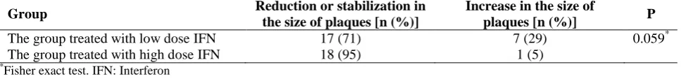
Table 3 compares the two groups in terms of the impact of drugs on the size of the plaque. During the course of treatment, six patients in each of the two groups suffered from relapse and the difference was not statistically significant (P = 0.633).
Table 2. Comparison of changes in plaque size based on the results of magnetic resonance imaging (MRI) in the two
groups treated with low dose interferon (IFN) and high dose IFN
Group Reduction or stabilization in
the size of plaques [n (%)]
Increase in the size of
plaques [n (%)] P
The group treated with low dose IFN 17 (71) 7 (29) 0.059*
The group treated with high dose IFN 18 (95) 1 (5)
*
Table 3. Comparison of side effects between two groups
Side effect Low dose IFN [n (%)] High dose IFN [n (%)] 2 P
Yes 21 (87.5) 18 (95.0) 0.658 0.417
No 3 (12.5) 1 (5.0)
IFN: Interferon
Side effects
Of all, 21 patients in the low dose IFN group and 18 patients in the high dose IFN group had some degrees of side effects, and the difference was not statistically significant (P = 0.417).
Table 4 shows the relationship between the levels of reduction in the number of MRI plaques in the two groups. Based on the results of Fisher’s exact test, the P value obtained from this relationship (P = 0.048) was significant.
The aim of this study was to determine the effect of low dose IFN and high dose IFN and compare their effects on changes of demyelination plaques in brain MRI of patients with RRMS.
Based on the results of this study, compared with low dose IFN, high dose IFN was more efficient in stopping and healing patients in terms of the number of plaques. In addition, compared with low dose IFN, high dose IFN had better performance in stopping and curing patients in terms of the reduction in the size of plaques.
Our study has similarities with some other studies that have been conducted in this field. In Bastianello, et al.’s study,14 a total of 520 patients with RRMS were selected and were treated using IFN-β-1a with two different doses. The results showed that subcutaneous IFN-β-1a clearly played a role in reducing MRI plaques; in addition, using a higher dose was more effective in the treatment of patients and reduction of the plaques. This study is in line with our study as it showed that higher doses of the drug were more effective. Our study is also consistent with Mori, et al.’s study15 which showed that high dose IFN had a better impact on the improvement of some
patients suffering from disorders caused by MS. The results of Lowery-Nordberg, et al.’s study16 also confirm our findings; they showed that high dose IFN had an impact on the levels of biological factors such as the PMP (CD31+) and PMP (CD54+) and it was also able to make changes in these markers.
The results of a study that was conducted by Hartung17 showed that treatment with high dose IFN is more effective for the prevention of relapse and it is the most important indicator. In a study by Schwid, et al.,18 which was conducted on the effects of IFN-β therapy in the management of relapsing MS, the results were consistent with the results of our study and showed that, compared with IM IFN-βa-1a 30 mcg QW, using SC IFN-β-1a 44 mcg TIW for the treatment of MS patients was associated with a significant reduction in clinical and imaging measures of disease activity over 1-2 years. In addition, the study also showed that patients who changed from low dose QW treatment to high dose TIW treatment experienced more benefits of treatment without a substantial increase in adverse events. The results of our study are different from Li, et al.’s study,19 as they reported that all types of IFN therapy can make changes in all parameters of the MRI. However, in our study, the difference in IFN dose was clear and that there were differences in the effects of high dose and low dose IFN.
In a systematic review study by Oliver, et al.,20 which investigated IFN-β treatments in adults with RRMS, the results indicate the high dose IFN therapy was more effective than lower doses in reducing relapse. This finding was not consistent with our results but it was in line with our study in terms of increased number of plaques and plaque stability.
Table 4. Comparison of reductions in the number of plaques based on the results of magnetic resonance imaging (MRI) in the two groups treated with low dose interferon (IFN) and high dose IFN
Group Reduction or stabilization in
the number of plaques [n (%)]
Increase in the number
of plaques [n (%)] P
The group treated with low dose IFN 14 (58) 10 (42) 0.048*
The group treated with high dose IFN 17 (89) 2 (11)
*Fisher exact test. Based on the results of Fisher’s exact test, the P value obtained from this relationship (P = 0.048) was significant.
In a study by Prosperini, et al.,21 121 patients with RRMS switched to high dose IFN-β and they were followed up for 2 years. The results of their study showed that switching from the low dose to the high dose IFN-β did not reduce the risk of further relapses or increased disability in the 2-year follow-up period. As a result, the findings of their study were different from ours. However, as in our study, they also recommended further studies to obtain more evidence.19 Unlike our results, in a study by Etemadifar, et al.13 no significant difference was observed between low dose IFN and high dose IFN in terms of the reduction in disease relapse.
As one of the limitations of this study, although clinical trial with randomization is the best way to test the hypothesis of this study, due to budgetary limitations and the long duration of the project, it became difficult for the researchers to use this method. A lack of implementation of clinical trials for this study may result in estimation errors. It is recommended to conduct clinical trial studies to investigate the effect of different pharmaceutical brands.
This study has three key messages: At first, the two drugs were similar in terms of reducing disease relapse and complications (including flu-like symptoms, injection site reaction, injection site redness, and slight increase in liver enzymes). In addition, both drugs were effective in controlling active and demyelinating plaques and preventing the activation of plaques. However, high dose IFN-β-1a was more effective in reducing the number and size of MRI plaques in patients with RRMS. Second, it is recommended
to conduct more properly designed clinical trials. To better assess the effects of low dose IFN drug especially for Iranian-manufactured IFNs, it is recommended to carry out similar studies with more patients and with a longer time periods to assess the reduction of disability, relapse, and complications and to evaluate the improvements in the results of brain and spinal cord MRI; such studies can also assess the effect of the time of initiating treatment process. Third, as a practical suggestion, it is recommended to use high dose IFN-β-1a for RRMS patients as high dose IFN-β-1a drug is more effective in reducing the number and size of MRI plaques.
The authors declare no conflict of interest in this study.
The authors thank the MS Society of Kurdistan for their contributions in this study and Research Affairs of Kurdistan University of Medical Sciences for the grant supporting of this study. This article provided based on the medical student thesis submitted to the Faculty of Medicine, Kurdistan University of Medical Sciences, Sanandaj, Iran.
1. Niedziela N, Adamczyk-Sowa M,
Pierzchala K. Epidemiology and clinical record of multiple sclerosis in selected countries: a systematic review. Int J Neurosci 2014; 124(5): 322-30.
2. Milo R, Miller A. Revised diagnostic criteria of multiple sclerosis. Autoimmun Rev 2014; 13(4-5): 518-24.
3. Tsang BK, Macdonell R. Multiple sclerosis- diagnosis, management and prognosis. Aust Fam Physician 2011; 40(12): 948-55.
4. Filippi M, Rocca MA, Barkhof F, Bruck W, Chen JT, Comi G, et al. Association between pathological and MRI findings in multiple sclerosis. Lancet Neurol 2012; 11(4): 349-60.
5. Pittock SJ, Noseworthy JH, Rodriguez M. MRI findings in benign multiple sclerosis
are variable. J Neurol 2007; 254(4): 539-41.
6. Li T, Xiao H, Li S, Du X, Zhou J. Multiple sclerosis: clinical features and MRI findings in Northern China. Eur J Med Res 2014; 19: 20.
7. Mahurkar S, Suppiah V, O'Doherty C. Pharmacogenomics of interferon beta and glatiramer acetate response: a review of the literature. Autoimmun Rev 2014; 13(2): 178-86.
8. Murdoch D, Lyseng-Williamson KA. Subcutaneous recombinant interferon-beta-1a (Rebif): a review of its use in relapsing-remitting multiple sclerosis. Drugs 2005; 65(9): 1295-312.
9. Sanford M, Lyseng-Williamson KA. Subcutaneous recombinant interferon-beta-1a (Rebif(R)): a review of its use in
the treatment of relapsing multiple sclerosis. Drugs 2011; 71(14): 1865-91. 10. Devonshire VA, Verdun di Cantogno E.
Review of subcutaneous interferon beta-1a, delivered via the electronic self-injection device RebiSmart, for the treatment of multiple sclerosis. Ther Deliv 2011; 2(11): 1455-65.
11. Vallittu AM, Halminen M, Peltoniemi J, Ilonen J, Julkunen I, Salmi A, et al. Neutralizing antibodies reduce MxA protein induction in interferon-beta-1a-treated MS patients. Neurology 2002; 58(12): 1786-90.
Neurosurg Psychiatry 1999; 66(2): 197-201.
13. Etemadifar M, Maghzi AH, Hoseinzadeh A. Comparing side effects of CinnoVex with Avonex in relapsing remitting multiple sclerosis patients. J Isfahan Med Sch 2009; 27(93): 93-101. [In Persian]. 14. Bastianello S, Giugni E, Amato MP, Tola
MR, Trojano M, Galletti S, et al. Changes in magnetic resonance imaging disease measures over 3 years in mildly disabled patients with relapsing-remitting multiple sclerosis receiving interferon beta-1a in the COGnitive Impairment in MUltiple Sclerosis (COGIMUS) study. BMC Neurol 2011; 11: 125.
15. Mori F, Kusayanagi H, Buttari F, Centini B, Monteleone F, Nicoletti CG, et al. Early treatment with high-dose interferon
beta-1a reverses cognitive and cortical plasticity de fi cits in multiple sclerosis. Funct Neurol 2012; 27(3): 163-8. 16. Lowery-Nordberg M, Eaton E,
Gonzalez-Toledo E, Harris MK, Chalamidas K, McGee-Brown J, et al. The effects of high dose interferon-beta1a on plasma microparticles: correlation with MRI parameters. J Neuroinflammation 2011; 8: 43.
17. Hartung HP. High-dose, high-frequency recombinant interferon beta-1a in the treatment of multiple sclerosis. Expert Opin Pharmacother 2009; 10(2): 291-309. 18. Schwid SR, Thorpe J, Sharief M, Sandberg-Wollheim M, Rammohan K, Wendt J, et al. Enhanced benefit of increasing interferon beta-1a dose and frequency in relapsing multiple sclerosis:
the EVIDENCE Study. Arch Neurol 2005; 62(5): 785-92.
19. Li DK, Zhao GJ, Paty DW. Randomized controlled trial of interferon-beta-1a in secondary progressive MS: MRI results. Neurology 2001; 56(11): 1505-13. 20. Oliver BJ, Kohli E, Kasper LH.
Interferon therapy in relapsing-remitting multiple sclerosis: a systematic review and meta-analysis of the comparative trials. J Neurol Sci 2011; 302(1-2): 96-105.
including population growth, aging, adoption of a sedentary lifestyle and poor dietary habits, increased disease-related risk factors and lack of public knowledge about stroke.6-8
Lack of knowledge regarding main clinical presentations of the disease is a major health problem which leads to prolonged time elapsed from the onset of stroke to hospitalization, late diagnosis and therefore delayed start of appropriate treatment. The knowledge and awareness of people about symptoms, warning signs and risk factors of stroke is crucial to prevent stroke by reducing the number of patients who are at a higher risk of the disease and to help patients to seek immediate medical care and receive timely diagnosis and life-saving treatment by the rapid detection of those at a higher risk of developing neurovascular events.8,9
While there have been major advances toward the management of stroke since few years ago, significant proportion of people are still unaware of stroke-related symptoms and risk factors.10,11 In addition to developing countries,7,8,10,12,13 in many developed nations like Canada,14 the USA,15-19 South Korea,20 Australia,21 France,22 and Denmark11 lots of stroke patients are not presented to the emergency department in timely manner to receive appropriate treatment due to their inadequate knowledge about major warning signs and risk factors of the disease.10 Cossi, et al.8 demonstrated that more than 33.0% of participants were able to recognize at least one stroke symptom, and more than 55.2% were aware of stroke risk factors. They found that paralysis or hemiplegia was the most frequent symptom identified by 34.4%, and hypertension was the major risk factor identified by 34.5% of individuals.
Although abundant studies have been conducted to survey the public knowledge of stroke in western nations, far too little attention has been paid to this important issue in developing countries, especially in Iran where the average age of population, epidemiologic features and risk factors of stroke, availability of information, education, training, and medical care facilities are different. In addition, considering the marked increase in the incidence of stroke in developing countries, especially in Iran,4,5 findings of such study are of great interest that help us to know the knowledge of our population about stroke and understand the extent of the problem. This will help us to adopt more effective, comprehensive, educational programs
to increase the public knowledge of stroke and therefore reduce the burden of stroke. This study was therefore conducted to evaluate the public awareness regarding risk factors and warning signs of stroke among a sample of Iranian population. To the best of our knowledge, this study is the first population-based survey that assesses the level and the factors related to stroke awareness among Iranian population.
Guilan province is located in the northern part of Iran and forms the southwest border of the Caspian Sea. The province extends over 14000 km2 and has inhabitants of about 2.5 million people. Rasht, the capital of Guilan, is the most populous and the largest city along the Caspian Sea coast.
This cross-sectional, population-based telephone survey was carried out between May and July 2012 in Rasht, Iran. The study was approved by the ethic and faculty research committee of the Guilan University of Medical Sciences (GUMS). A total of 649 households were randomly selected using a systematic randomization from the list of telephone numbers obtained from the information service of the telephone directory and then contacted by telephone call. To avoid probable selection bias, the participants were randomly selected from the three socioeconomically different districts of the city. Individuals who were 15 years or more and consented to participate in our research were initially included in the study. The other phone number was substituted when the eligible person was not available on the first phone call to answer our questions. The interviewers first introduced themselves and briefly explained the aim of the study to respondents and then asked them if they were interested to participate in this research. Two medical interns of GUMS were trained and given instructions to implement a telephone interview and clarify any ambiguous question if needed. Respondents’ answers were recorded without the direct intervention of interviewers. All questions were closed-ended and were asked in Persian.
n =Z1−α2 P(1−P)
d2
The questionnaire was modified to suit individual local socio-cultural condition. For assessing the content validity of questionnaire, 10 independent academic experts were invited to review questionnaire based on content validity ratio (CVR) indexes and content validity index (CVI). CVR was used for assessing the importance and accuracy of items. Based on Lawshe table, the CVR value of all items were higher than 62%. Therefore, all items considered as necessary items in the questionnaire. CVI was used for assessing congruency of each item. The CVI for each item was in range of 0.7-1.0. CVI less than 0.7 was unacceptable; 0.7-0.8 needed major revision; 0.8-0.9 needed minor revision and modification; and CVI ≥ 0.9 was acceptable without any revision. According to expert’s opinions, all questions had high CVI and CVR values for quantitative validity. Reliability of the questionnaire was also assessed using simultaneous method based on results of a pilot study (n = 25) (reliability more than 90%). The internal consistency of the questionnaire as calculated by Kuder-Richardson 20 coefficients was considered acceptable (α = 0.79). Kuder-Richardson 20 coefficients > 0.70 was considered acceptable for internal consistency.
In addition to demographic characteristics (i.e., age, gender, profession, and educational level), the questionnaire composed of 36 questions in three sections. The first section consisted of six closed-ended questions about the source of information and approach to stroke (i.e. stroke in relatives or friends, previous information about stroke, interest to have information about stroke, sources of information, recommended sources of information, and encountering patients with symptoms suggesting stroke). The second section included fifteen closed-ended questions evaluating the awareness of the participants about symptoms and warning symptoms (i.e., numbness or weakness of one side of the body, difficulty speaking or understanding speech, double or blurred vision, severe headache, and dizziness). The third section included fifteen closed-ended questions regarding the awareness of the participants about the risk factors (i.e., hypertension, hyperlipidemia, smoking, obesity, previous stroke, diabetes mellitus (DM), alcoholism, oral contraceptives, heart disease, and positive family history for stroke). The
individuals’ awareness of stroke warning signs and risk factors is classified into three categories: poor (equal or fewer than 5 correct answers), moderate (6-10 correct answers), and good (more than 10 correct answers). The overall awareness level was defined as a percentage score of the number of correct answers in all sections divided by the total number of answers.
Statistical analysis was done by SPSS for Windows (version 18, SPSS Inc., Chicago, IL, USA). Descriptive data were reported as percentages, frequencies, or mean ± standard deviation (SD). The normality of variable distribution was checked by the Kolmogorov-Smirnov test. Mann Whitney U Test was used to determine differences between mean values. Kruskal-Wallis Test was used to compare the frequencies of variables with more than two groups. Multiple linear regression model was used to examine the predictors of the overall level of stroke knowledge. Variables with a P-value ≤ 0.01 were included in the final stepwise model. P-value less than 0.05 was considered significant.
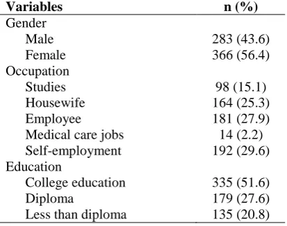
In this study, 649 subjects with the mean age of 32.0 ± 12.2 years (ranging from 15 to 80 years) were interviewed; and 75.0% of respondents were younger than 43 years old. Women constituted 56.4% of the study population; 29.6% of subjects were self-employed and 51.6% had academic education (Table 1).
Table 1. Demographic data of subjects (n = 649)
Variables n (%)
Gender
Male 283 (43.6)
Female 366 (56.4)
Occupation
Studies 98 (15.1)
Housewife 164 (25.3)
Employee 181 (27.9)
Medical care jobs 14 (2.2)
Self-employment 192 (29.6)
Education
College education 335 (51.6)
Diploma 179 (27.6)
Less than diploma 135 (20.8)
The most common sources of information in respondents were family (21.1%) and media (17.3%). Most subjects (65.5%) recommended “mass media” as the best source of information about stroke. When encountering a patient with symptoms consistent with stroke, 82.4% of subjects would notify emergency medical systems (EMS); 92.8% of them would refer the patient to a neurologist and 90.9% believed that the patient should immediately be transferred to a specialized healthcare center in less than 3 hours to receive adequate treatment. The source of information and approach of respondents about stroke is shown in table 2.
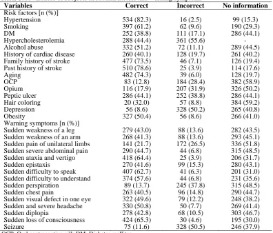
The awareness of participants toward risk factors and warning signs of stroke is shown in table 3. Hypertension (82.3%) and previous history of stroke (78.6%) were the major factors reported by participants, while oral contraceptive pill (OCP) (12.8%) and DM (38.8%) were not reported commonly. In addition, opium use (82.1%) and depression (91.0%) were the most common factors not correctly identified as major risk factors. The awareness about risk factors was poor in 48.8%, moderate in 39.9% and good in 11.3% of respondents.
Table 2. The source of information and approach of respondents about stroke (n = 649)
Variables n (%)
Stroke in relatives or friends
Yes 174 (26.8)
No 475 (73.2)
Previous information about stroke
Yes 362 (55.8)
No 287 (44.2)
Interest to have an information about stroke
Yes 597 (92.0)
No 59 (8.0)
Sources of information
Family members and friends 137 (21.1)
Television and radio 112 (17.3)
Reading book 38 (5.9)
Newspapers 15 (2.3)
Others 240 (37.0)
Multisource 107 (16.5)
Recommended sources of information
Mass media audiovisual 425 (65.5)
Educational booklets 100 (15.4)
Others 52 (8.0)
Multisource 72 (11.1)
Encounter patients with symptoms suggesting stroke
Telephone EMS 535 (82.4)
Refer to neurologist 602 (92.8)
Need medical help immediately (before 3 hours)
590 (90.9)
EMS: emergency medical systems
Table 3. Awareness of subjects about risk factors and warning symptoms
Variables Correct Incorrect No information
Risk factors [n (%)]
Hypertension 534 (82.3) 16 (2.5) 99 (15.3)
Smoking 397 (61.2) 62 (9.6) 190 (29.3)
DM 252 (38.8) 111 (17.1) 286 (44.1)
Hypercholesterolemia 288 (44.4) 361 (55.6) -
Alcohol abuse 332 (51.2) 72 (11.1) 289 (44.5)
History of cardiac disease 260 (40.1) 128 (19.7) 261 (40.2)
Family history of stroke 477 (73.5) 46 (7.1) 126 (19.4)
Past history of stroke 510 (78.6) 25 (3.9) 114 (17.6)
Aging 482 (74.3) 39 (6.0) 128 (19.7)
OCP 83 (12.8) 184 (28.4) 382 (58.9)
Opium 116 (17.9) 207 (31.9) 326 (50.2)
Peptic ulcer 286 (44.1) 252 (38.8) 286 (44.1)
Hair coloring 20 (32.0) 57 (8.8) 384 (59.2)
Depression 56 (8.6) 328 (50.2) 265 (40.8)
Obesity 327 (50.4) 56 (8.6) 266 (41.0)
Warning symptoms [n (%)]
Sudden weakness of a leg 279 (43.0) 88 (13.6) 282 (43.5)
Sudden weakness of an arm 268 (41.3) 88 (13.6) 293 (45.1)
Sudden pain of unilateral limbs 141 (21.7) 172 (26.5) 336 (51.8)
Sudden severe abdominal pain 290 (44.7) 44 (6.8) 315 (48.5)
Sudden ataxia and vertigo 418 (64.4) 25 (3.9) 206 (31.7)
Sudden epistaxis 270 (41.6) 99 (15.3) 280 (43.1)
Sudden difficulty to speak 407 (62.7) 41 (6.3) 201 (31.0)
Sudden difficulty to understand 374 (57.6) 44 (6.8) 231 (35.6)
Sudden perspiration 89 (13.7) 245 (37.8) 315 (48.5)
Sudden chest pain 263 (40.5) 96 (14.8) 290 (44.7)
Sudden visual defect in one eye 322 (49.6) 79 (12.2) 248 (38.2)
Sudden and severe headache 330 (50.8) 50 (7.7) 269 (41.4)
Sudden diplopia 278 (42.8) 68 (10.5) 303 (46.7)
Sudden loss of consciousness 424 (65.3) 30 (4.6) 195 (30.0)
Seizure 75 (11.6) 328 (50.5) 246 (37.9(
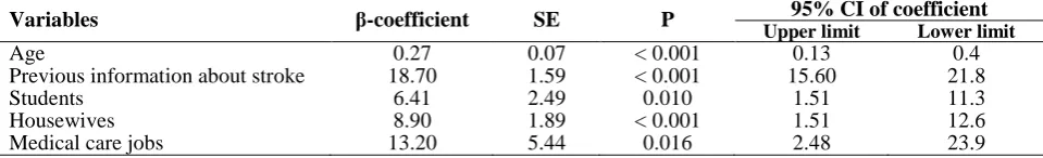
Table 4. Results of multiple linear regressions in variables related to subjects awareness
Variables β-coefficient SE P 95% CI of coefficient
Upper limit Lower limit
Age 0.27 0.07 < 0.001 0.13 0.4
Previous information about stroke 18.70 1.59 < 0.001 15.60 21.8
Students 6.41 2.49 0.010 1.51 11.3
Housewives 8.90 1.89 < 0.001 1.51 12.6
Medical care jobs 13.20 5.44 0.016 2.48 23.9
CI: Confidence interval; SE: Standard error
The loss of consciousness (65.5%), as well as vertigo and ataxia (64.4%), were reported as the most common warning signs of stroke. On the other hand, sudden perspiration (86.3%) and unilateral pain in limbs (78.3%) were the most correctly identified incorrect responses. In addition, the weakness of an arm (41.3%) and diplopia (42.8%) were less commonly identified as the warning signs of stroke. Totally, the awareness of stroke warning signs was poor in 51.8%, moderate in 34.8% and good in 13.4% of respondents.
The approach to patients with stroke was not significantly associated with age (P = 0.700), sex (P = 0.345), educational level (P = 0.084), job (P = 0.340), family history of ischemic stroke (P = 0.100), previous knowledge about stroke (P = 0.130), and the source of information (P = 0.060). However, the awareness of people regarding the risk factors of stroke was significantly related to age (P < 0.001), sex (P = 0.029), educational level (P = 0.006), job (P < 0.001), family history of ischemic stroke (P < 0.001), having previous knowledge about stroke (P < 0.001), and the source of information (P = 0.050). Moreover, the awareness of people about stroke warning signs was significantly associated with age (P < 0.001), sex (P = 0.008), educational level (P = 0.004), job (P < 0.001), family history of ischemic stroke (P < 0.001), having previous knowledge about stroke (P < 0.001), and the source of information (P = 0.018).
Totally, the overall mean percentage score of subjects’ awareness about risk factors and the warning signs of stroke was 54.4 ± 22.8 (ranging from 3.03 to 93.9). In addition, the total awareness in 75 % of participants was more than 70%. In total, the overall awareness of people about stroke was associated with gender (P = 0.017), educational level (P = 0.016), job (P = 0.001), family history of ischemic stroke (P = 0.001), previous knowledge about stroke (P = 0.001), and the source of information (P = 0.050). Multiple linear regressions showed that age (β = 0.277,
P < 0.001), the academic level of education (β = 6.41, P = 0.010), housewifery (β = 8.9, P < 0.001), jobs related to medical care and the previous information about stroke (β = 18.71, P < 0.001) were significant predictors of the overall awareness of patients about stroke (Table 4) as the overall awareness level of subjects increased by 0.27% in proportion to every year of increase in age. It also increased by 18.7% when people had previous information about stroke.
To propagate efficient treatment-seeking behavior and to bring the correct message appropriately, the assessment of public needs for information should precede the development and implementation of educational campaigns for the public.9 The early detection of stroke risk factors and warning signs has an important role in the prevention and management of patients with stroke.6 The lack of information about stroke can disarrange the prevention programs and delay the rapid medical intervention. This study was the first population-based telephone survey in Iran which assessed the public awareness of stroke, warning signs and risk factors in Rasht. This study has shown that the awareness about stroke, its risk factors and warning signs is adequate, and it can be related to significant factors such as education and the source of information.
further public education using various media sources including television, radio, newspaper, magazine, and educational pamphlets is needed to improve the awareness of community regarding stroke’s risk factors.
The loss of consciousness as well as vertigo and ataxia were the major warning signs of stroke identified by participants. However, a few number of respondents reported paresthesia and aphasia as warning signs. In addition, sudden chest pain and perspiration were reported by some respondents. In the only community-based, face-to-face interview survey conducted in Iran, Borhani Haghighi, et al. revealed that abdominal pain are one of the most commonly identified symptoms of stroke.6 While in another study in Korea,24 participants identified paresthesia as the main warning signs. Therefore, most of the educational efforts in future should be focused on increasing the awareness of Iranian community about stroke’s warning signs.
In our study, the mean percentage score of public awareness about risk factors and warning signs was 54.4% and it was more than 70.0% in 75.0% of cases. In a telephone survey by Pancioli, et al. 57% of subjects knew at least one warning sign and 68% of them named at least one risk factor.18 In the only community-based, face-to-face interview survey in Korea by Kim, et al.,24 62.0% reported at least one stroke symptom and 56.0% reported at least one risk factor for stroke in open-ended questioning. In Saudi Arabia, Alaqeel, et al.23 revealed that 21.7% of the respondents correctly chose ≥ 5 risk factors and made ≤ 1 error and 18.4% of the participants were able to correctly identify ≥ 3 symptoms of the list and make ≤ 1 error.
In another large population-based telephone survey, Sug Yoon, et al.21 found that 76.2% of Australian individuals could name one or more risk factors of stroke; however, just 49.8% of them could identify at least one stroke warning sign. Moreover, smoking and visual disturbance were two most common risk factors and symptoms of stroke listed by 39.4% and 24.1% of respondents, respectively. The high rate of correct answers in our study is likely to be related to use of closed-ended questions in our questionnaires, in contrast to most previous studies. Therefore, further studies will need to be performed to survey the public awareness when using open ended questionnaire.
In a study by Travis, et al. in the USA, 42.0% of persons would first call EMS if having a stroke.25
In our study, 82.4% of respondents would immediately call EMS, 92.8% would refer to a neurologist and 90.9% suggested receiving adequate treatment in less than three hours, when they see patients with symptoms suggesting stroke. There are several possible explanations for this relatively high percentage of correct responses, compared to similar studies. First, participation of more educated people in this study. Second, higher general medical knowledge of our population. Third, identifying loss of consciousness as the most common warning sign by participants. It is therefore likely that the fear of loss of consciousness sign alone may be related to high rate of calling EMS in our study.
The most common sources of information in our study were friends and then multimedia; moreover, the highest awareness was seen in respondents that studied books as sources of information. Kim, et al.24 revealed that the major source of information about stroke was television (59%), and the most reliable sources were the respondents' physicians (55%); however, among the respondents of 20 to 39 years of age, the Internet (37%) was the second greatest source of information. Alaqeel, et al.23 reported that 49.9% of respondents named mass media as the source of their knowledge. In a study by Stern, et al.26 in the USA, 657 adults were examined for the effectiveness of the slide/audio community education program lonely or accompanied by facilitation led by a trained individual. They reported that slide/audio program is effective in increasing the knowledge of stroke risk factors, warning signs, and necessary action but facilitation did not significantly affect the short-term acquisition of information.26 Different findings in source of information can be related to increased number of Internet users; also it showed that multimedia programs can be effective in all developing and developed countries.
high-income people.25 Borhani Haghighi, et al. showed that the attitude and knowledge were related to age, education and income but not to gender and domicile.6 Stern, et al. expressed that multimedia, family and friends, health professionals and educational campaigns can successfully increase stroke awareness. However, they also showed that race or educational level could not increase the knowledge.26 These differences can be related to cultural and other influential factors among different nations. According to the important effect of age and educational level in this study, more educational programs, especially in school age, should be planned in this region to increase the level of awareness of students.
As a limitation, limited-sample telephone-based survey instead of face-to-face interview was done in our study. Telephone call can affect the responses of subjects. Although interviewers were trained on how to avoid leading questions, the interviewer bias might have influenced the participant response. In addition, a number of people in this region might not have had access to telephone; thus, people with low socioeconomic status may not have been included.
This study concludes that the awareness of people about stroke, its risk factors and warning signs was adequate and can be related to significant factors such as education and the source of information. So it is suggested to program public multimedia and health education in academies and colleges in future to increase the knowledge and awareness of people.
The authors declare no conflict of interest in this study.
The authors would like to thank Guilan Trauma Research Center, and Dr. Kazemnezhad for his statistical advice.
1. Ferri CP, Schoenborn C, Kalra L, Acosta D, Guerra M, Huang Y, et al. Prevalence of stroke and related burden among older people living in Latin America, India and China. J Neurol Neurosurg Psychiatry 2011; 82(10): 1074-82.
2. Zhang Y, Chapman AM, Plested M, Jackson D, Purroy F. The incidence, prevalence, and mortality of stroke in France, Germany, Italy, Spain, the UK, and the US: A literature review. Stroke Res Treat 2012; 2012: 436125.
3. Hosseini AA, Sobhani-Rad D,
Ghandehari K, Benamer HT. Frequency and clinical patterns of stroke in Iran - Systematic and critical review. BMC Neurol 2010; 10: 72.
4. Fahimfar N, Khalili D, Mohebi R, Azizi F, Hadaegh F. Risk factors for ischemic stroke; results from 9 years of follow-up in a population based cohort of Iran. BMC Neurol 2012; 12: 117.
5. Azarpazhooh MR, Etemadi MM, Donnan GA, Mokhber N, Majdi MR, Ghayour-Mobarhan M, et al. Excessive incidence of stroke in Iran: evidence from the Mashhad Stroke Incidence Study (MSIS), a population-based study of stroke in the Middle East. Stroke 2010; 41(1): e3-e10. 6. Borhani Haghighi A, Karimi AA, Amiri
A, Ghaffarpasand F. Knowledge and attitude towards stroke risk factors, warning symptoms and treatment in an
Iranian population. Med Princ Pract 2010; 19(6): 468-72.
7. Campos-Sousa RN, Soares VY, Almeida KJ, Carvalho LI, Jacobina KS, Athayde Netto AE, et al. Knowledge of stroke among a Brazilian urban population. Arq Neuropsiquiatr 2007; 65(3A): 587-91. 8. Cossi MJ, Preux PM, Chabriat H, Gobron
C, Houinato D. Knowledge of stroke among an urban population in Cotonou (Benin). Neuroepidemiology 2012; 38(3): 172-8.
9. Nedeltchev K, Fischer U, Arnold M, Kappeler L, Mattle HP. Low awareness of transient ischemic attacks and risk factors of stroke in a Swiss urban community. J Neurol 2007; 254(2): 179-84.
10. Pandian JD, Jaison A, Deepak SS, Kalra G, Shamsher S, Lincoln DJ, et al. Public awareness of warning symptoms, risk factors, and treatment of stroke in northwest India. Stroke 2005; 36(3): 644-8. 11. Truelsen T, Krarup LH. Stroke awareness in Denmark. Neuroepidemiology 2010; 35(3): 165-70.
12. Al Shafaee MA, Ganguly SS, Al Asmi AR. Perception of stroke and knowledge of potential risk factors among Omani patients at increased risk for stroke. BMC Neurol 2006; 6: 38.
13. Evci ED, Memis S, Ergin F, Beser E. A population-based study on awareness of stroke in Turkey. Eur J Neurol 2007;
14(5): 517-22.
14. Ramsden VR, Shuaib A, Reeder BA, Khan K, Liu L. Risk factor awareness: A randomized telephone survey of public knowledge. Can J Public Health 1994; 85 Suppl 2: S57-S60.
15. Kleindorfer D, Khoury J, Broderick JP, Rademacher E, Woo D, Flaherty ML, et al. Temporal trends in public awareness of stroke: Warning signs, risk factors, and treatment. Stroke 2009; 40(7): 2502-6. 16. Becker K, Fruin M, Gooding T,
Tirschwell D, Love P, Mankowski T. Community-based education improves stroke knowledge. Cerebrovasc Dis 2001; 11(1): 34-43.
17. Rowe AK, Frankel MR, Sanders KA. Stroke awareness among Georgia adults: Epidemiology and considerations regarding measurement. South Med J 2001; 94(6): 613-8.
18. Pancioli AM, Broderick J, Kothari R, Brott T, Tuchfarber A, Miller R, et al. Public perception of stroke warning signs and knowledge of potential risk factors. JAMA 1998; 279(16): 1288-92. 19. Hux K, Rogers T, Mongar K. Common
perceptions about strokes. J Community Health 2000; 25(1): 47-65.
21. Sug Yoon S, Heller RF, Levi C, Wiggers J, Fitzgerald PE. Knowledge of stroke risk factors, warning symptoms, and treatment among an Australian urban population. Stroke 2001; 32(8): 1926-30. 22. Neau JP, Ingrand P, Godeneche G.
Awareness within the French population concerning stroke signs, symptoms, and risk factors. Clin Neurol Neurosurg 2009; 111(8): 659-64.
23. Alaqeel A, AlAmmari A, AlSyefi N, Al-Hussain F, Mohammad Y. Stroke awareness in the Saudi community living in Riyadh: prompt public health measures must be implemented. J Stroke Cerebrovasc Dis 2014; 23(3): 500-4. 24. Kim YS, Park SS, Bae HJ, Heo JH, Kwon
SU, Lee BC, et al. Public awareness of stroke in Korea: a population-based national survey. Stroke 2012; 43(4): 1146-9.
25. Travis LH, Flemming KD, Brown RD Jr, Meissner I, McClelland RL, Weigand SD. Awareness of stroke risk factors, symptoms, and treatment is poor in people at highest risk. J Stroke Cerebrovasc Dis 2003; 12(5): 221-7. 26. Stern EB, Berman M, Thomas JJ, Klassen
![Table 3. Comparison of side effects between two groups Side effect Low dose IFN [n (%)] High dose IFN [n (%)]](https://thumb-us.123doks.com/thumbv2/123dok_us/8750745.1748496/10.595.80.516.95.135/table-comparison-effects-groups-effect-low-dose-high.webp)