Open Access
Research article
Effects of aging and calorie restriction on the global gene expression
profiles of mouse testis and ovary
Alexei A Sharov
†1, Geppino Falco
†1, Yulan Piao
1, Suresh Poosala
2,
Kevin G Becker
2, Alan B Zonderman
2, Dan L Longo
3, David Schlessinger
1and
Minoru SH Ko*
1Address: 1Laboratory of Genetics, National Institute on Aging, National Institutes of Health, Baltimore, MD 21224, USA, 2Research Resources
Branch, National Institute on Aging, National Institutes of Health, Baltimore, MD 21224, USA and 3Laboratory of Immunology, National Institute
on Aging, National Institutes of Health, Baltimore, MD 21224, USA
Email: Alexei A Sharov - [email protected]; Geppino Falco - [email protected]; Yulan Piao - [email protected]; Suresh Poosala - [email protected]; Kevin G Becker - [email protected]; Alan B Zonderman - [email protected]; Dan L Longo - [email protected]; David Schlessinger - [email protected]; Minoru SH Ko* - [email protected] * Corresponding author †Equal contributors
Abstract
Background: The aging of reproductive organs is not only a major social issue, but of special interest in aging research. A long-standing view of 'immortal germ line versus mortal soma' poses an important question of whether the reproductive tissues age in similar ways to the somatic tissues. As a first step to understand this phenomenon, we examine global changes in gene expression patterns by DNA microarrays in ovaries and testes of C57BL/6 mice at 1, 6, 16, and 24 months of age. In addition, we compared a group of mice on ad libitum (AL) feeding with a group on lifespan-extending 40% calorie restriction (CR).
Results: We found that gene expression changes occurred in aging gonads, but were generally different from those in somatic organs during aging. For example, only two functional categories of genes previously associated with aging in muscle, kidney, and brain were confirmed in ovary: genes associated with complement activation were upregulated, and genes associated with mitochondrial electron transport were downregulated. The bulk of the changes in gonads were mostly related to gonad-specific functions. Ovaries showed extensive gene expression changes with age, especially in the period when ovulation ceases (from 6 to 16 months), whereas testes showed only limited related changes. The same trend was seen for the effects of CR: CR-mediated reversal of age-associated gene expression changes, reported in somatic organs previously, was limited to a small number of genes in gonads. Instead, in both ovary and testis, CR caused small and mostly gonad-specific effects: suppression of ovulation in ovary and activation of testis-gonad-specific genes in testis.
Conclusion: Overall, the results are consistent with unique modes of aging and its modification by CR in testis and ovary.
Published: 3 June 2008
BMC Biology 2008, 6:24 doi:10.1186/1741-7007-6-24
Received: 27 February 2008 Accepted: 3 June 2008
This article is available from: http://www.biomedcentral.com/1741-7007/6/24 © 2008 Sharov et al; licensee BioMed Central Ltd.
Background
Aging is a complex biological phenomenon characterized by a gradual and progressive loss of function in diverse organs and tissues. Global gene expression profiling is beginning to provide molecular signatures of aging in var-ious organs and tissues [1,2]. For example, aged muscles show a reduced expression of genes involved in energy metabolism and an increased expression of heat-shock proteins and oxidative-stress inducible genes [3]. The most recent analysis of human muscle samples has revealed the increased expression of genes associated with extracellular matrix, cell growth, complement activation, and cytosolic ribosomes, as well as the decreased expres-sion of genes involved in chloride transport and mito-chondrial electron transport [1]. Aged brain tissues show increased expression of genes associated with stress and the inflammatory response and decreased expression of genes associated with the ubiquitin-proteasome pathway and growth factors [2,4]. Aged heart shows increased mRNA levels for structural proteins and decreased mRNA levels for genes involved in fatty-acid metabolism [3]. Global gene expression profiling has also revealed that age-related gene expression changes in muscle are com-pletely [5] or partially [6] suppressed by calorie restriction (CR), which is a factor capable of increasing the lifespan in organisms, including rodents [7-9]. Similarly, it has been shown that age-related decrease in the induction of Hsp70 in response to heat stress in rat hepatocytes is par-tially reversed by CR [10].
Gonads, especially ovary, are of special interest in aging research, because the human ovary shows an especially demarcated effect of age: a reproducible decline of repro-ductive capacity in the mid-30 s, followed by menopause in the late 40 s to 50 s. Modern trends postponing child-bearing in developed countries are thus accompanied by increased infertility and other complications. Aging of gonads also has direct implications for longevity, as increased fertility is associated with decreased longevity in several species [11,12]. An increase in longevity in mutants is often accompanied by a reduction of fecundity, whereas delayed reproduction is often associated with longevity. For example, it has been shown that the abla-tion of germ cells increases the lifespan of the worm [13] (although not the fly [14]). Furthermore, the transplanta-tion of young ovary increases the lifespan in mouse, sug-gesting that ovaries can affect the lifespan of organisms, possibly through the production of hormones and other factors [15]. Gonads are also the focus of attention in var-ious theories of aging [16]. For example, an evolutionary theory of aging considers that aging occurs because the maintenance of individuals is not supported by natural selection after their reproductive periods [11]. The dispos-able soma (DS) theory suggests that when availdispos-able energy is limited, it is selectively reallocated from
repro-duction to somatic maintenance, thermogenesis, DNA repair and anti-oxidative functions [7,11,17].
Despite the importance of gonads in aging research, stud-ies of gonad aging by global gene expression profiling have been limited to partial studies of mouse oocytes [18] and ovary [19]. It has remained unclear whether gonads and somatic organs show similar patterns of gene expres-sion associated with aging and CR. In this study, we have carried out global gene expression profiling of testis and ovary from 1- to 24-month-old mice on ad libitum (AL) or CR diets. The whole-genome DNA microarray data pro-vide a resource for the analysis of combined effects of age, diet, and sex on gene expression in the gonads.
Results
Experimental design
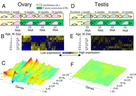
We analyzed gene expression patterns in ovaries and testes sampled from 1-, 6-, 16-, and 24-month-old C57BL/6 mice fed on AL and CR diets (Figure 1A and 1D). Mice were kept on an AL diet until 14 weeks of age and then split into AL and CR gonads [20]. Two individual animals for each condition were used for the microarray studies. We examined a total of 28 samples (two AL ovaries at each of 1, 6, 16, and 24 months; two CR ovaries at 6, 16, and 24 months; two AL testes at 1, 6, 16, and 24 months; and two CR testes at 6, 16, and 24 months). These 28 samples were hybridized on whole-genome oligonucleotide microarrays bearing approximately 44,000 probes, repre-senting 25,585 nonredundant genes with gene symbols [21] (Additional file 1). The same RNA samples were used for quantitative reverse-transcription polymerase chain reaction (RT-PCR) validation of selected genes.
The effects of CR on physiological status and overall sur-vival curve for C57BL/6 mice used in this study have been well established [20]. For example, it has been shown that 40% CR suppresses follicle cycles and ovulation in female C57BL/6 mice [22]. Survival data on the oldest C57BL/6 mice we used (24 months) are as follows: males AL (70%), females AL (60%), males CR (90%), and females CR (85%) [20]. The average lifespan of this strain on AL diet is around 30 months; the CR diet extends average lifespan up to 50% [20].
Global analysis of age-related changes in gene expression: major changes in ovary and minor changes in testis
limited changes in gene expression over time (Figure 1E and 1F). There was a major alteration of gene expression patterns in ovary between 6 and 16 months (Figure 1B and 1C), which corresponds to the time when ovulation ceases in mice [19]. A simple pair-wise comparison between samples from the youngest (1 month) and the oldest (24 months) animals also revealed a difference in the rate of change between testis and ovary. There were 4657 genes with significant differential expression between oldest (24 months) and youngest (1 month) females on AL diet; and 1724 of these genes had more than two-fold change in their expression. In contrast, there were only 326 genes with significant differential expression between the oldest (24 months) and youngest (1 month) males on AL diet; and only 122 of these genes had more than two-fold change in their expression. It was also clear from the heatmaps that the difference between AL and CR in each age group was modest. Thus, CR did
not suppress age-associated gene expression changes in testis and ovary.
To examine organ-specific genes, we first separated genes that were predominantly expressed in ovary or testis from genes that were expressed in both organs. There were 14,438 genes with differential expression between ovary and testis (false discovery rate (FDR) at most 0.1, at least 1.5-fold change in gene expression; see Additional file 3). Genes that had a more than two-fold difference in expres-sion between ovary and testis (N = 9993), are called 'ovary-associated' and 'testis-associated' genes throughout the analysis below.
Major trends in gene expression changes with age in ovary
To focus on the differences between young and old ovary, we compared a group of samples from young (1 and 6 months) females with a group of samples from old (16
Global patterns of gene expression in ovary and testis in mice of age from 1 to 24 months, and on an AL or a CR diet
Figure 1
Global patterns of gene expression in ovary and testis in mice of age from 1 to 24 months, and on an AL or a CR diet. (A) Experimental design for females; (B) two-dimensional heatmap of expression of 3000 most significant genes in ovary; (C) three-dimensional heatmap of expression of the same genes in ovary; (D) experimental design for males; (E) two-dimensional heatmap of expression of 3000 most significant genes in testis (819 genes overlapped with ovary); (F) three-dimen-sional heatmap of expression of the same genes in testis.
Ovary
Testis
1 month 6 months 16 months 24 months Newborn
RNA RNA RNA RNA
Calorie restriction (CR)
RNA RNA RNA RNA
Ad libitum (AL)
1 month 6 months 16 months 24 months Newborn
Low expression High expression
Age, M Diet Age, M
1 6 6 16 16 24 24
-AL CR AL CR AL CR
Diet
Genes Genes
1-AL 6-AL
6-CR 16-AL
16-CR 24-AL
24-CR
1-AL 6-AL
6-CR 16-AL
16-CR 24-AL
24-CR
A
B
C
D
E
F
1 6 6 16 16 24 24
and 24 months) females on AL diet. Based on one-factor ANOVA we found 3937 genes that were differentially expressed between young and old ovaries (FDR at most 0.1, at least 1.5-fold change in gene expression level); however, 323 of them did not pass the two-factor linear
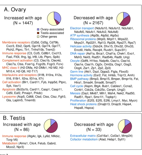
regression test (p < 0.05 for age variable) and therefore were not considered age-dependent. There were 1447 genes that increased and 2167 genes that decreased their expression in the ovary with age, respectively (Figure 2A and Additional file 4). To further characterize these genes,
Functional groups of genes affected by age
Figure 2
Functional groups of genes affected by age. (A) Ovary and (B) testis. Pie charts show the proportion of genes that were over-expressed either in ovary or testis by at least two-fold. Functional categories of genes were assembled from GO annota-tions and from PubMed.
Decreased with age
(N = 2167)
Increased with age
(N = 1447)
Membrane receptors(Cd3d, Cd3g, Cd79b,
Cxcr3, Ebi2, Gpr6, Gpr18, Gpr74, Gpr77, Ptch2, Ptprc, Tlr1, Tnfrsf10b, Trem2)
Immune response(C3, Ccl5, Cd8b1, Cxcl13,
Fasl, Flt3l, Ifng, Igj, Irf8, Ltb, Ptprc, Spib)
Complement activation (C3, Clec1b, Clec4d, Clec7a, Ctss, Fcer1g, Fcgr2b, Fcgr3, Fcrx)
MHC class II(DMa, DMb1, M2,
H2-M10.4, H2-Q8, H2-T17)
Interleukins and receptors(Il1f8, Il10ra, Il12a, Il16, Il18r1, Il2ra, Il31ra, Il7r)
Cell activation(Blr1, Card11, Fyb, Inpp5d, Ptprc, Syk, Vav1)
Apoptosis(Bcl2a1b, Card11, Casp1, Casp11,
Cd5l, Eaf2, Pmaip1, Prkdc)
Lysosome(Acp5, Cd68, Ctsd, Ctss, Ctsz, Fgfr3, Gla, Laptm5, Tmem9)
Electron transport(Ndufa10, Ndufa12, Ndufab1, Ndufb5, Ndufc1, Ndufc2, Ndufs8)
ATP synthesis(Atp5b, Atp5d, Atp5o)
Ribosomal proteins(Mrpl3, Mrpl11, Mrps22, Mrps31, Rpl22l1, Rpl13, Rpl28, Rps27l, Rps7) Helicase activity(Ddx24, Dhx15, Dhx32, Dhx33,
Ercc6l, Hells, Recql4, Ruvbl1, Supv3l1) DNA repair(Asf1a, Brca1, Chaf1a, Chek1, Exo1,
Msh3, Neil3, Rad51, Rad54b, Rad9, Ung) Oocyte(Gdf9, H1foo, Nalp4b, Oas1c, Oas1d,
Oas1e, Oas1h, Og2x, Omt2a, Oog1, Oog3, Oog4, Zar1, Zp1, Zp2, Zp3)
Germ line(Akt1, Dazl, Dppa3, Figla, Fbxo5) Hormone activity(Esr2, Fst, Inhbb, Trip13, Amh) BMP pathway(Bmp3, Bmp15, Bmper, Bmpr1b, Fst,
Htra1, Smad4, Smad6, Smad7)
Cell cycle(Aspm, Btg4, Bub1, Cables1, Ccna2, Ccnb1, Cdc2a, Cdc25c, Cenph, Ung2)
Meiosis(Dazl, Mki67, Mlh1, Msh4, Nek2, Rad50, Rad51, Rpa1, Smc1l1, Topbp1)
Proliferation (E2f3, E2f5, E2f6, Lmyc1, Myc, Mycn) Heat shock proteins(Dnajb13, Dnajc9, Hspa4,
Hspa8, Hspca) Ovary-associated
Testis-associated Other genes
A. Ovary
Increased with age
(N = 86)
Decreased with age
(N = 33)
Immune response(Atg4c, Igk, Ly6d, Nfkbiz, Tcrg-V6)
Metabolism(Alms1, Clic4, Fetub, Gabrd, Mccc2, Npr3)
we analyzed functional groups of genes in these sets based on gene ontology (GO) [23] terms (Additional file 5) and information gleaned from the literature. Examples of genes that showed the high differential expressions during aging and were grouped into different functional groups are presented in Additional file 6.
Groups of genes that increased their expression with age included 'receptor binding' (N = 43; mostly membrane receptors), 'defense response' (N = 83), and 'immune response' (N = 65); see Additional file 5. Examples of genes related to immune response included interleukins and interleukin receptors (Additional file 6A), and major histocompatibility complex (MHC) II (Additional file 6B). MHC-II genes are known to be expressed in the theca layer of growing ovarian follicles, and their expression is increased in postovulatory follicles [24]. The expression of apoptosis-related genes (for example, Bcl2a1b, Casp1, Casp11) and several tumor necrosis factor (TNF) recep-tors (Additional file 6E and 6F) also increased with age. Many genes were related to complement activation (innate immune response), including Fc receptors (Fcrx, Fcgr2b, Fcgr3, Fcer1g), complement components (C3, C1sb), lectins (Clec7a, Clec4d, Clec1b, Clec4a3, Lgals3; Additional file 6D), toll-like receptors (Tlr1, Tlr2, Tlr13), and cathepsins (Ctss, Ctsd, Ctsb, Ctsz).
Groups of genes that decreased their expression with age included 'mitochondrion' (N = 101), 'protein biosynthe-sis' (N = 98), 'RNA metabolism' (N = 91), 'DNA metabo-lism' (N = 128), 'cell cycle' (N = 119), and 'response to DNA damage' (N = 47) (Figure 2A, Additional files 5, 6J and 6L). The mitochondrion group included electron transport, adenosine triphosphate (ATP) synthesis, and mitochondrial ribosomal proteins. The protein biosyn-thesis group included cytosolic and mitochondrial ribos-omal proteins. Taken together, a general decline of metabolism is one of the main features of ovary aging.
Groups of genes that decreased their expression with age also included many oocyte-specific genes: Oog1, Oog3,
Oog4, Zp1, Zp2, Zp3, Nalp5 (Mater), Gdf9, Og2x, Omt2a,
H1foo, Oas1d, Zar1, Nalp4b, and Oosp1 (Figure 2A and Additional file 6G, H and 6I). Oogenesin3 (Oog3) showed the most dramatic decrease in expression (57.7-fold) from age 1 to 24 months. In our earlier comparison of young and old oocytes [18], expression of some of these genes in oocytes decreased with age (for example, Nalp5, Zp3) but no more than three-fold from 5–6 weeks to 42–45 weeks. The current data indicate a larger decrease of expression of these genes in the ovary (for example, 8-fold for Nalp5
and 5.3-fold for Zp3 within a comparable age interval from 1 to 16 months), which is most likely related to the reduction of the number of oocytes in ovaries with age [25]. Other groups of genes that decreased expression
with age were hormones and hormone receptors, germ-line-specific genes, genes associated with cell cycle, the bone morphogenetic protein (BMP) pathway, helicase activity, meiosis, and heat shock proteins (Figure 2A and Additional file 6J and 6K). Taken together, these gene expression data are consistent with the characteristic decrease in the number of oocytes and follicles in ovaries with age.
Major trends in gene expression changes with age in testis
We carried out a similar analysis on testis. Comparison of a group of samples from young (1 and 6 months) males with a group of samples from old (16 and 24 months) males on AL diet with one-factor ANOVA revealed 163 differentially expressed genes (FDR at most 0.1, at least 1.5-fold change in gene expression); however 41 of them did not pass the two-factor linear regression test (p < 0.05 for age variable) and therefore were not considered age-dependent. There were 86 genes that increased and 33 genes that decreased their expression in testis with age (Figure 2B and Additional file 7). Only 10 up-regulated genes and 9 down-regulated genes changed their expres-sion consistently in testis and ovary (Additional file 7), which indicates that major age-related changes of gene expression in testis are mostly organ-specific. Indeed, age-affected genes in testis included a large portion of testis-associated genes (see the pie charts in Figure 2B). Genes that increased their expression with age included immune response and metabolism (Figure 2B and Additional file 8A and 8B). Genes that decreased their expression in testis with age included extracellular matrix and cofactor metab-olism (Figure 2B; Additional files 8C and 9).
Effect of CR on gene expression in ovary
To focus on the differences between AL and CR in ovary, we compared a group of ovary samples from females on a CR diet with a group of ovary samples from females on AL diet using one-factor ANOVA as described in Methods and found 123 differentially expressed genes (FDR at most 0.1, at least 1.5-fold change). However, five of these genes did not pass the additional two-factor ANOVA test (p < 0.05 for diet factor) and therefore were not considered diet-dependent. This analysis confirmed our observation that CR affected a much smaller set of genes than aging in ovary (Figure 1B), and identified only 34 genes with increased expression and 84 genes with decreased expres-sion by CR (Figure 3A and Additional file 10).
Consistent with the suppression of ovulation by CR [22], genes that were suppressed by CR in the ovary included
Oog4, Zp2, Zp3, Nalp5, Gdf9, Og2x; see Additional file 6H, I and 6J). Although the difference was not significant based on our statistical criteria (FDR at least 0.1, at most 1.5-fold change), the average log-ratio (CR/AL) of the expression of 25 known oocyte-specific genes [29-32] was significantly positive (0.061 ± 0.015, p = 0.0006; see Addi-tional file 11). These data are consistent with the retention of a larger number of oocytes in ovaries of CR mice com-pared with ovaries of AL mice, as implicated by the sup-pression of ovulation under CR [22]. However, alternative explanations, such as structural changes other than oocyte numbers in CR ovaries, cannot be excluded.
The expression of metabolism-related genes in the ovary decreased under CR. For example, many mitochondrial genes, including those involved in electron transport, oxi-doreductase activity, as well as lipid and sterol-metabo-lism, decreased their expression in the ovaries of mice on a CR diet (Figure 3A and Additional files 12, 13D, and 13F). Cell adhesion gene expression was also suppressed by CR diet (Figure 3A and Additional files 12 and 13E). On the other hand, CR diet increased the expression of
metabolism-related genes (for example, Adipoq, Adpn,
Fabp4, Gsn, Mc2r, Pck1, Per2, Per3, Retn, Scd1; see Figure 3A and Additional files 12, 13B, and 13C), many of which are known to regulate body weight and food intake. For example, Retn and Scd1 cause insulin resistance [33,34] and Scd1 is induced by a fat-free diet [35]. Adiponectin (Adipoq) promotes fatty acid oxidation and glucose uptake and has anti-inflammatory and antiatherogenic effects [36,37]. Gelsolin (Gsn) has anti-apoptotic function [38].
Mc2r regulates steroidogenesis [39]. Pck1 stimulates hepatic glucose output [40]. Fabp4 is a transporter of fat molecules [41]. Activation of circadian clock genes (Per2,
Per3) may be related to the regular timing of food supply in CR mice. It has been shown that the Per2 gene is one of the major components of the ovarian circadian clock in rats [42].
As an alternative approach to analyzing the data, we com-pared gene expression levels in ovary between AL and CR conditions for each age group separately, because the above-mentioned analysis comparing all-age pooled data might miss genes responsive to CR in an age-dependent
Functional groups of genes affected by diet (AL versus CR)
Figure 3
Functional groups of genes affected by diet (AL versus CR). (A) Ovary and (B) testis. Pie charts show the proportion of genes that were over-expressed either in ovary or testis by at least two-fold. Functional categories of genes were assembled from GO annotations and from PubMed.
Metabolism regulation(Adipoq, Adpn, Bcl6, Gsn, Pck1, Retn, Scd1)
Transporter activity(Fabp4, Pkd2l2, Slco2b1, Slc7a10)
Rythmic process(Per2, Per3)
Lipid/steroid metabolism(Akr1c18, Ch25h, Gm2a, Haghl, Hsd11b2, Lhfp, Lsr)
Oxidoreductase activity(Akr1c18, Ch25h, Cyp4f14, Cyp11a1, Hao3, Hsd11b2, Kcnab3, Lsr,
Txndc2)
Electron transport(Cyp4f14, Cyp11a1, Fdx1, Hao3, Txndc2)
Cell adhesion(Agc1, Col3a1, Dok1, Epdr2, Icam4, Itga11, Pdlim2, Plau, Scarb1, Timp1)
Testis-associated(Apobec4, Nalp14, Pcsk1, Speer4b, Wdr17)
Aging(Kl)
A. Ovary
B. Testis
Increased with CR
(N = 34)
Decreased with CR
(N = 84)
Ovary-associated Testis-associated Other genes
Increased with CR
(N = 14)
Decreased with CR
(N = 4)
manner. As this analysis was based on two replications only, we expected to see a higher number of false posi-tives. To limit their numbers, we excluded genes that showed contradictory responses to CR at different ages (for details see Methods). By using pair-wise comparisons of log-expression mean values for each age group based on the error variance identified in ANOVA, we found that some metabolism-related genes (Acacb, Dgat2, Ces1, Ces3,
Pfkfb3, and Pdha1) were upregulated by CR only in old age (24 months; see Additional file 14). It has been reported that Dgat2 is regulated by Leptin and is involved in insulin resistance [43]. Acacb and Pfkfb3 are also involved in the regulation of metabolism [44-46]. In contrast, the expres-sion of relaxin receptor Lgr7, which stimulates appetite in rats [47] and dystrophin related protein Drp2 was upregu-lated by CR only in young females. Similarly, a large group of genes (N = 166) were downregulated by CR only in the ovary of young females (6 months). These genes included many metabolism genes (for example, Akr1c18,
Acox3, Ch25h, Cox6a1, Fdx1, Olr1, Pigt, Sdc1) and genes that are expressed in ovarian follicles (Cdkn1b, Cdkn2b,
Mif, S100a6, Saa1, and Star) [27,48-51]. This group of genes also included Sfrp4, which is expressed in corpus luteum [52], genes involved in extracellular matrix (Adamts4, Adamts12, Col3a1, Col4a1, Lamc1, Mmp2,
Mmp9, Sparc, Timp1), and nucleosome assembly (Hist1h2aa, Hist1h2bn, Hist1h4d, Hist1h4h, Hist1h4m,
Hist2h4, Myst3), which may be indirectly related to follicle growth and degradation. The decrease of expression of these genes is consistent with the repression of ovulation by CR in young females.
As mentioned in the introduction, it is of major interest whether CR can delay or abolish age-associated altera-tions of gene expression. However, the number of genes altered in the ovary both by age and CR over the entire lifespan was very small (N = 18; see Additional file 10), which suggests a lack of interaction between these proc-esses. However, we found more interaction between CR and aging by considering age-specific effects of CR in ova-ries (Figure 4 and Additional file 14). Among 35 genes affected by both age and CR at 6 months, the majority (91%, N = 32) changed their expression in the same direc-tion in old age and under CR (Figure 4A). As most of these
genes decreased with both age and CR (N = 30) and
included genes related to follicle growth and ovulation (see above), we suggest that CR at a young age causes pre-mature decline of ovarian function, especially repression of follicle growth and ovulation. However, at older ages CR caused some anti-aging effects in ovaries: of 20 genes whose expression depended on both age and diet at the age of 16 or 24 months, 13 genes changed their expression in the opposite direction in response to age and CR (Fig-ure 4 and Additional file 14). For example, Clec4d, Crb2,
Csprs, Lefty1, Mcpt5, Sfrp4, and Upk1b increased their expression with age but were suppressed by CR.
Effect of CR on gene expression in testis
We next focused on the differences between AL and CR in testis, and compared a group of samples from males on a CR diet with a group of samples from males on an AL diet using one-factor ANOVA (as described in Methods) and found 21 differentially expressed genes (FDR at most 0.1, at least 1.5-fold change). However, two of these genes did not pass the additional two-factor ANOVA test (p < 0.05 for diet factor) and therefore were not considered diet-dependent. There were 15 genes that increased and 4 genes that decreased their expression in the testes of mice on a CR diet (Figure 3B and Additional files 13G, H, I, and 15). There was no overlap between genes that responded to CR in ovary and testis, which implies that the effect of CR is specific to testis or ovary, and suggests that there are no universal effects of CR in gonads.
Among genes activated by CR in testis, the majority were testis-associated (93%, N = 14; Figure 3B and Additional file 14). These genes included two metabolism-related genes, Pcsk1, which is directly involved in converting proinsulin to insulin [53], and Apobec4, as well as genes involved in spermatogenesis (Nalp14, Speer4b) [54,55].
Analysis of age-specific responses of gene expression to CR in testis revealed additional genes affected by diet (Addi-tional file 16). To limit false positives, we excluded genes that showed contradictory responses to CR at different ages (for details see Methods). Genes that were suppressed by CR in testis at various ages included genes associated with the immune response (Ccl11, Ccl8, Clec4e, Clec7a,
Cxcl5, Gbp4, Ifit2, Osm, Tlr2, Tlr7) and cellular morpho-genesis (Creg1, Esm1, Krt1-17, Nov, Osm). The majority of genes activated by CR were testis-associated, especially at younger ages (6–16 months), whereas only a few testis-associated genes were suppressed by CR (Figure 4D). Over-represented functional groups of genes activated by CR included ion transport (Catsper4, Chrna9, Emid2,
Grin2b, Kcnn2, Kcnn3, Kcnu1, Sytl1) and carboxylic acid metabolism (Acot12, Cpt1b, Folh1, Gls2, Pdgfd, Si, Tdo2). These data clearly show that testicular functions are not harmed even under low-energy conditions. There were only 12 genes affected by both age and CR in testis, and the majority of them (N = 10) changed in opposite direc-tions with age and CR in the oldest group of males (24 months); see Figure 4B.
Expression of genes associated with aging, DNA repair, and heat shock response
in repair and control of DNA and the response to heat shock, because they are potential indicators of the aging process. The list of these genes (Additional file 17, N = 218) was obtained by combining information from GO annotations, Bioscience Corporation [57] and the litera-ture [58]. Examples of gene expression patterns for these genes are given in Additional file 18. A large portion of these genes contained testis-associated genes (47%, N = 103), and only a small portion of genes (10%, N = 22) was associated with ovary (Additional file 17). Expression of these genes is probably linked to reproducing germ cells, which are abundant in testis and absent in ovary. For
example, Fanconi anemia gene Fancd2 and breast cancer gene Brca2 had a higher expression in testis than in ovary (Additional file 18A). However, some genes were over-expressed in ovary, including the alpha thalassemia gene
Atrx, which is known to be involved in chromatin regula-tion [59] (Addiregula-tional file 18A). Sirt1 and Sirt2 had a higher expression in testis than in ovary, and Sirt6 was expressed more in ovary than in testis (Additional file 18B). Different sets of telomere-maintenance genes were over-expressed in ovary and testis (Additional file 18C). Testis had over-expression of Terf1 and Terf2, whereas ovary had a slightly higher expression of Tep1.
Age-specific effect of diet (AL versus CR)
Figure 4
Age-specific effect of diet (AL versus CR). (A), (B) Numbers of age-dependent genes which were also affected by diet at specific age from 6 to 24 months in ovary and testis, respectively. (C), (D) Proportion of genes that were over-expressed either in ovary or testis, respectively, by at least two-fold among genes that were increased or decreased by CR at specific age from 6 to 24 months.
Ovary
Testis
0 10 20 30 40
0 10 20 30 40
0% 20% 40% 60% 80% 100%
0% 20% 40% 60% 80% 100%
_
Expression
change
CR Age
+
_
+
_
+
_
+
A
B
C
Ovary
D
N
o
o
f genes
Age, M
6
16
24
Age, M
6
16
24
_
Expression
change
CR Age
+
_
+
_
+
_
+
Testis
_
_
_
_
_
_
+
+
+
CR effect
166
21
9
30
21
27
20
104
30
139
19
109
Ovary-associated
Testis-associated
Other genes
Genes:
Age, M
N genes
6
16
24
6
16
24
CR effect +
+
+
6
16
24
The majority of genes that were affected by aging in ovary decreased their expression with age (N = 67; for example,
Atr, Brca1, Brca2, Sirt6, and Fancd2) and only a few genes (N = 8; for example, Prkdc, Dnajc6, and Dnajb12) increased their expression with age (Additional file 17). This result was expected because most DNA repair genes are associated with germ-line cells, which decreased in numbers in ovaries with age. Only a few genes related to aging, DNA repair, and heat shock changed their expres-sion with age in testis, or with diet in either ovary or testis.
Discussion
Global expression profiling of nearly all genes has cap-tured genome-wide trends in gene expression during aging and its modification by CR in ovary and testis. The current study has revealed large age-related changes of gene expression in ovary compared with very limited changes in testis. In ovary, oocyte- and germ-line-specific genes decreased their expression with age, whereas genes related to membrane receptors and immune response increased their expression with age. The difference between ovary and testis reflects in part the dramatic changes of tissue composition in ovaries than in testes. During the course of life, ovaries undergo progressive changes of tissue compositions owing to changes in the number of follicles and atretic follicles, and in the amount of interstitial and stromal scar tissues, and vasculatures. It is thus important to point out that the gene expression profiles of a whole ovary reflect compounding effects of changes in tissue compositions and changes in gene expression of cells constituting each tissue type.
The current work presents the first global gene expression profile of aging testis, along with a comprehensive profile of aging ovary. Previous reports on aging testis in rodents are limited to a small number of genes (see, for example, [60,61]). Therefore, global gene expression profiles of aging testis in AL and CR conditions will provide useful information for further data mining. In the case of ovary, there have been some reports on global expression profil-ing of agprofil-ing ovary and oocytes. Age-associated gene expression changes detected in the current work are com-parable with those reported in previous reports. Of 545 genes with symbols that decreased their expression in adult ovaries (4–6 months) compared with newborn ova-ries [62], 73 showed decreasing expression with age in our experiments, and only 10 genes showed increasing expres-sion. Some discrepancies in these data sets were expected because different microarray platforms and ages of mice were used in different studies. For example, among the genes identified as differentially expressed in ovaries between 1.5- and 8-month-old mice [19], only a few genes (e.g., Gata3) were confirmed in our study.
Does gonad age in a similar manner to other somatic organs?
A long-standing view of 'immortal germ line versus mortal soma' poses an important question of whether the repro-ductive tissues age in a similar manner to the somatic tis-sues. Among the six functional categories of genes commonly altered in human muscle, kidney, and brain [1], our analysis revealed that only two categories consist-ently changed in ovary. Genes related to complement acti-vation were upregulated and genes related to mitochondrial electron transport chain were downregu-lated. The similarity in response of genes related to com-plement activation may be superficial, because many of these genes have ovarian-specific functions related to fol-licle development, ovulation, and corpus luteum forma-tion and degradaforma-tion [63,64] rather than defense against pathogens. As for other functional categories, genes asso-ciated with extracellular matrix and cytosolic ribosomes decreased their expression with age in ovary (Additional file 5), which is opposite to the trend in somatic organs, whereas the remaining two categories, cell growth and chloride transport, showed no consistent age-related trend in ovaries. In testis, we observed no significant changes in any of these functional groups of genes. Appar-ently the number of genes affected by age was limited. Taken together, these data indicate that aging of gonads generally shows a different pattern of gene expression changes than aging of somatic organs. Testis ages little by our criteria and thus seems to support the idea of immor-tal germ line versus morimmor-tal soma. Ovary shows dramatic age-associated changes in gene expression with some sim-ilarity to somatic organs, but the interpretation of the data is complicated by the fact that ovary aging is dominated by the progressive loss of follicles during the reproductive lifespan. Indeed, our earlier microarray study comparing oocytes isolated from young mice with oocytes isolated from old mice shows only moderate gene expression changes with age: of around 22,000 genes examined there were only 99 genes with a more than two-fold change between old and young oocytes [18].
Does CR delay age-associated gene expression changes in gonad?
compli-cated, especially for ovary, owing to the fact that CR represses ovulation [22]. Considering the significant effects of long-term arrest of cycles on ovary, it is most likely that the observed gene expression changes are the compounding results of direct CR effects on ovary and indirect ovulation suppression effects.
Is the effect of CR on gene expression in gonads similar to that in somatic organs?
Comparison of our results with published data on the effect of CR on gene expression in liver [65] and heart [66] showed that sets of genes affected by CR are mostly organ-specific. There were only a few common genes affected in a similar manner by CR in ovary and other organs. Three genes, Scd1, Pck1, and Amy1 (at 6 months), were activated by CR in both ovary and liver [65] and four genes, Scd1,
Gsn, Sfpq, and Inmt, were activated by CR in both ovary and heart [66]. As Scd1 was upregulated by CR in three organs (ovary, liver, and heart) and is known to be acti-vated by a fat-free diet [35], this gene could be a generic marker for CR. However, the major effects of CR in gonads were gonad-specific: suppression of ovulation in ovary and activation of testis-associated genes in testis. For example, Per2 and Per3 (circadian rhythm genes in ovary) were upregulated by CR (Additional file 13A), and are known to be positively associated with lifespan [67]. Although Per2 and Per3 seem to be ovarian markers for CR, it is also possible that the regular feeding schedule employed to implement CR, activated genes associated with circadian rhythm in ovary [68].
Regulated expression of adipokines in ovary
One interesting finding in the current work is the regu-lated expression of adipokines in ovary. Among four well-known adipokines, Leptin, Adiponectin (Adipoq), Resistin (Retn), and Visfatin, we found that Adipoq and Retn were expressed in ovary and highly induced by CR. Adipokines, secreted from adipocytes, have been implicated in female fertility and obesity [69]. Similarities between adipocyte and ovarian follicle cells have been suggested by the expression of adipocyte-specific type of fatty acid binding protein 4 (Fabp4) in granulosa cells undergoing apoptosis [70]. Expression of leptin in granulosa cells and theca cells has also been shown [71,72]. However, we found for the first time that Adipoq and Retn are expressed in the ovary. The possibility of detecting transcripts encoding adipok-ines from contaminated adipocytes can be excluded, because the ovary dissection carefully removed neighbor-ing adipose tissues, which was confirmed later by histo-logical examination.
Implications for theories of aging
The effect of CR on various organs is often discussed in relation to the theory of optimal energy allocation. According to the DS theory, species with low extrinsic
mortality (mammals) tend to invest more energy in soma maintenance under CR conditions (which may result in an increased lifespan), at the cost of low energy allocated to reproduction [7,11,17]. Our data on gene expression seem to support this theory for mouse ovary in two ways: first, by demonstrating that patterns of gene expression changes are mostly ovary-specific and are different from those in somatic organs, which indicates differences in resource allocation strategy between reproduction and somatic functions during animal life; and second, by showing a decrease of metabolism and follicle growth in young CR females. Owing to the competition of reproduc-tive and somatic organs for available energy, we expected that CR would affect different sets of genes, and indeed found that a set of genes affected by CR in ovary had a very limited overlap with a set of genes affected by CR in liver and heart.
In contrast, testis showed expression patterns different from ovary: CR did not suppress mitochondrial function, although some metabolism-related genes were sup-pressed. The majority of genes upregulated by CR were tes-tis-associated, suggesting that there is no tendency to sacrifice testicular functions under a CR diet. Thus, the strategy of energy allocation between somatic and repro-duction functions is different between sexes, and males tend to invest more energy in reproduction than into soma maintenance in CR conditions. Our data are consist-ent with the physiological observation that male fertility is only slightly reduced in rodents under CR [73]. Thus, evolutionary implementation of DS theory may be espe-cially dimorphic. This poses an interesting question. Is an additional explanation required to account for the differ-ence between males and females in terms of evolutionary basis of aging? For example, it is conceivable that under CR conditions a need to shut down reproductive function is much greater in females than in males, because the lives of both mother and fetus are at a risk in pregnant females. Some theories of aging may need to be reexamined in the light of new data reported in this paper.
Methods
Animalshoused. The mice were euthanized at 1, 6, 16, and 24 months of age, and organs were quickly removed and stored in RNAlater at -20°C. The CR mice were fed between 9 AM and 11 AM everyday, but were not fed on the final day and euthanized before noon. The AD mice were continuously supplied with an excess of food. The use of mice in this project was approved by the NIA-IRP Animal Care and Use Committee.
Microarray experiments
RNA extraction, labeling, and hybridization on a microar-ray were performed independently for each mouse with two replications for each combination of age, sex, and diet (14 microarrays were used for ovaries and 14 for testis). Two replications were sufficient for this study because the goal was to depict major trends in the change of gene expression with age and diet rather than to assess the indi-vidual variability of gene expression for each gene. The lat-ter task would require many more replications but it was beyond the scope of this work. Genes with high individual variability of their expression appeared not significant in our statistical analysis and therefore were ignored. Most of the analysis (except the age-specific effect of CR) is based on at least four samples which increased the reliability of results. Total RNAs were extracted from entire organ (ovary or testis). The tissue was processed by the Bead Beater (Bio-Spec, Bartlesville, OK) followed by RNA puri-fication using the RNeasy Mini Kit (Qiagen, Valencia, CA; Invitrogen). Total RNAs were labeled with Cy3-CTP. Fluo-rescently labeled microarray targets were prepared from 2.5 μg aliquots of total RNA samples using a Low RNA Input Fluorescent Linear Amplification Kit (Agilent). A reference target (Cy5-CTP-labeled) was produced from Stratagene Universal Mouse Reference RNA. Targets were purified using an RNeasy Mini Kit (Qiagen), and then quantified on a NanoDrop scanning spectrophotometer (NanoDrop Technologies). cRNA was hybridized to the NIA Mouse 44K microarrays v2.1 and v2.2 (whole genome 60-mer oligo arrays manufactured by the Agilent Technology: designs 012799 and 014117, respectively) [77]. All hybridizations compared one Cy3-CTP-labeled experimental target with the single Cy5-CTP-labeled uni-versal mouse reference (Stratagene) target which was used for normalization. Microarrays were hybridized and washed according to the Agilent protocol G4140-90030. Slides were scanned on an Agilent DNA Microarray Scan-ner, using standard settings, including automatic PMT adjustment. The microarray data discussed in this publica-tion have been deposited in NCBI Gene Expression Omnibus (GEO) [78] and are accessible through GEO Series accession number GSE7502. The data are also avail-able at the NIA Array Analysis software [79,80].
Statistical analysis
Mouse Gene Index (ver. mm7) software [82] using FDR at most 0.1 and enrichment ratio at least 1.5 as thresholds. Only nonredundant genes with gene symbols were used for analysis. Statistical significance was assessed using the hypergeometric distribution and FDR method, which was adjusted to account for redundant GO categories as described [83].
RT-PCR analysis
The same microarray hybridization RNA samples together with one additional biological replicate (total, N = 3) were used for quantitative reverse-transcription (RT)-PCR. The total RNA was DNAse treated (DNA-free, Ambion, Austin, TX, USA), annealed with random hexamer and reverse transcribed into cDNA with ThermoScript reverse tran-scriptase (Invitrogen, Carlsbad, CA, USA). PCR primer pairs were designed using Vector NTI software (Invitro-gen) and were tested on ovary or testis cDNA with SYBR Green PCR Master Mix (Applied Biosystems, Foster City, CA, USA). Each primer pair was run using a matrix of for-ward and reverse primers concentrations, and threshold cycle measurements were compared with dissociation curves to determine optimal primer concentrations with high amplicon specificity. Genes were analyzed by quan-titative RT-PCR on an ABI 7700 Sequence Detection Sys-tem (Applied BiosysSys-tems). Reactions were set up in a 25
μl volume containing 12.5 μl of SYBR Green PCR Master Mix (Applied Biosystems), 2.0 μl of primers, 0.5 μl of H2O and 10.0 μl of cDNA 5.0 ng/μl. The list of primers and relative concentrations are summarized in Additional file 19 and results are shown in Additional file 20. Ther-mal cycling was initiated with 2 min incubation at 50°C, followed by a two-step PCR amplification at 95°C for 15 s and 60°C for 40 s repeated 40 times. The amount of tar-get mRNA was determined from the appropriate standard curve and divided by the amount of H2A.2 mRNA for nor-malization.
Authors' contributions
AAS carried out the data analysis, prepared figures and tables, and wrote the manuscript. GF carried out RNA extraction and qRT-PCR, and contributed to writing the manuscript. YP carried out RNA labeling and microarray hybridization. SP supervised the maintenance of mouse colony, designed the study, and carried out and analyzed the histology. KGB designed the study and supervised the RNA collection. ABZ designed and supervised the study. DLL conceived, designed, and supervised the study, and reviewed the manuscript. DS supervised the study and edited the manuscript. MSHK supervised the study, con-tributed to the data analysis, and wrote and finalized the manuscript. All authors participated in the discussion and approved the final manuscript.
Additional material
Additional file 1
Table of normalized expression of nonredundant genes with symbols in ovary and testis.
Click here for file
[http://www.biomedcentral.com/content/supplementary/1741-7007-6-24-S1.xls]
Additional file 2
Principal Component Analysis (PCA) of expression of all genes in ovary (A) and testis (B) of mice from 1 to 24 months old on ad libi-tum (AL) or calorie restriction (CR) diet. In ovary, PC1 is associated with age; PC2 is related to an initial increase of gene expression followed by a decrease in the oldest age; PC3 represents the effect of calorie restric-tion, which is strongest at the age of 6 months. In testis, PC1 is related to age; PC2 is associated with CR but mostly in the oldest animals. The first two principal components accounted for 58% of variance in testis com-pared to 74% in ovary, which indicates that PCA patterns for testis were noisier than those for ovary due to very limited changes in gene expression values.
Click here for file
[http://www.biomedcentral.com/content/supplementary/1741-7007-6-24-S2.pdf]
Additional file 3
Table of genes that were differentially expressed between ovary and testis. Genes with at least two-fold over-expression in ovary (or testis) are called ovary – (or testis) associated genes.
Click here for file
[http://www.biomedcentral.com/content/supplementary/1741-7007-6-24-S3.xls]
Additional file 4
Table of genes with differential expression in ovary between young (1–6 months) and old (16–24 months) females on an AL diet.
Click here for file
[http://www.biomedcentral.com/content/supplementary/1741-7007-6-24-S4.xls]
Additional file 5
Table of GO annotations of genes with differential expression in ovary between young (1–6 months) and old (16–24 months) females on and AL diet.
Click here for file
Additional file 6
Gene expression changes in mouse ovarywith age from 1 to 24 months on ad libitum (AL) or calorierestriction (CR) diet. Panels A-F show genes whose expression increases with age; panels G-L show genes whose expression decreases with age. Genes were selected arbitrarily to represent each functional category, but most of them were genes with the greatest differential expression in each functional category. (A) interleukin-related genes, (B) MHC-II genes, (C) complement component genes, (D) C-lec-tin genes, (E) apoptosis-related genes, (F) TNF receptor genes, (G) oog-enesin genes, (H) zona pellucida genes, (I) other oocytes-specific genes, (J) cell cycle-related genes, (K) BMP-signaling genes, and (L) DNA repair genes.
Click here for file
[http://www.biomedcentral.com/content/supplementary/1741-7007-6-24-S6.pdf]
Additional file 7
Table of genes with differential expression in testis between young (1–6 months) and old (16–24 months) males on an AL diet.
Click here for file
[http://www.biomedcentral.com/content/supplementary/1741-7007-6-24-S7.xls]
Additional file 8
Gene expression changes in mouse testis with age from 1 to 24 months on ad libitum (AL) and calorie restriction (CR) diet. Panels A and B show genes whose expression increases with age; panels C and D show genes whose expression decreases with age. Genes were selected arbitrarily to represent each functional category, but most of them were genes with the greatest differential expression in each functional category. (A) immune response genes; (B) metabolism genes; and (C) extracellular matrix genes.
Click here for file
[http://www.biomedcentral.com/content/supplementary/1741-7007-6-24-S8.pdf]
Additional file 9
Table of GO annotations of genes with differential expression in testis between young (1–6 months) and old (16–24 months) males on an AL diet.
Click here for file
[http://www.biomedcentral.com/content/supplementary/1741-7007-6-24-S9.xls]
Additional file 10
Table of genes affected by calorie CR in ovary (ages 6–24 months com-bined, age effects subtracted).
Click here for file
[http://www.biomedcentral.com/content/supplementary/1741-7007-6-24-S10.xls]
Additional file 11
Table of the effect of CR on the expression of oocyte-specific genes in ovary (ages 6–24 months combined).
Click here for file
[http://www.biomedcentral.com/content/supplementary/1741-7007-6-24-S11.xls]
Additional file 12
Table of GO annotations of genes affected by CR in ovary (ages 6–24 months combined).
Click here for file
[http://www.biomedcentral.com/content/supplementary/1741-7007-6-24-S12.xls]
Additional file 13
Gene expression changes in mouse ovary (A-F) and testis (G-I) with
ad libitum (AL) or calorie restriction (CR) diet. Genes were selected arbitrarily to represent each functional category, but most of them were genes with the greatest differential expression in each functional category. (A) circadian rhythm genes, (B) metabolism regulating genes, (C) trans-porter activity genes, (D) lipid/steroid metabolism-related genes, (E) cell adhesion genes, (F) electron transport genes, (G) testis-associated genes, (H) aging-related gene, and (I) interferon responsive gene.
Click here for file
[http://www.biomedcentral.com/content/supplementary/1741-7007-6-24-S13.xls]
Additional file 14
Table of genes affected by CR in ovary at specific age.
Click here for file
[http://www.biomedcentral.com/content/supplementary/1741-7007-6-24-S14.xls]
Additional file 15
Table of genes affected by CR in testis (ages 6–24 months combined, age effects subtracted).
Click here for file
[http://www.biomedcentral.com/content/supplementary/1741-7007-6-24-S15.xls]
Additional file 16
Table of genes affected by CR in testis at specific age.
Click here for file
[http://www.biomedcentral.com/content/supplementary/1741-7007-6-24-S16.xls]
Additional file 17
Table of the expression of genes associated with aging, DNA repair, and heat shock response in ovary and testis.
Click here for file
[http://www.biomedcentral.com/content/supplementary/1741-7007-6-24-S17.xls]
Additional file 18
Expression of genes associated with aging and DNA damage control in ovary and testis in mice from 1 to 24 months old on ad libitum
(AL) or calorie restriction (CR) diet. Genes were selected arbitrarily to represent each category. (A) genes involved in human diseases, (B) sirtuin family genes, and (C) telomerase-related genes.
Click here for file
[http://www.biomedcentral.com/content/supplementary/1741-7007-6-24-S18.pdf]
Additional file 19
Table of primers for q-PCR.Click here for file
Acknowledgements
We would like to thank Nikita Orlov for generating Figure 1C and 1F with Matlab, Dawood Dudekula and Yong Qian for assistance in informatics, William H Wood III and Kirstin Smith for assistance with RNA extraction, and Chris Ottolenghi and Antonino Forabosco for useful discussions. The work was supported by the Intramural Research Program of National Insti-tute on Aging, NIH.
References
1. Zahn JM, Sonu R, Vogel H, Crane E, Mazan-Mamczarz K, Rabkin R, Davis RW, Becker KG, Owen AB, Kim SK: Transcriptional profil-ing of agprofil-ing in human muscle reveals a common agprofil-ing signa-ture. PLoS Genet 2006, 2:e115.
2. Weindruch R, Prolla TA: Gene expression profile of the aging brain. Arch Neurol 2002, 59:1712-1714.
3. Park SK, Prolla TA: Gene expression profiling studies of aging in cardiac and skeletal muscles. Cardiovasc Res 2005, 66:205-212. 4. Lee CK, Weindruch R, Prolla TA: Gene-expression profile of the
ageing brain in mice. Nat Genet 2000, 25:294-297.
5. Lee CK, Klopp RG, Weindruch R, Prolla TA: Gene expression pro-file of aging and its retardation by caloric restriction. Science
1999, 285:1390-1393.
6. Han E, Hilsenbeck SG, Richardson A, Nelson JF: cDNA expression arrays reveal incomplete reversal of age-related changes in gene expression by calorie restriction. Mech Ageing Dev 2000,
115:157-174.
7. Kirkwood TB, Shanley DP: Food restriction, evolution and age-ing. Mech Ageing Dev 2005, 126:1011-1016.
8. Weindruch R, Walford RL, Fligiel S, Guthrie D: The retardation of aging in mice by dietary restriction: longevity, cancer, immu-nity and lifetime energy intake. J Nutr 1986, 116:641-654. 9. Spindler SR: Use of microarray biomarkers to identify
longev-ity therapeutics. Aging Cell 2006, 5:39-50.
10. Heydari AR, You S, Takahashi R, Gutsmann A, Sarge KD, Richardson A: Effect of caloric restriction on the expression of heat shock protein 70 and the activation of heat shock transcrip-tion factor 1. Dev Genet 1996, 18:114-124.
11. Holliday R: Understanding Ageing New York: Cambridge University Press; 1995.
12. Partridge L, Gems D, Withers DJ: Sex and death: what is the con-nection? Cell 2005, 120:461-472.
13. Hsin H, Kenyon C: Signals from the reproductive system reg-ulate the lifespan of C. elegans. Nature 1999, 399:362-366. 14. Barnes AI, Boone JM, Jacobson J, Partridge L, Chapman T: No
exten-sion of lifespan by ablation of germ line in Drosophila. Proc Biol Sci 2006, 273:939-947.
15. Cargill SL, Carey JR, Muller HG, Anderson G: Age of ovary deter-mines remaining life expectancy in old ovariectomized mice.
Aging Cell 2003, 2:185-190.
16. Wu JM, Zelinski MB, Ingram DK, Ottinger MA: Ovarian aging and menopause: current theories, hypotheses, and research models. Exp Biol Med (Maywood) 2005, 230:818-828.
17. Kirkwood TB: Understanding the odd science of aging. Cell
2005, 120:437-447.
18. Hamatani T, Falco G, Carter MG, Akutsu H, Stagg CA, Sharov AA, Dudekula DB, VanBuren V, Ko MS: Age-associated alteration of gene expression patterns in mouse oocytes. Hum Mol Genet
2004, 13:2263-2278.
19. Zimon A, Erat A, Von Wald T, Bissell B, Koulova A, Choi CH, Bach-varov D, Reindollar RH, Usheva A: Genes invoked in the ovarian transition to menopause. Nucleic Acids Res 2006, 34:3279-3287.
20. Turturro A, Witt WW, Lewis S, Hass BS, Lipman RD, Hart RW:
Growth curves and survival characteristics of the animals used in the Biomarkers of Aging Program. J Gerontol A Biol Sci Med Sci 1999, 54:B492-B501.
21. Carter MG, Sharov AA, VanBuren V, Dudekula DB, Carmack CE, Nelson C, Ko MS: Transcript copy number estimation using a mouse whole-genome oligonucleotide microarray. Genome Biol 2005, 6:R61.
22. Nelson JF, Gosden RG, Felicio LS: Effect of dietary restriction on estrous cyclicity and follicular reserves in aging C57BL/6J mice. Biol Reprod 1985, 32:515-522.
23. Ashburner M, Ball CA, Blake JA, Botstein D, Butler H, Cherry JM, Davis AP, Dolinski K, Dwight SS, Eppig JT, for The Gene Ontology Consortium, et al.: Gene ontology: tool for the unification of biology. Nat Genet 2000, 25:25-29.
24. Barua A, Michiue H, Yoshimura Y: Changes in the localization of MHC class II positive cells in hen ovarian follicles during the processes of follicular growth, postovulatory regression and atresia. Reproduction 2001, 121:953-957.
25. Ottolenghi C, Uda M, Hamatani T, Crisponi L, Garcia JE, Ko M, Pilia G, Sforza C, Schlessinger D, Forabosco A: Aging of oocyte, ovary, and human reproduction. Ann N Y Acad Sci 2004, 1034:117-131. 26. Ndiaye K, Fayad T, Silversides DW, Sirois J, Lussier JG: Identifica-tion of downregulated messenger RNAs in bovine granulosa cells of dominant follicles following stimulation with human chorionic gonadotropin. Biol Reprod 2005, 73:324-333. 27. Hernandez-Gonzalez I, Gonzalez-Robayna I, Shimada M, Wayne CM,
Ochsner SA, White L, Richards JS: Gene expression profiles of cumulus cell oocyte complexes during ovulation reveal cumulus cells express neuronal and immune-related genes: does this expand their role in the ovulation process? Mol Endocrinol 2006, 20:1300-1321.
28. Richards JS, Sharma SC, Falender AE, Lo YH: Expression of FKHR, FKHRL1, and AFX genes in the rodent ovary: evidence for regulation by IGF-I, estrogen, and the gonadotropins. Mol Endocrinol 2002, 16:580-599.
29. Song JL, Wessel GM: How to make an egg: transcriptional reg-ulation in oocytes. Differentiation 2005, 73:1-17.
30. Andreu-Vieyra C, Lin YN, Matzuk MM: Mining the oocyte tran-scriptome. Trends Endocrinol Metab 2006, 17:136-143.
31. Tremblay K, Vigneault C, McGraw S, Morin G, Sirard MA: Identifi-cation and characterization of a novel bovine oocyte-specific secreted protein gene. Gene 2006, 375:44-53.
32. West MF, Verrotti AC, Salles FJ, Tsirka SE, Strickland S: Isolation and characterization of two novel, cytoplasmically polyade-nylated, oocyte-specific, mouse maternal RNAs. Dev Biol
1996, 175:132-141.
33. Gutierrez-Juarez R, Pocai A, Mulas C, Ono H, Bhanot S, Monia BP, Rossetti L: Critical role of stearoyl-CoA desaturase-1 (SCD1) in the onset of diet-induced hepatic insulin resistance. J Clin Invest 2006, 116:1686-1695.
34. Yura S, Sagawa N, Itoh H, Kakui K, Nuamah MA, Korita D, Takemura M, Fujii S: Resistin is expressed in the human placenta. J Clin Endocrinol Metab 2003, 88:1394-1397.
35. Mziaut H, Korza G, Elkahloun AG, Ozols J: Induction of stearoyl CoA desaturase is associated with high-level induction of emerin RNA. Biochem Biophys Res Commun 2001, 282:910-915. 36. Yamauchi T, Kamon J, Minokoshi Y, Ito Y, Waki H, Uchida S,
Yamas-hita S, Noda M, Kita S, Ueki K, Eto K, Akanuma Y, Froguel P, Foufelle F, Ferre P, Carling D, Kimura S, Nagai R, Kahn BB, Kadowaki T: Adi-ponectin stimulates glucose utilization and fatty-acid oxida-tion by activating AMP-activated protein kinase. Nat Med
2002, 8:1288-1295.
37. Ouchi N, Ohishi M, Kihara S, Funahashi T, Nakamura T, Nagaretani H, Kumada M, Ohashi K, Okamoto Y, Nishizawa H, Kishida K, Maeda N, Nagasawa A, Kobayashi H, Hiraoka H, Komai N, Kaibe M, Rakugi H, Ogihara T, Matsuzawa Y: Association of hypoadiponectine-mia with impaired vasoreactivity. Hypertension 2003,
42:231-234.
38. Leifeld L, Fink K, Debska G, Fielenbach M, Schmitz V, Sauerbruch T, Spengler U: Anti-apoptotic function of gelsolin in fas antibody-induced liver failure in vivo. Am J Pathol 2006, 168:778-785. 39. Haskell-Luevano C, Todorovic A, Gridley K, Sorenson N, Irani B,
Xiang Z: The melanocortin pathway: effects of voluntary exercise on the melanocortin-4 receptor knockout mice and
Additional file 20
Quantitative RT-PCR analysis of selected genes in ovary and testis in mice from 1 month to 24 months on AL or CR diet.
Click here for file
ACTH(1–24) ligand structure activity relationships at the melanocortin-2 receptor. Endocr Res 2004, 30:591-597. 40. Raab RM, Bullen J, Kelleher J, Mantzoros C, Stephanopoulos G:
Reg-ulation of mouse hepatic genes in response to diet induced obesity, insulin resistance and fasting induced weight reduc-tion. Nutr Metab (Lond) 2005, 2:15.
41. Gorbenko O, Filonenko V, Gout I: Generation and characteriza-tion of monoclonal antibodies against FABP4. Hybridoma (Larchmt) 2006, 25:86-90.
42. Fahrenkrug J, Georg B, Hannibal J, Hindersson P, Gras S: Diurnal rhythmicity of the clock genes Per1 and Per2 in the rat ovary. Endocrinology 2006, 147:3769-3776.
43. Suzuki R, Tobe K, Aoyama M, Sakamoto K, Ohsugi M, Kamei N, Nemoto S, Inoue A, Ito Y, Uchida S, Hara K, Yamauchi T, Kubota N, Terauchi Y, Kadowaki T: Expression of DGAT2 in white adipose tissue is regulated by central leptin action. J Biol Chem 2005,
280:3331-3337.
44. Oh W, Abu-Elheiga L, Kordari P, Gu Z, Shaikenov T, Chirala SS, Wakil SJ: Glucose and fat metabolism in adipose tissue of acetyl-CoA carboxylase 2 knockout mice. Proc Natl Acad Sci USA 2005,
102:1384-1389.
45. Winter A, van Eckeveld M, Bininda-Emonds OR, Habermann FA, Fries R: Genomic organization of the DGAT2/MOGAT gene fam-ily in cattle (Bos taurus) and other mammals. Cytogenet Genome Res 2003, 102:42-47.
46. Atsumi T, Nishio T, Niwa H, Takeuchi J, Bando H, Shimizu C, Yosh-ioka N, Bucala R, Koike T: Expression of inducible 6-phosphof-ructo-2-kinase/fructose-2,6-bisphosphatase/PFKFB3 isoforms in adipocytes and their potential role in glycolytic regulation. Diabetes 2005, 54:3349-3357.
47. McGowan BM, Stanley SA, Smith KL, White NE, Connolly MM, Thompson EL, Gardiner JV, Murphy KG, Ghatei MA, Bloom SR: Cen-tral relaxin-3 administration causes hyperphagia in male Wistar rats. Endocrinology 2005, 146:3295-3300.
48. Yang P, Roy SK: Transforming growth factor B1 stimulated DNA synthesis in the granulosa cells of preantral follicles: negative interaction with epidermal growth factor. Biol Reprod 2006, 75:140-148.
49. Son DS, Roby KF, Terranova PF: Tumor necrosis factor-alpha induces serum amyloid A3 in mouse granulosa cells. Endo-crinology 2004, 145:2245-2252.
50. Bayrak A, Oktay K: The expression of cyclin-dependent kinase inhibitors p15, p16, p21, and p27 during ovarian follicle growth initiation in the mouse. Reprod Biol Endocrinol 2003, 1:41. 51. Matsuura T, Sugimura M, Iwaki T, Ohashi R, Kanayama N, Nishihira J:
Anti-macrophage inhibitory factor antibody inhibits PMSG-hCG-induced follicular growth and ovulation in mice. J Assist Reprod Genet 2002, 19:591-595.
52. Hsieh M, Boerboom D, Shimada M, Lo Y, Parlow AF, Luhmann UF, Berger W, Richards JS: Mice null for Frizzled4 (Fzd4-/-) are infertile and exhibit impaired corpora lutea formation and function. Biol Reprod 2005, 73:1135-1146.
53. Shibasaki M, Bujo H, Takahashi K, Murakami K, Unoki H, Saito Y: Cat-alytically inactive lipoprotein lipase overexpression increases insulin sensitivity in mice. Horm Metab Res 2006,
38:491-496.
54. Westerveld GH, Korver CM, van Pelt AM, Leschot NJ, Veen F van der, Repping S, Lombardi MP: Mutations in the testis-specific NALP14 gene in men suffering from spermatogenic failure.
Hum Reprod 2006, 21:3178-3184.
55. Kamitani A, Yamada H, Kinuta M, Watanabe M, Li SA, Matsukawa T, McNiven M, Kumon H, Takei K: Distribution of dynamins in tes-tis and their possible relation to spermatogenesis. Biochem Biophys Res Commun 2002, 294:261-267.
56. Haigis MC, Guarente LP: Mammalian sirtuins – emerging roles in physiology, aging, and calorie restriction. Genes Dev 2006,
20:2913-2921.
57. Bioscience Corporation: SuperArray. [http://www.superar ray.com].
58. Wood RD, Mitchell M, Lindahl T: Human DNA repair genes, 2005. Mutat Res 2005, 577:275-283.
59. Gibbons R: Alpha thalassaemia-mental retardation, X linked.
Orphanet J Rare Dis 2006, 1:15.
60. Chen H, Luo L, Liu J, Brown T, Zirkin BR: Aging and caloric restriction: effects on Leydig cell steroidogenesis. Exp Gerontol
2005, 40:498-505.
61. Rocha JS, Bonkowski MS, Franca LR, Bartke A: Mild calorie restric-tion does not affect testosterone levels and testicular gene expression in mutant mice. Exp Biol Med (Maywood) 2007,
232:1050-1063.
62. Herrera L, Ottolenghi C, Garcia-Ortiz JE, Pellegrini M, Manini F, Ko MS, Nagaraja R, Forabosco A, Schlessinger D: Mouse ovary devel-opmental RNA and protein markers from gene expression profiling. Dev Biol 2005, 279:271-290.
63. Bukovsky A: Immune system involvement in the regulation of ovarian function and augmentation of cancer. Microsc Res Tech
2006, 69:482-500.
64. Kim YS, Kim MS, Lee SH, Choi BC, Lim JM, Cha KY, Baek KH: Pro-teomic analysis of recurrent spontaneous abortion: identifi-cation of an inadequately expressed set of proteins in human follicular fluid. Proteomics 2006, 6:3445-3454.
65. Dhahbi JM, Kim HJ, Mote PL, Beaver RJ, Spindler SR: Temporal link-age between the phenotypic and genomic responses to caloric restriction. Proc Natl Acad Sci USA 2004, 101:5524-5529. 66. Dhahbi JM, Tsuchiya T, Kim HJ, Mote PL, Spindler SR: Gene
expres-sion and physiologic responses of the heart to the initiation and withdrawal of caloric restriction. J Gerontol A Biol Sci Med Sci
2006, 61:218-231.
67. Froy O, Chapnik N, Miskin R: Long-lived alphaMUPA transgenic mice exhibit pronounced circadian rhythms. Am J Physiol Endo-crinol Metab 2006, 291:E1017-1024.
68. Feillet CA, Ripperger JA, Magnone MC, Dulloo A, Albrecht U, Challet E: Lack of food anticipation in Per2 mutant mice. Curr Biol
2006, 16:2016-2022.
69. Mitchell M, Armstrong DT, Robker RL, Norman RJ: Adipokines: implications for female fertility and obesity. Reproduction 2005,
130:583-597.
70. Nourani MR, Owada Y, Kitanaka N, Sakagami H, Hoshi H, Iwasa H, Spener F, Kondo H: Occurrence of immunoreactivity for adi-pocyte-type fatty acid binding protein in degenerating gran-ulosa cells in atretic antral follicles of mouse ovary. J Mol Histol
2005, 36:491-497.
71. Cioffi JA, Van Blerkom J, Antczak M, Shafer A, Wittmer S, Snodgrass HR: The expression of leptin and its receptors in pre-ovula-tory human follicles. Mol Hum Reprod 1997, 3:467-472. 72. Karlsson C, Lindell K, Svensson E, Bergh C, Lind P, Billig H, Carlsson
LM, Carlsson B: Expression of functional leptin receptors in the human ovary. J Clin Endocrinol Metab 1997, 82:4144-4148. 73. Merry BJ, Holehan AM: Serum profiles of LH, FSH,
testoster-one and 5 alpha-DHT from 21 to 1000 days of age in ad libi-tum fed and dietary restricted rats. Exp Gerontol 1981,
16:431-444.
74. Lustig A, Weeraratna AT, Wood WW 3rd, Teichberg D, Bertak D, Carter A, Poosala S, Firman J, Becker KG, Zonderman AB, Longo DL, Taub DD: Transcriptome analysis of age-, gender- and diet-associated changes in murine thymus. Cell Immunol 2007,
245:42-61.
75. Xu X, Zhan M, Duan W, Prabhu V, Brenneman R, Wood W, Firman J, Li H, Zhang P, Ibe C, Zonderman AB, Longo DL, Poosala S, Becker KG, Mattson MP: Gene expression atlas of the mouse central nervous system: impact and interactions of age, energy intake and gender. Genome Biol 2007, 8:R234.
76. Zahn JM, Poosala S, Owen AB, Ingram DK, Lustig A, Carter A, Weeraratna AT, Taub DD, Gorospe M, Mazan-Mamczarz K, Lakatta EG, Boheler KR, Xu X, Mattson MP, Falco G, Ko MS, Schlessinger D, Firman J, Kummerfeld SK, Wood WH 3rd, Zonderman AB, Kim SK, Becker KG: AGEMAP: a gene expression database for aging in mice. PLoS Genet 2007, 3:e201.
77. Carter MG, Hamatani T, Sharov AA, Carmack CE, Qian Y, Aiba K, Ko NT, Dudekula DB, Brzoska PM, Hwang SS, Ko MS: In situ – synthe-sized novel microarray optimized for mouse stem cell and early developmental expression profiling. Genome Res 2003,
13:1011-1021.
78. NCBI: Gene Expression Omnibus. [http://www.ncbi.nlm.nih.gov/ geo/].
79. Sharov AA, Dudekula DB, Ko MS: A web-based tool for principal component and significance analysis of microarray data. Bio-informatics 2005, 21:2548-2549.