Schaer
et al.
Schaeret al. Molecular Autism (2015) 6:42
R E S E A R C H
Open Access
Sex differences in cortical volume and
gyrification in autism
Marie Schaer
1*, John Kochalka
1, Aarthi Padmanabhan
1, Kaustubh Supekar
1and Vinod Menon
1,2,3Abstract
Background:Male predominance is a prominent feature of autism spectrum disorders (ASD), with a reported male to female ratio of 4:1. Because of the overwhelming focus on males, little is known about the neuroanatomical basis of sex differences in ASD. Investigations of sex differences with adequate sample sizes are critical for improving our understanding of the biological mechanisms underlying ASD in females.
Methods:We leveraged the open-access autism brain imaging data exchange (ABIDE) dataset to obtain structural brain imaging data from 53 females with ASD, who were matched with equivalent samples of males with ASD, and their typically developing (TD) male and female peers. Brain images were processed with FreeSurfer to assess three key features of local cortical morphometry: volume, thickness, and gyrification. A whole-brain approach was used to identify significant effects of sex, diagnosis, and sex-by-diagnosis interaction, using a stringent threshold ofp< 0.01 to control for false positives. Stability and power analyses were conducted to guide future research on sex differences in ASD.
Results:We detected a main effect of sex in the bilateral superior temporal cortex, driven by greater cortical volume in females compared to males in both the ASD and TD groups. Sex-by-diagnosis interaction was detected in the gyrification of the ventromedial/orbitofrontal prefrontal cortex (vmPFC/OFC). Post-hoc analyses revealed that sex-by-diagnosis interaction was driven by reduced vmPFC/OFC gyrification in males with ASD, compared to females with ASD as well as TD males and females. Finally, stability analyses demonstrated a dramatic drop in the likelihood of observing significant clusters as the sample size decreased, suggesting that previous studies have been largely underpowered. For instance, with a sample of 30 females with ASD (totaln= 120), a significant sex-by-diagnosis interaction was only detected in 50 % of the simulated subsamples.
Conclusions:Our results demonstrate that some features of typical sex differences are preserved in the brain of individuals with ASD, while others are not. Sex differences in ASD are associated with cortical regions involved in language and social function, two domains of deficits in the disorder. Stability analyses provide novel quantitative insights into why smaller samples may have previously failed to detect sex differences.
Keywords:Gyrification, Females, Cortical volume, Neuroimaging, Cerebral morphometry, Sex differences
Background
Autism spectrum disorder (ASD) is characterized by impaired social interactions, altered communication skills, and restricted interests or repetitive behaviors [1]. Recent estimates suggest that ASD affects one in 68 children in the US [2], with a strong sex-related bias. On average, the male to female ratio is estimated to be 4.3:1 [3]. This striking sex difference raises important questions regarding potential protective factors in
females [4, 5]. For instance, the extreme male brain theory of autism [6] links ASD etiology to the masaculi-nizing effects of fetal testosterone [7] and postulates that ASD represents an exaggerated expression of such masculinization. An alternate hypothesis is that activa-tion of the maternal immune system following infecactiva-tion may have a sex-specific effect on the developing fetal brain, targeting microglia [8] and increasing the risk of ASD [9]. However, these theories remain controversial and progress in the field has been hampered by the limited number of neurobiological studies on sex differences in affected individuals.
* Correspondence:[email protected]
1
Department of Psychiatry and Behavioral Sciences, Stanford University School of Medicine, Stanford, CA 94305, USA
Full list of author information is available at the end of the article
Little is known about neuroanatomical differences in ASD between males and females, because most studies of individuals with ASD are based on small samples with limited numbers of females. An important question is whether there are sex differences in brain structures as-sociated with the core phenotypes of the disorder: language, social communication, and repetitive and re-stricted behaviors and interests (RRBI). To the best of our knowledge, only four studies to date have specifically assessed sex differences in cortical morphometry of indi-viduals with ASD. Initial studies included fewer than 10 females with ASD [10, 11]. First, Bloss et al. [10] reported that girls with ASD show more anomalies in cerebral lobes volumes than affected boys. Two years later, Schumann et al. [11] similarly reported more severe structural anomalies in the amygdala in girls with ASD. More recent studies have examined local differ-ences in gray and white matter in larger samples of indi-viduals using voxel-based morphometry (VBM) [12, 13]. In a sample of 58 participants (n= 13 females with ASD), Beacher et al. [12] observed a significant sex-by-diagnosis interaction in the right inferior parietal lobe, with ASD individuals showing an attenuation of the typical male > female volumetric difference. The largest study to date examined 120 participants (n= 30 females with ASD) [13]. Using VBM, Lai and colleagues did not find any significant sex-by-diagnosis interac-tions in gray matter volume but reported several clus-ters of sex differences in white matter volume. Not surprisingly, none of the results published to date have been replicated, underscoring the substantial hetero-geneity of the ASD phenotype [14–16] and highlighting the need for samples with larger numbers of females.
Previous studies have been based on manual delinea-tion of the amygdala [11], semi-automated extracdelinea-tion of lobar volumes [10], or voxel-based morphometry [12, 13]. Importantly, none of these studies have used surface-based morphometry to provide a comprehen-sive characterization of sex differences in structural measures such as cortical volume, thickness, and gyri-fication. Compared to volume-based methods, surface-based methods more accurately reflect the cortical geometry and have proven to be more powerful and reliable in detecting effects, with fewer subjects required to achieve similar levels of significance [17, 18]. Surface-based methods also allow the distinction between cortical thickness [19] and gyrification [20], which provide com-plementary information about the timing and nature of disrupted neurodevelopmental processes (reviewed in [21]). Crucially, altered gyrification is thought to reflect early cortical development [22–25], whereas altered cortical thickness is associated with later cortical maturation during childhood and adolescence [26, 27]. To complement these specific measures, surface-based
measurements of local cortical volume can also be used to provide more direct comparison to results from pre-vious studies using voxel-based morphometry.
Here, we leverage a new, large, open-access dataset [28] to investigate sex differences in brain structure in ASD, using surface-based morphometry. This dataset (autism brain imaging data exchange (ABIDE)) is a con-sortium effort between 17 international sites sharing their neuroimaging data and collectively contains the largest sample of females with ASD available to date. After careful quality control, we matched each female with ASD (n= 53) for age and site with one male with ASD. We also matched typically developing (TD) males and females using the same procedures. We then used surface-based morphometry to measure local cortical volume, thickness, and gyrification at each of ~150,000 vertices per hemisphere. A whole-brain approach with stringent correction for multiple comparisons was used to assess the main effect of sex, main effect of diagnosis, and sex-by-diagnosis interaction. Finally, we used boot-strap procedures to examine the stability of our findings and carried out post-hoc power analyses based on the observed effect sizes to estimate the relationship be-tween observed statistical power and sample size.
internal states [42, 43], and/or pre-motor/motor areas densely connected with the striatum as a substrate for repetitive behaviors [44, 45].
Methods
Participants
Demographic, cognitive assessment, and structural MRI data from 539 individuals with ASD and 573 typical controls (age 6–56 years old), acquired across 17 inter-national sites, were obtained from the open-access ABIDE database [28]. For each of these sites, approval of the study protocol by the Institutional Review Board or an explicit waiver to provide fully anonymized data, was required by the ABIDE Consortium before data contri-bution [28]. A comprehensive list of all the review boards that approved the study is provided in the “Acknowledgements” section. Further, in accordance with Health Insurance Portability and Accountability (HIPAA) guidelines, the ABIDE Consortium ensured that all the datasets were fully anonymized, with no protected health information included. Diagnosis of ASD was performed using the Autism Diagnostic Observation Schedule (ADOS, [46]), the Autism Diag-nostic Interview—Revised (ADI-R, [47]), or both. Given the low prevalence of females with ASD, this large-scale dataset represents a unique opportunity to study a large sample of females with ASD. However, this unprecedented advantage comes at the expense of the need for combining MRI data from different sites, using different acquisition parameters and yielding dif-ferent data quality. To overcome this limitation, we used careful individual matching for site and age. These procedures are described below.
We first examined and processed the cerebral T1-weighted MRI acquisitions for all the 1112 participants, obtaining accurate three-dimensional cortical models for 945 participants (see details in the “Image Processing” section below). Exclusion criteria included apparent motion artifact, sub-optimal contrast impairing the tissue segmentation, or incompleteness of the structural acquisition. We then selected all females with ASD. The resulting 53 females with ASD were individually matched for site and for age with 53 males with ASD. Similarly, the 53 females with ASD were individually matched for site and for age with TD females and then TD males. Unfortunately, two sites were missing a matching TD female, so our final sample contained 51 TD females and 53 TD males. Data from the 210 selected in-dividuals was collected at 11 sites (Caltech:n= 16; CMU:
n= 12; KKI: n= 16; Leuven: n= 8; Max_Mun: n= 12; NYU: n= 40; OLIN: n= 11; Pitt: n= 16; UCLA: n= 23; UM:n= 24; Yale:n= 32; age range 8.1–39.3 years old). A detailed description of the final sample examined in the present study is provided in Table 1; the matching
procedure is described in detail in Additional file 1: Table S1. All 106 patients included in the present study had a clinical ASD diagnosis. Of the 91 for which the ADOS-G [46] or revised ADOS Gotham [48] scores were available, 5 did not meet the ASD criteria at the ADOS: an 18 year old female had an ADOS total that was 4 points below the cut-off, a 10 year old female had an ADOS Gotham score that was 3 points below the off, and 3 additional patients did not meet the cut-off by one point for either the ADOS-G or the ADOS Gotham scores (1 male, 2 females). These 5 patients were however largely above the cut-off scores for autism as measured by the ADI-R [47] (social domain 13–24; communication domain 11–19; RRB 5–12; onset of the anomalies 2–5).
Image processing
MRI images were processed with FreeSurfer (http:// surfer.nmr.mgh.harvard.edu, version 5.3) in each indi-vidual’s native space. Prior to cortical reconstruction, all images were resampled to a common isotropic voxel size of 1 × 1 × 1 mm. For each participant, cor-tical reconstructions were carefully inspected on a slice-by-slice basis and corrected by a single experi-enced FreeSurfer user (MS). Following thorough qual-ity control, 15 % of the acquisitions from the original ABIDE sample had to be excluded, mainly due to excessive head motion. Among the remaining good quality acquisitions (n= 945), the samples were selected via the matching process detailed above. Cerebral volumes were extracted using previously described procedures [49]. Briefly, the processing steps involved (1) removing non-brain tissue, (2) executing automatic segmentation of the subcortical gray matter struc-tures, and (3) extracting cortical surfaces [50, 51]. Both intensity and continuity data from the entire three-dimensional MR volume were used in the seg-mentation procedures, thus producing accurate repre-sentations of cortical thickness and volume. These procedures have been validated against histological studies [52] and have been shown to be reliable across scanner models and field strengths [53]. The reconstruc-tion process resulted in measurements for cortical volume, cerebral white matter volume, and subcortical volume. Supratentorial volume was also computed as the sum of cortical, cerebral white matter, and subcortical volumes. Intracranial volume was not extracted, as some sites did not include the entire cerebellum in their field of view.
is a surface-based measure of the degree of cortical folding that quantifies the amount of cortex buried within the sulcal folds in the surrounding circular re-gion. Inter-subject comparisons of the cortical vol-ume, thickness, and lGI values were performed through spherical registration of the surfaces to the
fsaverage template space, a transformation that mini-mizes metric distortion and allows for a highly reli-able point-to-point comparison of cortical measures between groups [54, 55]. Cortical volume and thick-ness maps were smoothed using a 10 mm full width at half maximum (FWHM) two-dimensional Gaussian kernel (yielding an overall degree of smoothness of 14.5 and 14.4 mm for volume and of 17.0 and 16.7 mm for thickness values, for the left and right hemisphere, re-spectively). AslGI measure is already intrinsically smooth, the data were only minimally smoothed (1 mm FWHM) to achieve a similar level of smoothness to the cortical volume and thickness data (final degree of smoothness in thelGI data was 16.3 and 15.9 mm for the left and right hemisphere respectively).
Statistical analyses
We used a general linear model (GLM) to estimate the effect of sex, diagnosis, and sex-by-diagnosis interactions on all neuroanatomical variables, including age as a co-variate. Cortical volume was also included as a covariate in the analyses of local cortical volume and gyrification to account for sex-related differences in brain scaling (see Table 2). Given that mean cortical thickness did not differ between males and females, we did not include any additional covariate in the cortical thickness ana-lysis (see Results). A statistical threshold of p< 0.01 (corrected for multiple comparisons using Monte Carlo simulations [56]) was used for all analyses, to provide stringent criteria to minimize false positives. Clusters with significant effects of diagnosis, sex, or sex-by-diagnosis interactions were further tested using two-by-two analysis of covariance (ANCOVA) analyses between the four groups. For the post-hoc analyses, a more permissive significance threshold is reported on the plots, with the followingpvalues provided in Figs. 1 and 2: *p< 0.05, **p< 0.01, ***p< 0.001.
Table 1Description of the study sample
ASD F ASD M TD F TD M p pASDF-ASDM pASDF-TDF pTD-TDM pASDM-TDM pASDF-TDM pASDM-TDF
Age 17.1 ± 8.3 17.2 ± 8.4 17.2 ± 7.6 17.1 ± 8.2 >.999 >.999 >.999 >.999 >.999 >.999 >.999
Cognitive variables
FSIQ 107.8 ± 17.1 102.4 ± 16.5 111.2 ± 12.1 107.8 ± 9.7 0.024 0.318 0.702 0.696 0.316 >.999 0.028
VIQ 106.4 ± 19.2 103.7 ± 15.7 110.7 ± 13.3 107.2 ± 11.2 0.207 0.880 0.633 0.776 0.761 0.995 0.215
PIQ 103.7 ± 16.8 102.0 ± 15.9 107.3 ± 12.7 106.2 ± 10.8 0.294 0.958 0.712 0.989 0.574 0.869 0.396
Autistic symptoms
ADOS total 11.3 ± 4.0 11.9 ± 3.9 0.533
ADOS social 7.7 ± 2.8 8.0 ± 2.9 0.663
ADOS comm. 3.7 ± 1.6 4.0 ± 1.4 0.409
ADOS RRB 2.1 ± 1.5 1.9 ± 1.3 0.591
ADI social 19.8 ± 5.9 20.8 ± 5.3 0.441
ADI comm. 15.5 ± 5.0 16.0 ± 4.3 0.683
ADI RRBI 5.8 ± 2.3 5.8 ± 3.0 0.896
As a result of missing information in the ABIDE dataset, the cognitive and clinical descriptions encompass the following sample sizes: FSIQ:n= 50 ASD F, 50 ASD M, 48 TD F, 51 TD M; VIQ:n= 43 ASD F, 43 ASD M, 42 TD F, 43 TD M; PIQ:n= 43 ASD F, 44 ASD M, 41 TD F, 45 TD M; ADOS (total, social, communication):n= 33 ASD F, 33 ASD M; ADOS stereotyped behaviors:n= 30 ASD F, 30 ASD M; ADI social, communication and RRBI:n= 38 ASD F, 42 ASD M
Table 2Brain volumes in the four groups
ASD F ASD M TD F TD M pdiag psex psex-by-diag
Supratentorial 982 ± 120 1,109 ± 124 981 ± 91 1,098 ± 105 0.695 <0.001 0.743
Cortical 493 ± 71 554 ± 72 490 ± 56 547 ± 66 0.561 (0.607) <0.001 (0.353) 0.826 (0.869)
Cerebral white 413 ± 62 470 ± 68 416 ± 49 470 ± 68 0.836 (0.279) <0.001 (0.571) 0.904 (0.746)
Subcortical 60.2 ± 5.0 65.6 ± 5.3 59.9 ± 4.9 64.6 ± 5.1 0.352 (0.306) <0.001 (0.224) 0.604 (0.671)
To evaluate the robustness of our findings with respect to sample size and to sampling bias related to inter-individual differences, we carried out a bootstrap ana-lysis building on the individual matching described above and in Additional file 1: Table S1. In this proced-ure, age-, site-, and sex-matched“quartets” made of one individual from each group were randomly subsampled
from the full cohort without replacement. For each new subsample, vertex-wise statistical analyses similar to those above were conducted to examine a significant sex-by-diagnosis interaction, using a statistical thresh-old most commonly used in structural imaging studies (p< 0.05, corrected and uncorrected). We simulated sample sizes ranging from 15 to 50 individuals in each
A B C
Fig. 1Main effect of sex: local cortical volume.aWhole-brain analyses (p< 0.01, corrected). In each hemisphere, one cluster at the pSTG/PT showed a relative increase in cortical volume in females compared to males, both within each diagnostic group (black signs) and across diagnostic groups (gray signs). For the post-hoc two-by-two analyses, the followingpvalues are depicted: *p< 0.05, **p< 0.01, ***p< 0.001.bBootstrapping analyses. The likelihood to observe both clusters was tested using a bootstrap procedure, simulating sample sizes ranging from 15 to 50 individuals in each group (totaln= 60–200). For a sample size of 30 females with ASD, the probability to observe the pSTG/PT clusters at the level ofp< 0.05 (corrected) was below 40 % for the right hemisphere and below 60 % for the left hemisphere.cPower analyses. Plot depicting the relationship between statistical power and sample size, computed a posteriori based on the effect sizes obtain in the full dataset
A B
C
group (with steps of 5), using 500 bootstrapped sub-samples for each sample size. The procedure was used to estimate the likelihood of finding the“true”result we observed in the full cohort from smaller samples.
Results
Demographic and cognitive profile
As detailed in Table 1, the four groups (females with ASD, males with ASD, TD females, TD males) did not differ in age (F= 0.002, p> 0.999), verbal IQ (F= 1.536,
p= 0.207), or performance IQ (F= 1.248, p= 0.294). However, the four groups differed in full scale IQ (F= 3.2,
p= 0.024), driven by higher full scale IQ in TD females compared to males with ASD (p= 0.028).
Autism symptoms
Males and females with ASD did not differ in overall severity of autism as measured with total scores at the ADOS (F= 0.393, p= 0.533). There were also no sex differences in social and communication deficits as well as restricted and repetitive behavior, as measured with ADOS and ADI-R (allps≥0.441).
Brain volumes
A series of 2 × 2 ANCOVA analyses, with supratentorial, cortical, white matter, and subcortical volumes as dependent variables, sex and diagnosis as fixed factors, and age as a covariate revealed a significant main effect of sex on all these measures (allps< 0.001) but not diagnosis (all ps≥0.352) or their interaction (all ps≥0.507). The main effect of sex was driven by significantly smaller supratentorial (F= 63.559,p< 0.001), cortical (F= 44.379,
p< 0.001), white matter (F= 52.145, p< 0.001), and subcortical (F= 51.954, p< 0.001) volumes in females, compared to males (Table 2). After correcting for differ-ences in the supratentorial volume, sex differdiffer-ences in the cortical (p= 0.353), white matter (p= 0.571), and subcor-tical (p= 0.224) volumes were not significant, suggesting that the observed sex-related differences were driven by differences in supratentorial volume. To account for the observed sex-related scaling in brain volumes, we cor-rected for cortical volume in subsequent analyses of local cortical volume and gyrification.
To assess whether cortical thickness measures also had to be corrected for a sex-related scaling effect, we conducted 2 × 2 ANCOVA analyses on mean cortical thickness for each hemisphere, which did not reveal any significant main effects of sex (left: p= 0.221, F= 1.510; right: p= 0.152, F= 2.069), diagnosis (left: p= 0.355,
F= 0.858; right: p= 0.596, F= 0.281), or their inter-action (left: p= 0.649, F= 0.207; right: p= 0.852, F= 0.035). We therefore did not introduce any additional covariates in the cortical thickness analyses.
Local cortical volume, thickness, and gyrification
Vertex-wise analyses of local cortical volume showed a significant main effect of sex, but not diagnosis or their interaction. The main effect of sex was observed in homologous regions of the posterior superior temporal cortex (pSTG) extending to the planum temporale (PT). For both clusters, we verified that the main effect of sex remained significant after including site as a covariate in the model. We also verified that the results were not altered when taking full scale intelligence quotient (FSIQ) into account. Females had larger cortical volumes in the right (5.95 cm2, cluster-wise p= 0.003, effect size = 0.347) as well as the left pSTG/PT clusters (8.3 cm2, cluster-wise p= 0.0001, effect size = 0.307). Post-hoc analyses demonstrated that the greater pSTG/PT cortical volume in females compared to males was signifi-cant in ASD (left: F= 21.934,p< 0.001; right:F= 10.315,
p= 0.002) as well as TD (left:F= 8.981, p= 0.003; right:
F= 15.561,p< 0.001) groups (Fig. 1a).
Cortical thickness analyses revealed no significant effects of sex, diagnosis, or sex-by-diagnosis interaction.
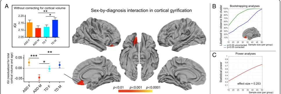
For local cortical gyrification (lGI), there was a signifi-cant sex-by-diagnosis interaction, but no main effects of sex or diagnosis. The significant sex-by-diagnosis inter-action was observed in one cluster that extended from the right ventromedial prefrontal cortex (vmPFC) to the orbitofrontal cortex (OFC) (7.99 cm2, cluster-wise
p= 0.004, effect size = 0.253; see Fig. 2a). We verified that the sex-by-diagnosis interaction remained signifi-cant in this cluster after including site as a covariate in the model. We also verified that the results remained unchanged when taking FSIQ into account. Post-hoc analysis revealed that among the four groups examined, after correction for volumetric differences, males with ASD had the lowest lGI while females with ASD had the highestlGI values, in the vmPFC/OFC.
To further assess the relevance of this vmPFC/OFC cluster, we performed additional whole-brain analyses to examine the effect of sex within the ASD group (ASD M
(7.04 cm2, cluster-wisep= 0.0022; see Additional file 2: Figure S1b). No cortical regions exhibited lower or higher gyrification in females with ASD compared to TD females (see Additional file 2: Figure S1c).
Taken together, our results from local gyrification analysis point to atypical sex differences in the vmPFC/OFC, mainly driven by a lGI reduction in males with ASD.
Stability analyses
Given the large individual differences in the ASD pheno-type, we next investigated whether the observed clusters of sex differences in cerebral morphometry depend on sample size. For this purpose, we performed a bootstrap procedure, sampling different sized subsets from the full cohort of 210 participants. We explored sample sizes ranging from 15 to 50 individuals from each of the four groups, generating 500 random samples for each. With each sample, vertex-wise statistical analyses similar to those above were performed, to assess the signifi-cance of main effect of sex on cortical volume and sex-by-diagnosis interaction on local gyrification. These simulation analyses used a statistical threshold of p< 0.05, and results were aggregated across sub-samples. The result of this bootstrap subsampling analysis yielded a cortical map with a frequency of observing a significant result for each vertex for each simulated sample size.
Analysis of all the individual cortical maps for the sex-by-diagnosis interaction revealed that, except the verti-ces in the right vmPFC/OFC region, no other cortical vertex survived statistical threshold (p< 0.05, corrected) in more than 10 % of the simulated samples, for any of the sample sizes studied (15 to 50, in increments of 5). For the main effect of sex, only one other small cluster in addition to the vertices in the bilateral STG region showed significance atp< 0.05 (corrected) in more than 10 % of simulated subsamples: a cluster in the right occipital pole showed a significant effect of sex in 12.4, 15.4, and 29 % of the simulated subsamples for sample sizes of 40, 45, and 50 participants respectively in each of the four groups. Of note, this small cluster was not significant in the final sample size of 210 participants, even at the threshold ofp< 0.05 (corrected).
We next examined the effect of sample size in the clusters that demonstrated a significant main effect of sex and sex-by-diagnosis interaction in the full dataset. For both the main effect of sex and the sex-by-diagnosis interaction, a dramatic drop in the likelihood to detect these significant clusters was observed as the sample size decreased (Figs. 1b and 2b). For instance, with 30 indi-viduals per group (total n= 120), the vmPFC/OFC cluster that showed a significant sex-by-diagnosis inter-action in the full sample survived statistical correction in
only ~50 % of the bootstrapped subsamples (Fig. 2b). For the same sample size, bootstrapping analyses re-vealed that the main effect of sex in the bilateral pSTG/ PT clusters was found in less than 60 and 40 % of the subsamples respectively (Fig. 2b). These stability analyses demonstrate that a sufficiently large sample size is re-quired to identify the effects we report.
Post-hoc power analyses
Finally, we conducted a posteriori power analyses using published algorithms [57] to compute the achieved power based on the sample size and effect size. These analyses yielded a power of 0.779 for the sex-by-diagnosis interaction in the right vmPFC/OFC, 0.930 for the left pSTG/PT cluster that showed a main effect of sex, and 0.977 for its right counterpart. The relationship between achieved power and sample size for the effect sizes observed in this study is plotted in Figs. 1c and 2c. Given an effect size of 0.253, achieving a power of 0.8 in the cluster of significant sex-by-diagnosis interaction would require a few more individuals than we had available: 55 individ-uals per group. For the main effect of sex however, power analyses revealed that the left cluster would reach a power of 0.8 with ~38 individuals per group (total n= 151), and the right cluster with 30 individ-uals per group (total n= 120). As only one study to date achieved a sample of 30 females with ASD with a total sample size of 120, the result of the stability and power analyses suggests that all previous studies examining sex differences in cerebral morphometry in ASD were underpowered.
Discussion
Neurotypical sex differences, characterized by larger volume in females compared to males in the posterior superior temporal cortex/planum temporale (pSTG/PT), were preserved in individuals with ASD. This suggests that sex differences in language acquisition [58, 59] and language processing [60–63] systems of the brain that are commonly reported in typically developing individ-uals might also be observed in individindivid-uals with autism. Preserved sex differences in cortical areas responsible for language processing also suggest that the mecha-nisms leading to language difficulties in ASD do not differ between affected males and females. In contrast, atypical sex-specific alterations of gyrification patterns were found in the orbitofrontal/ventromedial prefrontal cortex in individuals with ASD, with reduced gyrification observed in affected males only. Based on previous reports that lGI changes little with development [64], this pattern of altered gyrification points to early abnor-mal development of the orbitofrontal/ventromedial prefrontal cortex in males with ASD and provides a sex-specific biological substrate in a cortical region that forms part of the“social brain”[65, 40].
Preserved sex differences in the temporal cortex in ASD
A main effect of sex was observed in both groups, with larger pSTG/PT cortical volume in females relative to males. In these clusters, we did not observe any signifi-cant sex-by-diagnosis interaction, rather we found a similar pattern of relative volumetric increase in both TD females and females with ASD compared to TD males and males with ASD. This finding suggests that one of the brain structures that is most typically related to a core symptom of ASD, namely language difficulties, does not show any sex-specific differences.
In typically developing individuals, many studies have reported a relatively enlarged superior temporal gyrus (STG) in females compared to males [66–69]. A large body of literature relates sex differences in the anatomy and function of perisylvian structures (inferior frontal and superior temporal cortex, along with planum tem-porale) to differences in various aspects of language pro-cessing, including semantic [61], phonological [63, 62], and narrative processing [60]. In addition to these functional processing differences, language acquisition appears to follow a sex-specific trajectory: typically developing girls have been shown to mature more rapidly than boys in specific language skills including early communicative gestures, and expressive language [58]. These sex differences have been shown to persist until later in childhood [59] but tend to fade by adult-hood [70].
In individuals with ASD, studies including predomin-antly or only males have shown differences in STG structure [71, 72] and in STG activation during speech
processing [73]. Inter-individual variations in STG vol-ume [71] and in trajectories of volvol-umetric STG growth [72] were further related to history of language delay in males with ASD. Future studies are required to better understand the functional significance of preserved sex differences in the STG. In the meantime, based on extant functional imaging data, we suggest that our find-ings may reflect sex differences in language acquisition and processing in individuals with ASD. Although quan-titative studies measuring sex differences in language and communication skills have been inconsistent so far [74–76], some clinical studies suggest that girls with ASD acquire language skills faster than their male peers. For instance, girls with higher IQ tend to be diagnosed later than males, as their superior language skills during their first years of life might mask autistic symptoms [77, 78, 74]. Also, a study using retrospective parent reports has suggested that, at the age of 4 years old, girls with ASD present less severe communication difficulties compared to affected boys [79]. These studies suggest that the sex differences in language and communication seen in typical development, with faster acquisition of language and better communication skills in girls, might be relatively preserved in ASD. Future studies in-cluding more fine-grained measures of language skills and history of language development are required to better understand if preserved sex differences in pSTG/ PT cortical volumes relates to sex-related differences in trajectories of language acquisition in young children with ASD.
Altered sex differences in the ventromedial/orbitofrontal cortex in ASD
Functional neuroimaging studies, predominately with male participants, have shown altered function of the vmPFC/OFC region. First, task-based fMRI studies have reported altered activity of the vmPFC/OFC in predom-inantly male samples during theory of mind [88] and reward processing [89] including social reward [90], tasks. Second, resting state fMRI studies have identified altered connectivity in anterior nodes of the default-mode network [91–93, 40], which overlap with the vmPFC/OFC cluster identified in this study. Finally, oxy-tocin administration has been shown to increase both OFC activity and orientation to social stimuli in a sam-ple composed of 18 boys and 3 girls with ASD [94]. The fact that all these studies were conducted in samples composed mostly or entirely of affected males suggests altered function of the vmPFC/OFC in males with ASD but leaves the question of potential functional alterations of this region in affected females unanswered. Further research is needed to investigate whether the function of the vmPFC/OFC differs in females with ASD. Extant reports suggest that females with ASD have a greater desire to interact with others, tend to imitate their peers more, and develop better compensatory strategies to mask their difficulties relative to males (reviewed in [9]). Taken together, our findings of a sex-specific disrup-tion of cortical development in the vmPFC/OFC pro-vides a neuroanatomical template for further studies of sex differences in social cognition in males and females with ASD.
Developmental origins of the cortical sex differences
The surface-based morphometry method used here allowed for a better characterization of the nature of the cortical changes in ASD than previous studies. While the developmental mechanisms leading to altered cortical volume are not well understood, they are known to be related to complex age-dependent cortical matur-ation and aging processes [26, 27, 95]. Investigating the developmental origins of sex differences in cortical thickness in ASD will require careful analysis of the trajectory of developmental changes from early infancy. Our sample size, despite being the largest of its kind studied to date, did not allow us to examine sex differ-ences in trajectories of cortical changes with age. Iden-tifying sex differences in cortical thickness trajectories in typically developing individuals requires much larger sample sizes and a longitudinal design [96], and this may be true of ASD as well. An important ques-tion for future research is whether there are differences in the developmental trajectories of sex differences in STG regions identified in the present study.
In contrast, there is a large body of literature supporting the notion that modified gyrification re-sults from disruptions early in cortical development
[24, 25, 23, 22]. Based on the extant data, we suggest that the significant sex-by-diagnosis interaction in gyrification, with selectively reduced gyrification in the vmPFC/OFC of males with ASD, may have its origins in early development. The process of cortical folding starts at 16 weeks post-conception and ends within the first few months of life [64]. Altered patterns of cortical folding are thus assumed to result from adverse events that occur during this period. For instance, premature birth [24, 25], obstetric com-plications [23], or cardiac surgery during the first months of life [22] are known to affect gyrification. Another prenatal factor that is known to shape the brain of developing fetuses is the level of fetal testos-terone [97]. In a brain imaging study of children aged 8 to 11 years, Lombardo and colleagues assessed how variations in levels of fetal testosterone predicted local gray matter volume. Although their study did not distinguish between volume, gyrification, and thick-ness, they noted that increased fetal testosterone was related to patterns of both increased and decreased gray matter. In particular, the OFC showed a negative correlation between fetal testosterone level and gray matter volume. In light of the recent evidence that fetal steroidogenic activity is elevated in male children who will develop autism later on [7], our finding of altered vmPFC/OFC gyrification in males with ASD supports the idea that elevated fetal testosterone in males with ASD may influence early development of the brain in utero.
Stability and power analysis
a sample of 30 females with ASD (total n= 120), a sig-nificant sex-by-diagnosis interaction was only captured in 50 % of the simulated subsamples. The likelihood of observing a sex-by-diagnosis interaction in the vmPFC/ OFC cluster increased to 60 % with a sample of 35 females (total n= 140), and 90 % for a sample of 45 females with ASD (total n= 180). We also investigated this issue by computing post-hoc power based on the estimated effect size identified in our significant clusters. This analysis confirmed that samples of 30 to 55 females with ASD (total n= 120–220) are required to observe a significant main effect of sex as well as a sex-by-diagnosis interaction in the same clusters with a power of at least 0.8. Taken together, the simulation analysis and the power computation supports the view that identifying robust and reproducible findings probably requires samples of females with ASD much larger than previously studied.
Limitations
One of the limitations of our study is that the ABIDE cohort includes mainly individuals with high-functioning ASD (IQ range 61–147). Given the difficulty of acquiring MRIs from lower-functioning children and adolescents, the inclusion of only high-functioning affected individ-uals is a common limitation of imaging studies in ASD. A second limitation is that we did not have access to fine-grained measures of the clinical and cognitive phenotypes of the individuals with ASD included in the ABIDE cohort. With the available measures, the groups of males and females with ASD did not differ on the severity of autistic symptoms in social interactions, communication, or RRB, limiting our ability to assess whether the observed sex-by-diagnosis interaction has an impact on the observed behavioral phenotype. Finally, to achieve a sample size of 53 females with ASD, we had to merge cerebral MRI acquired at different sites with varying scanning parameters. To minimize the impact of the different sites on quality, we conducted thorough quality control, and matched all the females individually for age and site by“quartet”. Given the dearth of studies examining females with ASD to date, we suggest that our study offers a template for directing future studies examining sex differences in the disorder.
Conclusion
Using local cortical properties in a unique sample of 210 children, adolescents, and adults, we identified specific neuroanatomical features of typical sex differences that are preserved in individuals with ASD, as well as those that are not. In typically developing individuals, larger cortical volume in the pSTG/PT volume in females has been linked to sex differences in language processing and language acquisition [66–69]. This pattern of sex
differences was preserved in individuals with ASD, pointing to a neuroanatomical basis for clinical findings that females with ASD might acquire language faster than affected males [77, 78, 74, 79]. We also observed a significant sex-by-diagnosis interaction, characterized by reduced gyrification of the vmPFC/OFC region in males with ASD. Early prenatal or perinatal disruption in cortical folding development in this cortical region may underlie greater social deficits in males compared to females, with ASD. Future large-scale brain imaging stud-ies including more fine-grained assessments of social and language skills are required to replicate these findings and to better examine the relationship between sex differences in the brain structure and in behavioral and clinical phenotypes. Finally, our bootstrap analyses demonstrated that large sample sizes are required when examining sex differences in neuroanatomical features in ASD. The potential effect of sampling within a highly heterogeneous disorder further stresses the need for a better frame-work to divide individuals with autism into clinically, etiologically, and neurobiologically homogeneous sub-groups, a challenge that requires larger samples than currently available.
Additional files
Additional file 1: Table S1.Full description of the participants included in the present study.
Additional file 2: Figure S1.Comparison of gyrification patterns two-by-two (whole-brain analyses).
Abbreviations
ASD:autism spectrum disorders; DTI: diffusion tensor imaging; OFC: orbitofrontal cortex; pSTG: posterior superior temporal gyrus; PT: planum temporale; RRBI: repetitive and restricted behaviors and interests; TD: typically developing; vmPFC: ventromedial prefrontal cortex.
Competing interests
The authors declare that they have no competing interests.
Authors’contributions
MS, JK, and VM designed the study. MS carried out the image processing. MS and JK conducted the statistical analyses. MS drafted the manuscript. All authors (MS, JK, AP, KS, and VM) contributed to the interpretation of the results and to revisions and rewriting of the initial manuscript. All authors read and approved the final manuscript.
Acknowledgements
the IRB of the School of Medicine of New York University for the New York University Langone Medical Center (NYU), the Hartford Hospital Institution Review Committee for the Institute of Living at Hartford Hospital (OLIN), the IRB of University of California Los Angeles (UCLA), the local Ethics Committee for Biomedical Research at the Katholieke Universiteit Leuven for the University of Leuven (Leuven), the IRB of the University of Michigan (UM), the IRB of the University of Pittsburgh (Pitt), and the Human Investigation Committee of the Yale University (Yale). Efforts leading to the constitution of the ABIDE dataset and its sharing was primarily supported through funds to Adriana Di Martino (NIMH K23MH087770 and the Leon Levy Foundation) and funds to Michael P. Milham and the INDI team (gifts from Joseph P. Healy and the Stavros Niarchos Foundation to the Child Mind Institute, as well as by an NIMH award R03MH096321).
Author details 1
Department of Psychiatry and Behavioral Sciences, Stanford University School of Medicine, Stanford, CA 94305, USA.2Program in Neuroscience, Stanford University School of Medicine, Stanford, CA 94305, USA. 3Department of Neurology and Neurological Sciences, Stanford University School of Medicine, Stanford, USA.
Received: 10 March 2015 Accepted: 18 June 2015
References
1. American Psychiatric Association. Diagnostic and statistical manual of mental disorders. 5th ed. Arlington, VA: American Psychiatric Association; 2013. 2. Baio J. Prevalence of autism spectrum disorder among children aged
8 years—autism and developmental disabilities monitoring network, 11 sites, United States, 2010. MMWR Surveill Summ.
2014;63(2):1–21.
3. Fombonne E. The changing epidemiology of autism. J Appl Res Intellect Disabil. 2005;18:281–94.
4. Schaafsma SM, Pfaff DW. Etiologies underlying sex differences in autism spectrum disorders. Front Neuroendocrinol. 2014;35(3):255–71. doi:10.1016/j.yfrne.2014.03.006.
5. Baron-Cohen S, Lombardo MV, Auyeung B, Ashwin E, Chakrabarti B, Knickmeyer R. Why are autism spectrum conditions more prevalent in males? PLoS Biol. 2011;9(6):e1001081. doi:10.1371/journal.pbio.1001081. 6. Baron-Cohen S. The extreme male brain theory of autism. Trends Cogn Sci.
2002;6(6):248–54.
7. Baron-Cohen S, Auyeung B, Norgaard-Pedersen B, Hougaard DM, Abdallah MW, Melgaard L, et al. Elevated fetal steroidogenic activity in autism. Mol Psychiatry. 2014. doi:10.1038/mp.2014.48
8. Lenz KM, Nugent BM, Haliyur R, McCarthy MM. Microglia are essential to masculinization of brain and behavior. J Neurosci. 2013;33(7):2761–72. doi:10.1523/JNEUROSCI.1268-12.2013.
9. Lai M, Lombardo MV, Auyeung B, Chakrabarti B, Baron-Cohen S. Sex/gender differences and autism: setting the scene for future research. J Am Acad Child Adolesc Psychiatry. 2015;54(1):11–24. doi:10.1016/j.jaac.2014.10.003. 10. Bloss CS, Courchesne E. MRI neuroanatomy in young girls with autism:
a preliminary study. J Am Acad Child Adolesc Psychiatry. 2007;46(4):515–23. doi:10.1097/chi.0b013e318030e28b.
11. Schumann CM, Barnes CC, Lord C, Courchesne E. Amygdala enlargement in toddlers with autism related to severity of social and communication impairments. Biol Psychiatry. 2009;66(10):942–9. doi:10.1016/ j.biopsych.2009.07.007.
12. Beacher FD, Minati L, Baron-Cohen S, Lombardo MV, Lai MC, Gray MA, et al. Autism attenuates sex differences in brain structure: a combined voxel-based morphometry and diffusion tensor imaging study. AJNR Am J Neuroradiol. 2012;33(1):83–9. doi:10.3174/ajnr.A2880.
13. Lai MC, Lombardo MV, Suckling J, Ruigrok AN, Chakrabarti B, Ecker C, et al. Biological sex affects the neurobiology of autism. Brain.
2013;136(Pt 9):2799–815. doi:10.1093/brain/awt216.
14. Via E, Radua J, Cardoner N, Happe F, Mataix-Cols D. Meta-analysis of gray matter abnormalities in autism spectrum disorder: should Asperger disorder be subsumed under a broader umbrella of autistic spectrum disorder? Arch Gen Psychiatry. 2011;68(4):409–18. doi:10.1001/archgenpsychiatry.2011.27. 15. Lenroot RK, Yeung PK. Heterogeneity within autism spectrum disorders:
what have We learned from neuroimaging studies? Front Hum Neurosci. 2013;7:733. doi:10.3389/fnhum.2013.00733.
16. Haar S, Berman S, Behrmann M, Dinstein I. Anatomical abnormalities in autism? Cereb Cortex. 2014. doi:10.1093/cercor/bhu242.
17. Greve DN, Svarer C, Fisher PM, Feng L, Hansen AE, Baare W, et al. Cortical surface-based analysis reduces bias and variance in kinetic modeling of brain PET data. Neuroimage. 2014;92:225–36. doi:10.1016/j.neuroimage.2013.12.021.
18. Tucholka A, Fritsch V, Poline JB, Thirion B. An empirical comparison of surface-based and volume-based group studies in neuroimaging. Neuroimage. 2012;63(3):1443–53.
doi:10.1016/j.neuroimage.2012.06.019.
19. Fischl B, Dale AM. Measuring the thickness of the human cerebral cortex from magnetic resonance images. Proc Natl Acad Sci U S A.
2000;97(20):11050–5. doi:10.1073/pnas.200033797.
20. Schaer M, Cuadra MB, Tamarit L, Lazeyras F, Eliez S, Thiran JP. A surface-based approach to quantify local cortical gyrification. IEEE Trans Med Imaging. 2008;27(2):161–70. doi:10.1109/TMI.2007.903576.
21. Schaer M, Eliez S. Contribution of structural brain imaging to our understanding of the cortical development. Eur Psy Rev. 2009;2(1):13–6.
22. Schaer M, Glaser B, Cuadra MB, Debbane M, Thiran JP, Eliez S. Congenital heart disease affects local gyrification in 22q11.2 deletion syndrome. Dev Med Child Neurol. 2009;51(9):746–53. doi:10.1111/j.1469-8749.2009.03281.x. 23. Haukvik UK, Schaer M, Nesvag R, McNeil T, Hartberg CB, Jonsson EG, et al.
Cortical folding in Broca's area relates to obstetric complications in schizophrenia patients and healthy controls. Psychol Med. 2012;42(6):1329–37.
doi:10.1017/S0033291711002315.
24. Kesler SR, Vohr B, Schneider KC, Katz KH, Makuch RW, Reiss AL, et al. Increased temporal lobe gyrification in preterm children. Neuropsychologia.
2006;44(3):445–53.
25. Gimenez M, Junque C, Vendrell P, Narberhaus A, Bargallo N, Botet F, et al. Abnormal orbitofrontal development due to prematurity. Neurology. 2006;67(10):1818–22.
26. Shaw P, Kabani NJ, Lerch JP, Eckstrand K, Lenroot R, Gogtay N, et al. Neurodevelopmental trajectories of the human cerebral cortex. J Neurosci. 2008;28(14):3586–94. doi:10.1523/JNEUROSCI.5309-07.2008.
27. Shaw P, Greenstein D, Lerch J, Clasen L, Lenroot R, Gogtay N, et al. Intellectual ability and cortical development in children and adolescents. Nature. 2006;440(7084):676–9.
28. Di Martino A, Yan CG, Li Q, Denio E, Castellanos FX, Alaerts K, et al. The autism brain imaging data exchange: towards a large-scale evaluation of the intrinsic brain architecture in autism. Mol Psychiatry. 2013. doi:10.1038/mp.2013.78
29. Sacher J, Neumann J, Okon-Singer H, Gotowiec S, Villringer A. Sexual dimorphism in the human brain: evidence from neuroimaging. Magn Reson Imaging. 2013;31(3):366–75. doi:10.1016/j.mri.2012.06.007. 30. Ruigrok AN, Salimi-Khorshidi G, Lai MC, Baron-Cohen S, Lombardo MV, Tait
RJ, et al. A meta-analysis of sex differences in human brain structure. Neurosci Biobehav Rev. 2014;39:34–50. doi:10.1016/j.neubiorev.2013.12.004. 31. Luders E, Toga AW. Sex differences in brain anatomy. Prog Brain Res.
2010;186:3–12. doi:10.1016/B978-0-444-53630-3.00001-4.
32. Werling DM, Lowe JK, Luo R, Cantor RM, Geschwind DH. Replication of linkage at chromosome 20p13 and identification of suggestive sex-differential risk loci for autism spectrum disorder. Mol Autism. 2014;5(1):13. doi:10.1186/2040-2392-5-13.
33. Stone JL, Merriman B, Cantor RM, Yonan AL, Gilliam TC, Geschwind DH, et al. Evidence for sex-specific risk alleles in autism spectrum disorder. Am J Hum Genet. 2004;75(6):1117–23.
34. Yu J, He X, Yao D, Li Z, Li H, Zhao Z. A sex-specific association of common variants of neuroligin genes (NLGN3 and NLGN4X) with autism spectrum disorders in a Chinese Han cohort. Behav Brain Funct. 2011;7:13. doi:10.1186/1744-9081-7-13.
35. Chang SC, Pauls DL, Lange C, Sasanfar R, Santangelo SL. Sex-specific association of a common variant of the XG gene with autism spectrum disorders. Am J Med Genet B Neuropsychiatr Genet. 2013;162B(7):742–50. doi:10.1002/ajmg.b.32165.
36. Schwarz E, Guest PC, Rahmoune H, Wang L, Levin Y, Ingudomnukul E, et al. Sex-specific serum biomarker patterns in adults with Asperger's syndrome. Mol Psychiatry. 2011;16(12):1213–20. doi:10.1038/mp.2010.102.
38. Dere E, Dahm L, Lu D, Hammerschmidt K, Ju A, Tantra M, et al. Heterozygous ambra1 deficiency in mice: a genetic trait with autism-like behavior restricted to the female gender. Front Behav Neurosci. 2014;8:181. doi:10.3389/fnbeh.2014.00181.
39. Van Wijngaarden-Cremers PJ, van Eeten E, Groen WB, Van Deurzen PA, Oosterling IJ, Van der Gaag RJ. Gender and age differences in the core triad of impairments in autism spectrum disorders: a systematic review and meta-analysis. J Autism Dev Disord. 2014;44(3):627–35.
doi:10.1007/s10803-013-1913-9.
40. Gotts SJ, Simmons WK, Milbury LA, Wallace GL, Cox RW, Martin A. Fractionation of social brain circuits in autism spectrum disorders. Brain. 2012;135(Pt 9):2711–25. doi:10.1093/brain/aws160.
41. Pelphrey KA, Shultz S, Hudac CM, Vander Wyk BC. Research review: constraining heterogeneity: the social brain and its development in autism spectrum disorder. J Child Psychol Psychiatry. 2011;52(6):631–44. doi:10.1111/j.1469-7610.2010.02349.x.
42. Uddin LQ, Menon V. The anterior insula in autism: under-connected and under-examined. Neurosci Biobehav Rev. 2009;33(8):1198–203. doi:10.1016/j.neubiorev.2009.06.002.
43. Uddin LQ, Supekar K, Lynch CJ, Khouzam A, Phillips J, Feinstein C, et al. Salience network-based classification and prediction of symptom severity in children with autism. JAMA Psychiatry. 2013;70(8):869–79.
doi:10.1001/jamapsychiatry.2013.104.
44. Langen M, Bos D, Noordermeer SD, Nederveen H, van Engeland H, Durston S. Changes in the development of striatum are involved in repetitive behavior in autism. Biol Psychiatry. 2014;76(5):405–11.
doi:10.1016/j.biopsych.2013.08.013.
45. Delmonte S, Gallagher L, O'Hanlon E, McGrath J, Balsters JH. Functional and structural connectivity of frontostriatal circuitry in autism spectrum disorder. Front Hum Neurosci. 2013;7:430. doi:10.3389/fnhum.2013.00430.
46. Lord C, Risi S, Lambrecht L, Cook Jr EH, Leventhal BL, DiLavore PC, et al. The autism diagnostic observation schedule-generic: a standard measure of social and communication deficits associated with the spectrum of autism. J Autism Dev Disord. 2000;30(3):205–23.
47. Lord C, Rutter M, Le Couteur A. Autism diagnostic interview-revised: a revised version of a diagnostic interview for caregivers of individuals with possible pervasive developmental disorders. J Autism Dev Disord. 1994;24(5):659–85.
48. Gotham K, Pickles A, Lord C. Standardizing ADOS scores for a measure of severity in autism spectrum disorders. J Autism Dev Disord.
2009;39(5):693–705. doi:10.1007/s10803-008-0674-3.
49. Fischl B, Salat DH, Busa E, Albert M, Dieterich M, Haselgrove C, et al. Whole brain segmentation: automated labeling of neuroanatomical structures in the human brain. Neuron. 2002;33(3):341–55.
50. Dale AM, Fischl B, Sereno MI. Cortical surface-based analysis. I. Segmentation and surface reconstruction. NeuroImage. 1999;9(2):179–94.
51. Fischl B. FreeSurfer. Neuroimage. 2012;62(2):774–81. doi:10.1016/ j.neuroimage.2012.01.021.
52. Rosas HD, Liu AK, Hersch S, Glessner M, Ferrante RJ, Salat DH, et al. Regional and progressive thinning of the cortical ribbon in Huntington's disease. Neurology. 2002;58(5):695–701.
53. Han X, Jovicich J, Salat D, van der Kouwe A, Quinn B, Czanner S, et al. Reliability of MRI-derived measurements of human cerebral cortical thickness: the effects of field strength, scanner upgrade and manufacturer. Neuroimage. 2006;32(1):180–94.
54. Fischl B, Sereno MI, Dale AM. Cortical surface-based analysis. II: Inflation, flattening, and a surface-based coordinate system. NeuroImage. 1999;9(2):195–207.
55. Fischl B, Sereno MI, Tootell RB, Dale AM. High-resolution intersubject averaging and a coordinate system for the cortical surface. Hum Brain Mapp.
1999;8(4):272–84. doi:10.1002/(SICI)1097-0193(1999)8.
56. Hagler Jr DJ, Saygin AP, Sereno MI. Smoothing and cluster thresholding for cortical surface-based group analysis of fMRI data. Neuroimage.
2006;33(4):1093–103.
57. Faul F, Erdfelder E, Lang AG, Buchner A. G*Power 3: a flexible statistical power analysis program for the social, behavioral, and biomedical sciences. Behav Res Methods. 2007;39(2):175–91.
58. Eriksson M, Marschik PB, Tulviste T, Almgren M, Perez Pereira M, Wehberg S, et al. Differences between girls and boys in emerging language skills: evidence from 10 language communities. Br J Dev Psychol. 2012;30(Pt 2):326–43. doi:10.1111/j.2044-835X.2011.02042.x.
59. Bornstein MH, Hahn C, Haynes OM. Specific and general language performance across early childhood: stability and gender considerations. First Language. 2004;24(3):267–304.
60. Kansaku K, Yamaura A, Kitazawa S. Sex differences in lateralization revealed in the posterior language areas. Cereb Cortex. 2000;10(9):866–72. 61. Baxter LC, Saykin AJ, Flashman LA, Johnson SC, Guerin SJ, Babcock DR, et al.
Sex differences in semantic language processing: a functional MRI study. Brain Lang. 2003;84(2):264–72.
62. Burman DD, Bitan T, Booth JR. Sex differences in neural processing of language among children. Neuropsychologia. 2008;46(5):1349–62. doi:10.1016/j.neuropsychologia.2007.12.021.
63. Bitan T, Lifshitz A, Breznitz Z, Booth JR. Bidirectional connectivity between hemispheres occurs at multiple levels in language processing but depends on sex. J Neurosci. 2010;30(35):11576–85. doi:10.1523/JNEUROSCI. 1245-10.2010.
64. Armstrong E, Schleicher A, Omran H, Curtis M, Zilles K. The ontogeny of human gyrification. Cereb Cortex. 1995;5(1):56–63.
65. Frith CD. The social brain? Philos Trans R Soc Lond B Biol Sci. 2007;362(1480):671–8.
66. Good CD, Johnsrude I, Ashburner J, Henson RN, Friston KJ, Frackowiak RS. Cerebral asymmetry and the effects of sex and handedness on brain structure: a voxel-based morphometric analysis of 465 normal adult human brains. Neuroimage. 2001;14(3):685–700. doi:10.1006/nimg.2001.0857. 67. Sowell ER, Trauner DA, Gamst A, Jernigan TL. Development of cortical and
subcortical brain structures in childhood and adolescence: a structural MRI study. Dev Med Child Neurol. 2002;44(1):4–16.
68. Harasty J, Double KL, Halliday GM, Kril JJ, McRitchie DA. Language-associated cortical regions are proportionally larger in the female brain. Arch Neurol. 1997;54(2):171–6.
69. Schlaepfer TE, Harris GJ, Tien AY, Peng L, Lee S, Pearlson GD. Structural differences in the cerebral cortex of healthy female and male subjects: a magnetic resonance imaging study. Psychiatry Res. 1995;61(3):129–35. 70. Trofimova I. A study of the dynamics of sex differences in adulthood. Int J
Psychol. 2013;48(6):1230–6. doi:10.1080/00207594.2012.756981.
71. Lai MC, Lombardo MV, Ecker C, Chakrabarti B, Suckling J, Bullmore ET, et al. Neuroanatomy of individual differences in language in adult males with autism. Cereb Cortex. 2014. doi:10.1093/cercor/bhu211
72. Prigge MD, Bigler ED, Fletcher PT, Zielinski BA, Ravichandran C, Anderson J, et al. Longitudinal Heschl's gyrus growth during childhood and adolescence in typical development and autism. Autism Res. 2013;6(2):78–90. doi:10.1002/aur.1265.
73. Lai G, Schneider HD, Schwarzenberger JC, Hirsch J. Speech stimulation during functional MR imaging as a potential indicator of autism. Radiology. 2011;260(2):521–30. doi:10.1148/radiol.11101576.
74. Andersson GW, Gillberg C, Miniscalco C. Pre-school children with suspected autism spectrum disorders: do girls and boys have the same profiles? Res Dev Disabil. 2013;34(1):413–22. doi:10.1016/j.ridd.2012.08.025.
75. Harrop C, Shire S, Gulsrud A, Chang YC, Ishijima E, Lawton K, et al. Does gender influence core deficits in ASD? An investigation into social-communication and play of girls and boys with ASD. J Autism Dev Disord. 2014. doi:10.1007/s10803-014-2234-3.
76. Carter AS, Black DO, Tewani S, Connolly CE, Kadlec MB, Tager-Flusberg H. Sex differences in toddlers with autism spectrum disorders. J Autism Dev Disord. 2007;37(1):86–97. doi:10.1007/s10803-006-0331-7.
77. Giarelli E, Wiggins LD, Rice CE, Levy SE, Kirby RS, Pinto-Martin J, et al. Sex differences in the evaluation and diagnosis of autism spectrum disorders among children. Disabil Health J. 2010;3(2):107–16.
doi:10.1016/j.dhjo.2009.07.001.
78. Kopp S, Gillberg C. Girls with social deficits and learning problems: autism, atypical asperger syndrome or a variant of these conditions. Europeand Child Adolescent Psychiatry. 1992;1(2):82–99.
79. McLennan JD, Lord C, Schopler E. Sex differences in higher functioning people with autism. J Autism Dev Disord. 1993;23(2):217–27.
80. Watanabe H, Nakamura M, Ohno T, Itahashi T, Tanaka E, Ohta H, et al. Altered orbitofrontal sulcogyral patterns in adult males with high-functioning autism spectrum disorders. Soc Cogn Affect Neurosci. 2014;9(4):520–8.
doi:10.1093/scan/nst016.
82. Leopold A, Krueger F, Dal Monte O, Pardini M, Pulaski SJ, Solomon J, et al. Damage to the left ventromedial prefrontal cortex impacts affective theory of mind. Soc Cogn Affect Neurosci. 2012;7(8):871–80. doi:10.1093/scan/nsr071. 83. Vandekerckhove M, Plessers M, Van Mieghem A, Beeckmans K, Van Acker F,
Maex R, et al. Impaired facial emotion recognition in patients with ventromedial prefrontal hypoperfusion. Neuropsychology. 2014;28(4):605–12. doi:10.1037/neu0000057.
84. Kohls G, Chevallier C, Troiani V, Schultz RT. Social 'wanting' dysfunction in autism: neurobiological underpinnings and treatment implications. J Neurodev. 2012;4(1):10. doi:10.1186/1866-1955-4-10.
85. Wolf RC, Philippi CL, Motzkin JC, Baskaya MK, Koenigs M. Ventromedial prefrontal cortex mediates visual attention during facial emotion recognition. Brain. 2014;137(Pt 6):1772–80. doi:10.1093/brain/awu063. 86. Klin A, Jones W, Schultz R, Volkmar F, Cohen D. Visual fixation patterns during
viewing of naturalistic social situations as predictors of social competence in individuals with autism. Arch Gen Psychiatry. 2002;59(9):809–16.
87. Jones W, Carr K, Klin A. Absence of preferential looking to the eyes of approaching adults predicts level of social disability in 2-year-old toddlers with autism spectrum disorder. Arch Gen Psychiatry. 2008;65(8):946–54. doi:10.1001/archpsyc.65.8.946.
88. Murdaugh DL, Shinkareva SV, Deshpande HR, Wang J, Pennick MR, Kana RK. Differential deactivation during mentalizing and classification of autism based on default mode network connectivity. PLoS One. 2012;7(11):e50064. doi:10.1371/journal.pone.0050064.
89. Dichter GS, Felder JN, Green SR, Rittenberg AM, Sasson NJ, Bodfish JW. Reward circuitry function in autism spectrum disorders. Soc Cogn Affect Neurosci. 2012;7(2):160–72. doi:10.1093/scan/nsq095.
90. Richey JA, Rittenberg A, Hughes L, Damiano CR, Sabatino A, Miller S, et al. Common and distinct neural features of social and non-social reward processing in autism and social anxiety disorder. Soc Cogn Affect Neurosci.
2014;9(3):367–77. doi:10.1093/scan/nss146.
91. Uddin LQ, Supekar K, Lynch CJ, Cheng KM, Odriozola P, Barth ME et al. Brain state differentiation and behavioral inflexibility in autism†. Cereb Cortex. 2014. doi:10.1093/cercor/bhu161.
92. Jung M, Kosaka H, Saito DN, Ishitobi M, Morita T, Inohara K, et al. Default mode network in young male adults with autism spectrum disorder: relationship with autism spectrum traits. Molecular Autism. 2014;5:35. doi:10.1186/2040-2392-5-35.
93. von dem Hagen EA, Stoyanova RS, Baron-Cohen S, Calder AJ. Reduced functional connectivity within and between 'social' resting state networks in autism spectrum conditions. Soc Cogn Affect Neurosci. 2013;8(6):694–701. doi:10.1093/scan/nss053.
94. Gordon I, Vander Wyk BC, Bennett RH, Cordeaux C, Lucas MV, Eilbott JA, et al. Oxytocin enhances brain function in children with autism. Proc Natl Acad Sci U S A. 2013;110(52):20953–8. doi:10.1073/pnas.1312857110. 95. Storsve AB, Fjell AM, Tamnes CK, Westlye LT, Overbye K, Aasland HW, et al.
Differential longitudinal changes in cortical thickness, surface area and volume across the adult life span: regions of accelerating and decelerating change. J Neurosci. 2014;34(25):8488–98. doi:10.1523/JNEUROSCI.0391-14.2014. 96. Mutlu AK, Schneider M, Debbane M, Badoud D, Eliez S, Schaer M. Sex
differences in thickness, and folding developments throughout the cortex. Neuroimage. 2013;82:200–7. doi:10.1016/j.neuroimage.2013.05.076. 97. Lombardo MV, Ashwin E, Auyeung B, Chakrabarti B, Taylor K, Hackett G, et al.
Fetal testosterone influences sexually dimorphic gray matter in the human brain. J Neurosci. 2012;32(2):674–80. doi:10.1523/JNEUROSCI.4389-11.2012. 98. Cauda F, Geda E, Sacco K, D'Agata F, Duca S, Geminiani G, et al. Grey matter
abnormality in autism spectrum disorder: an activation likelihood estimation meta-analysis study. J Neurol Neurosurg Psychiatry. 2011;82(12):1304–13. doi:10.1136/jnnp.2010.239111.
99. Stanfield AC, McIntosh AM, Spencer MD, Philip R, Gaur S, Lawrie SM. Towards a neuroanatomy of autism: a systematic review and meta-analysis of structural magnetic resonance imaging studies. Eur Psychiatry. 2008;23(4):289–99. doi:10.1016/j.eurpsy.2007.05.006.
Submit your next manuscript to BioMed Central and take full advantage of:
• Convenient online submission
• Thorough peer review
• No space constraints or color figure charges
• Immediate publication on acceptance
• Inclusion in PubMed, CAS, Scopus and Google Scholar
• Research which is freely available for redistribution