Determination of ctxAB Expression in Vibrio cholerae Classical
and El Tor Strains using Real-Time PCR
Seyed Mahmoud Amin Marashi1,2, Ramazan Rajabnia3, Abbas Ali Imani Fooladi4, Zohreh Hojati5, Sharareh Moghim6, Bahram Nasr Esfahani6∗
1. Cellular and Molecular Biology Research Center, (CMBRC), Babol University of Medical Sciences, Babol, Iran.
2. Department of Microbiology and Immunology, Babol University of Medical Sciences, Babol, Iran.
3. Infectious Diseases &Tropical Medicine ResearchCenter, Babol University of Medical Sciences, Babol, Iran.
4. Applied Microbiology Research Center, Baqiyatallah University of Medical Sciences, Tehran, Iran.
5. Genetics Division, Biology Department, Faculty of Sciences, University of Isfahan, Isfahan, Iran.
6. Department of Microbiology, School of Medicine, Isfahan University of Medical Sciences, Isfahan, Iran.
Cholera is an infection of the small intestines caused by the bacterium V. cholerae. It is a major cause of health
threat and also a major cause of death worldwide and especially in developing countries. The major virulence
factor produced by V. cholerae during infection is the cholera toxin. Total mRNA extraction and reverse
transcription was performed for making ctxAB cDNA. Relative Real-Time PCR analysis showed unequal
enterotoxin production in V. cholerae strains. The results showed that, classical strain produces cholera toxin
more than El Tor strain.
Key words: Vibrio cholerae, RT-qPCR, ctxAB expression
∗
Corresponding author: Department of Microbiology, School of Medicine, Isfahan University of Medical Sciences, Isfahan, Iran. E-Mail: [email protected].
holera is one of the infectious diseases that
still happens in developing countries. The 8th
pandemic of cholera spreads from Southeast Asia
across the Middle East and into Central America
and Africa (1, 2). The important pathogenesis factor
in Vibrio cholerae is a potent enterotoxin, cholera
toxin, which causes the severe diarrhea of cholera
(3, 4). The cholera toxin is produced by V. cholerae
and CTXΦ phage corporation. Control of
enterotoxin gene expression seems to be complex,
so that environmental factors are very important in
its expression (5, 6). The environmental signals
affect TcpPH gene and cause its activation and
finally affect ToxT gene. The ToxT protein is the
most important agent for ctxAB toxin expression,
because ToxT protein attaches to toxbox region at
upstream of ctxAB gene and induces ctxAB toxin
expression (7, 8) (Fig 1). Beside, other signaling
systems such as ToxR, RS1, AphAB and quorum
sensing have positive or negative effects on ToxT
protein (9, 10). Moreover, H-NS protein has
negative effect on TcpPH and ToxT genes that
C
Submmited 5 March 2013; Accepted 18 March 2013
Fig 1. Diagram of the vibrio cholerae ToxR regulon and ctxAB expression, with permission from ASM
finally would decrease the ctxAB toxin production
(11). The RS1 region contains rstA, rstB and rstC
fragments which have positive effect on ToxR gene
and therefore increase ctxAB toxin production (12).
Bakhshi et al. reported several V. cholerae attacks
in Southwest and Southeast of Iran between
2005-2009 (13, 14). As the level of production of a
protein is somehow related to its mRNA quantity,
we therefore aimed to determine V. cholerae strains
that can produce more ctxAB toxin.
Material and Method
Bacterial strains and growth conditions
We used two standard strains named
V.cholerae O1 Classic ATCC 14035 & V. cholerae
O1 El Tor N16961. The isolates were confirmed by
biochemical and immunological tests. Serotyping
was performed using monoclonal O1 antiserum and
mono-specific Inaba and Ogawa antisera (Pasteur
institute, Paris, France). All selected strains were
cultured according to the AKI-SW method and
standard growth curve were drawn (15).
Isolation of RNA and RT-PCR
Approximately, 2×10⁸ cfu/ml from each strain, was used for total RNA extraction. Total
RNA was isolated from the strains isolated
randomly from each V. cholerae grown in AKI
medium using the RNeasy® Protect Bacteria Mini
Kit (Qiagen Inc, GMBH, Germany) and the
integrity and purity was checked. Equivalent
concentrations of total RNA from each strain were
selected as template for RT-PCR. cDNA synthesis
and PCR amplification were performed using
QuantiTec Reverse Transcription Qiagen kit
(Qiagen Inc, GMBH, Germany). RT-PCR was
performed in the presence of random primer at
42°C for 10 min. After cDNA synthesis, the ctxAB
and recA genes were PCR amplified for checking.
PCR amplification was performed for 35 cycles as
follows: initial denaturation at 94°C for 5 min, then
denaturation at 94°C for 30 sec, annealing at 60°C
for 30 sec, extension at 72°C for 30 sec. At the end
of the 35th cycle, reaction mixtures were left at
72°C for another 3 min. Five microliters of each
reaction mixture was loaded on a 1% agarose gel
and subjected to electrophoresis to confirm that the
unique amplified fragment correspond to the
expected ctxAB gene fragment and recA as
housekeeping gene (16).
Real-Time PCR
Prepared cDNA was quantified using SYBR
green I dye. Four primers were designed by
AlleleID 6 software,
5’-CAGTCAGGTGGTCT-TATGC-3’ (ctxAB-F) and
Fig 2. Melting curves of ctxAB and recA genes.
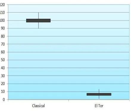
Fig 3. Level of cholera toxin production in classical and EL
Tor strains.
–ATTGAAGGCGAAATGGGCGATAG- 3’
(recA-F) and 5’ –TACACATACAGTTGGATTGCTTG
AGG- 3’ (recA-R) for housekeeping gene. Those
primers were specific to ctxAB and recA and
amplified a 115 & 106 bp respectively. SYBR
green Real-Time PCR assay was performed with a
20 µl PCR mixture volume containing 2x
QuantiTec SYBR Green PCR Master Mix (Qiagen
Inc, GMBH, Germany), 0.25 µM specific primer
sets, and 2 µl of cDNA sample. Amplification of
the primers, data acquisition, and relative analysis
were carried out in Chromo4 BioRad Real-Time
PCR. PCR reactions were performed as followings:
one cycle of 95 °C for 5 min, then 40 cycles of 95
°C for 15 sec, 60 °C for 30 sec. Following the
amplification, melting temperature analysis of the
PCR products was performed to determine the
specificity of the PCR. The standard curve was
established by using genomic DNA for each studied
gene to confirm that the primers amplified at the
same rate and to validate the experiment (55-95°C
with warming of 0.2°C per sec). Reverse
transcription and PCR positive controls (RNA and
DNA, respectively) and negative controls (distilled
water) were included in each run. The Real-Time
PCR reaction was performed twice assayed in
was used as a standard control.
Results
The specificity of each primer set for V.
cholerae was tested by PCR with genomic DNA
extracted via boiling. Only one size of amplicon
was obtained by PCR reaction for ctxAB and recA
genes when DNA from V. cholerae strains was
used. The amplicons obtained for each gene were
verified by sequencing. The presence of a single
PCR product was confirmed by Real-Time PCR for
each set of the primers using melting curve analysis
that resulted in a single product-specific melting
curve (Fig 2). The PCR efficiencies varied between
1.90 and 1.94. The relative expression ratio was
calculated for each gene of interest by a
mathematical model described by ∆CT method. The
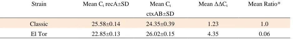
Cycle Threshold (CT ) results are showed in table 1.
Histogram and samples CT values are indicated in
Fig 3.
Discussion
In our study, the results are derived by using
“relative” method and ∆CT formula. By considering
that ctxAB primers have been carefully designed,
the amount of standard deviation results are close to
Table 1: Cycle threshold (Ct) results for the V.cholerae O1 Classic ATCC 14035 & V. cholerae O1 El Tor 62013
Strain Mean Ct recA±SD Mean Ct
ctxAB±SD
Mean ∆∆Ct Mean Ratio*
Classic
25.58±0.14 24.35±0.39 1.23 1.0
El Tor 22.85±0.13 26.02±0.15 4.35 0.06
*∆∆Ct was calculated as: ∆Ct (test) - ∆Ct (calibrator).Ratio=efficiency -∆∆Ct.
zero, the primer-dimer bands were not seen,
because the concentration of participating primers
in the reaction had been set up. Also, for more
accuracy and sensitivity, the PCR efficiency in
Real-Time PCR reactions were calculated and
replaced with ratio 2 in computational equations.
Dirita et al. showed that the level of cholera
toxin was higher in the classical strain compared to
El Tor strain because of the influence of toxR on
cholera toxin production in the classical strain (17).
In our study, in addition to Dirita et al. results, we
determined quantitative cholera toxin production
between classic and El Tor strains. Both classical
and El Tor strains have been shown to express
equivalent levels of ToxR. In contrast, the classical
strain expresses more ToxT, which has a higher
binding affinity to toxbox region, resulting in
higher expression of cholera toxin (18).
The comparison of the CT of the El Tor
strain with classical strain shows that toxin
production in El Tor strain is approximately 16-17
times lower than in the classical strain (P Value<0.05
for each). This result is consistent with other reports
because the amount of pathogenicity in classic
strain is more than El Tor strains (18). Furthermore,
histogram and samples CT results indicated that
toxin production in classical strain is higher than El
Tor strain (Fig 3). In conclusion, the results of our
study suggest that other factors modulate the
production of cholera toxin by regulating the CTX
cassette, supporting the idea that cholera toxin
production in V. cholerae classical and El Tor
strains is a multi-factorial phenomenon.
Acknowledgment
We are grateful to Dr. Bita Bakhshi for
shipment of the V. cholerae O1 classic ATCC
14035 and V. cholerae O1 El Tor ATCC N16961
strains.
References
1. Amin Marashi SM, Nasr Esfahani B, Tavakoli A, et al.
Simultaneous detection of integrase and antibiotic resistance
genes within SXT Constin in Vibrio cholerae O1 El Tor strains
isolated from Iran using multiplex-PCR. Iran J Basic Med Sci
2012;15:885-9.
2. Taneja N, Mishra A, Sangar G, et al. Outbreaks caused by
new variants of Vibrio cholerae O1 El Tor, India. Emerg Infect
Dis 2009;15:352-4.
3. Baptista MA, Andrade JR, Vicente AC, et al. The Amazonia
variant of Vibrio cholerae: molecular identification and study of
virulence genes. Mem Inst Oswaldo Cruz 1998;93:601-7.
4. Bravo L, Monte RJ, Ramirez M, et al. Detection of toxigenic
Vibrio cholerae 01 using polymerase chain reaction. Mem Inst
Oswaldo Cruz 1992;87:443-4.
5. Bakhshi B, Pourshafie MR, Navabakbar F, et al. Genomic
organisation of the CTX element among toxigenic Vibrio
cholerae isolates. Clin Microbiol Infect 2008;14:562-8.
6. Sa LL, Fonseca EL, Pellegrini M, et al. Occurrence and
composition of class 1 and class 2 integrons in clinical and
environmental O1 and non-O1/non-O139 Vibrio cholerae strains
from the Brazilian Amazon. Mem Inst Oswaldo Cruz;
105:229-32.
7. Withey JH, Dirita VJ. Vibrio cholerae ToxT independently
activates the divergently transcribed aldA and tagA genes. J
8. Withey JH, DiRita VJ. The toxbox: specific DNA sequence
requirements for activation of Vibrio cholerae virulence genes
by ToxT. Mol
Microbiol 2006;59:1779-89.
9. Klose KE. Regulation of virulence in Vibrio cholerae. Int J
Med Microbiol 2001;291:81-8.
10. Zhu J, Miller MB, Vance RE, et al. Quorum-sensing
regulators control virulence gene expression in Vibrio cholerae.
Proc Natl Acad Sci USA 2002;99:3129-34.
11. Matson JS, Withey JH, DiRita VJ. Regulatory networks
controlling Vibrio cholerae virulence gene expression. Infect
Immun 2007;75:5542-9.
12. Davis BM, Waldor MK. Filamentous phages linked to
virulence of Vibrio cholerae. Curr Opin Microbiol 2003;6:35-42.
13. Bakhshi B, Pourshafie MR. Assessing clonality of Vibrio
cholerae strains isolated during four consecutive years (2004 -
2007) in Iran. Scand J Infect Dis 2009;41:256-62.
of a single Vibrio cholerae clone in cholera outbreaks during
2005 in Iran. J Med Microbiol 2007;56:1615-9.
15. Iwanaga M, Yamamoto K, Higa N, et al. Culture conditions
for stimulating cholera toxin production by Vibrio cholerae O1
El Tor. Microbiol Immunol 1986;30:1075-83.
16. Amin Marashi M, Bakhshi B, Imani Fooladi AA, et al.
Quantitative expression of cholera toxin mRNA in Vibrio
cholerae isolates with different CTX cassette arrangements. J
Med Microbiol.
17. DiRita VJ, Neely M, Taylor RK, et al. Differential
expression of the ToxR regulon in classical and E1 Tor biotypes
of Vibrio cholerae is due to biotype-specific control over toxT
expression. Proc Natl Acad Sci U S A 1996;93:7991-5.
18. Gonzalez-Bonilla C, Gutierrez-Cogco L, Moguel-Pech L, et
al. [Evaluation of the ELISA method for cholera toxin
determination in Vibrio cholerae cultures]. Rev Latinoam
Microbiol 1994;36:273-6.