R E S E A R C H
Open Access
Comparison of three different methods of
internal sinus lifting for elevation heights of
7 mm: an ex vivo study
Aghiad Yassin Alsabbagh
1*, Mohammed Monzer Alsabbagh
1, Batol Darjazini Nahas
2and Salam Rajih
3Abstract
Background:Various techniques are available for elevating the sinus membrane. The aim of this study is to evaluate three methods of indirect sinus floor elevation regarding elevation heights of 7 mm on the outcomes of membrane perforation, length of perforation, and time required to perform the procedure.
Methods:Three different methods for indirect sinus lifting, bone added osteotome sinus floor elevation (BAOSFE), sinus floor elevation with an inflatable balloon, and crestal approach system (CAS kit) from OSSTEM, were assessed for their ability to lift the sinus without causing laceration of the Schneiderian membrane. The study was performed on 18 freshly slaughtered sheep heads (36 sinus lifts were done, 12 for each method). CBCT images of the heads were taken to assess the best location for the sinus lift. Then, the heads were bisected and the membrane was exposed from the medial aspect. After that, each method was performed. The intended elevation height was 7 mm. If the 7 mm were not reached, the maximum height of elevation was measured.
Results:The method used was significantly associated with the occurrence of perforation (pvalue = 0.014) where BAOSFE was associated with the largest number of perforations (58.4%,n= 7) compared to 8.3% and 8.3% for the balloon and CAS kit methods, respectively. The odds ratio for perforation occurrence from BAOSFE compared to the CAS kit was significant (OR = 0.091,p= .022). No significant odds ratio was found for the balloon method
compared to CAS kit. Additionally, the method used was significantly associated with time of operation and with the length of perforation (pvalue < 0.001) where CAS kit required the longest time and BAOSFE caused the biggest perforations.
Conclusions:The study shows that both the balloon and the CAS kit were superior to the BAOSFE in terms of safety in elevating the sinus membrane. Further, in vivo studies have to prove these findings.
Keywords:Maxillary sinus, Schneiderian membrane, Sinus floor elevation, Balloon elevation, Elevation height, CAS kit
Background
More than half of the implants placed in the posterior maxilla require sinus floor elevation (SFE) [1]. The need for this procedure is explained by continuous ridge re-sorption in an apical direction after tooth extraction combined with progressive sinus pneumatization in addition to poor bone quality that is frequently seen in the maxilla [2].
Sinus membrane perforation is considered the most common complication during sinus floor elevation pro-cedures, and its percentage varies according to the method used. Perforations happen either while fractur-ing the floor of the sinus or durfractur-ing the elevation of the mucosa [3, 4].
Crestal approach to the sinus kit (CAS kit) was intro-duced by OSSTEM implants (Osstem Implant Co., Bu-san, Korea) as a safe and effective method for sinus elevation with the advantage of using a reamer (the CAS drill) to perform the osteotomy in a conical shape and break the bony floor; however, only one questionnaire
* Correspondence:[email protected]
1Department of Periodontology, Damascus University Dental School,
Damascus, Syrian Arab Republic
Full list of author information is available at the end of the article
that assessed the satisfaction of dentists using the CAS KIT is available in the literature on this method [5]. Using an inflatable balloon for indirect sinus floor eleva-tion has been shown to be successful in elevating the mucosa for elevation heights of up to 10 mm [6, 7]. However, few studies in the literature compared this technique to others.
Lopez-Nino et al. studied the lamb as an ex vivo model for training in sinus floor elevation and concluded that the model is useful because of the similarities in the thickness of the lateral wall of the maxillary sinus and the thickness of the Schneiderian membrane between the models and the human standards [8].
Cone beam computed tomography (CBCT) can pre-cisely visualize the sinus complexity in 3D, with low irradi-ation to the patient. In implant dentistry, recent guidelines recommend the use of CBCT for three-dimensional treat-ment planning, especially prior to SFE for evaluating both residual alveolar and sinus conditions [9, 10].
Therefore, the two working hypotheses of our study were
“the CAS-Kit is safer than BAOSFE in breaking the sinus floor and the balloon is safer than BAOSFE in elevating the Schneiderian membrane”for elevation heights of 7 mm.
Methods The sample
To achieve our purposes, an experimental ex vivo study was carried. This research project was approved by the
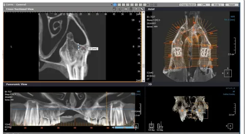
University of Damascus Local Research Ethics Committee (UDDS-3045PG.) and was funded by the Damascus Uni-versity Postgraduate Research Budget (97687027834DEN). The sinus floor elevations were done on 18 bisected heads of lambs aged between 6 and 12 months that were slaugh-tered in a maximum of 4 h before the procedures began. CBCT images of the heads were taken using the Picasso® Pro CBCT system (Vatech™, Seoul, South Korea) set at a voxel size of 0.2 mm, tube current of 5 mA, tube voltage of 83 KV with gray scale of 16 bit per pixel. A standard-ized position of the lamb’s heads was maintained by the correct head orientation in accordance with the 3D inter-secting planes of the red beam. Then, the images were an-alyzed for the best location to perform the sinus elevation where remaining bone height (RBH) is less than 5 mm on 3DOnDemand® programme (CyberMed, Finland) (Fig. 1). The RBH was measured from the apical tip of the buccal root on the third premolar which will be extracted to the floor of the sinus. The sample was randomized by generat-ing random numbers usgenerat-ing Research Randomizer software (http://www.randomizer.org/) [11] making sure that the same method was not done on the same lamb twice.
Visual assessment
After the extraction of the third premolar, the mesial side of the sinus was exposed (Fig. 2) in order to check the sinus for any perforations. The elevation height was measured using a depth gauge, and the intended
elevation height was 7 mm. If this height was not achieved, the maximum elevation was recorded. When a perforation of the membrane was present, its length was measured using a periodontal probe.
Sinus floor elevation methods BAOSFE
Bone blocks were harvested from the lamb’s head and made into soft bone particles using ACE bone mill®(ACE surgical Supply Co., Inc., Brockton, Ma, USA). For this technique, the osteotomy started with a pilot drill for 2 mm followed by burs with increasing diameter up to 3.2 mm. Then, osteotomes (FRIALIT-2 bone expander, Friadent, DENTSPLY Implants) were used to expand the osteotomy and to break the sinus floor after the addition of bone. The 4.5 mm osteotome was used to break the sinus floor and push continuous insertions of bone par-ticles. Every use of the osteotome to pack the bone is ex-pected to lift the sinus membrane for 1 mm [12].
Balloon sinus lift
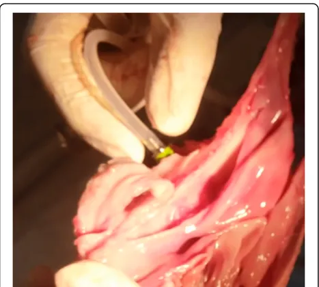
This approach starts like BAOSFE. The osteotomy is en-larged to 5.0 mm before the balloon (Zimmer Sinus Lift Balloon, Zimmer Dental Inc., California, USA) is inserted (Fig. 3). The sinus floor was broken with the 5 mm osteotome after the addition of bone. The sleeve of the balloon was inserted 1 mm beyond the sinus floor. The saline was injected slowly from the syringe into the balloon, so the balloon would inflate progressively (Fig. 4). The balloon was deflated, and the desired eleva-tion was checked if the elevaeleva-tion was not reached. The balloon was inserted again, and the process is repeated until the desired 7 mm are reached. One cubic centi-meter of saline is expected to lift the membrane for 6 mm [13].
CAS kit
The CAS kit consists of a set of safe end drills, metal stoppers, a depth gauge, a hydraulic lifter, bone graft
carrier, condenser, and a bone spreader (Fig. 5). The pro-cedure started with a 2-mm twist drill. The drills were used to enlarge the osteotomy and are stopped 1 mm short of the sinus floor. The sinus floor was broken with the 3.6 mm bur without going through the floor; a depth gauge was used to check the membrane integrity and to slightly lift the membrane. Then, the hydraulic lifter was inserted and stabilized (Fig. 6) and the saline solution is injected. 0.30 mL can elevate the membrane up to 3 mm [5]. The saline is drown out then injected again until the desired elevation is reached.
Statistics
Chi-square test was used to test the association between the three techniques and the occurrence of perforation whereas ANOVA (analysis of variance) was used to as-sess the association between method used and the two outcomes of the length of the perforation and the time of operation. Logistic regression of method used on the occurrence of perforation was employed to evaluate the odds of perforation for each method. P values equal to or smaller than .05 were considered to be significant. All
calculations were made using SPSS version 16 for Win-dows (SPSS®, Chicago, IL, USA).
Results
For the entire sample, the mean perforation length was (0.711 mm, SD = 1.4) and the mean time required to perform the procedure was (5.65 min, SD = 2.26), and out of the entire sample (N= 36), perforations happened in nine cases for a percentage of 25%.
Chi-square test showed a significant association between method used and the occurrence of perforation (chi-square statistic = 8.585, df = 2,pvalue = 0.014), as shown in Table 1. Also, ANOVA test showed a significant associ-ation between method used and the length of perforassoci-ation (F = 11.031, df = 2, 33, p value < 0.001) where the BAOSFE caused the largest mean length of perforations (3.42 mm) followed by the CAS kit and the balloon (0.5, 0.5 mm). As for the time required to perform the proce-dures ANOVA test showed a significant association be-tween method used and the time required to perform it (F= 1221.2, df = 2,33,pvalue < 0.001); CAS kit required the longest time (8.486 min) followed by the balloon then BAOSFE (5.393, 3.073 min) (Table 1).
Table 2 shows the results of logistic regression of method used on the occurrence of perforation, the odds ratio showed significant differences between the balloon technique and the BAOSFE (OR = 0.091,pvalue = 0.022), and between the CAS kit and the BAOSFE (OR = 0.091, pvalue = 0.022); however, no significant differences were found between the balloon and the CAS kit (OR = 1,0,p value = 1). It should be noted that the CAS kit was only able to lift the membrane for a maximum of 5 mm.
Discussion
Although the lateral sinus floor elevation is a proven clinically successful technique [14], the indirect SFE ap-proach is favorable among clinicians because it does not require a second surgery site and hence cause less trauma and discomfort for the patient [14–16]. However, Fig. 4The inflated balloon while elevating the sinus membrane
(The balloon is seen from the medial.)
this method has its drawbacks, such as a higher risk of membrane perforation, a decreased space for using sur-gical instruments, and limitation in elevation heights when using the conventional techniques [3, 16, 17].
The osteotome technique originally described by Tatum 1994 has been shown microscopically to elevate the sinus floor for 5 mm without causing perforations [18]. Thus, this technique should not be used when the intended elevation height is more than 5 mm [19]. Therefore, a need for trans-alveolar approach that can elevate the membrane safely and for elevation heights greater than 5 mm has risen, Tatum de-scribed a modified approach to his osteotome technique in which bone particles are pushed in the sinus. The addition of bone will prevent direct contact between the instruments and the membrane [20]. Recently, many methods for SFE have been described as an alternative for the osteotome technique. Most of this techniques fall under two categories: using an inflatable device such as a balloon or using hy-draulic pressure, both of which have been shown to reduce the rate of membrane perforation [6, 7, 13, 21, 22]. Soltan and Smiler described the use of the balloon and concluded that it is a highly successful and easy to perform procedure [6]. Recently, many systems have been developed which rely on hydraulic pressure to lift the sinus mucosa including the
Jeder-System (Jeder GmbH, Vienna, Austria) which consists of a drill with a chamber which is filled with saline solution. After the initial drilling is done, the drill is connected to a pump that produces high hydraulic pressure; the pressure is used to break the sinus floor and to lift the membrane [23]. Also, OSSTEM implants introduced the CAS kit as a method for preparing the osteotomy and elevating the membrane through hydraulic pressure.
Using a reamer instead of the osteotomes for breaking the sinus floor has the advantage of creating a thin bone shell that prevents direct contact between the drill and the Schneiderian membrane [24]. Moreover, using a reamer has been shown to cause less discomfort and nausea when compared to the osteotome technique as a result of the constant tapping of the osteotomes [25]. As a result, the CAS kit has the advantage over the BAOSFE and the balloon in preparing the osteotomy and breaking the sinus floor safely and with less compli-cations. Moreover, it was noted during our study that using a drill gives better feedback to the surgeon when breaking the sinus floor compared to the osteotome.
However, in our study, the CAS-kit was able to lift the membrane for a maximum of 5 mm. We believe that the saline pressure injected through the hydraulic lifter from a syringe is small and decreases gradually after leaving the lifter, whereas a study on the Jeder system showed a height gain of (9.2 ± 1.7 mm). This could be attributed to the high hydraulic pressure from the Jeder pump which is a machine that control the hydraulic pressure [23]. On the other hand, in our study, the balloon was able to lift the membrane for 7 mm in all cases; therefore, the balloon was bet-ter in elevating the mucosa.
Our study compared between three techniques for SFE for elevation heights of 7 mm. The 7 mm elevation height was chosen as a previous study by Stelzle et al. 2011 showed that BAOSFE caused perforations in the mucosa in all samples for perforation heights of 10 mm [7]. Therefore, we tried to set a threshold that might be achieved with internal sinus lifting techniques and be feasible in clinical practice. Perforations were checked using the three different methods: the mesial window, using a depth gauge, and the injection of saline solution through the osteotomy, which allowed for accurate re-cording of perforations.
Fig. 6The hydraulic lifter stabilized in the osteotomy before injecting the saline
Table 1The association between the methods used the following variables: occurrence of perforation, length of perforation, and the time of operation
BAOSFE BALLOON CAS kit Total Stats pvalue
Occurrence of perforation 7 (58.4%)N= 12 1 (8.3%)N= 12 1 (8.3%)N= 12 9 (25%)N= 36 × 2 = 8.585a 0.014
Length of perforation (mean) 3.42 mm 0.5 mm 0.5 mm 0.711 mm F= 11.031 0.0001
Time of operation (mean) 3.073 min 5.393 min 8.486 min 5.651 min F= 1221 0.0001
FANOVA test
a
The BAOSFE technique caused perforations in the membrane in 7 out of 12 cases with a percentage of 58.4. This result is consistent with many previous studies which state that this technique has a high rate of perforations when the RBH is less than 5 mm [2, 7, 26]. Also, all the perforations happened during the elevation process; how-ever, this percentage is different than that reported by Steltzle (100%) in a similar study as the intended elevation height was less by 3 mm in our study [7].
For the balloon technique, only one perforation hap-pened during the elevation process and the balloon was able to lift the membrane for 7 mm in all successful cases. This result supports various studies that showed a high success rate for this technique [6, 7, 13]; however, the osteotomy should be enlarged to 5 mm before inserting the balloon and this might limit the indications for this technique in thin ridges.
The CAS kit caused perforation of the Schneiderian membrane in one of the 12 cases (8.3%) which happened during the osteotomy. This is the first study to our knowledge to assess the CAS kit form OSSTEM im-plants since we found one published article that was a questionnaire sent to dentists who used the system to assess their satisfaction with the CAS kit, The study re-ported a membrane perforation rate of 4.1%. This per-centage is smaller than that reported in our study (8.3%); however, we believe that our method of checking perfo-rations is more accurate. Also, the difference in sample size may have contributed to the outcome [5].
Conclusions
Within the limitation of this study and that of an ex vivo study, we can accept our hypotheses that the balloon is better than the BAOSFE in elevating the membrane mu-cosa and the CAS kit is better than the BAOSFE in pre-paring the osteotomy and breaking the sinus floor for elevation heights of 7 mm. Further, in vivo studies need to be taken to prove these findings.
Authors’contributions
YSA conceived the study, held surgical procedures, and drafted the manuscript. AMM supervised the surgical procedures, reviewed, and approved the manuscript. DNB was responsible for the interpretation of the CBCT images and reviewed the manuscript. RS did the statistical analysis and reviewed the manuscript.
Competing interests
Aghiad Yassin Alsabbagh, Mohammed Monzer Alsabbagh, Batol Darjazini Nahas, and Salam Rajih declare that they have no competing interests.
Publisher’s Note
Springer Nature remains neutral with regard to jurisdictional claims in published maps and institutional affiliations.
Author details
1Department of Periodontology, Damascus University Dental School,
Damascus, Syrian Arab Republic.2Department of Orthodontics, Damascus
University Dental School, Damascus, Syrian Arab Republic.3Temple university,
Philadelphia, USA.
Received: 13 March 2017 Accepted: 29 August 2017
References
1. Seong WJ, Barczak M, Jung J, Basu S, Olin PS, Conrad HJ. Prevalence of sinus augmentation associated with maxillary posterior implants. The Journal of oral implantology. 2013;39(6):680–8.
2. Cawood JI, Howell RA. A classification of the edentulous jaws. Int J Oral Maxillofac Surg. 1988;17(4):232–6.
3. Tan WC, Lang NP, Zwahlen M, Pjetursson BE. A systematic review of the success of sinus floor elevation and survival of implants inserted in combination with sinus floor elevation. Part II: transalveolar technique. J Clin Periodontol. 2008;35(8 Suppl):241–54.
4. Cho SC, Wallace SS, Froum SJ, Tarnow DP. Influence of anatomy on Schneiderian membrane perforations during sinus elevation surgery: three-dimensional analysis. Pract Proced Aesthet Dent. 2001;13(2):160–3. 5. Kim YK, Cho YS, Yun PY. Assessment of dentists’subjective satisfaction with a
newly developed device for maxillary sinus membrane elevation by the crestal approach. Journal of periodontal & implant science. 2013;43(6):308–14. 6. Soltan M, Smiler DG. Antral membrane balloon elevation. The Journal of
oral implantology. 2005;31(2):85–90.
7. Stelzle F, Benner KU. Evaluation of different methods of indirect sinus floor elevation for elevation heights of 10 mm: an experimental ex vivo study. Clin Implant Dent Relat Res. 2011;13(2):124–33.
8. Lopez-Nino J, Garcia-Caballero L, Gonzalez-Mosquera A, Seoane-Romero J, Varela-Centelles P, Seoane J. Lamb ex vivo model for training in maxillary sinus floor elevation surgery: a comparative study with human standards. J Periodontol. 2012;83(3):354–61.
9. Benavides E, Rios HF, Ganz SD, An C-H, Resnik R, Reardon GT, et al. Use of cone beam computed tomography in implant dentistry: the Table 2The results of logistic regression of method used on the occurrence of perforation
Methods BAOFSE BALLOON CAS kit
Number of cases 12 12 12
Number of perforations 7 1 1
Percentage 58.4% 8.3% 8.3%
Comparison of methods regarding perforations (odds ratio)
Balloon\BOAFSE Balloon\CAS kit CAS kit\BAOFSE
Odds ratio 0.091 1 0.091
pvalue 0.022 1 0.022
Confidence interval Lower Upper Lower Upper Lower Upper
1.437 160.972 0.55 18.085 1.437 160.972
International Congress of Oral Implantologists consensus report. Implant Dent. 2012;21(2):78–86.
10. Harris D, Quirynene M. Guidelines for the use of diagnostic imaging in implant dentistry: update of the EAO clinical oral implants research. 2012. 11. Urbaniak GC, Plous S. Research randomizer (Version 4.0) [Computer
software], http://www.randomizer.org/2015. Available from: http://www. randomizer.org/. 2013.
12. Younes R, Nader N, Khoury G. Sinus grafting techniques: a step-by-step guide 2015.
13. Hu X, Lin Y, Metzmacher AR, Zhang Y. Sinus membrane lift using a water balloon followed by bone grafting and implant placement: a 28-case report. Int J Prosthodont. 2009;22(3):243–7.
14. Zitzmann NU, Scharer P. Sinus elevation procedures in the resorbed posterior maxilla. Comparison of the crestal and lateral approaches. Oral Surg Oral Med Oral Pathol Oral Radiol Endod. 1998;85(1):8–17. 15. Esposito M, Felice P, Worthington HV. Interventions for replacing missing
teeth: augmentation procedures of the maxillary sinus. The Cochrane database of systematic reviews 2014(5):CD008397.
16. Schwartz-Arad D, Herzberg R, Dolev E. The prevalence of surgical complications of the sinus graft procedure and their impact on implant survival. J Periodontol. 2004;75(4):511–6.
17. Emmerich D, Att W, Stappert C. Sinus floor elevation using osteotomes: a systematic review and meta-analysis. J Periodontol. 2005;76(8):1237–51. 18. Engelke W, Deckwer I. Endoscopically controlled sinus floor augmentation.
A preliminary report. Clin Oral Implants Res. 1997;8(6):527–31. 19. Sendyk W, Sendyk C. Reconstrução óssea por meio do levantamento do
assoalho do seio maxilar. São Paulo: Santos. 2002:109–22.
20. Summers RB. The osteotome technique: part 3—less invasive methods of elevating the sinus floor. Compendium. 1994;15(6):698, 700, 2–4 passim; quiz 10. 21. Pommer B, Watzek G. Gel-pressure technique for flapless transcrestal
maxillary sinus floor elevation: a preliminary cadaveric study of a new surgical technique. Int J Oral Maxillofac Implants. 2009;24(5):817–22. 22. Sotirakis EG, Gonshor A. Elevation of the maxillary sinus floor with hydraulic
pressure. The Journal of oral implantology. 2005;31(4):197–204. 23. Jesch P, Bruckmoser E, Bayerle A, Eder K, Bayerle-Eder M, Watzinger F. A
pilot-study of a minimally invasive technique to elevate the sinus floor membrane and place graft for augmentation using high hydraulic pressure: 18-month follow-up of 20 cases. Oral surgery, oral medicine, oral pathology and oral radiology. 2013;116(3):293–300.
24. Bae OY, Kim YS, Shin SY, Kim WK, Lee YK, Kim SH. Clinical outcomes of reamer- vs osteotome-mediated sinus floor elevation with simultaneous implant placement: a 2-year retrospective study. Int J Oral Maxillofac Implants. 2015;30(4):925–30.
25. Ahn SH, Park EJ, Kim ES. Reamer-mediated transalveolar sinus floor elevation without osteotome and simultaneous implant placement in the maxillary molar area: clinical outcomes of 391 implants in 380 patients. Clin Oral Implants Res. 2012;23(7):866–72.
![Fig. 3 a The balloon in a resting position. b The inflated balloon [12]](https://thumb-us.123doks.com/thumbv2/123dok_us/9571285.1940099/3.595.56.540.87.290/fig-balloon-resting-position-b-inflated-balloon.webp)