RESEARCH
Novel budding mode in
Polyandrocarpa
zorritensis
: a model for comparative studies
on asexual development and whole body
regeneration
Marta Scelzo
1†, Alexandre Alié
1†, Sophie Pagnotta
2, Camille Lejeune
1, Pauline Henry
1, Laurent Gilletta
1,
Laurel S. Hiebert
1,3, Francesco Mastrototaro
4and Stefano Tiozzo
1*Abstract
Background: In tunicates, the capacity to build an adult body via non-embryonic development (NED), i.e., asexual budding and whole body regeneration, has been gained or lost several times across the whole subphylum. A recent phylogeny of the family Styelidae revealed an independent acquisition of NED in the colonial species Polyandrocarpa zorritensis and highlighted a novel budding mode. In this paper, we provide the first detailed characterization of the asexual life cycle of P. zorritensis.
Results: Bud formation occurs along a tubular protrusion of the adult epidermis, the stolon, in a vascularized area defined as budding nest. The bud arises through a folding of the epithelia of the stolon with the contribution of undif-ferentiated mesenchymal cells. This previously unreported mode of bud onset leads to the formation of a double vesicle, which starts to develop into a zooid through morphogenetic mechanisms common to other Styelidae. The budding nest can also continue to accumulate nutrients and develop into a round-shaped structure, designated as spherule, which represents a dormant form able to survive low temperatures.
Conclusions: To understand the mechanisms of NED and their evolution, it is fundamental to start from a robust phylogenetic framework in order to select relevant species to compare. The anatomical description of P. zorritensis
NED provides the foundation for future comparative studies on plasticity of budding and regeneration in tunicates. Keywords: Ascidian, Evolution, Non-embryonic development, Tunicate, Vasal budding
© The Author(s) 2019. This article is distributed under the terms of the Creative Commons Attribution 4.0 International License (http://creat iveco mmons .org/licen ses/by/4.0/), which permits unrestricted use, distribution, and reproduction in any medium, provided you give appropriate credit to the original author(s) and the source, provide a link to the Creative Commons license, and indicate if changes were made. The Creative Commons Public Domain Dedication waiver (http://creat iveco mmons .org/ publi cdoma in/zero/1.0/) applies to the data made available in this article, unless otherwise stated.
Introduction
In addition to sexual reproduction and embryogenesis, a significant number of metazoan species are able to propagate asexually or regenerate completely and form new bodies through non-embryonic development (NED) [1–3]. Many species of tunicates, the sister group of vertebrates [4], are capable of asexual development and whole body regeneration [5]. Tunicates include solitary
species, which can only reproduce sexually, and colonial species that, in addition to sexual reproduction, are also able to propagate asexually and have extensive regenera-tive capabilities [5–7]. In tunicates, asexual reproduction and whole body regeneration occur by clonal replication whereby somatic tissues undergo a budding process that leads to the development of clonally related zooids. Cur-rent hypotheses for the phylogeny of the tunicates sug-gest many convergent acquisitions of NED across the whole subphylum [8–10], a view further supported by the diversity of tunicate budding modes. In fact, mechanisms of asexual development differ substantially among colo-nial species and involve differences in the body region where new buds appear, and differences in the types of
Open Access
*Correspondence: [email protected]
†Marta Scelzo and Alexandre Alié have contributed equally to this work 1 CNRS, Laboratoire de Biologie du Développement
de Villefranche-sur-Mer (LBDV), Sorbonne Université, 06230 Villefranche-sur-Mer, France
cells and tissues that contribute to the development of new zooids [11–15]. Diversity in budding mode likely contributes to the wide variation in colony organization and integration among zooids found across the subphy-lum [16].
In order to dissect the mechanisms of budding in ascidians and to understand the acquisition and loss of complex characters such as asexual propagation and whole body regeneration, it is necessary to broaden our knowledge of budding modes among different species, and to compare them in a phylogenetic per-spective relying on robust trees. Most of our current knowledge of tunicate NED comes from a limited
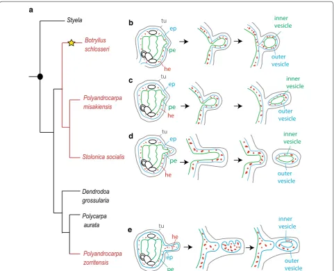
range of species. One of the most well-studied colo-nial ascidians belongs to the family Styelidae, Botryl-lus schlosseri (Pallas 1776). In this species, the bud originates from an evagination and thickening of two epithelia, the peribranchial epithelium and the sur-rounding epidermis, with the possible participation of mesenchymal cells [17]. The evagination arches pro-gressively to form a double vesicle (Fig. 1): the outer vesicle derives from the adult epidermis and gives rise to the epidermis of the new zooid, while the inner vesicle derives from the peribranchial epithelium and undergoes morphogenetic changes that eventually give rise to the internal organs of the functional new
Polycarpa aurata Dendrodoa grossularia
Polyandrocarpa misakiensis
Styela
Polyandrocarpa zorritensis Stolonica socialis
Botryllus schlosseri
ep
pe
he
inner vesicle
outer vesicle
a
b
c
d
e
outer vesicle
inner vesicle
outer vesicle
inner vesicle
outer vesicle inner vesicle
tu
pe
ep he
tu
he
tu
ep pe
tu
ep
pe
he
Fig. 1 Phylogeny of the family Styelidae and diversity of budding modes. a Simplified phylogenetic tree of Styelidae family (modify from Alié et al. 2018), showing the distribution of solitary species (shown in black) and colonial species (in red). The yellow star indicates the branches in which vascular budding evolved. b–d Schematic representation of peribranchial budding in Botryllus schlosseri (b), Polyandrocarpa misakiensis (c), and
zooid. Circulating mesenchymal cells aggregate within the folding epithelium and proliferate, participating in the formation of the organs [18–20]. The resulting bud remains attached to the colony by a short pedun-cle. This mode of reproduction, called peribranchial budding or blastogenesis, permits colonial growth and periodic zooidal renewal. In the family Styelidae, peri-branchial budding is the main form of asexual devel-opment, with some minor variations between species (Fig. 1). For instance, in Polyandrocarpa misakien-sis [21], mesenchymal cells are thought to transdif-ferentiate and integrate into the inner vesicle and the evaginated bud separates from the parent [22, 23]. In
Stolonica socialis (Hartmeyer 1903), the atrial epithe-lium and epidermis protrude and extend considerably from the zooid body forming a process called a stolon [24]. When the tip of the stolon reaches a certain mass, it is constricted off forming a bud disconnected from the maternal zooid [25].
In a recent phylogenetic reconstruction of the fam-ily Styelidae, we revealed that all the species under-going peribranchial budding belong to a single clade (Fig. 1, from [10]). Within this clade, the genera Bot-ryllus, Botrylloides, and Symplegma are also able to regenerate via a second budding mode, vascular bud-ding, in which the bud originates from the aggregation of circulating mesenchymal cells interacting with the vascular epithelium [26, 27]. The above-cited phylog-eny of Styelidae also revealed that the colonial species
Polyandrocarpa zorritensis (Van Name 1931) does not group with the clade of “peribranchial budders” but is rather more closely related to solitary styelid species (Fig. 1). These results suggest a convergent acquisition of NED in P. zorritensis, and prompted us to further describe this process [10, 28]. Preliminary histological analyses revealed that, as in other colonial styelids, a double-layered vesicle is formed prior to bud organo-genesis [10]. However, in P. zorritensis, the inner vesi-cle does not derive from peribranchial epithelium, but it originates from the epidermis of the circulatory sys-tem, which has ectodermal embryonic origin [7]. This undescribed mode of budding not only supports the
independent acquisition of coloniality and makes P.
zorritensis a key species for comparative studies aim-ing to understand the distribution of coloniality in the group, but also opens up new territory to explore the developmental mechanisms behind the capacity for budding.
In this paper, we provide the first detailed charac-terization of the asexual life cycle of Polyandrocarpa zorritensis and we describe the ontogenesis of this unreported form of ascidian budding that we name “vasal budding.”
Materials and methods
Sample collection and specimen husbandry
Colonies of Polyandrocarpa zorritensis were collected in the harbors of Taranto (Mare Piccolo, 40°28′48.29″N 17°16′4.33″E) and La Spezia (Assonautica Benedetti, 44°06′10.7″N 9°49′34.5″E) (Italy). Colonies in the field are composed of feeding zooids and often a layer of smaller orange globular non-feeding structures that we have termed spherules. The spherules were gently separated from the colony and stored in circulating sea water sys-tem at 11 °C (at salinity of 39–40‰). New zooids were grown by transferring the spherules to glass microscope slides at 24 °C. We use the term “hatching” for the emerg-ing of a new zooid from a spherule. The animals were maintained at 24 °C in open sea water systems and fed with a mix of living algae (Tisochrysis lutea) and concen-trated algae (Isochrysis 100 and Shellfish Diet 1800, Red Maricolture Inc).
Light and transmission electron microscopy (TEM)
Inclusion of Polyandrocarpa zorritensis specimens for paraffin sectioning and hematoxylin/eosin staining were performed as described in Alié et al. [10]. Samples for semi-thin and ultra-thin sectioning were fixed with a 3% solution of glutaraldehyde in sodium cacodylate buffer (pH 7.3), post-fixed with osmium tetroxide (OsO4) 1%
cacodylate buffer, dehydrated using acetone, and embed-ded in epoxy resin. An UltracutE Reichert ultrami-crotome was used for the sectioning. Semi-thin sections (1–2 µm) were stained with a solution of methylene blue 1% in sodium tetraborate + azur II 1% in water (v/v), and
images were analyzed on a ZEISS microscope Axio Imag-erA2. Ultra-thin sections (60–80 nm) were contrasted with uranyl acetate and lead citrate and observed under a transmission electron microscope TEM JEM 1400 JEOL, and imaged using a MORADA SIS camera (Olympus).
Effect of low‑temperature storage and spherule size on hatching and growth
To test whether storing the spherules at 11 °C had any negative effect on the hatching rate, two different batches of spherules were used. The first corresponds to spherules collected in Spring 2016 (April) and directly transferred to glass slides at 24 °C in the laboratory. The second batch corresponds to spherules that were kept at 11 °C in circu-lating sea water for 7 months before being transferred to 24 °C. After 7 days, the number of the zooids that were generated in relation to the number of spherules sowed was counted. After 13 days, the number of zooids bearing stolons was counted.
photometrically, by delineating their periphery manu-ally and measuring the area using ImageJ software [29]. To test the effect of spherule size, we arbitrarily split 45 spherules into three categories of size (“small” from 1 to 1.5 mm in diameter; “medium” from 1.5 to 2 mm in diameter; “big” 2 mm in diameter and larger) and then monitored the size of zooids derived from these spher-ules, considering also the number of stolons born by each zooid. Statistical significance was assessed using a Mann–Whitney test. To test the effect of budding nest size, 32 budding nests were followed during 6 days post-abscission (i.e., separation of the bud from its parental zooid by cutting the stolonial vessel with a razor blade) and the size of zooids was then measured. A linear regression curve was drawn using Excel software.
Results
Asexual life cycle of P. zorritensis in captivity
The budding process in Polyandrocarpa zorritensis has been briefly described by Brunetti and Mastrototaro [28] and by Alié et al. [10]; however, detailed descriptions of the colonial life cycle and bud histology are still miss-ing. Wild colonies are composed of a few hundred zooids clustered together and connected by a dense network of orange-colored stolons covered by a thick tunic (Fig. 2a, b). Most of these stolons have the shape of a pearl neck-lace, with spherical structures of 1 to 2.5 mm in diame-ter connected to each other by thin segments of stolon (Fig. 2c). Each of these spherules can be easily separated from the colony—their stiffness permits their manipu-lation without damage—and stored at 11 °C for up to several months without any detectable morphological alteration.
When transferred to glass slides at 24 °C, the spher-ules show morphological changes and eventually give rise to zooids. After 1 to 5 days, the spherule loses its round shape, becomes more elongated, and shows lighter orange coloration (Additional file 1: Figure S1); after 6 to 10 days two open siphons become visible (Fig. 2d) indicating that filter feeding activity has initiated. Up to 12 months at 11 °C, spherules are still able to “hatch” and develop into filtering zooids, although this process
is slightly slower than freshly collected ones (Additional file 2: Figure S2). When a colony is placed at 11 °C, most of the zooids regress and die, while only the spherules remain unaffected.
During the development from spherule to zooid, the body surface sprouts several stolons along which buds will appear. The stolons begin as small protrusions that allow the attachment of the zooid to the substrate (Fig. 2d). Usually, one or several of these body protru-sions keep growing producing elongated stolons running along the substrate (Fig. 2e). The size of a zooid as well as the number of the emitted stolons is correlated to the size of the spherule from which it develops (Additional file 3: Figure S3). The stolon consists of a monolayered epider-mal vessel covered with a thin layer of tunic. Circulating mesenchymal cells, hemocytes, flow through the stolonal lumen switching direction periodically (Additional file 4: Movie 1). Unlike the stolons of other ascidian species [30,
31], no central septum is present in the lumen of the P. zorritensis stolon. Along the whole stolon, anastomosed oval structures named ampullae grow parallel to the cen-tral vessel (Fig. 2e–e’). In some points along the stolon, the number of ampullae increases and their orientation changes, forming a tridimensional highly pigmented dome-like cluster, here named “budding nest” (Fig. 2f, g). Bud development begins at the center of these structures. Along a single stolon, it is possible to observe several budding nests, which emerge without any clear temporal or spatial order, but rather form upon substrate irregu-larities such as the angle of a Petri dish or scratches on a glass slide (personal observation).
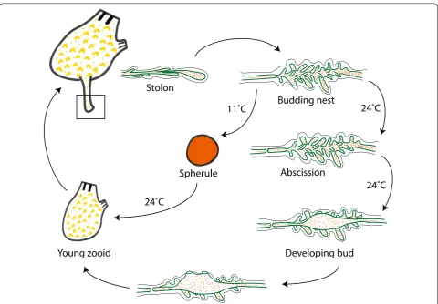
Bud development is triggered upon complete isola-tion of the budding nest from the colony, which occurs spontaneously by abscission of the stolonial epidermis and consequent interruption of hemocyte circulation between the colony and the nest (Fig. 2h). Under labo-ratory conditions, budding can be triggered by cutting the stolon with a razor blade. About 24 h after abscis-sion, the first signs of budding become externally vis-ible: the number of ampullae decreases and a new zooid grows in the center of the nest (Fig. 2i). Within 4–5 days after the abscission, all the ampullae have regressed and
(See figure on next page.)
i
j
k
a
b
c
*
d
e’
e
tunic
siphons
siphons
siphons
f
g
h
tunic
abscission
tunic
Developing
bud
stolon
stolon
ampulla
ampullae
stolon
Mature
budding nest
Spherule
Young
budding nest
Spherule
stolon
two siphons open at the top of the newly formed zooid (Fig. 2j), while the axis defined by the endostyle is always perpendicular to the substrate, the axis along the two siphons is established randomly in respect to the orienta-tion of the stolon. Just as we observed with spherules, the size of a zooid is proportional to the size of the nest from which it comes (Additional file 5: Figure S4). If abscission does not occur, the budding nest keeps growing, and if transferred at 11 °C, it eventually transforms into a dor-mant spherule (Fig. 2k).
The origin of budding: formation of the budding nest and the epidermal accumulation of reserve nutrients
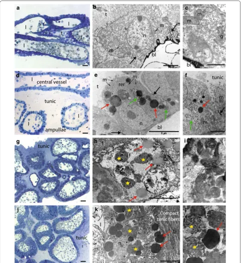
During formation of a budding nest, the epidermis of the stolonial vessel and its surrounding ampullae undergo drastic histological and cellular changes. Histological sec-tions confirmed the absence of a mesenchymal septum in the stolon. In the growing stolon, before nest formation, the stolonial wall is constituted by a monolayered epi-dermis composed of flat cells of 6–7 µm, with the highly electron-dense basal lamina facing the vascular lumen, while the apical side is covered with a layer of tunic 5–10 µm thick (Fig. 3a–c). Cells contain extensive RER (rough endoplasmic reticulum) and an abundant Golgi apparatus (Fig. 3c). At the basal pole, elaborated inter-cellular contacts connect the nearby cells (Fig. 3b, black arrows). The continuity with the zooid epidermis and the orientation of the epithelial cells confirm the epidermal nature of the stolon rather than peribranchial expansion as in other styelids (e.g., Stolonica socialis, [25]) (Fig. 3a).
When a budding nest starts forming, the tunic thickens (to around 100 µm) and ramifying ampullae accumulate around the central vessel (Figs. 2f, 3d). Epithelial cells of both the central vessel and the ampullae become round-ish, 10 µm thick and connected to neighbor cells by tight junctions at their basal halves (Fig. 3e, f, black arrows). RER becomes more prominent, mitochondria are abun-dant, and the cytoplasm is enriched in spherical electron-dense bodies of different dimensions (0.5–2 µm) (Fig. 3e, f, red arrows) in the vicinity of which very numerous smaller granules (around 0.5 nm) are also visible (Fig. 3e, f, green arrows).
When the budding nest reaches a dome shape of 1–2 mm in diameter, the number and density of ampul-lae clearly increase (Figs. 2g, 3g). At this stage, the central vessel and the ramifying ampullae are indistinguishable from each other in histological sections. The epider-mal cells change their original round shape and become rather cuboidal, 5–10 µm thick, and in close contact with their neighbors (Fig. 3h). RER, mitochondria, and Golgi become indistinguishable, while numerous of the above-mentioned spherical electron-dense bodies were observed (0.5–2 µm) (Fig. 3h, red arrows) in addition to
membrane-free inclusions with heterogeneous content (Fig. 3h, yellow stars) in which remnants of the Golgi apparatus are sometimes visible (Fig. 3i).
Budding nests and dormant spherules have very simi-lar histological and cytological properties (Fig. 3j–l), though the level of differentiation observed during nest formation is even more remarkable in spherules: their cytoplasm is completely filled with dark granules of 2–5 µm and with very amorphous and heterogeneous inclusions (Fig. 3k, l, red arrows and yellow stars, respec-tively), while smaller granules observed in budding nests (0.5–2 µm) are not visible anymore. The tunic fibers are more compacted (Fig. 3k), potentially conferring the characteristic spherule stiffness.
Vascular epidermis and mesenchymal cells participate in vasal bud formation
After abscission, the stolonial epidermis and mesenchy-mal cells contained in the budding nest undergo cellu-lar modifications and morphogenetic movements that eventually lead to the formation of a new individual. We called this process “vasal budding,” and we described the main ontological steps.
Step1: swelling
From 0 to 20 h post-abscission (hpa), budding starts by a phenomenon that we called “swelling” (Fig. 4a, b). A more or less spherical cavity about 300 µm in diam-eter (Fig. 4b) appears at the center of the nest, resulting from inflation of the stolonial vessel. This cavity, which is delimited by a monolayered epithelium, becomes polar-ized: cells at the top side (i.e., opposite to the substrate) become columnar (approximately 15 µm high × 5 µm wide), while cells at the bottom side (i.e., facing the sub-strate) are cuboidal (6–8 µm) (Fig. 4c, d, respectively). In comparison with the epithelia of the surrounding ampul-lae, the cells of the swelling cavity contain few inclusions and a higher nucleo-cytoplasmic ratio (compare ov and a
in Fig. 4d). Heterogeneous populations of mesenchymal hemocytes are visible inside the cavity, inside the connec-tions with ampullae, and inside the ampullae (Fig. 4b,d; Additional file 6: Figure S5a).
Step 2: invagination
n
t
l
t
l
a
b
c
bl
t
n
m
m
bl
t
l
l
l
l
m
g
m
d
e
f
bl
t
n
m
rer
m
m
tunic
l
l
l
m
g
h
n
t
i
j
k
ampullae
central vessel
Compact
tunic
tunic
tunic
Fig. 3 Histological and ultra-structural characterization of the stolon, budding nest, and spherule. a Transversal semi-thin section of a stolon. b A stolonial epithelial cell, with strongly electron-dense basal lamina (bl) and several mitochondria (m). The cells are connected by elaborated intercellular contacts (black arrows). c Detail of a stolonial epithelial cell, showing Golgi apparatus (g), basal lamina (bl), and mitochondria (m). d Longitudinal semi-thin section of a young nest. e Epithelial cells of the stolonial vessel, containing extensive rough endoplasmic reticulum (RER), large electron-dense granules (red arrows), and glycogen granules (green arrows). Tight junctions (black arrows) connect the neighboring cells. f Detail showing the intercellular junctions (black arrow), some small electron-dense granules (red arrow) and glycogen granules (green arrow). g Semi-thin sections of a mature nest. h Epithelial cell from a budding nest, containing spherical electron-dense granules (red arrow) and heterogeneous inclusions (yellow star). i Detail of the inclusions’ content. j Semi-thin section of a spherule. k An epithelial cell from a spherule, completely filled by heterogeneous inclusions (yellow stars) and electron-dense granules (red arrows). l Detail showing the two different types of inclusions present in the epithelial cells. (Scale bars. Semi-thin sections: 10 µm; b: 2 µm; c: 1 µm; e: um; f: 2 µm; h: 5 µm; i: 1 µm; k: 5 µm; l; 5 µm) (bl
side. In the course of invagination, the invaginating edges move toward each other (Fig. 4e, pink arrows), thus cov-ering the portion of epithelium located between them (Fig. 4e, orange star).
Step 3: fusion
Around 22 hpa, the invaginating edges eventually come into contact and fuse to each other (Fig. 4a, h). This con-sequently delineates a thin cavity that is the future lumen of the forming inner vesicle (Fig. 4h, i, green asterisk). Cells lining this forming lumen are elongated (20–25 µm high × 3–7 µm wide) and polarized, with clear apical tight junctions as well as an apical cilium (Fig. 4i, i’).
At the point of fusion, the epithelial organization of the invaginating epidermis is lost. A cluster of hemo-cytes appears around the closing aperture, between the prospective inner and outer vesicle (Fig. 4h, j). The vast majority of the clustering hemocytes show morpho-logical features of undifferentiated hemoblasts, i.e., high nucleo-cytoplasmic ratio, a prominent nucleolus, poorly developed RER and mitochondria (Fig. 4j, green arrow-head), whereas other cells contain few electron-dense granules (Fig. 4J, blue arrowhead). While the basal lam-ina is present on every epithelial cell of the inner and the outer vesicle (Additional file 6: Figure S5b), it is indis-cernible at the fusion point, so that the hemoblast cluster and the invaginating tissue constitute a morphologically indivisible unit without prominent cell junctions.
Step 4: double vesicle and initiation of organogenesis
Fusion finally results in the formation of a monolayered inner vesicle, where the apical side of the epithelia faces the lumen of the vesicle, and a monolayered outer vesi-cle, with the apical side of the epithelium facing the tunic (Fig. 4a, k). Mesenchymal cells remain visible between them, in the region where the fusion occurred (Fig. 4l). While the outer vesicle will give rise to the external epi-dermis of the new zooid, the inner vesicle folds and
undergoes organogenesis to give rise to the different organs of the zooid (Additional file 6: Figure S5c).
From this stage, regression of the surrounding ampul-lae begins. We have observed significant ultra-structural modifications in the ampullar epidermis, in which nutri-ents had accumulated during nest formation. At their basal side—facing the lumen—membranes of adjacent epithelial cells lose their integrity and therefore the cyto-plasm of all the cells that constitute the ampullar wall is in continuity (Fig. 4m, pink asterisks). In addition, the cytoplasmic electron-dense bodies are displaced toward the basal side of the cells (Fig. 4m). Finally, many mesen-chymal cells are in close contact with the ampullar wall (Fig. 4n). These cells contain large and empty vesicles, with a relatively small nucleus and sometimes a nucleo-lus, corresponding to the classical morphology of morula cells known in other ascidians. At some contact point between these cells and the epidermis, cell membranes are inconspicuous, so that cytoplasmic content of both partners seems to be in continuity (Fig. 4n’, blue arrow).
Discussion
Among tunicates, solely sexually reproducing species and species that adopt different forms of NED are scat-tered across the whole subphylum, suggesting that bud-ding and therefore coloniality have been acquired or lost several times [7, 10]. One colonial species in the family Styelidae, Polyandrocarpa zorritensis, shows a mode of budding that differs from other family members, which, along with its phylogenetic position [10], reinforces the hypothesis that NED has been acquired convergently in this species.
This paper describes the role of the vascular epider-mis and circulating hemocytes in this novel form of NED. We also show that despite the original nature of the budding tissue, bud of P. zorritensis conver-gently acquired some similarities with other tunicate buds, such as the double vesicle stage, the possible
(See figure on next page.)
participation of hemoblasts, the nutritive role of the epidermis and the existence of a dormant stage. The terms “stolonial budding” and “vascular budding,” which already designate budding modes in other tuni-cate species such as Stolonica socialis and Botryllus
primigenus [24, 27], are inappropriate for P. zorritensis.
Both of these budding modes involve cellular sources and early ontogenesis that differ from those we found in P. zorritensis. Therefore, given the peculiarities of P. zorritensis NED, we propose the term vasal budding. G
H
I
I’
a
b
c
d
e
f
g
h
i
i’
j
k
l
m
Swelling
In
vagination
Fusion
Double vesicl
e
c
d
f
g
i j
l
m tunic
tunic
iv ov
h
hemocytes
ov iv
tunic
tunic
hemocytes hemocytes
l
tunic h
lumen
l
t lumen
a
hemocytes Swelling
Invagination Fusion Double vesicle
n
n’
ov ov
ov a
tunic
lumen
lumen
*
*
*
*
*
a
*
*
*
tunic
Vasal buds originate from a novel combination of tissues
While tunicate budding modes are diverse in terms of tissues involved and early morphogenetic movements [7], most tunicate NED strategies converge morphologi-cally toward a common ontogenetic stage: a triploblastic vesicle formed by two monolayered polarized epithelia that sandwich a mesenchyme of circulating hemocytes [11, 15]. While the outer vesicle always originates from the parental epidermis, the anatomical source of the inner vesicle is highly variable across NED modes. Con-sequently, budding modes in tunicates have been catego-rized according to the tissues that give rise to the inner vesicle [16]. For instance, the inner vesicle can originate from: (1) the peribranchial epithelium (e.g., Botryllus,
Stolonica); (2) the epicardial tissue, with or without the participation of the intestinal epithelium (e.g., Diazona, Aplidium, and Polyclinum); (3) an endostyle outgrowth together with other mesenchymal cells (e.g., Salpa, Pyro-soma, and Doliolum); (4) the stolonial mesenchymal septum (e.g., Clavelina and Perophora) and (5) the circu-lating hemoblasts, as in the case of vascular budding (e.g., Subfamily Botryllinae) [26, 27, 32]. Here, we describe an additional, yet unreported budding mode that challenges the paradigm that “mature epidermis never gives rise to any tissue other than epidermis itself” [16]. Indeed, in P. zorritensis, both the outer and the inner vesicle derive from the parental epidermis through an invagination process. The peculiar onset of P. zorritensis vasal bud-ding supports the idea that among ascidians the budbud-ding capacity of a tissue may be uncoupled from its embryonic origin.
Studies of two different NED modes in Botryllus schlos-seri, peribranchial and vascular budding, show that the double vesicle stage exhibits the first spatial segregation of cell fate domains, notably for ectodermal and endoder-mal identities [33–35]. During vasal budding, the tissue differentiation and the organogenesis also start from the double vesicle stage, wherein organ primordia appears as foldings of the inner vesicle [10]. Therefore, it is possible that, despite the different onset, developmental modules responsible of the zooid morphogenesis are conserved in a convergently evolved NED.
The role of hemocytes in vasal bud ontogenesis
Formation of vasal buds in P. zorritensis involves a coor-dinated interplay between epithelial and mesenchymal (hemocyte) cells. In several tunicate species, hemocytes are known to participate in bud formation. In particu-lar, undifferentiated cells referred to as hemoblasts (or lymphocyte-like cells), which have been shown to pos-sess multi- or pluripotent stemness capacity, directly contribute to the formation of the inner vesicle [30, 36]. For instance, during stolonial budding of Perophora,
Ecteinascidia, and Clavelina the inner vesicle originates from hemoblasts arising from the mesenchymal septum [30, 37, 38], and during Botryllinae vascular budding the circulating hemoblasts give rise to the inner vesicle [27, 32, 39]. Moreover, during peribranchial budding of
Polyandrocarpa misakiensis, hemocytes appear to inte-grate into the growing bud by mesenchymal–epithelial transition [40, 41]. In P. zorritensis vasal budding, undif-ferentiated hemoblasts accumulate in the ridge formed by the invaginating epidermis during inner vesicle for-mation. In this area, the basal lamina is disrupted, the epithelial organization is lost and replaced by a mass of undifferentiated cells. Although based only on morpho-logical data, we hypothesize that hemoblasts integrate into the forming inner vesicle, contributing to its growth and potentially to its differentiation. Studies combining cell proliferation dynamics with molecular markers spe-cific for hemoblasts will be necessary to assay their role in the onset and/or differentiation of the vasal bud. It is unknown so far whether colonial tunicates possess par-ticular population(s) of hemocytes involved in asexual reproduction, which are absent in solitary species. Alter-natively, one can speculate that every tunicate possess homologous hemocyte populations, some of them being co-opted for a new function in NED. Morphological and molecular comparative studies on hemocytes at the sin-gle cell level would certainly help to solve this intriguing question.
The role of vascular epidermis and hemocytes in bud nutrition
Among tunicate NED, various populations of differenti-ated hemocytes function as reserve cells, i.e., they sup-ply nutrients at the onset of budding and during bud growth [25, 42, 43]. Although not functionally tested, this metabolic role of nourishing a bud physically and physiologically separated from the parental zooid has been documented in many species across the three major ascidian taxa, e.g., in trophocyte accumulation in winter stolons of Clavelina [44], in strobilated buds of Polycli-nidae [16] and in winter buds of Stolonica socialis and
Distomus variolosus [24]. In P. zorritensis, formation of the budding nest starts by a thickening of the ampullar epidermis that accumulates lipid and glycogen granules, together with amorphous membrane-free inclusions sim-ilar to lysosomal bodies described in other species [45–
as cytotoxic cells based on their phenoloxidase activity in different styelid species [53]. Therefore, we suggest that during vasal budding, the energy may be stored in epi-dermal cells of the ampullae rather than in trophocytes or reserve hemocytes. This is accomplished by glycogen and lipid accumulation and by self-digestion of these epi-dermal cells that release nutrients to the hemocoel and eventually to the growing buds. Such a nutritive func-tion of the bud epidermis and/or hemocytes has been convergently acquired in species whose buds are physi-cally detached from their parental zooids [24, 25, 31, 45]. Molecular comparison of anabolic and catabolic path-ways involved in nutrients accumulation and consump-tion may shed light on the mechanisms underlying this evolutionary convergence.
Co‑existence of propagating and dormant forms in P. zorritensis asexual life cycle
Previous authors have attempted to classify different tunicate NED modes according to their function within
the asexual life cycle of the colony. Mukai et al. [31] dis-tinguished between “buds which are concerned with the growth of the colony,” “buds which are destined to found new colonies,” and “buds whose function is primarily to survive adverse conditions.” The more popular classifi-cation by Nakauchi [54] recognized only two categories: “propagative budding”—used for colony growth—and “survival budding”—allowing colonies to survive adverse conditions or damages. To these two categories Turon [45, 55] added “rejuvenative budding” and “multiplica-tive budding,” respec“multiplica-tively, for synchronous regression/ renewal cycles and for production of new colonies. In the asexual life cycle of P. zorritensis, new zooid can arise either directly from the budding nest or from spherules (Fig. 5). Vasal buds of P. zorritensis seem to transcend all of the previously described functional categories, since they play (1) a survival role, in the form of the spherule, which allow for resistance to cold temperatures over win-ter [56], (2) a propagative role for colonial growth and expansion in the form of budding nest, and potentially
Abscission
11˚C
24˚C
24˚C
Stolon
Budding nest
Spherule
24˚C
Developing bud
Young zooid
(3) a colony-founding role through the dispersal of spher-ules, which are easily detachable from their mother col-ony by mechanical action, can be transported by water flow, and eventually allow colony multiplication.
Their behavior and ultrastructure suggests that P. zorri-tensis spherules represent a dormant stage. Dormancy, in general, is a change in physiological state during the life cycle in terms of morphology and metabolism that allows an organism to survive in hostile environments [57], and it appears to have evolved in almost all the groups of colonial animals. Dormancy has not been reported in any tunicate solitary species but it is present in different line-ages of colonial ascidians. It is intriguing to speculate that the cellular and molecular mechanisms underlying ascid-ian NED, such as the stem cells involved in asexual repro-duction and regeneration, were shared with dormancy. To our knowledge, P. zorritensis is the only known ascid-ian species where propagative buds and dormant stages coexist at the same time in the same colony. Therefore, P. zorritensis represents a striking model to study the rela-tionship between the evolution of NED and the evolution of dormancy.
Conclusion
In order to understand the evolution of complex traits like non-embryonic development and coloniality, it is necessary to identify suitable, closely related species in which to trace the gains, losses, and modifications of such characters [58]. Subsequently, one must provide detailed descriptions of their NED in order to uncover the cellular and molecular mechanisms that lie behind the budding processes. Due to the heterogeneity and the scattered phylogenetic distribution of NED within the Tunicata, this clade provides an excellent model to take such a comparative approach to understand the origins of NED. A recent phylogenetic analysis of the ascidian family Styelidae [10] highlighted P. zorritensis as an ame-nable model to compare budding mechanisms with NED modes in other more well-studied, closely related, models like in B. schlosseri [17, 32] and P. misakiensis [27]. The morphological description here forms a foundation for future molecular and cellular studies on the convergent origins on NED within the ascidians.
Additional files
Additional file 1: Fig. S1. Spherules at different degrees of transformation.
Additional file 2: Fig. S2. Effect of low-temperature storage on rate of budding and stolon production.
Additional file 3: Fig. S3. Relationship between the size of zooids and the size of spherules.
Additional file 4: Movie 1. Tip of a stolon with mesenchymal circulating cells.
Additional file 5: Fig. S4. Relationship between the size of zooids and the size of budding nests.
Additional file 6: Fig. S5. Details of a bud at stage 1, stage 2 and early bud organogenesis.
Authors’ contributions
M.S, A.A., and S.T. conceived and designed the study. M.S., A.A., S.P., C.L., P.H., and L.H. performed the experiments. L.G and F.M. provided P. zorritensis breed-ing support. M.S, A.A., and S.T analyzed the data and wrote the manuscript. All authors read and approved the final manuscript.
Author details
1 CNRS, Laboratoire de Biologie du Développement de Villefranche-sur-Mer
(LBDV), Sorbonne Université, 06230 Villefranche-sur-Mer, France. 2 Centre
Commun de Microscopie Appliquée, UFR Sciences, Faculté des Sciences del’Université de Nice - Sophia Antipolis, 06108 Nice, France. 3
Departa-mento de Zoologia, Instituto Biociências, Universidade de São Paulo, São Paulo 05508-090, Brazil. 4 Department of Biology and CoNISMa LRU, University
of Bari Aldo Moro, 70125 Bari, Italy.
Acknowledgements
We would like to thank S. Lotito, S. Collet and L. Gissat for technical assistance, the EMBRC France for the support with the aqua-culture infrastructure, the team of Assonautica (La Spezia) and Giovanni Chimienti (Taranto) for help collecting colonies of P. zorritensis.
Competing interests
The authors declare that they have no competing interests.
Availability of data and materials
Data sharing not applicable to this article as no datasets were generated or analyzed during the current study.
Consent for publication
Not applicable.
Ethics approval and consent to participate
Not applicable.
Funding
This work was supported by ANR (ANR-14-CE02-0019-01) and CNRS (PICS 07679) to S.T., and FAPESP-BEPE fellowship (2018/05923-3) to L.H.
Publisher’s Note
Springer Nature remains neutral with regard to jurisdictional claims in pub-lished maps and institutional affiliations.
Received: 6 February 2019 Accepted: 21 March 2019
References
1. Bell G. The masterpiece of nature: the evolution and genetics of sexual-ity. Berkeley: University of California Press; 1982.
2. Sköld HN, Obst M, Sköld M, Åkesson B. Stem cells in asexual reproduc-tion of marine invertebrates. In: Rinkevich B, Matranga V, editors. Stem cells in marine organisms. New York: Springer; 2009. pp. 105–137. 3. Brusca RC, Moore W, Shuster SM. Invertebrates, 3rd edition.
Sunder-land, Massachusetts, USA: Sinauer Associates, Inc., Publishers; 2016 4. Delsuc F, Brinkmann H, Chourrout D, Philippe H. Tunicates and not
cephalochordates are the closest living relatives of vertebrates. Nature. 2006;439:965–8.
6. Satoh N. Developmental biology of ascidians. Cambridge University Press, New York; 1994.
7. Stolfi A, Brown FD. Tunicata. In: Wanninger A, editor. Evolutionary devel-opmental biology of invertebrates 6: deuterostomia. Vienna: Springer. 2015; p. 135–204.
8. Delsuc F, Philippe H, Tsagkogeorga G, Simion P, Tilak M-K, Turon X, et al. A phylogenomic framework and timescale for comparative studies of tunicates. BMC Biol. 2018;16:39.
9. Kocot KM, Tassia MG, Halanych KM, Swalla BJ. Phylogenomics offers resolution of major tunicate relationships. Mol Phylogenet Evol. 2018;121:166–73.
10. Alié A, Hiebert LS, Simion P, Scelzo M, Prünster MM, Lotito S, et al. Conver-gent acquisition of nonembryonic development in styelid ascidians. Mol Biol Evol. 2018;35:1728–43.
11. Tiozzo S, Brown FD, De Tomaso AW. Regeneration and stem cells in ascid-ians. In: Bosh TC, editor. Stem cells from hydra to man. London: Springer; 2008. p. 95–112.
12. Berrill NJ. The Tunicata with an account of the British species. London: Quaritch; 1950.
13. Nakauchi M. Oozooid development and budding in the polyclinid ascid-ian, Parascidia flemingii (Urochordata). J Zool. 1986;208:255–67. 14. Millar RH. Budding in the ascidian Aplidium petrense Michaelsen. J Nat
Hist Ser. 1962;13:337–40.
15. Brien P. Blastogenesis and morphogenesis. Adv Morphog. 1968;7:151–203.
16. Berrill NJ. Regeneration and budding in tunicates. Biol Rev. 1951;26:456– 75. https ://doi.org/10.1111/j.1469-185X.1951.tb012 07.x.
17. Manni L, Zaniolo G, Cima F, Burighel P, Ballarin L. Botryllus schlosseri: a model ascidian for the study of asexual reproduction. Dev Dyn. 2007;236:335–52.
18. Nunzi MG, Burighel P, Schiaffino S. Muscle cell differentiation in the ascidian heart. Dev Biol. 1979;380:371–80. https ://doi.org/10.1016/0012-1606(79)90211 -2.
19. Tunicata B, Manni L, Zaniolo G, Burighel P. Ultrastructural study of oogen-esis in the compound ascidian. Acta Zool. 1994;75:101–13.
20. Akhmadieva AV, Shukalyuk AI, Aleksandrova YN, Isaeva VV. Stem cells in asexual reproduction of the colonial ascidian Botryllus tubaratus (Tuni-cata: Ascidiacea) text. Rus J Mar Biol. 2007;33:181–6.
21. Watanabe H, Tokioka H. Two new species and one possibly new race of social styelids from Sagami Bay, with remarks on their life history, espe-cially the mode of budding. Publ Seto Mar Biol Lab. 1972;XIX(5):327–45. 22. Kawamura K, Fujiwara S, Sugino YM. Budding-specific lectin induced in epithelial cells is an extracellular matrix component for stem cell aggre-gation in tunicates. Development. 1991;113:995–1005.
23. Kawamura K, Nakauchi M. Homeostatic integration of stem cell dynamics during palleal budding of ascidians. Zool Sci Zool Soc Jpn. 1991;8:p11–22. 24. Selys-Longchamps M. Sur le bourgeonnement des Polystiélinés Stolonica et Heterocarpa avec quelques notes sur l’anatomie de ces deux genres. Bull Sci Fr Belg. 1917;L(3):170–276.
25. Berrill NJJ. The development, morphology and budding of the ascidian Diazona. J Mar Biol Assoc UK. 1948;27:389–99
26. Gutierrez S, Brown FD. Vascular budding in Symplegma brakenhielmi and the evolution of coloniality in styelid ascidians. Dev Biol. 2017;423:152–69. https ://doi.org/10.1016/j.ydbio .2017.01.012.
27. Oka H, Watanabe H. Vascular budding, a new type of budding in botryl-lus. Biol Bull. 1957;112:225–40.
28. Brunetti R, Mastrototaro F. The non-indigenous stolidobranch ascidian polyandrocarpa zorritensis in the Mediterranean: description, larval morphology and pattern of vascular budding. Zootaxa. 2004;528:1. 29. Schneider CA, Rasband WS, Eliceiri KW. NIH image to ImageJ: 25 years of
image analysis. Nat Methods. 2012;9:671.
30. Freeman G. The role of blood cells in the process of asexual reproduction in the tunicate Perophora viridis. J Exp Zool. 1964;156:157–83.
31. Mukai H, Koyama H, Watanabe H. Studies on the reproduction of three species of Perophora (Ascidiacea). Biol Bull. 1983;164:251–66.
32. Ricci L, Cabrera F, Lotito S, Tiozzo S. Redeployment of germ layers related TFs shows regionalized expression during two non-embryonic develop-ments. Dev Biol. 2016;416:235–48.
33. Ricci L, Chaurasia A, Lapébie P, Dru P, Helm RR, Copley RR, et al. Identifica-tion of differentially expressed genes from multipotent epithelia at the onset of an asexual development. Sci Rep. 2016;6:27357.
34. Prünster MM, Ricci L, Brown FD, Tiozzo S. De novo neurogenesis in a budding chordate: co-option of larval anteroposterior patterning genes in a transitory neurogenic organ. Dev Biol. 2018. https ://doi.org/10.1016/j. ydbio .2018.10.009.
35. Prünster MM, Ricci L, Brown FD, Tiozzo S. Modular co-option of car-diopharyngeal genes during non-embryonic myogenesis. Evodevo. 2019;10:3. https ://doi.org/10.1186/s1322 7-019-0116-7.
36. Laird DJ, De Tomaso AW, Weissman IL. Stem cells are units of natural selection in a colonial ascidian. Cell. 2005;123:1351–60.
37. Dale Beers C. Some points in the bud development of a simple ascidian, Ecteinascidia turbinata Herdman. J Elisha Mitchell Sci Soc. 1923;39:92–4. 38. Brien P, Brien-Gavage E. Contribution à l’etude de la blastogenèse des
Tuniciers. III. Bourgeonnement de Clavelina lepadiformis. Miiller Rec Inst Zool Torley Rousseau. 1927;1:31–81.
39. Burighel P, Brunetti R, Zaniolo G. Hibernation of the colonial ascidian botrylloides leachi (Savigny): histological observations. Boll di Zool. 1976;43:293–301.
40. Tatzuke Y, Sunanaga T, Fujiwara S, Kawamura K. RACK1 regulates mes-enchymal cell recruitment during sexual and asexual reproduction of budding tunicates. Dev Biol. 2012;368:393–403.
41. Fujiwara S, Isozaki T, Mori K, Kawamura K. Expression and function of myc during asexual reproduction of the budding ascidian polyandrocarpa misakiensis. Dev Growth Differ. 2011;53:1004–14.
42. Fujimoto H, Watanabe H. Studies on the asexual reproduction in the poly-styelid ascidian, polyzoa vesiculiphora Tokioka. J Morphol. 1976;150:607– 21. https ://doi.org/10.1002/jmor.10515 00302 .
43. Berrill NJ, Cohen A. Regeneration in Clavelina lepadiformis. J Exp Biol. 1936;13:352–62.
44. Brien P. Embranchement des Tuniciers. Trait Zool Anat Syst Biol. 1928;11:553.
45. Turon X. A new mode of colony multiplication by modified budding in the ascidian Clavelina gemmae n. sp. (Clavelinidae). Invertebr Biol. 2005;124:273–83. https ://doi.org/10.1111/j.1744-7410.2005.00025 .x. 46. Rost-Roszkowska MM, Poprawa I, Chachulska-Zymełka A. Apoptosis
and Autophagy in the Midgut Epithelium of Acheta domesticus (Insecta, Orthoptera, Gryllidae). Zool Sci. 2010;27:740–5.
47. Lipovšek S, Letofsky-Papst I, Hofer F, Leitinger G, Devetak D. The evidence on the degradation processes in the midgut epithelial cells of the larval antlion Euroleon nostras (Geoffroy in Fourcroy, 1785) (Myrmeleontidae, Neuroptera). Micron. 2012;43:651–65.
48. Lobo-Da-Cunha A, Calado G. Histology and ultrastructure of the salivary glands in Bulla striata (Mollusca, Opisthobranchia). Invertebr Biol. 2008;127:33–44.
49. Sato K, Nakamura M, Tomita T, Toda M, Miyamoto K, Nozu R. How great white sharks nourish their embryos to a large size: evidence of lipid histotraphy in lamnoid shark reproduction. Biol Open. 2016;5:1211–5. 50. Angermuller S, Fahimi HD. Imidazole-buffered osmium tetroxide: an
excellent stain for visualization of lipids in transmission electron micros-copy. Histochem J. 1982;14:823–35.
51. Cima F, Peronato A, Ballarin L. The haemocytes of the colonial aplouso-branch ascidian Diplosoma listerianum: structural, cytochemical and functional analyses. Micron. 2017;102:51–64.
52. Hirose E, Shirae M, Saito Y. Ultrastructures and classification of circulat-ing hemocytes in 9 botryllid ascidians (chordata: ascidiacea). Zool Sci. 2003;20:647–56.
53. Ballarin L, Franchi N, Schiavon F, Tosatto SCE, Mičetić I, Kawamura K. Look-ing for putative phenoloxidases of compound ascidians: haemocyanin-like proteins in polyandrocarpa misakiensis and Botryllus schlosseri. Dev Comp Immunol. 2012;38:232–42.
54. Nakauchi M. Asexual development of ascidians : its biological signifi-cance, diversity, and morphogenesis. Am Zool. 1982;22:753–63. 55. Turon X. Periods of non-feeding in Polysyncraton lacazei (Ascidiacea:
Didemnidae): a rejuvenative process? Mar Biol. 1992;655:647–55. 56. Mastrototaro F, D’Onghia G, Tursi A. Spatial and seasonal distribution of
ascidians in a semi-enclosed basin of the Mediterranean Sea. J Mar Biol. 2008;88:1053–61.
57. Cáceres CE. Dormancy in invertebrates. Invertebr Biol. 1997;116:371–83. 58. Tiozzo S, Copley RR. Reconsidering regeneration in metazoans: an