IJPAR |Vol.6 | Issue 1 | Jan - Mar -2017 Journal Home page: www.ijpar.com
Research article Open Access
Design & in vitro evaluation of floating microspheres using misoprostol
K. Ranjith Kumar
1, D.V. R. N. Bhikshapathi
2*, B.Haarika
3 1Research Scholar, Mewar University, Chittorgarh, Rajasthan, India
2Research Supervisor, Mewar University, Chittorgarh, Rajasthan, India
3Sarojini Naidu Vanitha Pharmacy Maha Vidyalaya, Tarnaka, Secunderabad, TS, India
*Corresponding Author: D.V. R. N. Bhikshapathi
Email:[email protected]
ABSTRACT
The present research was aimed to prepare Misoprostol floating microspheres for sustained release using polymers such as sodium alginate and Hydroxy propyl methyl cellulose (HPMC K4M) by ionotropic gelation method. Drug and excipient compatibility studies were carried out by FT-IR and no interaction was observed. The prepared microspheres were evaluated for the Percent drug content, entrapment efficiency and In-vitro dissolution studies. Different formulations were prepared with different concentrations of polymers, among all the formulations F12 was selected as optimized formulation based on the micromeretic and physico chemical parameters including drug release studies. In vitro release study of formulation F12 showed 99.11% drug release after 12 h in a controlled manner, which is desired for disease like peptic ulcer. In vitro release profiles from optimized formulation F12 were applied on various kinetic models. The best fit with the highest correlation coefficient was observed in zero order and Higuchi model, indicating diffusion controlled principle. The marketed product IR tablet shows the drug release of 95.23% within 1 h. The results obtained from evaluation and performance study of different types of Misoprostol microspheres that system may be useful to achieve a controlled drug release profile may help to reduce the dose of drug, dosing frequency and improve patient compliance when compared with marketed product.
Keywords:
Misoprostol, Buoyancy, HPMC, Floating microspheres, Peptic ulcer.INTRODUCTION
Since the past three decades, the population of the GERD patients has recently been increasing. These situations are demanding to take drug for prolonged period of in multiple doses which causes non-compliance. To overcome this problem is to develop sustained or controlled release dosage
forms which will deliver the drug for upto 24 hrs and many drug molecules formulated as Gastroretentive Drug Delivery System (GRDDS) have been patented keeping in view its commercial success. Oral controlled release (CR) dosage forms have been extensively used to improve therapy of many important medications. The bioavailability
of drugs with an absorption window in the upper small intestine is generally limited with conventional pharmaceutical dosage forms. The residence time of such systems and thus, of their drug release into the stomach and upper intestine is often short. To overcome this restriction and to increase the bioavailability of these drugs, controlled drug delivery systems with a prolonged residence time in the stomach can be used [1].
Gastric emptying of dosage forms is an extremely variable process and ability to prolong and control the emptying time is a valuable asset for dosage forms, which reside in the stomach for a longer period of time than conventional dosage forms [2]. Several approaches are currently used to prolong gastric retention time. These include floating drug delivery systems, also known as hydrodynamically balanced systems, swelling and expanding systems, polymeric bioadhesive systems, modified-shape systems, high-density systems, and other delayed gastric emptying devices [3].
Floating microspheres are gastro-retentive drug delivery systems based on non-effervescent approach [4]. Gastric emptying of dosage forms is an extremely variable process and ability to prolong and control the emptying time is a valuable asset for dosage forms, which reside in the stomach for a longer period of time than conventional dosage forms [5].
Floating drug delivery system (FDDS) promises to be a potential approach for gastric retention. Floating microspheres are gastro-retentive drug delivery systems based on non-effervescent approach [6]. Floating microspheres have emerged as an efficient means of enhancing the bioavailability and controlled delivery of many
drugs the increasing sophistication of delivery technology will ensure the development of increasing number of gastro-retentive drug delivery systems to optimize the delivery of molecules that exhibit absorption window, low bioavailability, and extensive first pass metabolism [7].
Peptic ulcer disease, also known as a peptic ulcer or stomach ulcer, is a break in the lining of the stomach, first part of the small intestine, or occasionally the lower esophagus [8].
Misoprostol, a newly developed histamine H2 receptor antagonist, inhibits daytime as well as
night time gastric acid secretion, it has also exhibited gastroprotective activity. Which is used to treat gastric ulcers, Zollinger–Ellison syndrome, erosive esophagitis, gastro-oesophageal reflux disease and gastritis. Misoprostol has less bioavailability (80%) and lesser half life of 1.92 ± 0.94 hours [9]. The aim of present work is to design and in vitro evaluation of Misoprostol floating microspheres to enhance its bioavailability and prolonged residence time in stomach.
MATERIALS AND METHODS
Floating microspheres
Formulation
of
Misoprostol
Floating
microspheres
Misoprostol floating microspheres were prepared by ionic gelation technique using polymers Sodium alginate and HPMC K4M. Sodium bicarbonate was used as a floating agent and calcium chloride was used as crosslinking agent.
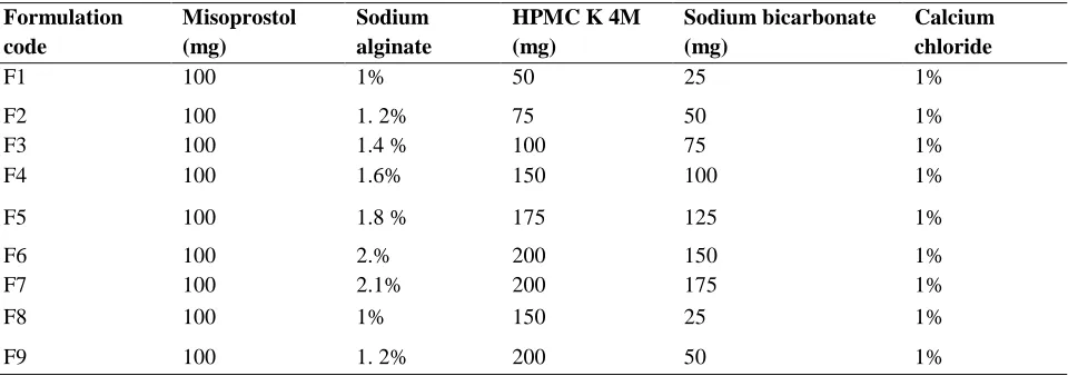
Table 1: Formulation trials of Misoprostol Floating microspheres Formulation code Misoprostol (mg) Sodium alginate
HPMC K 4M (mg)
Sodium bicarbonate (mg)
Calcium chloride
F1 100 1% 50 25 1%
F2 100 1. 2% 75 50 1%
F3 100 1.4 % 100 75 1%
F4 100 1.6% 150 100 1%
F5 100 1.8 % 175 125 1%
F6 100 2.% 200 150 1%
F7 100 2.1% 200 175 1%
F8 100 1% 150 25 1%
F10 100 1.4% 250 75 1%
F11 100 1.6% 300 100 1%
F12 100 1.8% 350 125 1%
F13 100 2.0% 400 150 1%
F14 100 2.2% 450 150 1%
Procedure
Floating alginate microspheres of Misoprostol were prepared by ionic gelation technique using different proportion of polymers as shown in table 1. A solution of sodium alginate solution is prepared weighed quantity of drug and HPMC K4M was triturated to form fine powder, and then added to above solution. Sodium bicarbonate, a gas forming agent was added to this mixture.
Resultant solution was extruded drop wise with the help of syringe and needle into 100ml aqueous calcium chloride solution and stirred at 100 rpm. After stirring for 10 minutes the obtained microspheres were washed with water and dried at 60 degrees -2 hours in an hot air oven and stored in dessicator [10].
Evaluation
of
Misoprostol
floating
microspheres
Micromeretic properties like particle size, angle of repose, bulk density, Tapped density, Compressibility index, Hausner’s ratio and evaluation parameters like Swelling index, Drug entrapment efficiency and % yield, In vitro
dissolution studies and percentage buoyancy studies were performed.
In vitro
drug release studies
In vitro drug release studies for developed Misoprostol microspheres were carried out by using dissolution apparatus II paddle type (Electrolab TDL-08L). The drug release profile was studied in 900 ml of 0.1 N HCl at 37± 0.50C temperature at 100 rpm. The amount of drug release was determined at different time intervals of 0, 1, 2, 3, 4, 6, 8, 10 & 12 hours by UV visible spectrophotometer (Shimadzu UV 1800) at 220nm.
Percentage buoyancy of Misoprostol floating
microspheres
In vitro floating ability can be determined by calculating percentage buoyancy and performed in USP type II dissolution test apparatus by spreading the floating microspheres in 0.1N HCL buffer containing the surfactant at 100 revolutions per minute (rpm) at 37± 0.5ᵒ C. After specific intervals of time, both the fraction of microspheres (floating and settled microspheres) is collected and buoyancy of the floating microspheres is determined by using formula [11].
Weight of floating microspheres
% Floating Microspheres = --- X 100 Initial weight of floating microspheres
Kinetic modeling of drug release
In order to understand the kinetics and mechanism of drug release, the result of the in vitro dissolution study of floating microspheres were fitted with various kinetic equations like Zero order as cumulative percentage released Vs. time, First order as log percentage of drug remaining to be released Vs. time, Higuchi’s model cumulative percentage drug released Vs. square root of time.r² and K values were calculated for the linear curves obtained by regression analysis of the above plots. To analyze the mechanism of drug release from the tablets the in vitro dissolution data was fitted to
zero order, first order, Higuchi’s release model and Korsmeyer – Peppas model.
Drug excipient compatibility studies
The drug excipient compatibility studies like Fourier Transmission Infrared Spectroscopy (FTIR), Differential Scanning Calorimetry (DSC) method and SEM were performed.
Stability studies
stability studies (REMI make). Accelerated Stability studies were carried out at 40 0C / 75 % RH for the best formulations for 6 months. The
microspheres were characterized for the percentage yield, entrapment efficiency and cumulative % drug released during the stability study period.
RESULTS AND DISCUSSION
Figure 1: Misoprostol floating microspheres
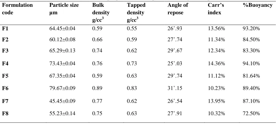
All the formulations were evaluated for their various physical parameters like particle size, bulk density, tapped density, angle of repose, Carr’s index and % buoyancy and found to be within the
results. The formulation F12 shows best results like particle size 65.45±0.09µm, bulk density of 0.74 g/cc³, angle of repose 25˚02, compressibility index 9.90% and % buoyancy of 98.50%.
Table 2: Micromeretic properties of Misoprostol floating microspheres Formulation
code
Particle size µm
Bulk density
g/cc3
Tapped density
g/cc3
Angle of repose
Carr’s index
%Buoyancy
F1 64.45±0.04 0.59 0.55 26˚.93 13.56% 93.20%
F2 60.12±0.08 0.66 0.59 27˚.74 11.34% 84.50%
F3 65.29±0.13 0.74 0.62 29˚.67 12.34% 83.30%
F4 73.43±0.04 0.76 0.73 25˚.03 14.36% 94.10%
F5 67.35±0.04 0.59 0.63 29˚.74 11.12% 81.64%
F6 79.67±0.09 0.89 0.83 31˚.15 10.23% 89.40%
F7 45.45±0.09 0.77 0.62 26˚.54 13.95% 87.10%
F9 81.22±0.11 0.79 0.75 28˚.70 11.03% 75.80%
F10 83.34±0.10 0.68 0.84 30˚.24 12.34% 76.40%
F11 78.45±0.21 0.67 0.72 26˚.91 11.90% 85.30%
F12 65.45±0.09 0.74 0.85 25˚.02 9.90% 98.50%
F13 77.23±0.19 0.85 0.73 28˚.54 10.34% 89.40%
F14 81.67±0.13 0.79 0.74 27˚.91 13.94% 92.20%
The results of Percentage yield, entrapment efficiency and swelling index floating microspheres of all formulations were within the limits shown in
Table 3. The Percentage yield, entrapment efficiency
and swelling index of F12 was found to be 98.50%, 98.56% and 98.10% respectively.
Table 3: Percentage yield, entrapment efficiency, Invitro cumulative % drug release and swelling index of
Misoprostol microspheres
Formulation code Percentage
Yield
Entrapment efficiency
Swelling index
F1 90.09% 91.09% 91.76%
F2 81.12% 82.23% 88.78%
F3 83.23% 80.56% 84.34%
F4 93.87% 93.30% 94.23%
F5 88.30% 86.20% 88.34%
F6 86.30% 84.10% 83.78%
F7 85.30% 83.30% 87.12%
F8 86.42% 84.30% 82.23%
F9 81.56% 72.89% 79.34%
F10 76.76% 83.78% 81.45%
F11 89.78% 85.78% 84.89%
F12 98.50% 98.56% 98.10%
F13 85.30% 81.30% 83.89%
F14 85.30% 80.88% 87.90%
Table 4: In vitro cumulative % drug release of Misoprostol floating microspheres
Time (Hrs) F1 F2 F3 F4 F5 F6 F7
0 0% 0% 0% 0% 0% 0% 0%
1 10.09% 22.08% 15.11% 12.09% 22.10% 16.10% 14.31%
2 19.05% 25.07% 23.12% 20.50% 25.11% 24.30% 21.15%
3 27.06% 30.11% 30.13% 28.03% 30.23% 30.20% 28.19%
4 35.08% 38.20% 38.90% 36.50% 38.20% 39.40% 37.23%
6 50.09% 51.30% 49.90% 51.60% 51.30% 53.80% 51.73%
8 66.20% 63.30% 61.20% 67.40% 63.30% 68.60% 66.46%
10 80.90% 69.90% 71.20% 82.80% 73.30% 73.90% 78.45%
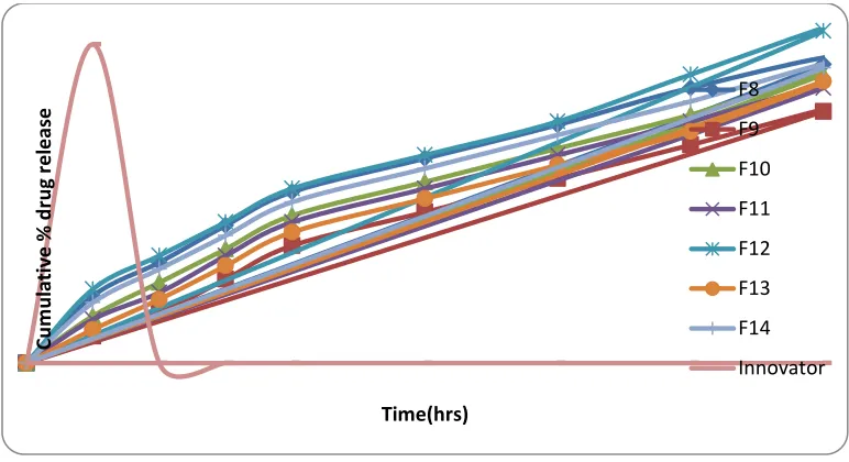
Figure 2: In vitro cumulative % drug release of Misoprostol floating microspheres
Table 5:In vitro cumulative % drug release of Misoprostol floating microspheres formulation
Time (Hrs) F8 F9 F10 F11 F12 F13 F14 Marketed product
0 0% 0% 0% 0% 0% 0% 0% 0%
1 22.12% 15.12% 20.23% 22.09% 13.09% 15.62% 21.63% 95.23%
2 25.40% 23.09% 24.80% 25.40% 21.50% 23.01% 32.01% --
3 30.01% 30.89% 31.30% 30.78% 29.20% 29.11% 37.11% --
4 38.20% 38.90% 44.40% 38.20% 37.60% 38.24% 44.83% ---
6 51.30% 49.90% 51.60% 51.30% 52.80% 52.83% 57.7% ---
8 63.35% 61.20% 60.30% 63.30% 68.50% 67.03% 64.6% ---
10 69.90% 70.10% 70.60% 69.91% 83.90% 72.22% 75.56% ---
12 87.16% 83.56% 85.45% 89.42% 99.11% 85.36% 87.76% --
C
u
m
u
lat
iv
e
%
d
ru
g
re
lea
se
Time(hrs)
F1
F2
F3
F4
F5
F6
Figure 3: In vitro cumulative % drug release of Misoprostolfloating microspheres
Release order kinetics of Roxatidine floating optimized formulation (F13)
Table 6: Release order kinetics of optimized formulation of floating microspheres
Formula Code Zero Order First Order Higuchi Korsmeyer-Peppas
R2 K R2 K R2 K R2 N
F12 0.967 8.015 0.766 0.131 0.950 35.26 0.979 2.184
From the above results it is apparent that the regression coefficient value closer to unity in case of zero order plot i.e.0.967 indicates that the drug release follows a zero order mechanism (Table no 6). This data indicates a lesser amount of linearity when plotted by the first order equation. Hence it can be concluded that the major mechanism of drug release follows zero order kinetics.
Further, the translation of the data from the dissolution studies suggested possibility of understanding the mechanism of drug release by
configuring the data in to various mathematical modeling such as Higuchi and Korsemeyer-Peppas plots.
The mass transfer with respect to square root of the time has been plotted, revealed a linear graph with regression value close to one i.e. 0.950 starting that the release from the matrix was through diffusion. Further the R2 value obtained from the Korsmeyer plots i.e. 0.959 suggest that the drug release from floating tablet was anomalous Non fickian diffusion
Cu
m
u
lativ
e
%
d
ru
g
re
le
ase
Time(hrs)
F8
F9
F10
F11
F12
F13
F14
Drug excipient compatibility studies
Fourier Transform Infrared Spectroscopy (FTIR)
Figure 4: FT-IR spectrum of pure drug Misoprostol
Figure 5: FTIR spectrum of physical mixture
Figure 6: FT-IR spectrum of Misoprostol optimized formulation F12
The IR spectrum of Misoprostol pure drug showed peaks at 2930 1, 1735 1, 1381 cm-1, and 1050 cm-1 which represented various bending and stretching vibrations of the different groups present in the drug molecule. Overall there
formulation was observed when compared to pure drug , indicating absence of any interaction.
Scanning electron microscopy studies
SEM of Misoprostol Floating microspheres
The external and internal morphology of controlled release microspheres were studied by Scanning Electron Microscopy.
Misoprostol Floating microspheres
Figure 9: Scanning electron micrograph of Misoprostol floating microspheres
The SEM of microspheres shows a hollow spherical structure with a rough surface morphology. Some of microsphere showed dented surface structure but they showed good floating ability on medium indicated intact surface (Fig 9). The shell of microspheres also showed some porous structure it may be due to release of carbon dioxide.
Stability studies
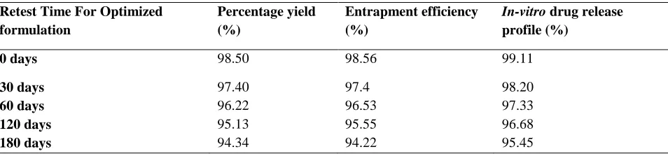
Optimized formulation was selected for stability studies on the basis of high cumulative % drug release. Stability studies were conducted for 6 months according to ICH guidelines. From these results it was concluded that, optimized formulation is stable and retained their original properties with minor differences which depicted in Table 6.
Table 6:Stability studies of optimized floating microspheres F12
Retest Time For Optimized formulation
Percentage yield (%)
Entrapment efficiency (%)
In-vitro drug release profile (%)
0 days 98.50 98.56 99.11
30 days 97.40 97.4 98.20
60 days 96.22 96.53 97.33
120 days 95.13 95.55 96.68
180 days 94.34 94.22 95.45
CONCLUSION
In the present study, an attempt was made to prepare different types of Misoprostol floating microspheres, which were characterized for particle
F12 showed 99.11% release after 12 h in a controlled manner. The in vitro release profiles from optimized formulations were applied on various kinetic models. The best fit with the highest correlation coefficient was observed in Higuchi model, indicating diffusion controlled principle. The marketed product shows the drug release of
95.23% up to1 h. FT-IR study confirmed the absence of drug-polymer interaction. From the results it can be concluded that the drug release from the floating microspheres was controlled by the polymer proportion. Prepared Misoprostol floating formulation showed best appropriate balance between buoyancy and drug release rate.
REFRENCES
[1]. Chaudhari A, Jadhav KR and Kadam VJ., Microspheres as a nasal drug delivery system, International Journal of Drug Discovery, 5(1), 2010, 8-17.
[2]. Shweta A, Javed A, Ahuja A, Khar Roop K, Baboota S. Floating Drug Delivery Systems: A Review, AAPS PharmSciTech 6(3), 2005, 372-390.
[3]. Dave Brijesh S, Amin Avani F, Patel Madhabhai M. Gastroretentive Drug Delivery System of Ranitidine Hydrochloride:Formulation and In Vitro Evaluation. AAPS Pharm Sci Tech 5(2), 2004, 1-6.
[4]. Gattani Y. S, Kawtikwar P. S, Sakarkar D. M. Formulation and evaluation of Gastro retentive Multiparticulate Drug delivery system of Aceclofenac, Int J ChemTech Res 1(1), 2009, 1-10.
[5]. Arora Shweta, Ali Javed, Ahuja Alka, Khar Roop K. AAPS Pharm Sci Tech; 6(3), 2005, E372–E390. [6]. Singh B, Kanoujia J, Pandey Manisha, Saraf Shubhini A. Int J PharmTech Research 2, 2010, 1415-1420. [7]. Gattani Y. S, Kawtikwar P. S & Sakarkar D. M. Formulation and evaluation of Gastro retentive Multiparticulate
Drug delivery system of Aceclofenac. Int J Chem Tech Res 1, 2009, 1-10. [8]. Najm, WI. "Peptic ulcer disease." Primary care 38(3), 2011, 383–94.
[9]. Patil S, Talele GS. Gastroretentive mucoadhesive tablet of Misoprostol for controlled release and enhanced bioavailability. Drug Deliv. 22(3), 2015, 312-9.
[10].Najmuddin M, Sachin S, Asgar A, Patel V, Khan T. Formulation and In vitro Evaluation of Floating Microspheres of Ketoprofen Prepared by Emulsion Diffusion Method. Int J Pharmacy and Pharma Res 2, 2010, 13-19.