R E S E A R C H
Open Access
Comparison of two different combined test
strips with fluorescent microspheres or
colored microspheres as tracers for
rotavirus and adenovirus detection
Na Jiang
1†, Lei Shi
1†, Jieping Lin
1†, Lifang Zhang
1, Yanxia Peng
1, Huiying Sheng
2, Ping Wu
1*and Qingjun Pan
1*Abstract
Background:Rotavirus (RV) and enteric adenovirus (AdV) mainly cause infantile infectious gastroenteritis. Several separate test methods for the detection of RV or AdV are currently available, but few tests are able to
simultaneously detect both RV and AdV viruses, especially in primary medical institutions.
Methods:The present study was mainly designed to compare the performance of two combined test strips for the detection of RV and AdV: a rotavirus–adenovirus strip with fluorescent microspheres for tracers (FMT); and the CerTest rotavirus–adenovirus blister strip with colored microspheres for tracers (CMT). To test the strips cultures of RV, AdV and from other enteric pathogens were used, in addition to 350 stool specimens from 45 symptomatic patients with gastrointestinal infections.
Results:Detection thresholds for RV and AdV cultures using serial dilutions showed that the sensitivity of FMT was significantly higher than that of CMT (bothP< 0.05). Specificity evaluation demonstrated that with culture mixtures of Coxsackie (A16), ECHO (type30), and entero- (EV71) viruses there was no detection of cross reaction using the two test strips, i.e., all the results were negative. With regard to the detection of RV in 350 clinical specimens, the total coincidence rate was 92.9%, the positive coincidence rate was 98.2%, and the negative coincidence rate was 90.8%. With regard to AdV detection, the total coincidence rate was 95.4%, the positive coincidence rate was 95.2%, and the negative coincidence rate was 95.5%.
Conclusions:FMT performed better than CMT with regard to the combined detection of RV and AdV.
Keywords:Rotavirus, Adenovirus, Fluorescent microspheres, Colored microspheres, Sensitivity, Specificity
Background
Rotavirus (RV) and enteric adenovirus (AdV) infections mainly result in acute infantile gastroenteritis, transmitted via the fecal–oral route. The symptoms include watery diarrhea, vomiting, headaches, fever, and gastralgic ab-dominal spasms [1, 2]. Symptoms may appear 1–2 days after viral infection, and persist for 1–10 days after the de-velopment of gastroenteritis, but different viral infections have different durations (RV approximately 3 days; AdV
5–8 days) [3], and the clinical features differ. Therefore, differential diagnosis is very important, especially for early diagnosis and treatment. Symptoms may also manifest in adults [4,5]. RV and AdV account for the first and second most common viral infections among children under 5 years, respectively [6–8]. Therefore, RV and AdV are tested for simultaneously during the clinical diagnosis of viral gastroenteritis [4,5].
Several methods for detecting RV and AdV are cur-rently available: 1) viruses were cultured in cells, and im-munological or molecular biological methods are used to identify the viruses [9]; 2) immunological methods are used directly to detect the presence of antigens or IgM and IgG antibodies in the specimens [10–12]; and 3)
* Correspondence:[email protected];[email protected]
†Equal contributors
1Clinical Research Center, Institute of Nephrology, Affiliated Hospital of
Guangdong Medical University, Zhanjiang, Guangdong, China Full list of author information is available at the end of the article
molecular biological methods are used directly to detect antigens [10, 13]. Methods 1 and 3 are limited with re-spect to technology and equipment, especially in pri-mary medical institutions and field tests. With regard to Method 2, tests based on gold immunochromatographic assays, in which colloidal gold is used as the tracer uti-lized in antigen–antibody reactions, are simple, fast, re-quire no special equipment or professionals, and have been used extensively in several fields including medi-cine and food safety [14–16]. However, this method has low sensitivity [17], and may fail to detect low titers of RV and AdV.
An emerging detection technique uses fluorescent mi-crospheres [18, 19] instead of colloidal gold particles [20]. Reaction bands can be observed with the naked eye following illumination with an ordinary UV lamp or spe-cial equipment. This technique is comparatively sensitive and stable [21, 22]. High-resolution spectra that are recognizable by the naked eye can be obtained from stimulated microspheres, with sensitivities higher than those of colloidal gold particles [23].The relevant prod-ucts are currently at the basic research and development stage, and are based on fluorescent microspheres (mainly for single-item detection) [24–26]. Colored microspheres are also used as detection tracers. They can be produced in any color (e.g., black, blue, orange, gray, red, pink, green, violet, or yellow) and in various sizes (most from 10μm to 1000 μm) from materials including polyethyl-ene, silica, and glass [27, 28]. However, the sensitivity of colored microsphere tracers is not well known, and will be evaluated in the present study.
The objective of the present study was to compare the two different methods—using fluorescent microspheretra-cers (FMT) or colored microspheretramicrospheretra-cers (CMT)—and to demonstrate the efficiency of FMT in relation to CMT in combined RV–AdV detection. This will provide a more reliable mean of detecting RV and AdV infections, and will have a significant clinical effect on the proper diagnosis and therapy of viral gastrointestinal infections, especially in primary medical institutions.
Materials and methods
Viruses
Viruses RV (Wa strain), AdV (type 40), Coxsackie (A16), ECHO 116 (type 30), and entero- (EV71) viruses were multiplied in cell cultures with MA-104 cells, Hela cells, Hep-2 cells, RD cells and Vero cells using DMEM (Dul-becco’s Modified Eagle Medium) culture media or RPMI culture media with 10% Fetal Bovine Serum (FBS), re-spectively, and stored in the laboratory.
Clinical samples
Clinical specimens were collected from 350 symptomatic patients with viral gastrointestinal infections from the
in- and out-patient wards at the Affiliated Hospital of Guangdong Medical University, and from the Guangzhou Women and Children Medical Care Center between March 2014 and February 2016.The study participants included 214 males and 136 females of mean age 16.2 ± 14.7 years. All the patients provided written informed consent to par-ticipate. The study was approved by the Ethics Committee of the Affiliated Hospital of Guangdong Medical University (LL20140317486).
All the authors had access to information that could identify individual participants during and after data collection.
Combined rotavirus–adenovirustest strips
The combined rotavirus–adenovirus fluorescent micro-spheres test strip (FMT) was provided by Guangzhou Ruimu Biotechnology Co., Ltd. (Guangzhou, China). The tracers were fluorescent microspheres (phenylethene luminative monomeric copolymers; approximate diam-eter 100 nm) with excitation and emission wavelengths of 365 nm and 485 nm, respectively (Zhuhai Yongru Co., Ltd., Zhuhai, China). The detection process was performed according to the manufacturer’s instructions, and the test mode is shown in Fig.1A.
A CerTest rotavirus–adenovirus blister test strip (CMT) was provided by CerTest Biotec S.L. (Zaragoza, Spain). The tracer is a colored microsphere (red for RV and AdV lines; blue for the control line). The detection process was performed according to the manufacturer’s instructions, and the test mode is shown in Fig.1B.
Rotavirus and adenovirus real-time reverse transcription polymerase chain reaction (real-time RT-PCR) kits
Rotavirus and adenovirus real-time RT-PCR detection kits were purchased from Guangdong Huayin Pharma-ceutical Technology Co., Ltd. (Guangzhou, China).
Sensitivity evaluation
RV and AdV were diluted and tested using the two strips. The initial concentrations of RV and AdV were 3.6 × 10⁵ plaque-forming units (PFU)/mL and 1.4 × 10⁵ PFU/mL, respectively. Three drops (150μL each) of the viral culture mixture were simultaneously applied to the sample loading well of the test strips. The results were visible within 10 min.
Evaluation of specificity
For specificity evaluation cultures of Coxsackie (A16), ECHO (type 30), and entero- 157 (EV71) viruses were used.
Preparation of clinical samples
and blood, etc. A thin tube was used to aspirate three drops (150μL each) of the mixture, which was simultan-eously applied to each of the test cards.
Statistical analyses
The total coincidence rate, the positive coincidence rate, and the negative coincidence rate of CMT and FMT were calculated to evaluate detection in the clinical specimens [29]. Statistical analysis was subjected to a Student’s t test. P< 0.05 was considered statistically significant. Statistical analysis was performed with SPSS 15.0.
Results
Sensitivity evaluation of CMT and FMT
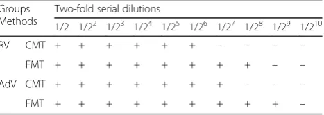
After two-fold serial dilutions of the RV cultures to 1/27 (28 × 102 PFU/mL), the CMT results were nega-tive, whereas viruses were still detectable with FMT, even the dilutions to 1/28 (14 × 102 PFU/mL), the FMT results were still positive; After two-fold serial dilutions of the AdV cultures to 1/28 (5.4 × 10² PFU/ mL), the CMT results were negative, whereas viruses were still detectable with FMT, even the dilutions to 1/29 (2.7 × 10² PFU/mL), the CMT results were still positive. Statistical analysis showed the sensitivity of FMT was significantly higher than that of CMT (both P< 0.05) (Table 1).
Specificity evaluation of CMT and FMT
The detection results from the two test strips (Table 2) showed that the culture mixture comprising Coxsackie (A16), ECHO (type30), and entero- (EV71) viruses had no cross reaction, i.e., all the results were negative.
Detection of RV and AdV in clinical specimens using CMT and FMT
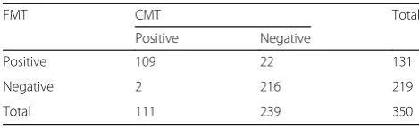
Both strips were used to test 350 specimens, and the re-sults are shown in Tables3and4.
For RV detection, the total coincidence rate of CMT and FMT was 92.9% [(109 + 216)/350)]. Compared to CMT, the positive coincidence rate of FMT was 98.2% [109/(109 + 2)], and the negative coincidence rate of FMT was 90.8% [216/(216 + 22)]. For AdV detection, the total coincidence rate of CMT and FMT was 95.4% [(59 + 275)/ 350)]. Compared to CMT, the positive coincidence rate of FMT was 95.2% [59/(59 + 3)], and the negative coinci-dence rate of FMT was 95.5% [275/(275 + 13)].
In addition to the two test strips, a rotavirus real-time RT-PCR detection kit was also used to detect RV in the clinical specimens [30]. The results showed that 20 of the 22 specimens that tested negative with CMT but positive with FMT, tested positive using the real-time RT-PCR detection method, and neither of the two speci-mens that had previously tested positive with CMT but negative with FMT gave a positive result.
Furthermore, in addition to the two test strips, an adenovirus real-time RT-PCR detection kit was also used to detect AdV in the clinical specimens [31]. The results showed that of the 13 specimens that had previously
Fig. 1Test modes of the two combined detection test strips. Fluorescent microspheres for tracers (FMT): Control line (C); Detection line (T) including the enteric adenovirus (AdV) line (near the sample loading well) and the rotavirus (RV) line. Interpretation of results: after the test procedures, double positive (left) and negative (right) results were detected. Colored microspheres for tracers (CMT): Control line (blue); Detection line (red) including the AdV line (near the sample loading end) and the RV line. Interpretation of results: after the test procedures, double positive (left) and negative (right) results were detected
Table 1Comparison of the two test strips results at two-fold serial dilutions of the RV and AdV cell cultured samples
Groups Methods
Two-fold serial dilutions
1/2 1/22 1/23 1/24 1/25 1/26 1/27 1/28 1/29 1/210
RV CMT + + + + + + – – – –
FMT + + + + + + + + – –
AdV CMT + + + + + + + – – –
tested negative with CMT but were positive using FMT, 11 tested positive with real-time RT-PCR, and none of the three specimens that had previously tested positive with CMT but negative with FMT tested positive using real-time RT-PCR.
Discussion
The use of colloidal gold test strips for the detection of RV and AdV is gradually increasing, both in China and abroad. However, owing to technological limitations, there are very few combined detection products. The CerTest rotavirus–adenovirus blister test strip (CMT) is popular in clinical practice and scientific research [32, 33]. However, CMT strips do sometimes fail to detect vi-ruses (false negative results) during clinical tests owing to deficiencies in the colored microsphere technique, es-pecially when there are insufficient quantities of RV and AdV in the stool specimens. Thus, there is a huge de-mand for lateral flow immunochromatographic tech-niques with highly sensitive fluorescent microspheres as tracers. Currently, there are few manufacturers of com-bined RV–AdV fluorescent microspheredetection test strips in China and abroad.
In the present study, CMT and real-time RT-PCR were used as the reference products to evaluate the detection performance of combined RV–AdV fluorescent sphere detection test strips. CMT uses colored micro-spheres (red and blue, etc.) as the tracers, which produce significant results when used to detect diagnos-tic reagents in vitro. The efficiency of the test trips can them be determined. Thus, colored latex microsphere immunoassays have gradually attracted attention in the industry [34–36].
RV is the most common cause of severe acute gastro-enteritis in children [1], and the most common entero-pathogens have been extensively studied in China [37–39]. In the present study, when a combined RV–AdV FMT strip was used to detect viruses in the clinical specimens, the rate
of positive RV detection in the 350 clinical specimens was 37.4% (131/350), and the rate of positive AdV detection was 20.6% (72/350). Such high infection rates demonstrate that it is necessary to detect both RV and AdV in clinical specimens from patients with gastroenteritis in China.
The results show that the combined RV–AdV FMT de-tection strips were significantly more sensitive than the CMT strips. Although the FMT technique seems to be more sensitive, it has some limitations that must be con-sidered, such as the risk of false positives. Therefore, cau-tion should be exercised, especially during the deteccau-tion of weakly positive specimens [40]. Low specimen numbers also constitute a limitation. Hence, there is a requirement for further development by medical institutions, and for large-scale clinical specimen verification. There was cross reaction with the other common viral intestinal infections with FMT. The test results should be obtained within 30 min of specimen collection to provide timely, accurate, and reliable diagnoses in clinical settings.
Conclusions
Both FMT and CMT were able to detect RV and AdV with high specificity, although FMT had higher detection sensitivity than CMT.
Abbreviations
AdV:adenovirus; CMT: colored microsphere as the tracer; FMT: fluorescent microsphere; PFU: plaque forming unit; Real-Time RT-PCR: Real-Time Reverse Transcription-PCR; RV: Rotavirus
Acknowledgements
We wish to thank Dr. Haiyan Xiao (Augusta University, Augusta, GA, USA) for modification of the manuscript in English.
Funding
The research is supported by Science and Technology Development Funds of Guangdong, China (no. 2016A020220018) and Medical Scientific Research Foundation of Guangdong Province, China (no. A2015535).
Availability of data and materials
The datasets used and analyzed in the current study are available from the corresponding author upon reasonable request.
Authors’contributions
NJ, LS and JL performed experiments and made the figs. LZ, YP and HS analyzed results. PW and QP designed the research and wrote the paper. All authors read and approved the final manuscript.
Ethics approval and consent to participate
Procedures were reviewed and approved by the Committee of the Affiliated Hospital of Guangdong Medical University.
Table 2Comparison of the test results with other viruses
Methods Coxsackie virus (A16) ECHO virus (type 30)
Enterovirus (EV71)
CMT – – –
FMT – – –
Table 3Comparison of the test results of RV clinical specimens with the two test strips
FMT CMT Total
Positive Negative
Positive 109 22 131
Negative 2 216 219
Total 111 239 350
Table 4Comparison of the test results of AdV clinical specimens with the two test strips
FMT CMT Total
Positive Negative
Positive 59 13 72
Negative 3 275 278
Consent for publication
Not applicable.
Competing interests
The authors declare that they have no competing interests.
Publisher’s Note
Springer Nature remains neutral with regard to jurisdictional claims in published maps and institutional affiliations.
Author details
1Clinical Research Center, Institute of Nephrology, Affiliated Hospital of
Guangdong Medical University, Zhanjiang, Guangdong, China.2Division of
Endocrinology and Metabolism, Guangzhou Women and Children’s Medical Centre, Guangzhou, Guangdong, China.
Received: 8 November 2017 Accepted: 27 February 2018
References
1. Parashar UD, Hummelman EG, Bresee JS, Miller MA, Glass RI. Global illness and deaths caused by rotavirus disease in children. Emerg Infect Dis. 2003;9:565–72. 2. Wilhelmi de Cal I, Mohedano del Pozo RB, Rotavirus S-FA. Other viruses
causing acute childhood gastroenteritis. Enferm Infecc Microbiol Clin. 2008; 26(Suppl 13):61–5.
3. Uhnoo I, Olding-Stenkvist E, Kreuger A. Clinical features of acute gastroenteritis associated with rotavirus, enteric adenoviruses, and bacteria. Arch Dis Child. 1986;61(8):732.
4. Elhag WI, Saeed HA, Omer el FE, Ali AS. Prevalence of rotavirus and adenovirus associated with diarrhea among displaced communities in Khartoum, Sudan. BMC Infect Dis. 2013;13:209.
5. Fletcher S, Sibbritt D, Stark D, Harkness J, Rawlinson W, Andresen D, et al. Descriptive epidemiology of infectious gastrointestinal illnesses in Sydney, Australia, 2007-2010. Western Pacific surveillance and response journal : WPSAR. 2015;6:7–16.
6. Hatherill M, Levin M, Lawrenson J, Hsiao NY, Reynolds L, Argent A. Evolution of an adenovirus outbreak in a multidisciplinary children's hospital. J Paediatr Child Health. 2004;40:449–54.
7. Ozsari T, Bora G, Kaya B, Yakut K. The prevalence of rotavirus and adenovirus in the childhood gastroenteritis. Jundishapur J Microbiol. 2016;9:e34867. 8. Ramani S, Kang G. Viruses causing childhood diarrhoea in the developing
world. Curr Opin Infect Dis. 2009;22:477–82.
9. Lee JH, Lee GC, Kim JI, Yi HA, Lee CH. Development of a new cell culture-based method and optimized protocol for the detection of enteric viruses. J Virol Methods. 2013;191:16–23.
10. Rovida F, Campanini G, Sarasini A, Adzasehoun KM, Piralla A, Baldanti F. Comparison of immunologic and molecular assays for the diagnosis of gastrointestinal viral infections. Diagn Microbiol Infect Dis. 2013;75(1):110. 11. Peret TC, Durigon EL, Candeias JM, Stewien KE, Candeias JA. A combined
staphylococcal coagglutination assay for rapid identification of rotavirus and adenovirus (COARA). J Virol Methods. 1995;52:265–72.
12. Brandt CD, Kim HW, Rodriguez WJ, Thomas L, Yolken RH, Arrobio JO, et al. Comparison of direct electron microscopy, immune electron microscopy, and rotavirus enzyme-linked immunosorbent assay for detection of gastroenteritis viruses in children. J Clin Microbiol. 1981;13:976–81. 13. Liu J, Kabir F, Manneh J, Lertsethtakarn P, Begum S, Gratz J, et al.
Development and assessment of molecular diagnostic tests for 15 enteropathogens causing childhood diarrhoea: a multicentre study. Lancet Infect Dis. 2014;14:716–24.
14. Pengsuk C, Chaivisuthangkura P, Longyant S, Sithigorngul P. Development and evaluation of a highly sensitive immunochromatographic strip test using gold nanoparticle for direct detection of vibrio cholerae O139 in seafood samples. Biosens Bioelectron. 2013;42:229–35.
15. Le T, Yan P, Xu J, Hao Y. A novel colloidal gold-based lateral flow immunoassay for rapid simultaneous detection of cyromazine and melamine in foods of animal origin. Food Chem. 2013;138:1610–5. 16. Chou SF. Development of a manual self-assembled colloidal gold
nanoparticle-immunochromatographic strip for rapid determination of human interferon-gamma. Analyst. 2013;138:2620–3.
17. Xie QY, Wu YH, Xiong QR, Xu HY, Xiong YH, Liu K, et al. Advantages of fluorescent microspheres compared with colloidal gold as a label in immunochromatographic lateral flow assays. Biosens Bioelectron. 2014;54:262–5. 18. Song C, Zhi A, Liu Q, Yang J, Jia G, Shervin J, et al. Rapid and sensitive detection of beta-agonists using a portable fluorescence biosensor based on fluorescent nanosilica and a lateral flow test strip. Biosens Bioelectron. 2013;50:62–5. 19. Xu W, Chen X, Huang X, Yang W, Liu C, Lai W, et al. Ru(phen)3(2+) doped silica
nanoparticle based immunochromatographic strip for rapid quantitative detection of beta-agonist residues in swine urine. Talanta. 2013;114:160–6. 20. Oliver C. Conjugation of colloidal gold to proteins. Methods Mol Biol. 2010;
588:369–73.
21. Aizik G, Waiskopf N, Agbaria M, Levi-Kalisman Y, Banin U, Golomb G. Delivery of liposomal quantum dots via monocytes for imaging of inflamed tissue. ACS Nano 2017. doi:https://doi.org/10.1021/acsnano.7b00016. PubMed PMID:28196324.
22. Li Z, Wang Y, Wang J, Tang Z, Pounds JG, Lin Y. Rapid and sensitive detection of protein biomarker using a portable fluorescence biosensor based on quantum dots and a lateral flow test strip. Anal Chem. 2010;82:7008–14. 23. Cong L, Takeda M, Hamanaka Y, Gonda K, Watanabe M, Kumasaka M, et al.
Uniform silica coated fluorescent nanoparticles: synthetic method, improved light stability and application to visualize lymph network tracer. PLoS One. 2010;5:e13167.
24. Zhang X, Wu C, Wen K, Jiang H, Shen J, Zhang S, et al. Comparison of fluorescent microspheres and colloidal gold as labels in lateral flow Immunochromatographic assays for the detection of T-2 toxin. Molecules. 2015;21:E27.
25. Aref MW, Akans E, Allen MR. Assessment of regional bone tissue perfusion in rats using fluorescent microspheres. Bone reports. 2017;6:140–4. 26. Qu JB, Xu YL, Liu Y, Wang Y, Sui Y, Liu JG, et al. Inherently fluorescent
polystyrene microspheres for coating, sensing and cellular imaging. Colloids Surf B: Biointerfaces. 2017;152:475–81.
27. Kumakura M, Kaetsu I. Polymeric microspheres for immunoresearch. Immunol Commun. 1984;13:119–25.
28. Hahn YK, Chang JB, Jin Z, Kim HS, Park JK. Magnetophoretic position detection for multiplexed immunoassay using colored microspheres in a microchannel. Biosens Bioelectron. 2009;24:1870–6.
29. Mei L, Wu P, Ye J, Gao G, Shao L, Huang S, et al. Development and application of an antigen capture ELISA assay for diagnosis of Japanese encephalitis virus in swine, human and mosquito. Virol J. 2012;9:4. 30. Dung TT, Phat VV, Nga TV, My PV, Duy PT, Campbell JI, et al. The validation
and utility of a quantitative one-step multiplex RT real-time PCR targeting rotavirus a and norovirus. J Virol Methods. 2013;187:138–43.
31. Sarantis H, Johnson G, Brown M, Petric M, Tellier R. Comprehensive detection and serotyping of human adenoviruses by PCR and sequencing. J Clin Microbiol. 2004;42(9):3963.
32. Akan H, Izbirak G, Gurol Y, Sarikaya S, Gunduz TS, Yilmaz G, et al. Rotavirus and adenovirus frequency among patients with acute gastroenteritis and their relationship to clinical parameters: a retrospective study in Turkey. Asia Pac Fam Med. 2009;8:8.
33. Choi SW, Park CH, Silva TM, Zaenker EI, Guerrant RL. To culture or not to culture: fecal lactoferrin screening for inflammatory bacterial diarrhea. J Clin Microbiol. 1996;34:928–32.
34. Walter B, Bauer R, Gaser E, Zwiener U. Validation of the multiple colored microsphere technique for regional blood flow measurements in newborn piglets. Basic Res Cardiol. 1997;92:191–200.
35. Bauer R, Walter B, Wurker E, Kluge H, Zwiener U. Colored microsphere technique as a new method for quantitative-multiple estimation of regional hepatic and portal blood flow. Exp Toxicol Pathol. 1996;48:415–20. 36. Oohara K, Usui A, Murase M, Tanaka M, Abe T. Regional cerebral tissue
blood flow measured by the colored microsphere method during retrograde cerebral perfusion. J Thorac Cardiovasc Surg. 1995;109:772–9. 37. Bai Y, Dai YC, Li JD, Nie J, Chen Q, Wang H, et al. Acute diarrhea during army
field exercise in southern China. World J Gastroenterol. 2004;10:127–31. 38. Qu M, Deng Y, Zhang X, Liu G, Huang Y, Lin C, et al. Etiology of acute
diarrhea due to enteropathogenic bacteria in Beijing, China. The Journal of infection. 2012;65:214–22.
39. Li Y, Xie X, Shi X, Lin Y, Qiu Y, Mou J, et al. Vibrio parahaemolyticus, southern coastal region of China, 2007-2012. Emerg Infect Dis. 2014;20:685–8. 40. Yang Q, Gong X, Song T, Yang J, Zhu S, Li Y, et al. Quantum dot-based