ABSTRACT
WALTERS, JAD ANTHONY. Structure, Folding, and Assembly of (Pro)caspase-3. The Role of the Dimer Interface in Active Site Formation. (Under the direction of A. Clay Clark).
Effector procaspase-3 plays a vital role in carrying out the final steps of programmed cell death, leading to the destruction of the cell. Because dimerization of (pro)caspase-3 is essential for enzyme stability and activity, it is important to understand the structural details of the interactions at the dimer interface.
We show a single site in the dimer interface of (pro)caspase-3 can be used to activate or inhibit the enzyme. The results presented here demonstrate activation of procaspase-3, in the absence of intersubunit linker cleavage, which generates a constitutively active,
uninhibitable enzyme that is very efficient in killing both healthy and diseased mammalian cells. Moreover, inhibition may also be achieved utilizing the same site and the structural studies reveal two novel pathways of inhibition. Overall, these studies show how the interactions at the dimer interface in procaspase-3 are essential in formation of a competent active site.
In addition to the procaspase-3 interface studies, several crystallographic studies are presented which are aimed at elucidating a structure of procaspase-3. Finally, a
comprehensive protocol for carrying out equilibrium folding studies and determining
conformational stabilities of macromolecules is provided. All together, this work has lead to exciting and novel discoveries in the field of apoptosis, including new mechanisms to
Structure, Folding, and Assembly of (Pro)caspase-3. The Role of the Dimer Interface in Active Site Formation
by Jad A.Walters
A dissertation submitted to the Graduate Faculty of North Carolina State University
in partial fulfillment of the requirements for the degree of
Doctor of Philosophy
Biochemistry
Raleigh, North Carolina March, 2009
APPROVED BY:
_______________________________ ______________________________ A. Clay Clark Robert B. Rose
Chair of Advisory Committee
_______________________________ ______________________________ Paul Wollenzien Carol K. Hall
DEDICATION
BIOGRAPHY
ACKNOWLEDGMENTS
I would like to thank Dr. A. Clay Clark for his support throughout the years. He has been a wonderful teacher and supervisor and I would not be the scientist I am today without his amazing guidance. He was sincerely a better supervisor than I could have ever imagined.
I would also like to thank my committee members, Dr. Bob Rose, Dr. Paul
Wollenzien and Dr. Carol Hall. Their support has been wonderful and I truly appreciate all of the great advice and feedback from them. Additionally I would like to thank Dr. Carla Mattos for the great collaborations and advice.
I owe a great deal of thanks to Dr. Paul Swartz. He was essentially a second advisor, and like Clay, I would not be the scientist I am today without his great guidance. Thank you Paul for teaching me everything from X-ray crystallography to repairing car transmissions!
you.
TABLE OF CONTENTS
Page
1. LIST OF TABLES………...………..……….…..………..xi
2. LIST OF FIGURES………..………....xiii
3. CHAPTER I. Introduction……....…………...……….………1
• The Significance of Programmed Cell Death………2
• Apoptotic Pathways………...3
• Mediators of Apoptosis………..3
• Caspase Structure and Activation………..5
• Characterization of the Caspase Interfaces………...………...11
• Project Goals……….………...13
4. CHAPTER II. A Constitutively Active and Uninhibitable Caspase-3 Zymogen………….. Efficiently Induces Apoptosis…………....………15
ABSTRACT……….…….……….…...……….……...16
INTRODUCTION..……….………….……….………...16
RESULTS……….………20
• Caspase-3 V266E interface mutants are active before processing………...20
• V266E interface mutants kill mammalian cells more efficiently than WT caspase-3………..……….24
• The intrinsic activity of interface mutants in transfected cells is……….
lower than that of WT………..32
• XIAP is a poor inhibitor of D3A,V266E……….38
• The X-ray crystal structure of V266E………..45
• Conserved water molecules in the dimer interface………..49
• Models of (in)active procaspase-3………...52
DISCUSSION………...62
EXPERIMENTAL PROCEDURES…..………...68
SUPPLEMENTAL EXPERIMENTAL PROCEDURES……….77
ACKNOWLEDGEMENTS………..80
5. CHAPTER III. Structural Details of Allosteric Inhibition Mechanisms………... in Caspase-3……….……….…81
ABSTRACT……….…….……….…...……….……...81
INTRODUCTION..……….………….……….………...82
RESULTS……….………89
• Crystal structures of the V266H series……….………...89
• The cause of Tyr195 displacement………..95
• The effects of Tyr195 displacement………97
• Intramonomer Inhibition………..98
• Intermonomer Inhibition………101
• Direct Inhibition……….109
• Enzyme Activity...117
DISCUSSION………...124
EXPERIMENTAL PROCEDURES…..………...127
6. CHAPTER IV. Thermodynamic, Enzymatic and Structural Effects of Ablating………….. a Salt Bridge at the Base of Loop 4 in (Pro)caspase-3.……….…130
ABSTRACT……….…….……….…...………...….131
INTRODUCTION..……….………….……….…………...….133
RESULTS……….………...136
• Structural and functional probes of equilibrium unfolding………...…136
• Equilibrium unfolding of procaspase-3 variants………...…137
• Denaturation is four-state………..138
• Crystal structures of the loop 4 mutants………147
DISCUSSION……….………....159
EXPERIMENTAL PROCEDURES…..……….163
7. CHAPTER V. Crystallization of Procaspase-3………...……….…170
RESULTS……….………...171
• Procaspase-3 variants……….………171
• Crystallization Conditions……….173
• Crystal structure of D28A and D9A,D28A………176
• Crystallization of procaspase-3, D3A……….178
6. CHAPTER VI. Practical Approaches to protein folding and assembly:………
Spectroscopic strategies in thermodynamics and kinetics………….…193
ABSTRACT……….…….……….…...………...….194
INTRODUCTION..……….………….……….…………...….195
EQUILIBRIUM UNFOLDING……….197
• Practical Considerations………197
INSTRUMENTATION……….………200
• Fluorescence emission………..200
• Circular dichroism………201
PREPARATION OF 10 M UREA STOCK……….………..203
CONFIRM THAT THE PROTEIN IS COMPLETELY UNFOLDED…….…………206
ESTABLISH EQUILIBRATION TIMES AND REVERSIBILTY FOR THE FOLDING REACTION.……….………...208
• Method 1.………..208
• Method 2.………..211
EQUILIBRIUM UNFOLDING………..212
INTERPRETATION OF EQUILIBRIUM UNFOLDING CURVES………213
• Monomeric Models…….………..216
• Dimeric Models………...219
DATA ANALYSIS……….229
• Equilibrium Constants and Fraction of Species………....229
CONCLUSIONS………..………...…….232
FUTURE DIRECTIONS………..………...235
REFERENCES………..………...………237
APPENDICES………..………..………..246
APPENDIX A………..248
• Making Morphing Movies In Rigimol ………...248
APPENDIX B………..255
• LSQMAN and MOLEMAN………... ………...255
• Brute Force Alignment in LSQMAN………256
• Renumbering PDB files with MOLEMAN………...256
• Renaming Chain IDs with MOLEMAN………...257
• Morphing in LSQMAN……….257
APPENDIX C • Useful Commands in Pymol……….260
LIST OF TABLES CHAPTER I
Table 1. Diseases Associated With Dysfunctions in Apoptosis………2
CHAPTER II
Table 1. XIAP Inhibition Constants for Caspase-3 Interface Mutants………39 Table 2. Summary of Data Collection and Refinement Statistics for Caspase-3(V266E)…..61
CHAPTER III
Table 1. Summary of Data Collection and Refinement Statistics………..87-88 Table 2. Structures Used in the Analysis of the Lys137-Glu190 Interaction………108 Table 3. Catalytic Parameters of the Caspase-3 Interface Mutants………...122
CHAPTER IV
Table 1. Thermodynamic Parameters for the L4 Variants……….143
CHAPTER V
Table 1. Generalized List of Crystallization Conditions for Procaspase-3………....175 Table 2. D3A,V266E Expression Profile………...188
CHAPTER VI
LIST OF FIGURES CHAPTER I
Figure 1. Pathways of Apoptosis………...4
Figure 2. Organization of and classification of the caspase subfamilies………...6
Figure 3. Schematic representation of the caspase maturation process……….7
Figure 4. (Panel A) Homology model of procaspase-3………...9
(Panel B) Active conformation of caspase-3……….……9
Figure 5. Movements of the active site loops upon maturation………...10
CHAPTER II Figure 1. (Panel A) Mutants of caspase-3 described in the text………...18
Figure 2. (Panel B) Labeling the caspase-3 V266E mutants by affinity based probes.……...22
Figure 3. (Panel C) Determining the substrate specificity of recombinant caspase-3… …… mutants………..25
Supplemental Figure 1. Dilution Studies of the V266E Interface Mutants………...…..23
Figure 4. (Panel A) Cell viability monitored by Annexin V staining in HEK293A cells using various caspase-3 mutants……..…………...………26
Figure5. (Panel B) Western blots of cellular lysates from panel A………28
Supplemental Figure 2. RT-PCR Reactions of DNA-free-RNA Isolated from………... Transfected HEK293A………..29
Figure 7. (Panel B) Hypotonic Jurkat lysates were immuno-depleted of endogenous………....
caspase-3 and reconstituted with recombinant caspase-3……….33 Supplemental Figure 3. Relative DEVD-ase Activity of the Culture Medium………...
from Cells Transfected with Caspase-3 Mutants………..37 Figure 8. (Panel A) WT Caspase-3 lysates display higher DEVD-ase activity than …………..
those of V266E interface mutants………34 Figure 9. (Panel B) Kinetics of DEVD-ase activity in lysates prepared from cells
transfected with WT or caspase-3(V266E) at various time periods……36 Figure 10. (Panel C) XIAP is a poor inhibitor of D3A, V266E……… 39 Figure 11. (Panel D) Interface mutants were inhibited by baculovirus p35 ………..… 41 Supplemental Figure 4. (Panel A) Co-expression of XIAP with the Caspase-3 V266E………. Mutants Cannot Rescue Cells from Apoptosis………….42 Supplemental Figure 4. (Panel B) Western blotting of the samples from Panel A …………42 Figure 12. (Panel A) Amino acids in the dimer interface of caspase-3 relative ……… ……...
to the two active sites. ……….……… 46 Figure 12. (Panel B) Amino acids in the dimer interface of caspase-1 relative… ………
to the two active sites……..……… ……….…… 46 Figure 13. (Panel C) Interactions in L2’with L4 and the P4 binding site……….…………..47 Supplemental Figure 5. (Panel A) Co-transfection of caspase-3 (V266E) with ………
Figure 14. (Panel A) Conserved waters in the dimer interface of WT caspase-3.…..……… 50 Figure 14. (Panel B) Waters in the dimer interface of caspase-3, V266E……….…….……50 Figure 15. (Panel C) Comparison of the dimer interface of V266E with that of caspase-1…51 Figure 15. (Panel D) V266E modeled using the rotamer found in caspase-1…………...…51 Supplemental Figure 6. (Panel A) Alignment of amino acid sequences showing
conservation of the safety catch. ………..……....67 Supplemental Figure 6. (Panel B) Alignment of amino acid sequences showing………...
conservation of residues in β-strand 8 in the interface...67 Figure 16. (Panel A) Model of inactive procaspase-3………...53 Figure 17. (Panel B) The blocking segment of IL-B prevents insertion of………...
active site loop 3 from monomer A………..………..55 Figure 18. (Panel C) The blocking segment of IL-A prevents insertion of ………..
active site loop 3 from monomer B………..………..56 Figure 19. (Panel D) Hydrophobic cluster in the inactive procaspase-3………..
centered about V266……….…..59 Figure 19. (Panel E) The V266E mutation disrupts the hydrophobic cluster………….….59 Figure 20. (Panel F) Comparison of the IL in the inactive and active procapase-3……….60
CHAPTER III
Figure 4. (Panel C) The crystal structure of the V266H variant………...93
Figure 5. (Panel D) The crystal structure of the Y197C,V266H variant…….…………...94
Figure 6. (Panel A) Structural details of intramonomer inhibition….………...96
Figure 7. (Panel B) Steric clash between Phe128 and Met61……...………....99
Figure 7. (Panel C) Movement in Met61 result in the lost interaction …… ………... between Thr62 and His121……….…………...99
Figure 8. (Panel D) Movement of the residues in the intramonomer mechanism … ………. result in the disorder of loop 1 ………...100
Figure 9. (Panel E) Relief of steric constraints in the dimer interface of the ……… ……… V266H structure restores order to the capase-3 structure.……..……..102
Figure 10. (Panel A) Structural details of intermonomer inhibition...……….103
Figure 11. (Panel B) Lost interactions affecting the stability of the loop bundle……..…....105
Figure 12. (Panel C) Electron density in the WT and V266H structures highlighting order in the Lys137 side chain……….…….………106
Figure 13. (Panel D) L2’ is truncated in the V266H structure…………...………...107
Figure 14. (Panel A) Residues involved in the direct inhibition mechanism..………..110
Figure 15. (Panel B) The alternate conformation of Glu124 in the R164G variant..………112
Figure 16. (Panel A) Conserved waters in the dimer interface of WT caspase-3…..……...114
Figure 16. (Panel B) Waters in the dimer interface of caspase-3, V266H….………….…..114
Figure 17. (Panel A) A plot of the initial velocity (υo) vs. substrate concentration… ……….. for wild-type caspase-3 and caspas-3 interface mutants.……...…….119
Figure 19. (Panel C) A plot of the initial velocity (υo) vs. substrate concentration ..…………..
for the residues involved in direct inhibtion…..……..……….121
CHAPTER IV
Figure 1. Concentration dependence of equilibrium unfolding of ………..
procaspase-3(C163S,E246A)……….………139 Figure 2. Concentration dependence of equilibrium unfolding of ………..
procaspase-3(C163S,K242A)...……….………140 Figure 3. Concentration dependence of equilibrium unfolding of ………..
procaspase-3(C163S,K242A,E246A)………141 Figure 4. (Panel A) Regions of (pro)caspase-3 affected by removing the………...
Lys242 – Glu246 ionic interaction………145 Figuer 5. (Panel B) Schematic representation of the crystal packing …… ………… ………
of caspase-3 in the orthorhombic space group I222………..146 Figure 6. Interactions between L2’ and L4 in the WT structure are
absent in the K242A, E246A and K242A,E246A structures………148 Figure 7. (Panel A) Hydrophobic interactions involving Lys242 and helix 4...…...……....150 Figure 8. (Panel B) Removal of Glu246 eliminates the Glu246 – Lys242
salt bridge and disrupts the hydrophobic involving……….
Lys242 and helix 4……….………….…..151 Figure 9. (Panel C) Interactions across the interface in the WT structure……… …………..
Figure 10. (Panel D) New interactions across the interface in the K242A,E246A …………..
structure between residues on helix 5 and the C-termini ……….…..154
Figure 11. A plot of the initial velocity (υo) vs. substrate concentration ………... for wild-type caspase-3 and K242A,E246A mutant…...……….155
Figure 12. Putative interactions in the inactive procaspase-3 structure involving Lys242 and Glu246………..………...….……157
CHAPTER V Figure 1. Variants of procaspase-3 described in the text………...172
Figure 2. Setup for crystallizing caspase-3………....174
Figure 3. Crystal packing of caspase-3 in the orthorhombic space group I222 leaves a large unoccupied region at the prodomain………..177
Figure 4. (Panel A) Comparison of the C2 and I222 lattice groups………..180
Figure 5. (Panel B) Crystal morphologies of the C2 and I222 space groups………181
Figure 6. (Panel A) SDS PAGE analysis of D3A and D3A,V266E………...………182
Figure 6. (Panel B) D3A crystals analyzed by SDS PAGE………...…182
Figure 7. Diagrams of the monoclinic space group C2 using different unique axes of rotation………...186
Figure 8. SDS PAGE analysis of the D3A,V266E expression profile………...189
Figure 9. Crystal morphologies of D3A,V266E……….191
CHAPTER VI
Figure 1. (Panel A) Emission spectra of excitation at 280 nm………..………...202 Figure 2. (Panel B) Urea denaturation curve indicating the pre-transition,………
transition, and post-transition regions………..215 Figure 2. (Panel C) Normalized data showing three probes used in the……….
unfolding experiments………..215 Figure 3. (Panel D) Noncoincidence of the unfolding curves when monitored ………
by different techniques……….218 Figure 3. (Panel E) Three-state monomer unfolding curve……….218 Figure 4. (Panel A) Two-state dimer unfolding curve …………..………..………...221 Figure 5. (Panel B) Three-state monomer unfolding curve with a………...
monomeric intermediate……….……….………….222 Figure 5. (Panel C) Three-state monomer unfolding curve with a …… ……….
dimeric intermadiate……….………….222 Figure 6. (Panel D) Four-state dimer unfolding curve………..225
APPENDIX C
CHAPTER I
The Significance of Programmed Cell Death
Apoptosis, or programmed cell death, plays a critical role in maintaining cellular homeostasis. The controlled process of apoptosis is used to neatly remove unwanted cells during development and those cells which pose a threat to the integrity of the organism. The balance between cell death and cellular proliferation is a tightly regulated process and dysfunction in the apoptotic pathway is implemented in several pathological conditions (Table 1).
Through excess apoptosis
AIDS
T lymphocytes
Neurodegenerative diseases
Alzheimer’s disease
Amyotrophic lateral sclerosis Parkinson’s disease
Retinitis pigmentosa Epilepsy
Haematologic disease
Aplastic anaemia
Myelodysplastic syndrome T CD4+ lymphocytopenia G6PD deficiency
Tissue damage
Myocardial infarction Ischaemic renal damage Polycystic kidney Through inhibition of apoptosis
Cancer
Colorectal Glioma Hepatic
Neuroblastoma
Leukaemias and lymphomata Prostate
Autoimmune diseases
Myastenia gravis
Systemic lupus erythematosus
Inflammatory diseases
Bronchial asthma
Inflammatory intestinal disease Pulmonary inflammation
Viral infections
Adenovirus Baculovirus Chamond et al., 1999
Apoptotic Pathways
Apoptosis is triggered through two major signaling pathways, the intrinsic and extrinsic pathways. The extrinsic pathway is initiated by ligation of transmembrane death receptors (Fas, TRAIL and TNF) (Boatright, 2003). Triggering of these death receptors leads to the recruitment of initiator procaspase-8/10, via the Fas-associated death domain protein (FADD). The interaction between the death effector domains (DED) of FADD and procaspase-8/10 results in cleavage and activation of the procaspase. Upon its release into the cytoplasm, caspase-8/10 cleaves and activates downstream executioner caspases -3, -6 and -7, which ultimately leads to the destruction of the cell.
The intrinsic pathway is initiated by the release of cytochrome c from the mitochondria in response to cytotoxic stress. Cytochrome c binds to apoptotic protease activating factor-1 (Apaf-1) in the presence of dATP, which results in formation of the Apaf-1/caspase-9 complex referred to as the apoptosome. Activated caspase-9
specifically targets and activates procaspase-3 (Budihardjo, 1999). Both pathways to caspase activation are shown in Figure 1.
Mediators of Apoptosis
Caspase Structure and Activation
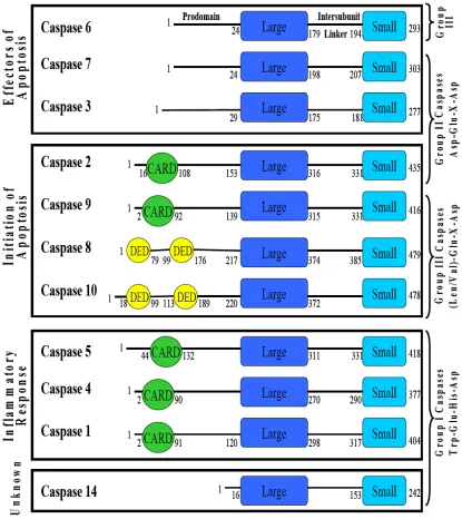
All caspases are organized in a similar manner, consisting of an N-terminal prodomain, a large subunit, an intersubunit linker and a small subunit. Typically, the prodomain of executioner caspases are shorter, while the prodomains of initiator and inflammatory caspases are longer, as they contain either a CARD (caspase recruitment domain) domain or a DED (death effector domain) domain. The prodomain of
procaspase-3 has been shown to aid in assembly of the enzyme, acting as an
intramolecular chaperone (Feeney, 2005) and the prodomain in initiator an inflammatory caspases aids in dimerization by binding to adaptor proteins upon initiation of apoptosis, thereby increasing the local concentration of the procaspase. This is known as the induced proximity model and is discussed further in Muzio et al., 1998 and Pop et al., 2006.
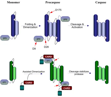
In the monomeric state, all of the caspases share common structural features consisting of a six-stranded beta-sheet core flanked by alpha-helices. Each monomer, composed of the large and small subunit (Figure 3), contains one active site, but remains inactive until the minimum requirement of dimerization is met. Once dimerized, a consistent β-sheet extends the length of the protein.
Figure 3. Schematic representation of the caspase maturation process. Effector caspases
are found in the cell as stable dimers. The red arrows indicate the three cleavage sites in procaspase-3 and the active site is indicated by a diamond. Cleavage at D9 and D28 remove the prodomain and cleavage at D175 in the intersubunit linker allows rearrangement of the active site loops, yielding mature caspase-3. Initiator and inflammatorycaspases are found in the cell as stable monomers. Dimerization is coupled to activation, thus, while cleavage (red arrows) stabilizes the enzyme, it is not necessary for activation.
pro
D175
Folding &
Dimerization Cleavage & Activation
pro
pro
D9 D28
Monomer Procaspase Caspase
pro
CARD
pro
CARD
Cleavage stabilizes protease Assisted Dimerization
CARD
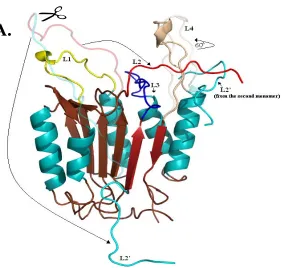
L3 (residues 198-213) and L4 (residues 247-263). Of the five loops that comprise the active site, one, L2’, is contributed by the second monomer, thus, it becomes apparent why dimerization is an absolute requirement for activation. In addition to cleavage at Asp175 in the intersubunit linker, two more cleavages occur at Asp9 and Asp28 which remove the prodomain (Figure 3).
The cleavage event in the intersubunit linker at Asp175 creates two of the five active site loops, L2 and L2', which are then liberated to undergo the necessary conformational changes needed for proper active site configuration. The structural changes observed in L2 allow the catalytic cysteine to adopt a more favorable position, which is now readied for nucleophilic attack of the substrate. In addition, L4 rotates
60°towards the active site and L3 moves toward the core of the protein, forming the
substrate binding pocket. L2' from the opposing monomer flips ~180° and wraps behind
L4 and making contacts with both L2, L4 and the S4 site of the substrate (Figures 4 and 5). The interaction between loops L2, L2’, L3, and L4 is known as the loop bundle, coined by Shi (Shi, 2001). Several hydrogen bonds are made
Figure 4. (A) Homology model of procaspase-3. The active site loops of one monomer are indicated. L1 (yellow), L2 (red), L2’ (cyan), L3 (blue) and L4 (light brown) (B) Active conformation of caspse-3. One monomer is composed of a large subunit (dark brown β-sheet) and a small subunit (2 dark red anti-parallel β-strands) L2’ is contributed by the small subunit of monomer 1 to the active site of monomer 2. The interactions between loops L2, L2’ and L4 form the loop bundle.
A
Monomer
Figure 5. (A) The movements of the active site loops are highlighted. Loops shown as semi-transparent are of procaspase-3 and the full color loops are in the active
configuration, caspase-3. For clarity, only one monomer is shown, however, L2’ from the second monomer is shown. Arrows indicate movement of the loops during maturation. The change in color from cyan to red in the intersubunit linker represents where cleavage occurs.
Characterization of the Caspase Interfaces
All caspases are initially produced in the cell as quiescent zymogens. It is known that dimerization is necessary for acquisition of activity in caspases. However, there are variations in activity levels of the unprocessed procaspase between the subfamilies. For instance, initiator and inflammatory caspases are stable monomers in the cell (Figure 3). Upon dimerization, the unprocessed enzyme displays activity levels comparable to that of the mature enzyme. This is because activity is coupled to dimerization rather than chain cleavage in initiator caspases. This feature may explain why initiator caspases are stable monomers; the ability to use dimerization to regulate enzymatic activity. However, procaspase-3 and the other effector caspases are stable dimers (Figure 3). Bose et al. show the stability of procaspase-3 is contributed largely by dimerization, approximately 18 kcal/mol of the total conformational free energy of 25 kcal/mol (Bose and Clark, 2001). For this reason, effector caspases utilize a different mechanism to regulate enzymatic activity, cleavage of the intersubunit linker, allowing rearrangement of the active site loops as discussed previously. Thus, unprocessed effector caspases display extremely low levels of activity when compared to the processed enzyme.
rely on electrostatic interactions across the dimer interface to stabilize the dimer. On the other hand, a hydrophobic interface is conserved among effector caspases-3, -6, and -7, while initiator caspases-2, -8, -9, and -10 display both hydrophobic and hydrophilic residues.
The hydrophobic residues in the dimer interface of caspase-3 provide a plausible explanation as to why the enzyme is a stable dimer, to shield the hydrophobic core from solvent. In contrast, caspase-1 contains a glutamate (E390) at the equivalent position of valine 266 in caspase-3. In addition, an insertion adjacent to glutamate, R391, creates a β -bulge disrupting hydrogen bonds across the interface. As expected, this negative design element (Richardson and Richardson, 2002) impedes dimer formation since the exposed β-bulge will only interact with another procaspase-1 monomer with a complementary binding surface. Caspase-9 represents a unique situation. At the core of the protein, equivalent to the V266 position in caspase-3, is F404. Just as tyrosine resides next to valine in caspase-3 (Y197), it is also found in this same position in caspase-9, Y345 (ref). This is an important structural feature to note because the presence of these numerous bulky residues in the caspase-9 interface creates an environment where only one
active site (Renatus, 2002). These structural details provide further explanation for the presence of a monomeric or dimeric caspases in solution (For a more in depth discussion on caspase interfaces see (MacKenzie and Clark, 2008)).
Project Goals
Differences in oligomeric properties and activity of the unprocessed enzyme of the caspase subfamilies bring about an interesting point. Why are some caspases stable monomers, while other caspases stable dimers? Moreover, what is unique to
inflammatory and initiator caspases that allows the unprocessed dimer to obtain such high levels of activity? Previous studies showed that insertion of glutamate (as seen in
procaspase-1) at position 266 in the dimer interface of procaspase-3 resulted in an pseudo-activated procaspase-3 in-vitro. That is, cleavage of the intersubunit linker is no longer necessary for activation in this variant. Conversely, substitution of the same residue, Val266, with histidine (as seen in procaspase-9) abrogates activity. Thus, the same site appears to act in a positive or negative allosteric manner in procaspase-3.
The first three chapters of this discussion focus on characterizing the allosteric site at the dimer interface of (pro)caspase-3. Chapter one focuses on elucidating the mechanism of activation in the procaspase-3 (V266E) variant. In addition, several in-vivo
importance of these interactions for proper active site formation and stability and
demonstrate a direct linkage between the active site and the dimer interface. Chapter four examines the effects of disrupting interactions at the base of L4 in (pro)caspase-3. The results emphasize the need for these contacts as removing them results in decreased stability and activity. Chapter five explains, in detail, the aspects of equilibrium unfolding experiments. Sample preparation, experimental procedures and data analysis are
CHAPTER II
A CONSTITUTIVELY ACTIVE AND UNINHIBITABLE CASPASE-3 ZYMOGEN EFFICIENTLY INDUCES APOPTOSIS
Jad Walters1, Cristina Pop2, Fiona L Scott2, Marcin Drag2,3, Paul Swartz1, Carla Mattos1, Guy S. Salvesen2 and A. Clay Clark1
1Department of Molecular and Structural Biochemistry, North Carolina State University,
Raleigh, NC 27695, USA
2Program in Apoptosis and Cell Death, The Burnham Institute for Medical Research,
10901 N Torrey Pines Rd, La Jolla, CA 92037, USA
3Current address: Division of Medicinal Chemistry and Microbiology, Faculty of
ABSTRACT
The zymogen of caspase-3 has essentially zero activity until it is cleaved by initiator caspases during apoptosis. However, a mutation of V266E in the dimer interface activates the protease in the absence of chain cleavage. We show that low concentrations of the pseudo-activated procaspase-3 kill mammalian cells rapidly, and importantly, this protein is not cleaved nor is it inhibited efficiently by the endogenous regulator XIAP. The 1.63Å structure of the variant demonstrates that the V266E mutation is accommodated at the dimer interface to generate an enzyme with essentially the same activity and specificity as wild type caspase-3. Structural modeling predicts that the interface mutation prevents the intersubunit linker from binding in the dimer interface, which allows the active sites to form in the procaspase in the absence of cleavage. The direct activation of procaspase-3 through a conformational switch rather than by chain cleavage may lead to novel therapeutic strategies for inducing cell death.
INTRODUCTION
Caspase activation, more than any other event, defines a cellular response to
N-terminal prodomain and a catalytic domain, which itself comprises a large subunit and a small subunit (Figure 1). In all procaspases, the large and small subunits are covalently connected by a sequence of amino acids referred to as the intersubunit linker (IL), which is cleaved during maturation. The mature caspase functions as a dimer of heterodimers with two active sites, each comprised of five loops: L1-L4 originate from one
heterodimer, and L2' originates from the second heterodimer. Both active sites have the same nomenclature and in each monomer of the procaspase L2 and L2’ are covalently connected in the IL (for review, see (MacKenzie and Clark, 2008)).
Figure 1. Procaspase-3(D3A,V266E) is Enzymatically Active without Cleavage of the Intersubunit Linker.
Mutants of caspase-3 described in the text. The interface mutation V266E was designed in the context of wild-type caspase-3 (WT) and the uncleavable
procaspase-3(D9A,D28A,D175A), called D3A. Low expressions generate “one-chain” procaspase-3 (Pro-WT). Over expression generates the “two-chain” caspase-3 by automaturation.
Pro-WT
D
3A
V266E
D
3A,V266E
D9A D28A D175A
V266E
V266E
D9A D28A D175A
Pro IL
WT
active site loops interact with residues in the interface (reviewed in (MacKenzie and Clark, 2008) The importance of the dimer interface was described by Wells and
coworkers, who showed allosteric inhibition by the binding of compounds in the interface of the mature caspase. The inhibitors prevent packing of the active site loops in the dimer interface and stabilize a form of the protein with a disorganized active site, similar to that of the procaspase (Hardy et al., 2004; Scheer et al., 2006).
In addition to small molecule binding, mutations in the allosteric site of the dimer interface were shown to affect active site formation in procaspase-3 (Pop et al., 2003). In one case, replacement of V266 with glutamate increased the activity by 60-fold,
representing a pseudo-activation of the zymogen. Notably, the increase in activity did not require cleavage of the IL but rather occurred via conformational changes in the intact zymogen. This is an important consideration because the results demonstrated that procaspase-3 indeed can gain a substantial amount of catalytic activity without cleavage of the polypeptide chain. In contrast to initiator procaspases, however, the inactive conformation of procaspase-3 is preferred, and subsequent chain cleavage stabilizes the active conformer by allowing new contacts to form in several active site loops (Feeney et al., 2006; Mittl et al., 1997; Rotonda et al., 1996). The previous in vitro studies did not address whether an increase in procaspase-3 activity would correlate to an increased cell death.
apoptotic pathways (Ashkenazi et al., 1999; Reed, 2006), most of the current therapies are upstream of caspase activation and often require combined treatments to be effective (Diehl and Behringer, 2006; Diehl et al., 2003). Ultimately, however, these therapies indirectly induce the activation of caspase-3. Because there is a larger pool of quiescent procaspase-3 in most cancer cells compared to normal cells (Putt et al., 2006), directly targeting procaspase-3 could lead to more effective therapy since effector caspases are the terminal proteases in the cell death cascade. We show here that pseudo-activated procaspase-3(V266E) rapidly kills mammalian cells via apoptosis and is not inhibited efficiently by XIAP, the cytosolic inhibitor of mature caspase-3. In addition, we determined the X-ray crystal structure of caspase-3(V266E) to gain a better
understanding of the structural changes caused by the mutation. We also present two models of procaspase-3 in the inactive and active conformations in order to examine the conformational switch in the procaspase. Together, the models suggest a mechanism of activation triggered by the V266E mutation that yields active enzyme without cleavage of the IL. Overall, the results show that the interface should be considered a potential target for cancer therapy, where identification of small molecules that bind to the interface of procaspase-3, resulting in its activation, may be a viable alternative to current therapies.
RESULTS
Caspase-3 V266E interface mutants are active before processing
studies, Western blotting analysis showed that the high enzyme activity of D3A,V266E could not be explained by alternately cleaved protein because the enzyme activity was only ~3-4 fold lower than that of the mature caspase-3. We examined this further by reacting the mutants with an active site probe developed for caspases, biotin-EVD-AOMK, which labels the catalytic cysteine contained in a competent active site. As shown in Figure 2 (top panel), the probe covalently labeled the large subunit of WT (positive control) but not the intact single chain wild-type procaspase-3 (negative
control). Importantly, single chain (uncleaved) D3A,V266E was labeled nearly as well as the large subunit of cleaved V266E and WT. Because D3A,V266E was shown previously to have enzymatic activity equal to that of the mature V266E (Pop et al., 2003), these results clearly show that the V266E mutation allows for activity of single chain caspase-3 in the absence of cleavage in the IL, at least on small synthetic substrate.
Although the V266E mutation does not change the oligomeric properties of caspase-3
at higher protein concentrations (μM range) (Pop et al., 2003), we examined whether the
mutation weakens the dimer interface at lower protein concentrations by performing dilution experiments coupled with enzyme activity measurements (supplemental Figure 1). The data follow a unimolecular mechanism, suggesting that, at least in the presence of the substrate, the caspase-3 mutants were stable dimers in the pM range of protein
concentration.
Supplemental Figure 1. Dilution Studies of the V266E Interface Mutants.
peptide library containing aspartate fixed in the P1 position (Figure 3). The data show that there are no substantial differences between the specificity of WT and the V266E mutants for the P2-P4 positions, where maximum activity was obtained for the tetrapeptide
sequence DEVD. We also tested the enzymes for cleavage of glutamate in the P1 position rather than aspartate, and we found that the ratios of the initial velocities for glutamate
versus aspartate cleavage were ~2 times lower for the V266E mutants compared to that of
wild-type mature caspase-3 (data not shown). Overall, the data show that the substrate specificities of the V266E mutants are very similar to that of wild-type caspase-3 and suggest that the active site of the activated procaspase resembles that of the mature caspase.
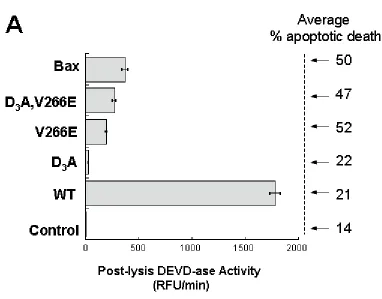
Figure 4. V266E Mutants Kill Cells More Efficiently than the Wild-type Caspase-3. HEK293A cells were transiently transfected with 1 μg total flag-tagged caspase-3 DNA (or 50 ng Bax/0.95 μg empty vector), and annexin V positive cells were quantified 24 hours later by FACS. Z-VAD-FMK (100 μM) or DMSO was added to the cultures 2 hours post-transfection. The values represent an average of three independent
resulted in robust cell death measured by annexin V staining (~50%), which exceeded that produced by WT and D3A (~20%). The loss in cell viability produced by the interface mutants was as pronounced as that produced by Bax, a cytotoxic protein that initiates the intrinsic apoptotic pathway at the mitochondrial level (Figure 4). In all cases, the levels of apoptosis were decreased in the presence of a caspase inhibitor, Z-VAD-FMK, suggesting that the increased levels of cell death were dependent on caspase activity. When protein production was monitored by Western blotting, only the WT and D3A species were detected by their reactivity to anti-FLAG antibodies (Figure 5).
Compared to the endogenous protein, the levels of WT were about five to ten-fold higher in transfected cells, as judged by the anti-caspase-3 immunoblot. The interface mutants could not be detected even after prolonged exposures of the FLAG immunoblots or after immunobloting with anti-cleaved caspase-3 antibodies (data not shown). Thus, the full-length procaspase-3 observed in the cells transfected with the interface mutants (Figure 5, top panel) represents the endogenous protein. Immunoblots using cell lysates prepared at earlier time points post-transfection gave the same results (data not shown). However, RT-PCR reactions showed the presence of mRNA for the caspase variants (supplemental Figure 2), demonstrating that the genes were transcribed. The parsimonious explanation for the lack of immunostaining of transfected V266E variants is that cells expressing them die before sufficient protein can accumulate – suggesting a lethal nature of the interface mutation.
Supplemental Figure 2. RT-PCR Reactions of DNA-free-RNA Isolated from Transfected HEK293A.
5). Cleaved PARP exits the nucleus and resides in the cytosol, so we examined the presence of cleaved PARP in mRIPA-soluble lysates. As expected, PARP was cleaved in all cells displaying high annexin V staining, and its cleavage was suppressed by Z-VAD-FMK (Figure 5). Cleaved PARP appeared to be in higher amounts in cells transfected with WT, which may indicate a higher degradation rate of cleaved PARP in cells transfected with the interface mutants or with Bax. In support of this, Western blot analysis against total nuclear PARP showed that full-length PARP was completely depleted in cells transfected with the interface mutants versus cells transfected with WT (data not shown).
Overall the data show that the constitutively active caspase-3 interface mutants were more efficient in killing transfected cells in culture than was wild-type caspase-3.
The interface mutants kill cells independently of endogenous caspase-3
Because the substrate specificity is the same for the V266E variants and WT (Figure 1C), it is likely that the mutants cleave the same substrates as caspase-3 during apoptosis. Alternatively, it is possible that the interface mutants activate the endogenous caspase-3, which then amplifies the downstream proteolytic events – although this is unlikely given that procaspase-3 is typically a poor substrate for active caspase-3 (Stennicke, 2000). To examine the latter possibility, we transfected MCF7 cells, which lack endogenous caspase-3 (Janicke et al., 1998), with plasmids containing the interface mutants, and we measured apoptosis by annexin V staining (Figure 6). Although the transfection
Figure 6. Caspase-3 V266E Mutants Kill Cells Independently of the Presence of Endogenous Caspase-3.
the pattern of cell death observed in the 293A cells (Figure 2A) was reproduced in the MCF7 cells (Figure 6). One should note that Bax, the positive control, was less toxic in MCF7 cells than were the interface mutants due to the lack of endogenous caspase-3, the main effector caspase. In all cases, the number of apoptotic cells diminished in the presence of the caspase inhibitor, Z-VAD-FMK.
Similarly, recombinant V266E cleaved caspase substrates in the absence of caspase-3 when added to crude cellular extracts. We examined hypotonic lysates from Jurkat cells that were immunodepleted of the endogenous caspase-3 and reconstituted with
recombinant caspase-3 proteins. The lysates were analyzed by Western blotting for cleavage of ICAD (Figure 7). We observed that ICAD was cleaved efficiently by the active WT and V266E proteins, and it remained unprocessed upon addition of the less active procaspase-3 (Figure 7). The cleavage of ICAD was judged by the disappearance of the full-length protein for which the antibodies were developed. Lysate prepared from cells treated with zVAD-FMK showed no DEVD-ase activity (not shown).
The intrinsic activity of interface mutants in transfected cells is lower than that of WT
Figure 8. WT Caspase-3 Lysates Display Higher DEVD-ase Activity than Those of V266E Interface Mutants, and Uncleavable Caspase-3(V266E,D3A) is Inhibited Poorly by XIAP.
transfected with the interface mutants (same lysates as in Figure 2). Surprisingly, the lysates containing WT showed the highest DEVD-ase activity (Figure 8). We expected to correlate this high activity to a high percentage of cell death in culture (see Figure 2A), which was not the case. In contrast, the lysates containing transfected interface mutants or Bax displayed less than 15% of the DEVD-ase activity of the WT lysates (Figure 8), despite the high amounts of Annexin V positive cells present in the cell culture (Figure 2A). This activity represented about half of that expected based on the intrinsic activities of the proteins, where the V266E variants have 3-4-fold lower activity than WT (Pop et al., 2003), but the results correlated to the lower accumulation of the proteins in the cell (Figure 5).
Figure 9. (B) Kinetics of DEVD-ase activity in lysates prepared from cells transfected with WT or caspase-3(V266E) for the indicated time periods. The average percentage of annexin V positive cells is shown for the corresponding lysates 24 hours post
Supplemental Figure 3. Relative DEVD-ase Activity of the Culture Medium from Cells Transfected with Caspase-3 Mutants.
medium (supplemental Figure 3), but the ratio between the activity of V266E mutants and the activity of WT reflected the ratio found in the lysates (Figure 8). Therefore, the “missing” DEVD-ase activity was not in the cellular medium.
XIAP is a poor inhibitor of D3A,V266E
Figure 10. (C) Caspase-3 mutants (300 pM) were incubated with XIAP at the indicated concentration in XIAP assay buffer (see Methods) for 30 minutes at 37 °C, and the remaining activity was tested against Ac-DEVD-AFC (100 μM). The activity rates were plotted as percentage of the maximum velocity in the absence of the inhibitor. The data were fit to an equation describing the enzymatic activity in the presence of a reversible competitive inhibitor (see Methods), and results of the fits are shown in Table 1. Table 1. XIAP Inhibition Constants for Caspase-3 Interface Mutants
Caspase-3 Protein IC50 (nM) Ki (nM) % Maximum Inhibition WT 7.4 ± 0.8 1.75 ± 0.25 97 ± 2
(Scott et al., 2005). In contrast, D3A,V266E was inhibited less efficiently by XIAP (Figure 10), displaying a Ki value of ~8-fold higher in comparison with WT (Table 1). The data also showed that D3A,V266E could be inhibited by XIAP only up to ~70% of its maximum activity, compared to ~97% for WT (Table 1). This information, translated to the intracellular environment where the XIAP concentration does not exceed ~50-70 nM (Rehm et al., 2006), suggests that the uncleaved mutant will be mostly uninhibited in the cell. Because D175 in the IL of procaspase-3 is a poor substrate for cleavage by mature caspase-3 (Stennicke, 2000 and Liu et al., 2005), we suggest that the V266E mutants remain unprocessed in the cell at low concentration, effectively avoiding inhibition by XIAP. This explains the early and robust cell death upon transfection with caspase-3 interface mutants in comparison to WT. We have ensured that the active sites of the interface mutants were equally competent for binding other inhibitors that did not require interaction with the cleaved form of caspase-3. For example, we show that the interface mutants were inhibited by baculovirus p35 (Figure 11) similarly to WT caspase-3.
Supplemental Figure 4. Co-expression of XIAP with the Caspase-3 V266E Mutants Cannot Rescue Cells from Apoptosis.
(A) HEK293A cells were transfected with 2 μg total DNA (1 μg caspase DNA + 1 μg XIAP DNA for co-transfections) and after 24 hours the cells were quantified for annexin V staining.
Supplemental Figure 5. Poor Inhibition of Caspase-3 Interface Mutants by XIAP. (A) Co-transfection of caspase-3(V266E) with XIAP at various DNA ratios.
accumulate the transfected XIAP. However, if the ratio of V266E to XIAP plasmids was varied during co-transfection, we observed that XIAP inhibited cell death only when present in large excess versus the caspase plasmid (>5:1 XIAP:caspase) (supplemental Figure 5).
Overall, the data show that the interface mutants and wild type caspase-3 are dimeric at low concentrations (supplemental Figure 1), that dimeric V266E and
D3A,V266E mutants are constitutively active before processing (Figure 1B), and that the unprocessed V266E mutant is not efficiently inhibited by XIAP (Figure 10). By contrast, the levels of procaspase-3 must accumulate at least 5-fold over the endogenous level (Figure 5) before autocleavage occurs, and the cleaved and active caspase-3 is neutralized by the endogenous XIAP inhibitor. We estimate that, in the absence of XIAP, the
The X-ray crystal structure of V266E
In order to examine structural changes caused by the replacement of V266, we crystallized and determined the structure of cleaved V266E to 1.63 Å (Table 2). The motivation for replacing V266 with glutamate was prompted by the hydrophilic dimer interface of caspase-1, which contains E390 (equivalent to V266 in caspase-3) and R391.
The latter residue is an insertion that results in a β-bulge in β-strand 8, and as a negative
design element (Richardson and Richardson, 2002) it presumably prevents the formation of indiscriminate oligomers from the monomeric procaspase-1. E390 forms a salt-bridge with R286 (R164 in caspase-3), which is on L2, neutralizes the positive charge from that residue, and appears to stabilize the active site (Figure 12).
Figure 12. Interfaces of Caspase-3 V266E and of Caspase-1.
(A) Amino acids in the dimer interface of caspase-3 relative to the two active sites. The positive charge of R164 (active site L2) is neutralized by E124.
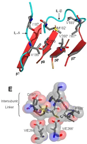
V266E, we observe electron density starting with K186. Importantly, in WT caspase-3 H185’ forms two H-bonds with T245 at the C-terminus of helix 5. The contacts across the interface anchor the base of L4 with L2', and these interactions are not observed in the mutant (Figure 13). Disruption of contacts at the C-terminus of helix 5 was shown
previously to decrease the activity of procaspase-3 (Feeney et al., 2004).
Second, the dimer interface of V266E has features in common with WT caspase-3 and with caspase-1, although E266 in the caspase-3 variant utilizes a different rotamer than does E390 in caspase-1. In this case, E266 interacts with R164 through a water molecule rather than through direct interactions as observed in caspase-1. Interestingly, if
the E390 is replaced with an aspartate in caspase-1, the salt bridge with R286 is preserved but is mediated by a water molecule (Datta, 1996). In WT, the positive charge of R164 is neutralized by E124, and these contacts are maintained in the mutant (Figures 11 and 14). When the structure is superimposed with that of caspase-1, one finds that only one of the two active sites aligns well (Figure 15). The presence of the inserted residue R391 in caspase-1, described above, allows E390 to adopt a rotamer that moves the carboxylate closer to R286 so that the two side chains are within H-bonding distance (2.8 Å). When we modeled V266E with the same rotamer as that of caspase-1, we found steric clashes between E266 and E266’, across the dimer interface, and between E266 and Y197, on the
adjacent β-strand (Figure 15). This issue is alleviated in caspase-1 because of the
amino acids and optimizing favorable interactions with neighboring residues and nearby waters. Based on this, one might think that the two active sites are not equivalent. However, we did not observe cooperativity between the active sites, as the Hill coefficient was one (data not shown).
Conserved water molecules in the dimer interface
On one side of the interface, α-helices 5 and 5’ border β-strands 8 and 8', while the
opposite face has a water-filled cavity (see (MacKenzie and Clark, 2008) for review). Moreover, several of the water molecules found in the central cavity are conserved. We examined over 5,000 crystallographic water molecules in twenty caspase-3 structures with resolution of 2.5 Å or better, including proteins from our own studies as well as those deposited in the Protein Data Bank. We considered water molecules that were within 1.4 Å of one another to be conserved, using a previously published method (Dechene, et al 2009). The structures included in our conserved water analysis were obtained from crystals with symmetry of various space groups formed under different crystallization conditions and differing in the number of molecules per asymmetric unit. Overall, the analysis identified several conserved water molecules throughout the structure of caspase-3, but we focus here on six conserved hydration sites in the dimer interface, which are found in a pseudo-planar arrangement approximately 3.8 Å above the V266/V266’ side chains (Figure 14).
Figure 14. V266E Changes the Dimer Interface.
(A) Dimer interface of WT caspase-3 demonstrating neutralization of the positive charge of R164 and six conserved water molecules.
crystallographic water molecules to provide a H-bonding network bridging the two active sites. Wat192 and Wat192’ in this network also H-bond to R164 and R164’, respectively. The water network is disrupted in V266E, where Wat219 and Wat219’ are removed, and the four central water molecules move to prevent clashes with E266 (Figure 14).
Interestingly, the carboxylates of E266 and of E266’ displace Wat108 and Wat108’, respectively, and maintain the planar contacts with Wat192 and Wat192’. Overall, this arrangement preserves the bonding network between R164 and R164’ but removes H-bonds contributed by Y197 and Y197’.
Models of (in)active procaspase-3
Our previous biochemical data for D3A,V266E (Pop et al., 2003), as well as that presented here, show that the V266E mutation effectively shifts the zymogen to an active conformer. Currently there are no structures for procaspase-3, so we generated homology models of the putative active and inactive zymogen in order to examine the
conformational switch. Due to their high sequence identity, we modeled inactive wild type procaspase-3 after procaspase-7 (Chai et al., 2001; Riedl et al., 2001a) and energy minimized the structures as described in Methods to assure that our final model is
Figure 16. Homology Models of (In)Active Procaspase-3.
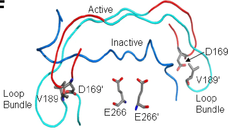
buried in the interface, making contacts with several interface residues and shielding the hydrophobic core from water (Figure 16). With the IL bound at the interface, the loop bundle comprised of D169 cannot form. As described previously (Feeney et al., 2006), contacts centered about D169 (on L2) are critical for stabilizing the active site in the zymogen as well as the mature caspase-3. The carboxylate of D169 interacts across the dimer interface with the amide nitrogens of V189’ and E190’ (from L2’), and the
backbone atoms of D169 form two H-bonds with those of K260 (in L4). Replacement of D169 with alanine was shown to abolish activity in WT and in D3A (Feeney et al., 2006), demonstrating that the loop bundle is important to procaspase activity as well. As a result, our model for the active procaspase-3, described below, includes the active site
stabilizing contacts provided by the loop bundle. D192 in caspase-7, the equivalent of D169, also is critical for procaspase-7 function (Denault & Salvesen, 2006).
Figure 17. (B) Superposition of inactive procaspase-3 (green residues) and of procaspase-7 (yellow) shows the blocking segment of IL-B, residues 184-189
(procaspase-3 numbering), prevents insertion of active site loop 3 from monomer A. For clarity, only one residue from the blocking segment of procaspase-7 is shown (semi-transparent sticks, Tyr211) while all of the residues of the blocking segment of
Figure 18. (C) Superposition of procaspase-7 (yellow) and of inactive procaspase-3 (green) reveals a blocking segment involving residues 179-180 (procaspase-3 numbering) of IL-B and V189 of IL-A preventing insertion of L3 in active site of monomer B. L3 (white ribbon, WT; blue ribbon, D3A,V266E) cannot insert into the interface until the
segment” because the residues prevent active site L3 from inserting into the interface and thus prevent formation of the substrate binding pocket (Riedl et al., 2001a). In addition, steric clashes between the side-chain of H185 (IL-B) and the backbone atoms of G202 (L3) and R164 (L2) (chain A) likely prevent rotation of L2 from chain A into the active conformation, where the side-chain of R164 rotates into the dimer interface to intercalate between P201 and Y197 and the catalytic C163 rotates into the S1 site. Similar steric constraints are observed in procaspase-7 (Figure 17). In the active procaspase-3, as well as upon cleavage of the IL to yield caspase-3, removal of the blocking segment of IL-B from the interface allows P201 from chain A to move ~6 Å into the active conformation, thus, forming the substrate binding pocket for active site A. For active site B, a blocking segment also prevents insertion of P201 and the substrate binding pocket, but the
interactions differ due to the asymmetric nature of the inactive procaspase-3 dimer. In this case, V189 (IL-A) resides in the region occupied by P201 (chain B) in the active caspase-3, so this residue behaves similarly for both ILs. However, the remainder of the blocking segment is comprised of the backbone atoms of residues 179-182 from IL-B, which also reside in the region occupied by P201-Y203 of the active caspase-3. Thus, although the mechanisms differ somewhat for each active site, the substrate binding loops of both active sites are prevented from inserting into the interface because the interface is occupied in the inactive procaspase-3 by segments of IL-A and IL-B.
resulting from the mutation. The hydrophobic cluster is comprised of seven residues: M182', from IL-B, and I187, I187’, V189, V189’, V266, and V266’ from both monomers (Figure 19). Based on this analysis, we suggest that V266E activates the procaspase by disrupting the hydrophobic cluster and introducing steric clashes with M182’ and I187’ (Figure 19) and essentially expelling the IL from the dimer interface – the first and most important step in activation. Because the glutamate side chain is about 2.5 Å longer than that of valine, the IL is prevented from rebinding in the interface after its release, and the protein conformation shifts to the active state. It is important to note that the
Figure 19. (D) Hydrophobic cluster in the inactive procaspase-3 centered about V266. Residues in the IL of both monomers contribute to the cluster.
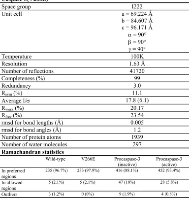
Table 2. Summary of Data Collection and Refinement Statistics for Caspase-3(V266E)
Space group I222
Unit cell a = 69.224 Ǻ
b = 84.607 Ǻ c = 96.171 Ǻ
α = 90°
β = 90°
γ = 90°
Temperature 100K
Resolution 1.63 Ǻ
Number of reflections 41720
Completeness (%) 99
Redundancy 3.0
Rsym (%) 11.1
Average I/σ 17.8 (6.1)
Rwork (%) 20.17
Rfree (%) 23.54
rmsd for bond lengths (Ǻ) 0.005
rmsd for bond angles (Ǻ) 1.2
Number of protein atoms 1939
Number of water molecules 297
Ramachandran statistics
Wild-type V266E Procaspase-3
(inactive) Procaspase-3 (active) In preferred
regions
235 (96.7%) 233 (97.9%) 416 (88.1%) 452 (93.4%)
In allowed regions
5 (2.1%) 5 (2.1%) 47 (10%) 28 (5.8%)
Outliers 3 (1.2%) 0 (0%) 9 (1.9%) 4 (0.8%)
Rmerge = ΣhΣi│I(h,i) - I(h)│/ ΣhΣiI(h,i), where I(h,i) values are symmetry-related intensities and I(h) is the mean intensity of the reflection with unique index h. Rwork = Σh║Fobs│-│
F-calc║/Σ│Fobs│, where Fobs and Fcalc are observed and calculated structure factors,
respectively. Rfree = ΣT║Fobs│-│Fcalc║/ΣT│Fobs│, where T is a test data set of 10% of the
not possible in the procaspase. The second supplemental movie shows the conformational switch between inactive and active procaspase-3.
DISCUSSION
We showed previously that the V266E mutation in the procaspase-3 dimer interface resulted in pseudo-activation of the enzyme (Pop et al., 2003), where the activity of the mutant was only three- to four-fold lower than that of the mature caspase. Importantly, the increase in enzyme activity did not require cleavage of the IL at D175, and there was no further increase in activity upon maturation. The data suggested that the pseudo-active procaspase may be effective in inducing apoptosis in mammalian cells, although it was not clear how activation occurred in the mutant, nor was it clear what threshold activity of procaspase-3 would be required to carry out apoptosis.
We show here that the variant (pro)caspase-3 kills mammalian cells rapidly compared to wild-type caspase-3. D3A,V266E is active in the cell upon translation, and before processing at D175, cleaves substrates similarly to the mature caspase. These features are important for two reasons. First, endogenous procaspase-3 is a poor substrate for
L2’, which serves as a second binding region for XIAP, remains sequestered in the procaspase.
From the data presented here, we estimate that less than 5 nM activated procaspase-3 is sufficient to carry out apoptosis. This threshold is much lower than the total cellular concentration of caspase-3, which is ~100 nM in apoptotic Jurkat (Pop et al., 2001) and HEK293 cells (Stennicke et al., 1998). The concentration of the zymogen appears to be higher than that of mature caspase-3 (Saunders et al., 2000), although HeLa cells are estimated to contain approximately 120 nM procaspase-3 (Rehm et al., 2006). Under normal cellular conditions, XIAP concentrations of 50-60 nM (Rehm et al., 2006) are sufficient to inhibit the small amount of activated caspase-3, and XIAP targets the protein for degradation by the proteasome (Suzuki et al., 2001). The induction of apoptosis leads to massive activation of endogenous procaspase-3, which overwhelms the control system and results in rapid cell death (Albeck et al., 2008). Because XIAP does not efficiently inhibit D3A,V266E, a low concentration of the pseudo-activated procaspase is sufficient for rapid cell death.
The interface mutation has little effect on the structure of the mature caspase, but it appears to have a larger effect on that of the procaspase. Our structural data show that L2’ is disordered in V266E, and conserved water molecules are displaced at the interface. At present, it is not clear how a mutation in the interface affects ordering of L2’, since the regions that are affected are >25 Å from the site of the mutation. Although residues in L2’ H-bond with the P4 carboxylate of the substrate, removal of those interactions do not
in a water-filled cavity, with the closest water molecules approximately 3.5 Å from the hydrophobic side chains, well within van der Waals contacts. As described by Wells and co-workers (Hardy et al., 2004), this region of the interface encompasses a site for binding of allosteric inhibitors, which prevent active site formation, in general, by
preventing the insertion of the substrate binding loop (L3) into the active site cavity. This is important because part of L3 extends into the dimer interface and forms new contacts with residues in L2 in addition to forming the base of the substrate binding pocket. The allosteric binding pocket is occluded in the procaspase due to the positioning of the IL in the interface, and the “blocking segment” of the IL prevents insertion of L3 into the active site. Our data suggest that the inhibition is alleviated in D3A,V266E so that the IL is removed from the allosteric site, allowing the active site loops to organize. Thus, the allosteric site linked to inhibition of the active caspase also stabilizes the inactive procaspase.
A comparison of the active and inactive procaspase-3 (supplemental movie 2) shows that the conformational switch primarily involves amino acids in the IL and active sites, where the bulk of the protein is not involved in the switch. From an analysis of the two
forms, we estimate a change in solvent accessible surface area (ΔASA) of 4,587 Å2 upon
switching to the active conformer. Using data derived from the dependence of the free energy of unfolding on denaturant concentration (the m value), Pace and coworkers
demonstrated a strong correlation between ΔASA and the number of amino acid residues
involved in the transition (Myers et al., 1995). From the relationship (ΔASA (Å2) = -907
approximately 59 amino acid residues. These values can be accounted for by
conformational changes in the IL (17 amino acids) and L3 (15 amino acids) from both monomers.
The inactive conformer appears to be stabilized by hydrophobic and charge-charge interactions across the dimer interface, and these stabilizing contacts are disrupted in the active conformer. Upon removal from the interface binding site, the IL moves across the surface of the protein to form the critical contacts contributed by D169, and the active conformer is stabilized primarily by backbone H-bonds between the two ILs and those that form at D169. Stabilizing interactions in the mature caspase, contributed by L2, L2’, L3, and L4, are unable to form in the active procaspase. The lack of these stabilizing interactions may explain, in part, why the inactive procaspase-3 is favored. A comparison of caspase-3 sequences in other organisms demonstrates that the safety-catch and
hydrophobic elements are conserved in all vertebrates (supplemental Figure 6A). In
addition, residues in β-strand 8 in the interface are nearly absolutely conserved in
vertebrates (supplemental Figure 6B), suggesting that the mechanisms described here for human procaspase-3 are conserved in other species as well.
Learning to selectively manipulate the level of apoptosis is expected to lead to
therapeutic strategies for a number of diseases in which the dysregulation of apoptosis is a common factor. Cancer cells, for example, typically have gained the ability to
therapy and lower drug resistance since effector caspases are the terminal proteases in the cell death program. Our data suggest that small molecules could bind to the dimer