A Survey of various Techniques for the
Detection of Optic Disc
A.Padma1, Dr.M.Sivajothi2, Dr.M.Mohamed Sathik3
Assistant Professor, Department of Computer Science, Sri Para Sakthi College for Women, Courtallam, India1
Associate Professor, Department of Computer Science, Sri Para Sakthi College for Women, Courtallam, India2
Principal, Department of Computer Science, Sadakathullah Appa College, Tirunelveli, India3
ABSTRACT: Eyes are one of the most important sensory organs in the human body as it renders vision. The obstruction of blood flow in optic disc cause changes because the optic disc is the entry point where the blood vessels supply the blood to retina. The eye diseases can be detected by locating the optic disc. Several methodologies are followed to diagnose the optic disc. Some eye diseases cause blindness without any significant symptoms. The progression of this eye impairment can be prevented by early diagnosis. The main aim of this survey is to analyze various approaches implemented for optic disc detection
KEYWORDS: optic disc; Eye diseases; optic cup; eye sight; segmentation;
I. INTRODUCTION
The human eye is an organ of vision. A vital organ of vision plays a very important role not only in life but also in the Human body. The human eye is the organ which gives the sense of sight, and allows to learn more about the surrounding world than that can be done with any of the other four senses. Eyes play a vital role in our day to day lives. Clear and bright eye sight makes this world a better place to live in. “The eyes are the windows to the soul” is an expression that is often used to describe the deep connection one feels when looking into another’s eyes [1].The eye permits us to look and recognize the shapes, colours, and dimensions of objects in the world by processing the light they reflect or emit.
There are several eye diseases which cause blindness without significant symptoms. In order to find such diseases, a detailed study about the anatomy of eye is needed. It is necessary to find the location of optic disc (OD) since it is the entrance location of optic nerve through which blood flows to retina. The changes in Optic disc, optic cup, and blood vessels are detected and eye diseases can be diagnosed using various techniques.
II .RELATED WORK
.
Fig .1 Optic Disc
Jun Cheng proposed a method for optic disc detection which contains three steps namely superpixel generation, feature extraction and classification of superpixel as disc or non disc as in [2].Superpixels are generated using simple Linear Iterative clustering method.Features are computed from five channel maps and center surround difference maps.A support vector machine classifier is utilised to classify the pixels as disc or non disc region.The elliptical hough transform is used for fitting the elliptical shape and Active shape model (ASM) is used to fine tune the disc boundary.
H.Yu proposed a methodology [3] which consists of three phases namely OD size estimation, OD localization and OD boundary segmentation. The background normalized image is obtained by applying image illumination correction.A binary template is taken to locate the OD canditates. The disc which is given in white is assigned a value 1 and the black background is assigned a value 0. The OD canditates are selected from the template matching results.The OD canditate with maximum contrast in the region is determined as the OD location using directional matched filtering.The vessels and bright regions present in the peripapillary region are removed using alternating sequential filtering and morphological reconstruction. The OD segmentation is done using fast hybrid level set method which provides more accurate detection of disc boundary.
III. ANATOMY OF AN EYE AND EYE DISEASES
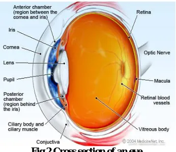
The anatomy of an eye is explained as in [4].The eye focuses light onto a specialized layer of cells, called the the retina, to create an image. Fig 2 shows the parts of an eye prominently.
The conjunctiva is a thin, transparent layer of tissues wrapping the front of the eye, including the sclera and the inside of the eyelids. The conjunctiva controls bacteria and foreign material from getting behind the eye. The cornea is the transparent, obvious layer at the front and middle of the eye. The main principle of the cornea is to aid focus light as it enters the eye.
The anterior chamber angle is situated where the cornea meets the iris. The trabecular meshwork is important because it is the area where the aqueous humour drains out of the eye. If the aqueous humour cannot properly drain out of the eye, the pressure can form inside the eye, producing optic nerve damage and eventually vision loss, a condition known as glaucoma.
The iris, which is the coloured portion of the eye, directs the amount of light that enters the eye. The iris is a ring shaped tissue with a central cavity, which is called the pupil. The posterior chamber is the fluid-filled area directly behind the iris but in front of the lens. The fluid that fills this chamber is called as the aqueous humour which helps to nourish the cornea and the lens. The lens is a transparent, flexible structure that is found just behind the iris and the pupil. A ring of muscular tissue, called the ciliary body, encloses the lens and is attached to the lens by fine fibres, called zonules.
The vitreous cavity is found behind the lens and in front of the retina. It is filled with a gel-like fluid, called the vitreous humour. The vitreous humour assists to retain the shape of the eye. The retina has two kinds of cells that instigate chemical reactions. These cells are called photoreceptors and the two different types of cells are the rods and cones. Rods are more sensitive to light and Cones allow people to view colour, but need more light.
The macula is positioned in the central part of the retina and has the maximum concentration of cones. It is the part of the retina that is responsible for offering sharp central vision. The choroid is a film of tissue that situates among the retina and the sclera. It is mainly made up of blood vessels. The choroid supports to nourish the retina. The optic nerve, a bunch of over one million nerve fibres, is responsible for transmitting nerve signals from the eye to the brain. These nerve signals hold information for processing by the brain.
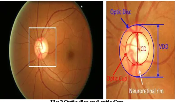
Fig 3 Optic disc and optic Cup
IV.SURVEY OF METHODOLOGIES
Aliaa Abdel [5] presented a method to automatically detect the position of optic disc. Illumination equalisation and adaptive histogram equalisation methods were utilized to standardise luminosity and contrast throughout the image. An algorithm was implemented to match the expected directional pattern of the retinal blood vessels. A Gaussian matched filter was applied for the segmentation of retinal vessels and the optic disc centre was traced by thinning the segmented vessels and filtering using local intensity.
Angel Suero [6] presented a methodology for tracing optic disc in retinal images. The background was homogenized in resized intensity images to correct the non uniform illumination. The brighter regions of OD were enhanced using morphological operations. The brightest area of the centroid was identified and the location of the OD was found
Deepali A. Godse [7] presented a novel algorithm for automatic localization of optic disc in retinal images, healthy as well as normal. Green channel histogram was employed to calculate the threshold. By applying this threshold the brighter regions within the images were detected. Area criterion and density criterion were applied to find the centre of OD.
Adhitya Kusuma Whardana [8] proposed an approach for the detection of optic disc and blood vessel using K-Means clustering method. A disadvantage was found that the cutting blood vessels lead to lack of clarity.
Sandra Morales [9] proposed a method for the automatic detection of optic disc. Principal Component Analysis (PCA) was applied on RGB fundus image to get the greyscale image in which the different structures are differentiated clearly. The vessels are removed using morphological operations. Stochastic Watershed Transformation was implemented to find the location of optic disc for automatic and accurate detection.
Hidayath Ullah [10] presented a new and robust technique for the automatic detection of optic disc in retinal images. The RGB image is transformed into HSI space. A median filter is applied to decrease the amount of shadowiness. The ensuing HSI components are changed to RGB to obtain the pre-processed image from the green channel. The Otsu’s binarization algorithm is executed on green channel to separate it as foreground and background. Area filtering is used to detect the false area. The Dilation operator is utilized to track down the boundary of optic disc. The filling operation is performed to trace the OD region enclosed by closed contour.
Neelam Sinha [11] presented a novel method based on the fact that ODs exhibit similar characteristics such as circular shape. The pre-processing steps such as equalization, enhancement, edge detection and morphological operations are not required to detect the location of optic disc. The circular outline of optic disc is marked in red channel image or greyscale image. The region where the blood vessels and nerves converge is identified as the circular shape. The sub image that contains OD at the centre is identified by locating the peak value of the coefficients
Prasad N. Maldhure [12] proposed a technique for optic disc and optic cup segmentation to detect glaucoma. Adaptive histogram equalization technique is used to increase the contrast of the colour image. Gabor filters are used for texture feature extraction. These two parameters are handled as a key feature to classify each super pixel as disc or non disc super pixel.SLIC algorithm is employed for super pixel generation. Same parameters are utilised for optic cup segmentation. The value of CDR is evaluated and compared with threshold value for the detection of glaucoma. Murugan. R [13] employed a new automated methodology to detect the optic disc in retinal images. The RGB image is changed into its LAB component. This image is made smooth with the help of bilateral smoothing filter. Line operator is used for further filtering. Processes such as gray orientation and binary map orientation are carried out. The presence of OD is identified from the maximum image variation in the resultant image. The regions other than OD are blurred by means of 2D circular convolution. The OD is identified with mathematical steps like peak classification, concentric circle design and image difference calculation.
R. Preethi Rajaiah [15] achieved the detection of glaucoma by optic disc boundary detection and optic cup segmentation. Features are obtained by means of linear discriminant analysis. The input image is augmented using adaptive histogram equalization. The vessels that are extracted from the retinal image are eradicated with Inpainting technique. The optic disc boundary is found by employing morphological operations on the resultant image. The optic cup segmentation is carried out in the green channel which is got from the input image. Watershed transformation is utilized for cup segmentation.
Oakar Phyo [16] presented a mathematical morphology method for the detection of optic disc and blood vessels. The input RGB image is converted into HSI colour space. The noise included in the image is diminished by median filter. The resultant image of the median filter is improved with adaptive histogram equalization. The closing morphological operator is operated to connect the closer objects together. The output image is binarized by thresholding. The filling operator is utilized to fill the holes in the image and the consequent image is reconstructed using morphology reconstruction. The optic disc region is located based on Otsu algorithm that is applied on the difference between the original image and the reconstructed image.
Gilbert Lim [17] built an accurate and reliable method for optic disc and optic cup segmentation. A vessel mask is gained by trench detection. These vessels are eliminated from the optic disc patch based inpainting. The red and green channels are obtained from the inpainted image and the channel histograms are equalized. The vessel mask is skeletonised. The values of raw kink strength map is first normalized and then expanded. A Gaussian blur is applied and the converted image is got. Each pixel is categorized one by one with convolutional neural network (CNN). Sukanya. R [18] proposed an efficient method for the segmentation of retinal images. The median filter is applied to remove the noise from the input retinal image. The sharp edges are preserved and detected using Andy operator. Transformation and morphological operations are used for enhancement. Clustering algorithms are used for categorizing vessel and non vessel part. Support Vector Machine (SVM) is used as a classifier to recognize the optic disc. Nivedha. S [19] developed a technique for the automatic recognition of optic disc. Principal Component Analysis (PCA) is implemented on the input RGB image to get grey image which shows the different structures of the retina obviously. The vessels are eliminated using inpainting technique. Morphological operators are used to extract the relevant structures of the resultant image. The centroid of the grey image is evaluated based on generalised distance function. The location of optic disc is found by applying stochastic watershed transformation that represents the objects of interest and separation of boundaries between objects.
Cemal kose [20] developed a simple statistic technique for the detection of optic disc and macula.The background image of healthy parts of retina is extorted from retinal image. The extracted image is extended for the entire image and the background image is generated. The blood vessels and degenerated areas are eradicated and the vertical and horizontal histograms are evaluated. The optic disc is traced and the macula is located based on the location of optic disc. A standard deviation image is created in standard deviation method for the detection of optic disc. The blood vessels and the degenerated areas are eliminated and the histograms are computed for the detection of macula.
Mrinal Haloi [21] recommended a technique is in which no separate pre processing steps are required. The optic disc is identified from the saliency map that can capture important variation of local structure. After finding the optic disc, a probabilistic latent semantic analysis (PLSA) based unsupervised technique was utilized to confirm whether the region is the optic disc or not. The location of optic disc is found using Fuzzy-KNN classification technique.
Tamilarasi [22] exploited a new approach for the automatic detection of OD contour. The grey scale image is acquired from input retinal image through the process of principal component analysis. Mathematical operations are utilized to eliminate the blood vessels. The generalized distance function, the stochastic watershed and geodesic transformation are put into operation to segment the optic disc in precise manner.
applied on grey image and the noise was reduced without blurring the sharp edge. The edges are detected with canny edge detection. The optic disc was found by circular hough transform.
Sinthanayothin [25] detected the OD by recognizing the area with the highest mean difference between neighbouring pixels using a window size up to that of OD. The images were preprocessed using an adaptive local contrast enhancement method which was employed to the intensity component.
Osareh [26] employed a model based (template matching) approach to roughly locate the OD. To begin with, the images were normalized by utilizing histogram specification and then the OD region from 25 colour standardized images was averaged to create a gray level template. The normalized correlation coefficient was manipulated to locate the most perfect match among the template and all the candidate pixels in the known image. Abdel-Ghafar [27] employed the Circular Hough Transform (CHT) to find the OD which has a nearly globular shape. The retinal vasculature in the green band image was hidden using the closing morphological operator. The sobel operator and a simple threshold were used to remove the edges in the image. CHT was finally applied to the edge points and the largest circle was located reliably to match to the OD.
Aquino [28] employed an approach to trace a pixel belonging to the OD region. The boundary is segmented by means of morphological operators, edge detection methods and circular Hough Transform.
S.Lu and J.H.Lim [29] proposed a method in which line operator is used to acquire circular brightness shape of the OD, since the utmost and least variation along the linear operator has a particular pattern to find the OD.
IV. CONCLUSION
In this paper, the importance and anatomy of the human eye is presented. In addition to this, numerous existing approaches for the identification of optic disc to diagnose the eye diseases proposed by different researchers are presented. The performances of these methods can be measured using metrics such as sensitivity, specificity, accuracy, precision rate etc. These methods have their own advantages and disadvantages depending upon the input image taken. From this study, it is observed that the filters and morphological operations are performed to overcome the segmentation problems caused due to the presence of blood vessels.
REFERENCES
1. Molly Blakely ,The importance of sightAvailable:https://www.marveloptics.com/blog/the-importance-of-sight-andvision-molly-blakely/ 2. J. Liu, Y. Xu, F. Yin, D. W. K. Wong, N. M. Tan,D. Tao, C. Y. Cheng, T. Aung, and T. Y. Wong, ‘Superpixel classification based optic disc
and optic cup segmentation for glaucoma screening’, IEEE Tran Med Imaging, vol. 32, no. 6, pp. 1019–1032, 2013.
3. H.Yu, E.S.Barriga, S. Echegaray, M.S. Pattichis, W. Bauman and P. Soliz , ‘Fast Localization and Segmentation of Optic disc in Retinal Images using Directional matching Filtering and Level Sets’, IEEE Transaction on Information Technology in Biomedicine, Vol. 16, No 4,pp. 644-657, 2012
4. Andrew A. Dahl, William C. Shiel, (July 2016)Anatomy of the eye http://www.emedicinehealth.com/anatomy_of_the_eye/article_em.htm
5. Aliaa Abdel-Haleim Abdel-Razik Youssif, Aref Zaki Ghalwash, and Amr Ahmed Sabry Abdel-Rahman Ghoneim,’Optic Disc Detection From Normalized Digital Fundus Images By Means of a Vessels’ DirectionMatched Filter’, IEEE TRANSACTIONS ON MEDICAL IMAGING, Vol.27, Issue 1, JANUARY 2008.
6. Angel Suero, Diego Marin, Manuel E. Gegundez-Arias, and Jose M.Bravo ‘Locating the Optic Disc in Retinal Images Using Morphological Techniques’, IVVBBIO 2013 Proceedings, 2013.
7. Deepali A. Godse and Dr. Dattatraya S.Bormane,’Automated Localization of Optic Disc in Retinal Images’, International Journal of Advanced Computer Science and Applications, Vol.4, Issue 2, 2013.
8. Adithya Kusuma Whardana and Nanik Suciati,’A Simple Method for Optic Disk Segmentation from Fundus Image’ , I.J.Image, Graphics and Signal Processing, Issue 11, pp. 36-42, 2014.
9. Sandra Morales, Valery Naranjo, David Perez, Amparo Navea, Mariano Alcaniz,’Automatic Detection Of Optic Disc Based on PCA and Stachastic Watershed’, EUSIPCO, 2012.
10. Hidayath Ullah, Zahoor Jan, Rashid Jalal Qureshi and Bilal Shams, ‘Automated Localization of Optic Disc in Colour Fundus Images’, World Applied Sciences Journal, Vol.28, Issue 11, pp. 1579-1584, 2013.
11. Neelam Sinha, R, Venkatesh Babu,’Sparse Representation for Optic Disk
Detection’,http://www.serc.iisc.ernet.in/~venky/Papers/optic_disk_spcom12.pdf
12. Prasad N. Maldhure, Prof.V.V Dixit,’Glaucoma Detection Using Optic Cup and Optic Disc Segmentation’, International Journal of Engineering Trends & Technology, Vol.20, Issue 2, pp. 52-55, 2015.
14. Jaspreet Kaur, Dr. H.P. Sinha,’Automated Localisation of Optic Disc and Macula from Fundus Images’, International Journal of Advanced Research in Computer Science and Software Engineering, Vol.2, Issue 4, pp. 242-249, 2012.
15. Preethi Rajaiah R, John Britto R ,’Optic Disc Boundary Detection and Cup Segmentation for prediction of Glaucoma’, International Journal of Science, Engineering and Technology Research, Vol.3, Issue 10, pp. 2665-2672, 2014.
16. Oakar Phyo, AungSoe Khaing, ’Automatic Detection of Optic Disc and Blood vessels from Retinal Images using Image Processing Techniques’, International Journal of Research in Engineering and Technology, Vol.3, Issue 3, pp. 300-307, 2014.
17. Gilbert Lim ,Yuan Cheng, Wynne Hsu, Mong Li Lee,’Integrated Optic Disc and Cup Segmentation with Deep Learning’, ICTAI '15 Proceedings of the 2015 IEEE 27th International Conference on Tools with Artificial Intelligence, pp. 162-169, 2015.
18. Sukanya. R, Ganga Holi,’Energy Efficient Routing Algorithms for Mobile Ad Hoc Networks–A Survey’, International Journal of Computer Science Engineering, Vol.4, Issue 3, pp. 102-109, 2015.
19. Nivedha S, Dinesh V,’Automatic Detection of Optic Disc for the Extraction of Ocular Structure’, International Journal of Computer Science and Mobile Computing, Vol.3, Issue 2, pp. 689-695, 2014.
20. Cemal Kose, Cevat Ikibas,’Statistical Techniques for Detection of Optic Disc and Macula and parameters Measurement in Retinal Fundus Images’, Journal of Medical and Biological Engineering, pp. 395-404, 2010.
21. Mrinal Haloi, Samarendra Dandapat, and Rohit Sinha,’An Unsupervised Method for Detection and validation of the Optic Disc and the Fovea’, arXiv:1601.066008 , 2016.
22. Tamilarasi, Duraiswamy, ’Ensemble System for Optic Disc and Macula Detection’, International Journal of Advanced Research in Computer Engineering & Technology, Vol.3, Issue 1, pp. 228-232, 2014.
23. Eunhwa Jung and Kyungho Hong, ’Optic Disc Detection and Blood Vessel Diameter Estimation in Patients with Diabetic Mellitus using Digital Color Fundus Images’, Indian Journal of Science and Technology, Vol.8, Issue 18, 2015.
24. Vijaya R. Patil, Vaishali Kumbhakarna, Dr. Seema Kawathekar, ’Detection of Optic Disc in Retina using Digital Image Processing’, International Journal of Computer Techniques, Vol.2, Issue 1, pp. 20-23, 2015.
25. C. Sinthanayothin,J.F. Boyce, H.L. Cook and T.H.Williamson, ’Automated localization of Optic Disc, Fovea and Retinal blood Vessels from Digital Colour Fundus Images’, Br.J.Opthalmol.,Vol.83,pp. 902-910,1999
26. Osareh A, Mirmehd M, Thomas B, Markham R. ,’Comparison of colour spaces for optic disc localisation in retinal images’. In Proceedings 16th International Conference on Pattern Recognition, Quebec City, Quebec, Canada, pp 743–746, 2002
27. R. A. Abdel-Ghafar, T. Morris, T. Ritchings and I.Wood,’Progress Towards Automated Detection and Characterization of the Optic Disc in Glaucoma and Diabetic Retinopathy’, Med. Inform. Internet Med., vol. 32, no. 1, pp.19-25 March 2007. DOI: 10.1080/14639230601095865 28. Arturo Aquino, Manuel Emilio Gegundez, Diego Marin, ’Automated Opt ic Disc Detection in Retinal Images of Patients with Diabetic
Retinopathy and Risk of Macular Ederma’, International Journal of Medical, Health, Bioengineering and Pharmaceutical Engineering, Vol.3, Issue 12, pp. 353-358, 2009
29. Shijian Lu, Joo Hwee Lim,’Automatic Optic Disc Detection from Retinal Images by a Line Operator’, IEEE Transactions on Biomedical Engineering, Vol.58, Issue 1, pp. 88-94, 2011.
BIOGRAPHY