Implementation of FPNN for the Classification
of Brain Tumor
R.Anand1,
Dr.G.Umamaheswara Reddy
2M.Tech (Communication Systems], Dept. of ECE, Sri Venkateswara University College of Engineering,
Tirupati, India1
Professor, Dept. of ECE, Sri Venkateswara University College of Engineering, Tirupati, India2
ABSTRACT: The most significant part of the human system is brain. The most rigorous diseases in the medical science is Brain Tumor. An effective analysis is a key concern for the radiologists in premature phase of tumor growth.The complex characteristics of the MRI images which gives the high intensive, divergent and uncertain boundaries make the enlarging of the tumor difficult. Physician’s interpretation of brain tumors may lead to misclassification sometime. So, an automated system is necessary. To address these problem, tumor segmentation method for MRI images which separates tumorous cell from healthy tissues has been carried out by K means clustering and Fuzzy C means clustering. From the segmented part, features are extracted to classify as tumorous or non-tumorous. The feature extraction methods are given by Intensity, Intensity Histogram and GLCM. The experimental results gives that among intensity based feature extraction methods GLCM (Gray Level Co-Occurrence) method is exhibiting better results among others. For the intensity related features MATLAB tool is used. The Accuracy of the classified data is given by the confusion matrix created from the extracted features. All the operations are performed against BRATS dataset.
KEYWORDS: MRI, GLCM, K-Means clustering, Fuzzy C means clustering, confusion matrix, MATLAB.
I. INTRODUCTION
The necessity for an automated system to classify and detect the tumour in medical images is very much needed. As it directly deals with the life of human being, the task require a higher amount of accuracy. In this paper, the BRATS data set of different Brain MRI images were collected. The idea behind this paper is to obtain different intensity based features for brain tumour and to produce the output of the study as base for the brain tumour classification purposes.Among the several extracted features from the MRImedical images [4] our consideration is to the textural features [5]. These features are calculated from the MATLAB tool, as the image processing task texture features are the most required.
Alteration and variation in the surface of an image is defined as a texture. We can also define texture as the distribution of gray levels in a neighbourhood. Medical images of non-tumour and tumour type can be found and classified quickly by the physician through analysing intensity based features of the medical images. Bio-medical imaging includes neural network, support vector machine [10] and classifiers [6]. In recent years, brain tumour occurrence is on rise. Due to lack of knowledge, many tumours are not detected at early stages. So that low grade gliomas become high grade gliomas. Once the symptoms appears its too late to treat the tumour.
There are two types of tumours: 1. Benign Tumour -Tumour in which expansion is not abrupt and does not affect neighbouring healthy cells and 2. Malignant Tumour -Tumour in which growth increases with the passage of time and affect neighbouring healthy cells resulting in the cause of death.
II. LITERATURE SURVEY
In literature survey different techniques are discussed to classify and identify the presence and nature of the tumor. Most of the literature survey is done on feature extraction process and the neural network analysis. Textural features for brain tumor classification using GLCM shows the maximum accuracy of greater than 90% by VrushenPawar and Zulpel. Gray Level Co-occurrence Matrix (GLCM) technique is used for feature extraction. Extracted features are then fed to neuro-fuzzy classifier for normal and abnormal MRI image classification. Experimental result demonstrates, about 50-60% improvement in iteration time and the accuracy level compared to the existing neuro classifier
III.PROPOSED ALGORITHM
The proposed algorithm starts with converting of the .mha file format of MRI image to required data format. The .mha file extension is related to the meta-image format, a special medicine image format used in the insight segmentation. The converted image is changed to grey scale image and then processed to the next steps.
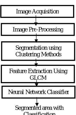
Figure 1: DATASET The proposed model consists of three major steps.
1. Pre-processing 2. Feature extraction
3. Classification using feed forward neural network
Figure 2: Proposed Model Image Pre-Processing
Segmentation using Clustering Methods
Feature Extraction Using GLCM
Neural Network Classifier
A. IMAGE ACQUISITION:
The proposed model was evaluated against the dataset BRATS. Through some basic authentication process the dataset is acquired. For the evaluation of the proposed model each image is of size 256 x 256.all the images are processed as grey scale image. The entries of the grey scale images are ranging from 0 to 255, where 255 is the pure white and 0 shows total black colour. All the entries varies from black to white.
For the processing of the above method the image of the different patients are stored in to the database in order to identify their sources.
B. IMAGE PRE-PROCESSING:
In this step, image is enhanced to get the finer details and noise is removed. In MRI the noise is caused due to fluctuations of magnetic field in the coil. Morphological operation is preferred to extract the brain portion from the skull before the process starts. The image quality is enhanced by removing noise and anti-blurring from the image. In the proposed model Gaussian Filter [7] is followed for the pre-processing task.
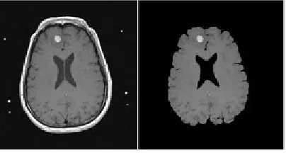
Figure 3: Pre-Processed Image C. IMAGE SEGMENTATION METHODS
a) K means segmentation
In k-means clustering method, number clusters to be defined as k. Initial cluster center for each cluster is assigned. The cost measure between data point and each of the cluster centers are calculated, and the data point is assigned to cluster that have the minimum cost measure. Cluster center is updated to new, based on mean value. The above process until the mean convergence takes place.
K-Means Algorithm:
1. Number of cluster is taken as K. 2. Selection of cluster is random.
3. Calculate mean or center of the cluster.
4. The distance between each pixel to each cluster center is calculated. 5. Calculate the mean and update the new cluster center.
6. Repeat the process from step 3 until the center does not move. b) Fuzzy C means Algorithm
The medical image is considerably fuzzy, then this is the most effective algorithm for the clustering. Without a training step conditions, the fuzzy methods can capture pixel closeness in the same region as the method is typically intensity-based method, such as morphological operations and thresholding. The data is represented by the means of membership function ranging from 0 to 1. This method is also called as soft clustering. This method is suitable for overlapping clusters.
= −
(1)
Where m-real number above 1,
Mij – Degree of membership of xi in the cluster j, xi– D-dimensional data measure,
The update of membership Mijand the cluster centers Rjare given by
= 1
∑
(2)
=∑ −
∑
(3)
The process ends when
Maxi | − | < (4)
Where
δ= termination value or constant between 0 and 1 K= Number of iteration steps
The algorithm contain following steps: 1. Initialize = [ ] matrix ( )
2. At k-step: calculate the centers vectors ( )= [ ]with ( )
=∑ −
∑
(5)
3. Update ( ) and ( )
= 1
∑
(6)
4. If − | < then STOP; otherwise return to step 2
= 1
∑
(7)
D. FEATURE EXTRACTION METHODS
In an image the visual contents are captured by feature extraction technique to preserve the raw image in its normalized form. Texture features of MRI image can be taken through co-occurrence matrix. The extracted set of features allows the classifier to distinguish between normal and abnormal pattern.
GLCM Features
As the textural features of a medical image [13] can be extracted from the GLCM, which always focus on the pixel intensity level of the neighboring pixel [6]. Five co-occurrence matrix are constructed in four spatial orientations horizontal, right diagonal, vertical and left diagonal (00,450,900and 1350) and fifth matrix is constructed [9] on the mean of preceding four matrices. The features are given by
1. CONTRAST
Contrast is the separation between the darkest and brightest area in an image.
= , ( − ) ,
2. CLUSTER SHADE
ℎ = + − − ,
( , )
3. ENERGY
It provides the sum of squared elements in the GLCM. It is also known as uniformity or the angular second moment.
= ( , ) ,
4. SUM OF SQUARE VARIANCE
It puts high weight that differs from the average value of P(i,j).
= ( , )(1− ) ,
Other features are also used in the paper to compare with the GLCM features. Those are Intensity Histogram Features and intensity based Features. In Intensity histogram [8] the features that are calculated for our statistics are smoothness, third moment, entropy and uniformity. The histogram is plotted based on extracted values which discriminates two classes of brain tumour images as benign or malign. N is the number of intensity level. For the purpose of the pattern matching and pattern recognition the simplest available element is pixel which follow the first order statistics elements. The features are mean, median, mode and standard deviation.
E. NEURAL NETWORK CLASSIFIER
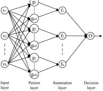
A neural network classifier is used to detect circumscribed tumour. The number of neurons at the input layer side are indicated by the number of images in the database and the output layer with one neuron indicates whether the MRI is a tumour or not and the hidden layer changes according to the number of rules that give the best recognition rate for each group of features.
Figure 4: Probabilistic Neural network
In the proposed model feed forward supervised neural network is considered which is given as Fuzzy Probabilistic Neural Network Classifier. It is a combination of two techniques consisting of Fuzzy cluster means and the Probabilistic neural network algorithm. It consists of Input layer, Hidden layer, pattern layer/summation layer and output layer. Pattern layer performs the classification. The probability density function used here is approximated by parsen estimator and the classification accuracy increase as number of training samples increases.Here for each of the input vector a ranking level is enervated and the highest rank holding vector is classified as winner. The pattern layer classifies the input vector only if there is a highest degree of match between the input vector and the training vector. This networks uses the bayesian theory .This is used to compromise fact that it is worst to misclassify an input vector that belongs to class A than to misclassify the vector that belongs to class B.
Where =Prior probability of occurrence of pattern in class And = cost associate with classifying vectors
( ) = 1 (2 )
−2( − ) ( − ) (9)
Where = ith Training Pattern from Class A n- Dimension of input vector
σ= smoothing parameter (corresponding to standard deviation of Gaussian distribution)
IV.MEASURE FOR PERFORMANCE EVALUATION
For the performance evaluation several types of techniques are commonly used for the proposed method. The measures mainly includes Analysis accuracy (AC) and Mathews Correlation coefficient (MCC) are calculated from the confusion matrix. Actual and predicted classes of the proposed method are described by the confusion matrix. MCC is used to measure the quality of binary classification. It returns a value from -1(inversion prediction) to +1(perfect prediction).
= ( + )
( + + + )
= ( − )
( + )( + )( + )( + )
Where TP=True Positive, TN= True Negative, FP= False positive and FN= False Negative.
V. EXPERIMENTAL RESULTS
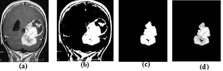
In the proposed method the image taken is pre-processed for the enhancement, by removing the noise and then separating from skull. The image sent for processing is grey scale image. In image segmentation Fuzzy C means clustering is used as the data does not fully belong to an individual cluster and the degree of belongingness of a data point to a particular cluster is given by the degree of membership, which is very important tool for the classification of malignant tumor.
Figure 5(a): original image, 5(b) Threshold image, 5(c):Segmented image, 5(d): Fuzzy Cmeans
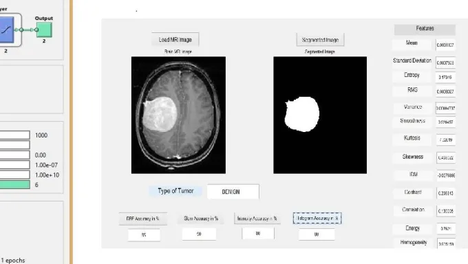
Figure 6: Training of Feed Forward network Figure 7: GUI OF proposed Model
Figure 10 shows the GUI of the proposed model along with cluster image, extracted features value, tumour part. The accuracy is calculate from the confusion matrix. The featured wise confusion matrix is give in the tabular for with the true positive, true negative, false positive and false negative.
Table1: Confusion matrix based on GLCM Features Figure 11: Dialogue Box showing Malignant Tumour
The above proposed method is also compared with the WEKA tool, intensity based, intensity histogram based which shows that the GLCM Feature extraction tool have the highest accuracy. This can be shown in figure 12.
Actual
Predicted
Cancer (positive)
Normal (negative) Cancer
(Positive)
17 2
Cancer (Negative)
VI. CONCLUSION AND FUTURE WORK
In the field of Bio-Medical applications, Image processing plays a vital role. The proposed system is developed for the diagnosing of tumor from magnetic resonance imaging pictures of brain. Diagnosing is done in many phases. In pre-processing stage filtering is performed on brain MR images. Fuzzy c means clustering method used for segmentation of the MR image. The texture features are extracted from the MR images of grey scale from GLCM method, these extracted features are given as input to artificial neural network for classifying MR image into benign or malignant tumorous image. In terms of future work the method can be extended to sub-classification of malignant tumor based on the training sets.
REFERENCES
[1] B.Sathees Kumar and Dr. R. AnbuSelvi “Feature Extraction Using Image Mining Techniques to Identify Brain Tumur”, IEEE sponsored conference on innovation in information Embedded and Communication systems ICIIECS’15 978-1-4799-6818-3/15
[2] NitishZulpl and Vrushsen Pawr2 "GLCM Textural Features for Brain TumorClassification",IJCSI International Journal of Computer Science issues,Vol. 9, Issue 3, No 3, May 2012
[3] M. C. Clark, L. O. Hall, D. B. Goldgof, L.P.Clarke,R. P. Velthuizen, and M. S.Silbiger, "MRI Segmentation using Fuzzy Clustering
techniques",IEEE Engineering in Medicine and Biology, pp. 730-742, 1994.
[4] Chang R.F., Wu W.1., Moon W.K., Chou Y.H., ChenD.R., "Support vector machines for diagnosis of breasttumors on US images", Academic Radiology,2003,10(2), p. 189-197.
[5] Chang R.F., Wu W.1., Moon W.K., Chou. Y.H., Chen.D.R., "Improvement in breast tumor discrimination bysupport vector machines and
peckle-emphasis textureanalysis", Ultrasound in Medicine and Biology, 2003,29(5), p. 679-686.
[6] M.Kasthuri and S.Britto Ramesh Kumar,"Multilingual Phonetic Based Stem Generations", SecondInternational Conference on Emerging research inComputing, Information Communication andApplications (ERCICA-2014) ,Volume: I, 31-02 August2014
[7] W. Chu, C. 1. Ong, and S. S. Keerthi "An improvedconjugate gradient scheme to the solution of least squaresSVM", IEEE Transactions onNeural Networks 16(2):pp. 498-501, 2005.
[8] V. Thavavela, 1.Jaffer Basha, R.Murugesan,"Anovel intelligent wavelet domain noise filtrationtechnique: Application to medical images", ExpertSystem with Application, 2010.
[9] Abe, Shigeo I Inoue, Takuya, " FuzzySupportVector Machines for Pattern Classification" Neuralnetworks, 200 I. Proceedings. IJCNN ,0 I
[10] M.Haralick, H.KShanmugam, andDinstein,"Texture features for image forimageclassification," IEEETrans. on Syst. ManCybernet.,vol. 3, no. 6,
pp. 610-621, 1973.
[II] Graham Sexton, KeshavDahal. "An adaptiveensemble classifier for mining concept drifting datastreams," Elsevier, Expert Systems withApplications,40, pp 5895-5906, 2013.
[12] Yongqiao Wang, Shouyang Wang, and K. K.Lai,"Anew fuzzy support vector machine toevaluatecreditrisk", IEEE Trans. on Fuzzy Systems, vol. 13, no. 6, pp.820-831, 2005.
[13] Shu-Hsien Liao, Pei-Hui Chu, Pei-Yuan Hsiao."Data mining techniques and applications A decadereview from 2000-2011," Elsevier, ExpertSystemswithApplications, 39, pp 11303-11311, 2012.
75 80 85 90 95 100
Histogram j48 Intensity Proposed
[14] Mr.Deepak, C.Dhanwani, Prof.MahipM.Bartere."Survey on various techniques of Brain TumorDetectionfrom MRI mages" InternationalJournalsofComputational Engineering Research, Vol4.IssueJanuary 2014, l.Issn 2250-3005.