Gene therapy for
ocular angiogenesis
Jam es W B Bainbridge
A thesis submitted for the degree of
Doctor of Philosophy, University of London
Department of Molecular Genetics
Institute of Ophthalmology
ProQuest Number: 10013346
All rights reserved
INFORMATION TO ALL USERS
The quality of this reproduction is dependent upon the quality of the copy submitted.
In the unlikely event that the author did not send a complete manuscript and there are missing pages, these will be noted. Also, if material had to be removed,
a note will indicate the deletion.
uest.
ProQuest 10013346
Published by ProQuest LLC(2016). Copyright of the Dissertation is held by the Author.
All rights reserved.
This work is protected against unauthorized copying under Title 17, United States Code. Microform Edition © ProQuest LLC.
ProQuest LLC
789 East Eisenhower Parkway P.O. Box 1346
abstract
This thesis describes a programme of work to develop local gene transfer of angiostatic proteins for the treatment of retinal and choroidal neovascular disorders. Current treatments are of limited efficacy and associated with significant adverse effects. Characterisation of the molecular and cellular events involved in angiogenesis has led to the identification of a number of angiostatic molecules with potential therapeutic value. The systemic administration of small molecule angiostatic proteins risks significant systemic adverse effects and if delivered intraocularly their half-life is short. Local gene transfer, however, offers the possibility of targeted, sustained and regulatable expression of angiostatic proteins to the retina after a single procedure to introduce a vector to an intraocular site. In order to investigate the potential value of this approach relevant animal models of retinal and choroidal angiogenesis were established and optimised. Viral vectors including adenoviral, adeno-associated viral and lentiviral vectors, expressing reporter genes and angiostatic proteins were
produced. Reporter gene studies were performed to determine the kinetics of transgene expression following intraocular vector delivery in the models. Gene delivery of a vascular endothelial growth factor (VEGF) inhibitor, soluble Flt-1, significantly reduced neovascularisation in models of both retinal and choroidal angiogenesis, and had no apparent adverse effect in normal animals. Targeting of transgene expression to sites and periods of active neo vascul ari sation was achieved by the incorporation of a hypoxia-responsive promoter into the gene construct. Pre-clinical studies of reporter gene transfer in a large animal model following subretinal delivery of a recombinant adeno-associated viral vector demonstrated efficient sustained reporter gene expression in cells of the outer retina. The findings of these studies were used to develop a
contents
Abstract... 2
Contents... 3
Figures and tab les... 10
Abbreviations...13
Publications...15
Acknowledgements...16
1 Introduction...17
1.1 Clinical signifîcance of ocular angiogenesis... 17
1.1.1 Proliferative diabetic retinopathy...17
1.1.2 Neovascular age-related macular degeneration... 19
1.1.2 Angiogenesis elsewhere in the eye... 21
1.2 Molecular biology of ocular angiogenesis; potential targets for angiostatic therapies... 22
1.2.1 Vascular endothelial growth factor... 25
1.2.2 Pigment epithelium-derived factor... 28
1.2.3 Growth hormone and insulin-like growth factor...28
1.2.4 Transforming growth factor-beta... 29
1.2.5 Fibroblast growth factors... 29
1.2.6 Platelet-derived growth factor...30
1.2.7 Matrix metalloproteinases... 30
1.2.8 Integrins... 31
1.2.9 Urokinase plasminogen activator... 32
1.2.10 Angiostatic steroids... 32
1.2.11 Angiostatin and endostatin... 33
1.2.12 Angiopoietins and Tie receptors... 34
1.2.13 Nitric oxide...34
1.2.14 Pathogenesis of angiogenesis in diabetic retinopathy...35
1.2.15 Pathogenesis of angiogenesis in age-related macular degeneration... 37
1.4 Rationale for ocular gene transfer of angiostatic proteins... 39
1.4.1 Vectors for ocular gene transfer... 40
1.4.2 Factors influencing ocular gene transfer...47
1.4.3 Applications of ocular gene transfer...48
1.4.5 Strategies for angiostatic gene transfer... 51
1.5 Aims of the thesis...52
2 Materials and methods...53
2.1 Reagents... 53
2.2 Amplification of plasmid DNA in bacteria... 53
2.2.1 Transformation of competent cells... 53
2.2.2 Amplification and recovery of recombinant plasmid DNA...53
2.2.3 Quantification of nucleic ac id ...54
2.3 DNA analysis... 54
2.3.1 Restriction enzyme digestion of plasmid DNA...54
2.3.2 Electrophoresis of DNA...54
2.4 Cloning in plasmid vectors...54
2.4.1 Creating appropriate DNA fragments... 54
2.4.2 Isolation of DNA fragments from agarose gels...55
2.4.3 DNA ligation...55
2.4.4 Confirmation of successful plasmid ligation... 56
2.5 Polymerase chain reaction (PCR)...56
2.6 Tissue culture... 57
2.6.1 Cell lines and viruses... 57
2.6.2 Culture of cell lines...57
2.6.3 Splitting and counting cells... 57
2.6.4 Long-term storage of c e lls ... 57
2.7 Manufacture of replication defective AAV... 58
2.7.1 Production of AAV...58
2.7.2 Purification of A A V ... 59
2.7.3 Concentration of A A V ... 59
2.8 Titration of AAV preparations... 60
2.8.1 Extraction of viral DNA from AAV particles... 60
2.8.2 Preparation of dot b lo t... 60
2.8.3 Preparation of probe... 61
2.8.4 Hybridisation of membrane... 61
2.10 Western blot for demonstration of of sFlt-1 expression by AAY-CM
V-sFlt-1 in vitro...63
2.11 Enzyme-linked immunosorbant assay for quantification of AAV-mediated expression of sFlt-1 in vivo... 63
2.10 Mouse fundus im aging... 64
2.10.1 A nim als... 64
2.10.2 Mouse fundus photography... 64
2.10.3 Mouse fundus fluorescein angiography... 65
2.11 Intraocular delivery of vector suspensions... 65
2.12 Preparation of retinal and choroidal flatmounts... 66
2.12.1 Fluorescein-dextran perfused whole retina flatmounts... 66
2.12.2 Fluorescein-dextran perfused whole sclera-choroid-RPE flatmounts 66 2.12.3 Whole retina flatmounts for detection of GFP expression...66
2.13 Histological methods...67
2.13.1 Paraffin wax sectioning...67
2.13.2 Cryo-sectioning...67
2.13.3 Evaluation of GFP fluorescence in cryosections...67
2.13.4 Staining of sections by periodic acid and Schiff’s reagent...68
2.13.5 Staining of sections by haematoxlyin and eosin...68
2.13.6 Immunostaining of vascular endothelial cells by anti-von Willebrand factor antibody... 68
2.13.7 Identification of cone photoreceptor cells in canine retina by peanut agglutinin staining...69
2.14 Buffers... 69
3 Angiostatic gene transfer in experimental retinal
neovascularisation... 72
3.1 Models of retinal neovascularisation... 72
3.2 Murine model of ischaemia-induced retinal neovascularisation... 74
3.2.1 Induction of ischaemia and retinal neovascularisation...74
3.2.2 Analysis of retinal neovascularisation... 76
3.3 Reporter gene expression by adenovirus and adeno-associated virus vectors in murine model of retinal neovascularisation... 81
3.3.1 Adenovirus-mediated expression in ischaemia-induced retinopathy... 83
3.3.2 AAV-mediated expression in ischaemia-induced retinopathy...84
3.4 Gene tran sfer of soluble V EG F receptor sFlt-1 by adenovirus and AAV
vectors in m urine model of retinal neovascularisation...87
3.4.1 Construction and production of rAAV vector expressing the soluble VEGF receptor sFlt-1...87
3.4.2 Determination of adenovirus-mediated and AAV-mediated in-vivo expression of sF lt-1... 90
3.4.3 Evaluation of angiostatic effect of Ad-mediated and AAV-mediated sFlt-1 expression in experimental retinal neovascularisation...91
3.4.4 Evaluation of adverse effects of Ad-mediated and rAAV-mediated sFlt-1 expression in normal animals...93
3.5 Discussion... 95
3.5.1 Murine model of ischaemia-induced retinal neovascularisation... 95
3.5.2 Reporter gene expression by Ad and AAV vectors in murine model of retinal neovascularisation... 96
3.5.3 Gene transfer of soluble VEGF receptor sFlt-1 by adenovirus and rAAV vectors in murine model of retinal neovascularisation...98
4 Angiostatic gene transfer in experimental choroidal
neovascularisation...102
4.1 Development of a model of choroidal neovascularisation...102
4.1.1 Application of laser in jury ...104
4.1.2 In vivo fluorescein angiography...106
4.1.3 FlTC-dextran perfused choroidal flatmounts...107
4.1.4 Histological evaluation of laser-induced choroidal neovascularisation .... 108
4.1.5 Image quantification of choroidal neovascularisation... 110
4.1.6 Reproducibility of laser-induced choroidal neo-vascul ari sation... 112
4.2 AAV-mediated an d lentivirus m ediated gene tran sfer in laser-induced choroidal n eovascularisation... 115
4.2.1 Production of AAV and lentiviral vectors... 115
4.2.2 Subretinal vector delivery in mice... 116
4.2.3 Laser-induction of choroidal neovascularisation... 117
4.2.4 Evaluation of GFP reporter gene expression...117
4.3 AAV-mediated gene tran sfer of the soluble V EG F receptor sFlt-1 in experim ental choroidal neovascularisation...119
4.3.1 Evaluation of angiostatic effect of rAAV-mediated sFlt-1 expression in experimental choroidal neovascularisation... 119
4.3.2 Long-term evaluation of potential adverse effects of AAV-mediated sFlt-1 expression in normal animals... 121
4.5 Discussion... 122
4.5.2 AAV-mediated and lenti virus-mediated reporter gene transfer in laser-
induced choroidal neovascularisation...124
4.5.3 AAV-mediated gene transfer of the soluble VEGF receptor sFlt-1 in experimental choroidal neovascularisation... 126
4.5.4 Evaluation of toxicity due to rAAV-mediated gene transfer of sFlt-1 in normal animals...128
5 Regulation of gene expression in experimental retinal and
choroidal neovascularisation... 131
5.1 Introduction...131
5.2 HRE-driven gene expression in experimental retinal neovascularisation 133 5.2.1 Construction of AAV.HRE.GFP...133
5.2.2 Delivery of AAV vectors in experimental retinal neovascularisation 134 5.2.3 Analysis of GFP expression in retinal whole-mounts and cryosections.... 134
5.3 HRE-driven expression in experimental choroidal neovascularisation.... 137
5.3.1 Delivery of AAV vectors in experimental choroidal neovascularisation.. 137
5.3.2 Analysis of GFP expression in retinal cryosections...138
5.4 Discussion... 140
5.4.1 Targeting of cellular transduction and regulation of gene expression 140 5.4.2 HRE-driven expression in experimental retinal and choroidal neovascularisation... 142
5.4.3 Conclusions... 143
6 Adeno-associated virus-mediated gene transfer to the
canine retina...145
6.1 Introduction...145
6.2 Vector production and delivery... 147
6.2.1 Vector construction, virus production and determination of titre ... 147
6.2.2 Surgery and anaesthesia... 148
6.3 Evaluation of reporter gene expression in vivo...151
6.4 Evaluation of retinal function by electroretinography... 153
6.4.1 Effect of subretinal AAV delivery on electroretinogram...153
6.4.2 Effect of subretinal PBS delivery on retinal function... 155
6.5 Histological examination... 157
6.5.1 Reporter gene expression... 157
6.6 Serology and immune responses... 161
6.7 Discussion... 162
6.7.1 Surgical technique for subretinal delivery of vector suspension... 162
6.7.2 AAV-mediated reporter gene expression... 163
6.7.3 AAV-mediated transduction of cone photoreceptor cells...164
6.7.4 Immune responses following subretinal delivery of AAV.CM V.GFP 165 6.7.5 Evaluation of retinal function following subretinal delivery of AAV.CM V.GFP... 167
6.8 Conclusion... 168
7 Discussion...170
7.1 Conclusions... 170
7.1.1 Time-courses of vector-mediated expression in experimental retinal and choroidal neovascularisation...170
7.1.2 Tissue patterns of transduction in experimental retinal and choroidal neovascularisation... 171
7.1.3 Efficacy of angiostatic gene transfer in experimental retinal and choroidal neovascularisation... 173
7.1.4 Regulation of gene expression in experimental retinal and choroidal neovascularisation... 174
7.1.5 Gene transfer in intermediate animal models...174
7.1.6 Rationale for a clinical trial of angiostatic gene transfer...174
7.2 Development of proposal for clinical trial of angiostatic gene transfer to the ey e... 175
7.2.1 Selection of appropriate target condition for clinical trial of angiostatic gene transfer... 176
7.2.2 Selection of angiostatic molecule for clinical trial of angiostatic gene transfer... 177
7.2.3 Selection of vector for clinical trial of angiostatic gene transfer... 177
7.2.5 Vector-related toxicity...178
7.2.6 Toxicity of the expressed protein...180
7.2.7 Ethical and regulatory issues...181
7.3 Future directions... 182
7.3.1 Experimental models of retinal and choroidal neovascularisation 182 7.3.2 Novel strategies for gene-based therapies... 184
Appendix: Proposal for a phase-1 clinical trial of gene
therapy for neovascular age-related macular degeneration
... 189
figures and tables
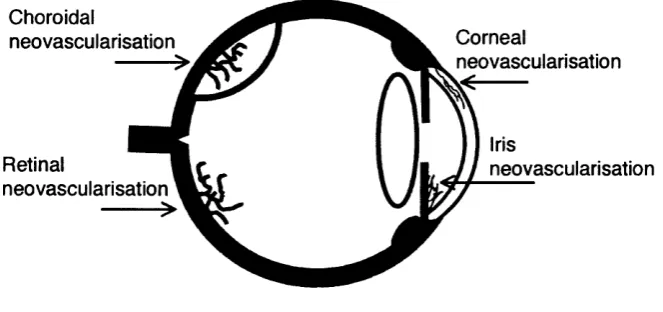
Figure 1.1 Schematic section of the eye showing common sites of intraocular
neovascularisation... 18 Figure 1.2 Fundus photographs showing retinal and choroidal neovascularisation 19 Figure 1.3 Anterior segment photographs showing neovascularisation of iris and
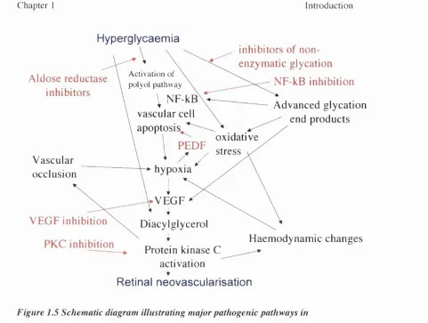
cornea...22 Figure 1.4 Schematic diagram illustrating stages of angiogenesis...24 Figure 1.5 Schematic diagram illustrating major pathogenic pathways in proliferative
diabetic retinopathy...36 Figure 3.1 Custom-built perspex chamber for controlled exposure of mouse pups to
hyperoxia... 75 Figure 3.2 Representative in-vivo fundus fluorescein angiograms of animals at p i 9,
120 seconds after intraperitoneal injection of sodium fluorescein... 76 Figure 3.4 Histological sections of retinas mice at p l 9 ... 79 Figure 3.5 Quantification of retinal neovascularisation in murine ischaemia-induced
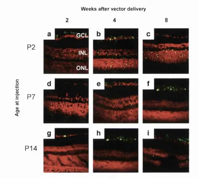
retinopathy... 80 Figure 3.6 Intravitreal delivery of viral vector... 82 Figure 3.7 Ad-mediated reporter gene expression following intravitreal injection 83 Figure 3.8 AAV-mediated reporter gene expression following intravitreal injection.. 84 Figure 3.9 AAV-mediated expression following intravitreal vector delivery in normal
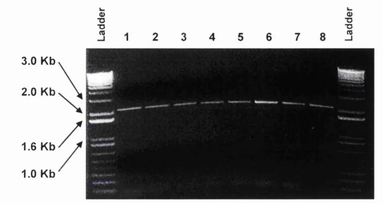
mice at different postnatal ages...86 Figure 3.10 PCR of sFlt-1 cDNA from Ad.CMV.sFlt-1 construct...88 Figure 3.11 Restriction enzyme digest of psFlt-l-NI by BamHI and Not 1... 89 Figure 3.12 Western blot confirming production of sFlt-1 by 293T cells following
infection by AAV.CMV.sFlt-1...90 Figure 3.13 Representative FITC-perfused retinal flatmounts at p l 9 ...92 Figure 3.14 Quantification of angiostatic effect of local sFlt-1 gene transfer on
Figure 3.15 Determination of potential toxicity resulting from gene transfer of sFlt-1 in normal (non-ischaemic) m ice...94 Figure 4.1 Diode laser induction of choroidal neovascularisation using a slit-lamp
mounted delivery system...105 Figure 4.2 Mouse fundus photographs showing effect of diode laser injury... 106 Figure 4.3 In vivo fundus fluorescein angiograms showing effect of laser injury 107 Figure 4.4 Fluorescence microscopy of FlTC-dextran perfused choroidal flatmounts
following laser-induced rupture of Bruch’s membrane...108 Figure 4.5 Histological analysis of effect of laser-injury... 109 Figure 4.6 Image quantification of area of hyperfluorescence at sites of laser injury on
in-vivo fluorescein angiography... 111 Figure 4.7 Extent of experimental neovascular lesions induced by different
combinations of laser settings over tim e...114 Figure 4.8 Delivery of vector suspension to the subretinal space in m ice...116 Figure 4.9 GFP reporter gene expression in laser-induced choroidal neo-vascul ari sati on
two weeks following subretinal injection of vector... 118 Figure 4.10 Effect of subretinal delivery of AAV.CMV.sFlt-1 on laser-induced
choroidal neovascularisation... 120 Figure 4.11 Effect of AAV.CMV.sRt-1 in normal animals... 121 Figure 5.1 CMV-driven and HRE-driven AAV-mediated GFP expression after
intravitreal vector delivery in a murine model retinal neovascularisation: retinal flatmounts... 135 Figure 5.2 CMV-driven and HRE-driven GFP expression following intravitreal vector
delivery in murine model of ischaemia-induced retinal neovascularisation: fluorescence micrographs of retinal cryosections...136 Figure 5.3 CMV-driven and HRE-driven GFP in laser-induced choroidal
neovascularisation: fluorescence microscopy of retinal cryosections 139 Figure 6.1 Fundus photographs of mouse and canine eyes... 147 Figure 6.2 Surgical technique for subretinal delivery of vector suspension in dogs.. 150 Figure 6.3 Fundus photograph and in-vivo fundus GFP fluorescence following
Figure 6.4 Electroretinography 8 weeks following subretinal delivery of
AAV.CMV.GFP in normal 6-week-old beagles... 154 Figure 6.5 Electroretinography 10 and 20 weeks following subretinal delivery of PBS
in normal 6-week-old beagles... 156 Figure 6.6 Fluorescence micrographs of retinal cryosections 6 weeks following
subretinal delivery of AAV.CMV.GFP vector to right e y e ... 158 Figure 6.7 Fluorescence micrographs of cryosections 6 weeks following subretinal
delivery of AAV.CMV.GFP... 158 Figure 6.8 AAV-mediated transduction of cone photoreceptors; scanning laser
confocal micrographs of sections of outer retina... 160
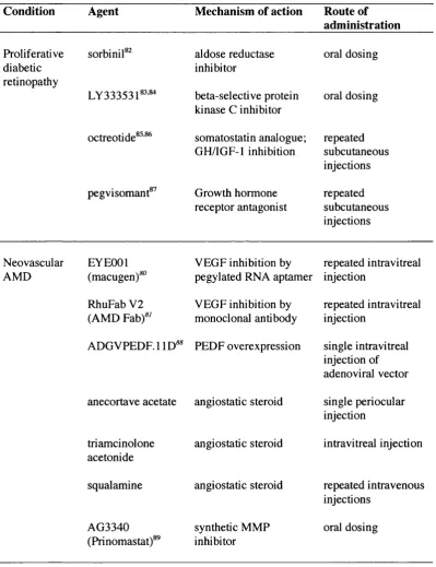
Table 1.1 Experimental molecular therapies for retinal and choroidal
abbreviations
AAV adeno-associated virus
Ad adenovirus
AGE advanced glycation end products AMD age-related macular degeneration CMV cytomegalovirus
cPPT central polypurine tract ECM extracellular matrix ERG electroretinogram
eNOS endothelial nitric oxide synthase FGF fibroblast growth factor
HV feline immunodeficiency virus
GH growth hormone
GFP green fluorescent protein
GTAC Gene Therapy Advisory Committee HIF-1 hypoxia-inducible factor-1
HIV human immunodeficiency virus HRE hypoxia-response element ICAM-1 intercellular adhesion molecule-1 IGF-1 insulin-like growth factor-1 iNOS inducible nitric oxide synthase MCP-1 monocyte chemoattractant protein-1 MMP matrix metalloproteinase
NO nitric oxide
NOS nitric oxide synthase NP-1 neuropilin-1
RPE retinal pigment epithelium SDT spontaneously diabetic Torii SIV simian immunodeficiency virus TGF-beta transforming growth factor-beta TIMP tissue inhibitor of metalloproteinases
publications
Work presented in this thesis has contributed to the following publications.
1. JW B Bainbridge, C Stephens, K Fowler, C Demaison, A Halfyard, AJ Thrasher and RR All. In-vivo gene transfer to the mouse eye using an HIV- based lentiviral vector: efficient long-term transduction of comeal endothelium and retinal pigment epithelium. Gene Therapy 2001 ;8:1665-1668
2. MB Reichel, JW B Bainbridge, D Baker, AJ Thrasher, SS Bhattacharya, RR All. An immune response after intraocular administration of an adenoviral vector containing a beta galactosidase reporter gene slows retinal degeneration in the rd mouse. Br J Ophthal 2001 ;85: 341-344
3. GM Sarra, C Stephens, F Schlictenbrede, JW B Bainbridge, AJ Thrasher, PJ Luthert, RR All. Kinetics of transgene expression in mouse retina following subretinal injection of recombinant adeno-associated virus. Vision Research 2002;42: 541-549
4. JW B Bainbridge, A Mistry, M De Alwis, E Paleog, A Baker, AJ Thrasher and RR All. Inhibition of retinal neovascularisation by gene transfer of soluble VEGF receptor sFlt-1. Gene Therapy 2002;9: 320-326
5. JW B Bainbridge, H Jia, D Selwood, RR All, and I Zachary. Peptides Encoded by exon 6 of VEGF inhibit VEGF-induced angiogenesis in vitro and ischaemic retinal neovascularisation in vivo. Biochem Biophys Res Commun 2003; 302(4):793-9
6. JW B Bainbridge, A Mistry, K Binley, M De Alwis, A Thrasher, S Naylor, R All. Hypoxia-regulated transgene expression in experimental retinal and choroidal neovascularisation Gene Therapy 2003; I 0 : 1049-54
7. JW B Bainbridge, A Mistry, FC Schlichtenbrede, A Smith, C Broderick, M De Alwis, A Georgiadis, PM Taylor, M Squires, C Sethi, D Charteris, AJ
Thrasher, D Sargan and RR Ali. Stable rAAV-mediated transduction of rod and cone photoreceptors in the canine retina. Gene Therapy -in press
acknowledgements
I am indebted to Robin Ali for inspiring me to embark on this project and for his guidance, support and encouragement throughout.
A number of individuals made essential personal contributions. In particular, I am grateful to A jay Mistry for his guidance and assistance with the cloning of constructs and production of vectors. I also thank Mahesh De Alwis for vector production, Frank Schlichtenbrede for electroretinography and Cathryn Broderick for serological
analysis. Alexander Smith and Angus MacNeil were invaluable sources of advice and help. I am grateful to Adrian Thrasher, Shomi Bhattacharya and David Hunt for their advice and support.
Much of the work presented is the result of collaborations. Ewa Paleog and Andrew Baker were involved with the adenoviral studies, Katie Binley and Stuart Naylor with the hypoxia-response element, and Ian Zachary with the VEGF peptides. David Sargan and Polly Taylor were involved in the canine studies, and David Charteris and
Charanjit Sethi contributed data on retinal gene transfer in cats.
I should like to thank The Wellcome Trust for funding the project.
Chapter 1 Introduction
1
Introduction
1.1 Clinical significance of ocular angiogenesis
Angiogenesis is the formation of new vasculature by the out-sprouting of vascular endothelial cells from existing vessels. This process is critical for embryonic development, growth, endometrial and placental proliferation, wound healing, and revascularisation of ischaemic tissues. Angiogenesis is also a central feature of many important diseases including cancer, rheumatoid arthritis, atherosclerosis and ocular neovascularisation. Pathological angiogenesis occurs in retinopathy of prematurity (ROP), proliferative diabetic retinopathy and age-related macular degeneration which are the leading causes of blindness in infants, individuals of working age and the elderly respectively.' ^ Neovascularisation in these conditions causes visual loss through increased vascular permeability leading to retinal oedema, vascular fragility resulting in haemorrhage, or fibro-vascular proliferation with tractional and
rhegmatogenous retinal detachment. Pathological angiogenesis occurring at other intraocular sites including the cornea and iris can also result in loss of vision through loss of comeal transparency and glaucoma respectively (Figure 1.1). Although
neovascularisation tends to occur at a relatively late stage in the course of many ocular disorders it is nonetheless a highly attractive target for therapeutic intervention since it represents a final common pathway in processes that are multifactorial in aetiology and is the event that typically leads directly to visual loss.
1.1.1 Proliferative diabetic retinopathy
Chapter 1 Introduction
Choroidal
neovascularisation a Corneal
neovascularisation
Iris
Retinal ^
\ Iw. J I
neovascularisationneovascularisation
Figure 1.1 Schematic section o f the eye showing common sites o f intraocular neovascularisation
Retinal neovascularisation is typically seen in ischaemic retinopathies and extends pre-retinally into the vitreous cavity. Neovascularisation o f the choroid is most commonly due to age-related macular degeneration and often extends subretinally.
occlusion and ischaemia. The subsequent hypoxia-induced upregulation of angiogenic growth factors results in neovascularisation that extends from the inner retinal surface into the vitreous gel (Figure 1.1).'^ Complications of neovascular proliferation are the major causes of persistent severe visual loss vision in diabetes through haemorrhage into the vitreous(Figurel.2b) or associated tractional retinal detachment.^ Although such complications of diabetes may be prevented by rigorous control of blood glucose^^ and blood pressure® this can be difficult to achieve in practice. The current conventional treatment for established proliferative diabetic retinopathy is pan-retinal laser photocoagulation.’ This technique is able to induce regression of retinal
C h a p te r 1 Introduction
A lth o u g h isch aem ia-in d u ced retinal n eo v ascu larisatio n is m o st freq u en tly seen in
d ia b etic eye d isease, it is also a central feature o f o th er c o m m o n retin o p ath ies
in clu d in g retin o p ath y o f p rem atu rity , retinal v ascu lar o c c lu sio n , sick le cell d isease,
ca ro tid artery d isease and o c u la r in flam m ato ry diso rd ers.
Figure 1.2 Fundus photographs showing retinal and choroidal neovascularisation a. Normal fundus: b. Retinal neovascularisation in diabetic retinopathy. Fine new vessels are visible along the superoternporal vascular arcade (arrow) and vitreous haemorrhage, presumably originating from new vessels at the optic disc (arrowhead) extends inferiorly; c. Choroidal neovascularisation in age-related macular
degeneration. Drusen (open arrow) and a ring o f lipid exudates (arrow) are evident at the macula. A subtle grey area centrally (arrowhead) is evidence o f the presence o f a choroidal neovascidar complex.
1.1.2 Neovascular age-related macular degeneration
A g e -related m acu lar d eg en eratio n (A M D ) is the m o st c o m m o n cau se o f b lin d n ess in
in d u strialized c o u n tries, w ith an estim ated in cid en ce o f 2 0 0 0 0 new cases an n u ally in
the U K and p rev alen ce o f 1.9% in p eople o ld er than 50 y e a r s . T h e p ath o g en esis o f
A M D is not well u n d ersto o d but in v o lv es a b n o rm alities o f the ex tra c e llu lar m atrix at
the level o f B ru c h ’s b asem en t m em brane.'^ C horoidal n e o v a sc u la risa tio n in this
co n te x t m ay be the resu lt o f h y p o x ia/isch aem ia o f o v e rly in g retinal p igm ent epithelial
(R P E ) cells, due e ith e r to the th ick en in g o f B ru c h ’s m e m b ran e o r to a b n o rm alities o f
Chapter 1 Introduction
choroidal p e r f u s io n ,le a d in g to the expression of pro-angiogenic cytokines. Exudation or haemorrhage from resulting neovascular complexes (Figure 1, Figure 2) that extend from the choroidal vasculature through breaks in Bruch’s membrane account for 80% of severe visual loss in this condition.
The established treatment modalities for choroidal neovascularisation in AMD comprise thermal laser photocoagulation and photodynamic therapy. These ablative approaches can offer short-term benefit to certain subgroups of patients^'^ but are associated with significant adverse e f f e c t s . L a s e r photocoagulation can be beneficial for the small minority of patients who have well-demarcated ("classic") neovascular lesions that do not extend beneath the fovea.^'^ ^ Photocoagulation destroys photoreceptor cells, however, creating a central scotoma such that treatment of subfoveal lesions results in destruction of the foveal photoreceptors with permanent loss of central vision.^'* Photodynamic therapy is a non-thermal process that aims to selectively destroy new blood vessels by irradiation following the intravascular administration of a photosensitising agent. Since this approach does not result in the destruction of overlying photoreceptor cells it can be effective in patients with subfoveal neovascularisation and delays loss of vision in a subgroup of patients with predominantly classic subfoveal lesions. Both photocoagulation and photodynamic therapy are designed to eliminate abnormal blood vessels, but since these treatments fail to address the underlying stimuli for blood vessel growth they are associated with high rates of persistent and recurrent disease. Following photocoagulation the
cumulative proportion of treated eyes with recurrent/persistent choroidal
neovascularisation is approximately 50% after 3 years^^ and is accompanied by an increased frequency of severe visual loss.^^ Following photodynamic therapy, persistence and recurrence of neovascularisation are also common and frequently require further treatments.
Chapter 1 Introduction
re-activiation or regrowth.^^ Transpupillary thermotherapy has been observed to cause closure of choroidal neovascular membranes^°^' but has not been shown to confer a benefit to vision in a controlled trial.
The surgical excision of subfoveal choroidal neovascularisation may stabilize or improve visual acuity in selected cases.^^ Visual prognosis is most dependent on the integrity of the subfoveal RPE-Bruch’s-choriocapillaris complex after removal of the CNV.^^ RPE incorporation into subfoveal membranes remains a limiting factor in AMD because surgical excision is typically complicated by concurrent removal of RPE.^^ RPE patch autotransplants^^ and human fetal RPE allografts^^ offer the potential means to repopulate the subretinal space but do not directly address the problems of CNV recurrence following surgical excision.^^ Excision of CNV in disorders other than AMD is more encouraging,^^ at least in part because the RPE Bruch’s-choriocapillaris complex is often preserved after surgery in these conditions. Excision of subfoveal CNV in punctate inner choroidopathy^® and the presumed ocular histoplasmosis syndrome^^ has been promising. Even in these conditions, however, CNV recurrence is a major limiting factor, occurring in up to 44 % of patients.'^ Experimental surgical procedures are being developed to reposition the neurosensory fovea away from abnormal RPE and/or choroid to a new location that has presumably healthier retinal RPE and choriocapillaris in order to recover or preserve foveal function. Translocation of the fovea away from such subfoveal choroidal neovascularisation also permits the removal of the choroidal neovascularisation or its destruction by laser
photocoagulation with minimal risk to the fovea.'^^ Recurrent neovascularisation is common, however, following such procedures, often occurs at the new fovea and is an important cause of vision loss."*^
1.1.2 Angiogenesis elsewhere in the eye
C h a p te r 1 Introduction
cau se loss o f vision through asso ciated clo su re o f the irid o -co rn eal d rain ag e angle
resu ltin g in raised in trao cu lar pressu re and g lau co m ato u s o p tic neuro p ath y .
N eo v ascu larisatio n o f the co rn e a can o ccu r in resp o n se to a n u m b e r o f d ifferen t insults
in clu d in g traum a, infection, in flam m atio n and c o n tact lens w ear and results in loss of
norm al corneal tra n sp a re n c y .^ T h is project, h o w ev er, is d irected to w ard s n eo v ascu lar
diso rd ers o f the retina and ch o ro id since it is th ese co n d itio n s that are resp o n sib le fo r
the m ajo r p ro portion o f visual loss du e to angiogenesis."^
b
1
Figure 1.3 Anterior segment photographs showing neovascularisation o f iris and cornea
a. Iris neovascularisation: b. Corneal neovascularisation
1.2 Molecular biology of ocular angiogenesis; potential
targets for angiostatic therapies
S ince a v ailab le treatm en ts fo r o c u lar n eo v ascu lar d iso rd ers are lim ited , there is a clear
clinical n eed fo r the d ev e lo p m e n t o f novel ap p ro ach es th a t are d irected ag ain st the
underly in g p ro -an g io g en ic stim uli so as to ach iev e a su stain ed th erap eu tic effect. T he
d e v e lo p m e n t o f such treatm en ts d ep en d s on a c le a r u n d e rstan d in g o f the m o le c u la r and
cellu lar p ro cesses inv o lv ed in an g io g en esis.
Chapter 1 Introduction
Early studies of neovascularisation in the retina contributed significantly to the understanding of the pathogenesis of angiogenesis. In 1948 Michael son investigated retinal vascular development and pathological retinal neovascularisation using perfusion techniques in human and cat fetuses."^^ He showed that the formation of retinal capillaries was predominantly a function of the retinal veins and described the capillary free zone that existed around arteries both in the immature and in the mature retina. It was on the basis of these observations that he formed the hypothesis that there existed in the retina a factor capable of affecting the growth of new blood vessels both in development and in disease. He argued that this factor, possibly of a biochemical nature, is present in the extra-vascular tissue of the retina in a concentration gradient that determined the extent of capillary growth from veins in development and in disease, and suggested that the stimulus for new vessel growth was associated with the metabolic demand of the retinal tissue.
C h a p te r Introduction
has also been im plicated in the d e v e lo p m e n t o f ch oroidal n eo v ascu larisatio n .'^ T h e
cy to k in e v ascu lar endothelial g ro w th fa c to r (V E G F ) has been id en tified as a m ajo r
ca n d id ate fo r M ic h a e lso n ’s v a so fo rm ativ e fa c to r th ough it ap p ears that an g io g e n e sis is
reg u lated not by a single fa cto r but by a b alan ce b etw een local stim u lato rs in clu d in g
V E G F and in h ib ito rs such as p ig m en t ep ith e liu m -d e riv e d fa c to r (PEDF).*’’
Receptor
1) Angiogenic cytokines eg VEGF, FGF-2
2) Matrix degradation
3) Endothelial cell proliferation and migration
4) Vessel maturation
5) Pericyte recruitment
Figure 1.4 Schematic diagram illustrating stages o f angiogenesis
A n g io g en esis is a com plex m u lti-ste p p ro cess that in v o lv es the o u t-sp ro u tin g o f
v ascu lar en d o th elial cells fro m e x istin g v essels th ro u g h en d o th elial cell p ro liferatio n
and m ig ratio n , e x tracellu lar m atrix re m o d ellin g and c ap illary tube fo rm atio n (F ig u re
1.4). T h is p ro cess is con tro lled by c o m p lex in teractio n s betw een grow th facto rs,
ex tra c e llu lar m atrix and c e llu lar c o m p o n e n ts, the net o u tc o m e being d eterm in ed by the
balan ce o f an g io g en ic and a n g io static e le m e n ts .^ A n u m b e r o f grow th fa c to r m o lecu les
are involved in the control o f a n g io g e n e sis and the th e ra p e u tic m an ip u latio n o f on e o r a
co m b in atio n o f these offers the p otential m ean s to control n e o v ascu larisatio n in the
C hapter 1 Introduction
eye. C ytokines that have been effectively targeted in experim ental m odels include vascular endothelial growth factor, insulin-like grow th facto r-1, pigm ent epithelium - derived factor, m atrix m etalloproteinases, angiostatin, endostatin, angiopoietin and integrins. A dditional targets in diabetic retinopathy include aldose reductase, advanced glycation end products, protein kinase C and nuclear factor kappa B. A lthough there are at present no established m olecular treatm ents for proliferative diabetic retinopathy or choroidal neovascularisation, a num ber o f experim ental m olecular therapies are currently under evaluation in clinical trials (Table 1.1).
1.2.1 Vascular endothelial growth factor
V EG F is a potent endothelial cell-specific m itogen that plays a critical role in
angiogenesis.^'’^^ A 46kD A hom odim eric glycopeptide, V EG F exists in three isoform s in the m ouse and four isoform s in the hum an as a result o f alternative splicing. V EG F is expressed by several different ocular cell types including pigm ent epithelial cells, pericytes, vascular endothelial cells, neuroglia and ganglion c e l l s , ' a n d in specific spatial and tem poral patterns during retinal development.^^ E xpression o f V EG F is upregulated by hypoxia in vitro^^ and in vivo^"^ through a hypoxia-inducible facto r-1 (H IF-1) transcriptional element.^^ V EG F isoform s act via specific ^ 5 - l i k e receptors, Flt-1 and Flk-1 /K D R, w hich are high-affînity receptor tyrosine kinases expressed on vascular endothelial cells that on binding V EG F phosphorylate a num ber o f proteins including phospholipase Gy. This leads to the form ation o f diacylglycerol (DAG), activation o f protein kinase C (PKC) and ultim ately to endothelial cell proliferation, m igration, and increased vasoperm eability. Placental grow th factor (PIGF) is a m em ber of the V EG F fam ily that stim ulates endothelial proliferation in vivo and angiogenesis in vitro and appears to potentiate the angiogenic effect o f V EG F either by enhancing its expression or by the form ation o f h e t e r o d i m e r s A soluble truncated form o f the V EG F receptor F lt-1, sFlt-1, is the only know n endogenous specific inhibitor o f VEGF.^^’^ sFlt-1, which lacks the m em brane-proxim al im m unoglobulin-like dom ain, the trans-m em brane spanning region and the
Chapter 1 Introduction
activity results from sequestration of VEGF, to which it binds with high affinity, and from formation of inactive heterodimers with membrane-spanning isoforms of the VEGF receptors Flt-1 and KDR/^^'
VEGF plays a central role in a number of angiogenic disorders including
tumorigenesis“ and rheumatoid arthritis® in addition to retinal neovascular disease. VEGF levels are increased in experimental models of retinal ischaemia,® ® in patients with proliferative diabetic retinopathy,^^® retinopathy of prematurity,® retinal vein occlusion,® and in choroidal neovascularisation.® Injection of recombinant VEGF protein into the eyes of non-human primates produces neovascularisation of the iris.^° and overexpression of VEGF by photoreceptors in transgenic mice causes
neovascularization in the retina.^' Adenovirus-mediated VEGF overexpression in the outer retina causes neovascularisation in the subretinal space.^^ Conversely, antagonists of VEGF inhibit experimental neovascularisation in the iris^ retina,^^’®’^'^ and choroid.^^
The central role of VEGF in angiogenesis makes it an attractive target molecule for angiostatic strategies both in the eye and elsewhere. The endogenously expressed soluble VEGF receptor, sFlt-1, has attracted particular attention for its potential therapeutic role in the control of n e o v a s c u la ris a tio n O th e r anti-VEGF strategies to control ocular neovascularisation in experimental models have included neutralising anti-VEGF monoclonal antibodies^^^, soluble VEGF-receptor chimeric proteins,® oligonucleotides,^'*^^ and inhibition of a VEGF-specific protein kinase.^^ Two
Chapter 1 Introduction
Table 1.1 Experimental molecular therapies for retinal and choroidal
neovascularisation evaluated in clinical trials
Condition Agent Mechanism of action Route of
administration Proliferative diabetic retinopathy sorbinil®^ LY333531^’^ aldose reductase inhibitor beta-selective protein kinase C inhibitor
oral dosing
oral dosing
octreotide®^’^ somatostatin analogue; GH/lGF-1 inhibition
repeated subcutaneous injections peg vi soman t^ Growth hormone
receptor antagonist repeated subcutaneous injections Neovascular AMD EYEOOl (macugen)^^
VEGF inhibition by pegylated RNA aptamer
repeated intravitreal injection
RhuFab V2 (AMD Fab)"'
VEGF inhibition by monoclonal antibody
repeated intravitreal injection
ADGVPEDF.llD"" PEDF overexpression single intravitreal injection of adenoviral vector anecortave acetate angiostatic steroid single periocular
injection triamcinolone
acetonide
angiostatic steroid intravitreal injection
squal amine angiostatic steroid repeated intravenous injections
AG3340 (Prinomastat)"^
synthetic MMP inhibitor
Chapter 1 Introduction
1.2.2 P igm ent ep ithelium -derived fa c to r
Pigm ent epithelium -derived factor (PEDF) is a soluble angiostatic protein secreted by retinal pigm ent epithelial cells in the developing and adult retina. It is a non-inhibitory m em ber o f the serine protease inhibitor (serpin) superfam ily o f proteins^ and w as first described for its neurotrophic properties.^' PED F induces neuronal differentiation of retinoblastom a cells^' and prom otes neurite-outgrow th and survival o f cerebellar granule neurons. The PED F gene is closely linked to an autosom al-dom inant locus for retinitis pigm entosa and it has been suggested that PED F m ay act as a survival factor for photoreceptors.^^ PED F interacts with receptors present on photoreceptors^^ and protective effects against photoreceptor death in m ouse m odels o f inherited retinal degeneration^'^ and after light damage^^ have been described. The angiostatic effect o f PED F may be the result o f prom otion of endothelial cells apoptosis.^^ PED F expression is dow n-regulated in hypoxia and its loss appears to play a perm issive role in ischem ia- driven retinal neovascularisation.^^ The com bination o f angiostatic and neuro-
protective properties o f PED F m ake this a potentially attractive candidate for the control o f neovascularisation in age-related m aculopathy since degeneration o f the retina is also a typical feature o f this condition. A clinical trial o f PED F for
neovascular A M D is currently ongoing.^*
1.2.3 G row th horm one and insulin-like grow th fa c to r
C hapter 1 Introduction
B oth local and system ic expression o f IGF-1 appear to contribute to its intraocular le v e ls /^ IGF-1 inhibition by a receptor antagonist suppresses experim ental retinal neovascularisation.^* A lthough a clinical trial o f the grow th horm one receptor
antagonist pegvisom ant has not dem onstrated regression o f diabetic retinopathy,*^ trials o f GH/IGF-1 axis inhibition using som atostatin analogues suggest that this approach may offer a beneficial effect in diabetic retinopathy*^ *^ and m ight also be effective in choroidal neovascularisation.
1.2.4 Transform ing grow th factor-beta
The transform ing grow th factor-beta (TG F-beta) superfam ily is a large fam ily o f m ultifunctional factors that regulate various cellular functions, including cellular proliferation, m igration, differentiation, apoptosis and extracellular m atrix production. The m echanism by w hich TG F-beta stim ulates angiogenesis is not well understood, but it appears to have an effect on the regulation o f V E G F gene expression at the transcriptional l e v e l 'a n d may also act by recruiting inflam m atory cells capable o f stim ulating angiogenesis d ir e c tly .T G F - b e ta 1 dow n-regulates vascular endothelial grow th factor receptor 2 (FLK) expression in vascular endothelial c e l l s .T G F - b e t a is expressed in ocular tissues including the cornea, ciliary epithelium , lens epithelium , retina, and blood vessels and appears to play a role in choroidal neovascularisation, being upregulated in experim ental models'®^ and in hum an s p e c im e n s .B lo c k a d e o f TG F-beta using a soluble TG F-beta receptor inhibits com eal angiogenesis.'®^
1.2.5 Fibroblast grow th factors
Chapter 1 Introduction
intrinsic tyrosine kinase activity and second messengers such as the mitogen-activated protein kinases. Intracellular FGFs might also have a direct biological role within the nucleus; the absence of a signal sequence to direct their secretion and their ability to traffic to the nucleus are unique structural features that may be relevant to the regulation of their a c tiv itie s .B o th FGF-1 and FGF-2 are involved in retinal development and angiogenesis’" and appear to have neurotrophic effects on retinal c e l l s . A l t h o u g h FGF-2 induces angiogenesis in the choroid'"’""^ it appears to be neither necessary nor sufficient alone for the development of retinal
neovascularization."^
1.2.6 Platelet-derived growth factor
Platelet-derived growth factor (PDGF) is a 45 kDa single chain polypeptide that has migratory and proliferative effects on retinal glial and endothelial cells’’^ PDGF is upregulated during ischaemia -induced retinal neovascularisation"’ and is also
implicated in the induction of proliferative vitreoretinopathy, being upregulated both in experimental models'^” and in human disease.’^’ ’^^
1.2.7 Matrix metalloproteinases
Matrix metalloproteinases (MMPs) are a family of zinc-binding, calcium ion- dependent, neural endopeptidases that function as proteolytic enzymes in the
degradation processes of the extracellular matrix (ECM)'“ and have important roles in development, wound healing and angiogenesis. Their inactive precursors are activated locally by proteolytic removal of amino acid terminal ends and their activity is further regulated at the transcriptional level by cytokines. The tissue inhibitors of
Chapter 1 Introduction
preferentially degrade basement membrane components such as type IV collagen. Angiogenesis is disrupted by PEX, a non-catalytic C-terminal hemopexin domain of MMP-2 with integrin binding a c t i v i t y .M M P s - 7 ,- 9 and -12 may block
angiogenesis by converting plasminogen to angiostatin which is one of the most potent angiogenesis antagonists.'^^ TIM Ps-1,-2,-3 and possibly TIMP-4 inhibit
n e o v a s c u la r is a tio n .C r itic a l roles for MMPs and TIMPs are implicated in ocular angiogenic disorders. MMPs-2 and -9 are implicated in both retinal'^' and choroidal neovascularisation. TIMP-3 is synthesised by RPE cells'^^ and is present in Bruch’s membrane and choroid in association with the extracellular matrix. Mutations in the gene encoding TIMP-3 have been described in Sorsby’s fundus dystrophy, a
maculopathy that is characterised by thickening of Bruch's basement membrane and choroidal neovascularisation. Matrix degradation is an attractive target for angiostatic therapy because it represents a critical step and final common pathway in angiogenesis. AG3340 (Prinomastat), an oral synthetic inhibitor of MMP-2, -9,-13 and -14,
inhibited tumour growth and angiogenesis in a variety of preclinical models but was ineffective in clinieal trials for choroidal neovascularisation in AMD.^
1.2.8 Integrins
Chapter 1 Introduction
1.2.9 Urokinase plasminogen activator
The urokinase plasminogen activator (uPA) system facilitates the movement of cells through a matrix, including migration of endothelial cells during angiogenesis, by initiating an extracellular cascade of proteolysis that involves the activation of plasminogen and MMPs."”’ *'^' uPA is a serine protease secreted by vascular cells as a single chain inactive form that is proteolytically converted to an active form that modulates angiogenesis by remodelling of extracellular matrix. In addition to
dissolution of matrix, proteolytic cascades activate certain growth factors such as TGF- beta or K 1-5'"^^ and release others such as FGF-2 that are sequestered on the cell surface or within the ECM, thus contributing to the evolution of a migratory or invasive cell phenotype. uPA is also able to modulate endothelial cell adhesion, migration and proliferation responses in a non-proteolytic fashion through signalling involving its specific cell surface receptor uPAR. uPAR itself lacks intracellular signalling domains and such signals are believed to be transduced via interactions between uPA/uPAR and in te g r in s .T h e uPA system has been implicated in the development of human diabetic neovascular membranes."^ A secreted version of the amino-terminal fragment (ATF) of murine uPA antagonizes uPA binding to uPAR. ATF inhibits cell invasion in vitro and angiogenesis in experimental tu m o u r s ,b u t this approach has yet to be applied to ocular neovascular disease.
1.2.10 Angiostatic steroids
Chapter 1 Introduction
ceils.A d m in iste re d intravenously, squalamine significantly improves intraocular neovascularisation in experimental m o d e l a n d a clinical trial of squalamine is currently underway to evaluate its effect in neovascular AMD.
1.2.11 Angiostatin and endostatin
Plasminogen is an inactive proenzyme that is converted by plasminogen activators to the active enzyme, plasmin, which is central to the fibrinolytic cascade. Angiostatin is a 38 kD internal fragment of plasminogen that comprises four kringle domains (kringle
1-4) and is an endogenous circulating inhibitor of angiogenesis first identified in mouse tumour models.*^ Its anti-angiogenic effect appears to result from specific inhibition of endothelial cell proliferation,'^ and may involve an interaction with the integrin alpha(v)beta(3)'^^ and increased endothelial cell apoptosis. The anti
proliferative activity of angiostatin on endothelial cells is shared by kringle 1, kringle 2, and kringle 3, but probably not by kringle 4, and its potency is increased when kringle 4 is removed.'^ Kringle 5 of plasminogen appears to be more potent than angiostatin in and may act through down-regulation of VEGF and up-regulation of PEDF.'^ Intravitreal injection of Kringle 5 reduces experimental ischaemia-induced retinal neovascularisation in vivo.'^'
Endostatin is a 20 kDa C- terminal fragment of collagen X Vlll released by elastase a c tiv ity .E n d o statin specifically inhibits endothelial proliferation in vitro and potently inhibits angiogenesis and tumor growth in vivo. Its mechanism of action is zinc-dependent and may involve blocking VEGF receptor signalling, interaction with integrins or induction of endothelial cell apoptosis.'^ Both angiostatin and
Chapter 1 Introduction
1.2.12 Angiopoietins and Tie receptors
Tie receptors are endothelium specific receptor tyrosine kinases. The Tiel receptor is required for the structural integrity of endothelial cells, but its ligand has yet to be identified. The Tie2 receptor has been implicated in stabilisation and maturation of vessels through the action of an agonist ligand, angiopoietin 1 (A ngl) and an antagonistic ligand, Ang2. A ngl mediates vessel maturation and remodelling and is essential for normal vascular development in the mouse'^. It promotes survival of endothelial cells but not chemotaxis or proliferation and its effect appears to result from the protection of endothelial cells against a p o p to s is .A n g l also appears to confer resistance to VEGF-mediated vascular leakage.'^ Ang2 antagonises the Tie2 receptor and its expression is upregulated during physiologic and pathologic
neovascularisation’^^ sTie-2, a synthetic soluble receptor of angiopoietin inhibits angiogenesis and has potential therapeutic value. Angl is also of potential therapeutic value for reducing microvascular leakage in diseases in which the leakage results from chronic inflammation or elevated VEGF and, in combination with VEGF, for
promoting the growth of non-leaky vessels.’^
1.2.13 Nitric oxide
anti-Chapter 1 Introduction
a n g io g e n ic d e p e n d in g on the different NOS isoform involved. There is evidence that eNOS plays a pro-angiogenic role*^'^ that involves the prom otion o f endothelial cell m igration and differentiation/^^ possibly via the upregulation o f alpha(v)beta(3) i n te g r in s .E le v a tio n o f eNOS activity in correlation w ith angiogenesis has been extensively reported and eNOS inhibitors may offer a valuable angiostatic effect. The role o f iNOS in angiogenesis rem ains controversial. A lthough iNOS is expressed in the ischaem ic r e t i n a 'i t s effect can be anti-angiogenic.'^* iNOS may be responsible for m isdirecting angiogenesis in ischaem ic retinopathy aw ay from the ischaem ic retina and into the v itr e o u s .I n h ib i tio n o f iNOS m ight then offer a very attractive
opportunity to control o f vitreal neovascularisation by facilitating appropriate re vascularisation o f hypoxic retina.
1.2.14 P ath o genesis o f an g io g en es is in d iab etic retinopathy
In diabetic retinopathy the m echanism s by w hich hyperglycaem ia leads to the upregulation o f angiogenic grow th factors (review ed in detail elsewhere'*") are com plex and involve activation o f the polyol pathway, non-enzm atic glycation and oxidative stress; retinal hypoxia is the result o f subsequent haem odynam ic changes, m icrovascular occlusion and vascular cell apoptosis (Figure 1.5). This pathw ay offers a num ber o f potential opportunities for specific m olecular therapeutic intervention. H yperglycaem ia activates the polyol pathw ay in w hich glucose is converted to sorbitol by endothelial aldose reductase leading to capillary cell death. A lthough inhibition o f aldose reductase reduces the high g lucose-ind uced death o f retinal capillary cells in v/fro,'*' the effect o f aldose reductase inhibitors in clinical trials o f diabetic retinopathy is inconclusive.*^ H yperglycem ia also leads to the irreversible form ation o f advanced glycation end products (A G Es) w hich in turn generate oxygen-derived free radicals that contribute tow ards increased oxidative stress and them selves increase autocrine retinal V EG F expression,'*^ and inhibition o f A G E form ation is another potential therapeutic strategy to target angiogenesis.'*^ '*^ A ctivation o f endothelial protein kinase C (PKC) by diacylglycerol, synthesised either de novo in chronic
C h ap ter Introduction
Hyperglycaemia
A ld o se reductase
inhibitors
Vascular occlusion
V E G F inhibition
PKC inhibition
A c tiv a tio n o f
p o ly o l p ath w ay
^ NF-kB
vascular cell
apoptosis,
1
P E D F/ / /
hypoxia1
V E G F
;
D iacylglycerol I
Protein kinase C
oxidative stress
inhibitors o f
non-en zym atic glycation
N F-kB inhibition
A dvanced glycation end products
H aem odynam ic ch an ges
activation
Retinal neovascularisation
Figure 1.5 Schematic diagram illustrating major pathogenic pathways in proliferative diabetic retinopathy
Potential strategies fo r molecular therapeutic intervention are indicated (red arrows).
platelet-activating factor expression with leukocyte activation, thro m b u s form ation,
h a e m o d y n a m ic changes, and increased vascular cell p erm eability and proliferation.
Interventions that increase diacylglycerol m etab o lism or inhibit P K C is ozym es
am eliorate the functional c o n seq u en ces o f D A G - P K C activation in experim ental
m odels o f diabetes'®'^ and reduce the extent o f isch aem ia-in d u ced experim ental o cu lar
n e o v a s c u la ris a tio n .'^ Clinical trials to evaluated the effect o f the P K C inhibitor
LY333531 in diabetic retinopathy are in p r o g r e s s . D e a t h o f retinal capillary cells
can o ccur by apoptosis especially in the context of fluctuating glucose levels; A G E s
m ay induce activation of n uclear facto r k appa B (N F -k B ), a m olecule that appears to
be an im portant signaling m olecule in retinal vascular cell s u r v i v a l . I n h i b i t i o n of
Chapter 1 Introduction
NFkB activation offers a potential strategy to prevent retinal hypoxia through the inhibition of vascular cell apoptosis.'^
Retinal neovascularisation may occur as a result of ischaemia in conditions other than diabetes, for example retinal vascular occlusion and carotid stenosis. In retinal vein occlusion neovascularisation appears to occur through hypoxia-induced upregulation of VEGF ex p re ssio n .T issu e hypoxia appears to be a key step in triggering
neovascularisation in experimental retinal vein o c c lu s io n ,a n d retinal VEGF expression is upregulated in both experimental vein occlusion in pigs'^ and in ischaemic retinal vein occlusion in patients.^
Retinal neovascularisation may also be a feature of inflammatory eye disease. This may result from associated vascular occlusion leading to ischaemia and hypoxia- induced VEGF upregulation. This mechanism is clearly involved in occlusive vasculitides such as Bechet’s disease and may also be the major mechanism in other conditions such as sarcoid u v eitis.A ltern ativ ely , certain inflammatory cytokines are believed to have a direct angiogenic effect, and retinal neovascularisation may develop in patients with chronic uveitis in the absence of retinal ischaemia. In this situation regression of new vessels may be induced by immuno suppression alone.'^
1.2.15 Pathogenesis of angiogenesis in age-related macular
degeneration
Choroidal neovascularisation in AMD appears to be the combined result of local upregulation of angiogenic cytokines in association with an underlying abnormality of the extracellular matrix between RPE cells and the choroid. Though choroidal
Chapter 1 Introduction
choroidal neovascularisation including high myopia, pseudoxanthoma elasticum and Sorsby’s fundus dystrophy.'” Patients with Sorsby’s fundus dystrophy have mutations in the gene encoding TlMP-3, a protein that is involved in regulation of extracellular matrix turnover. Extracellular matrix deposition along Bruch’s membrane and increased production of FGF-2 in RPE cells can both be caused by oxidative stress.'” Choroidal neovascularisation may be the result of upregulated expression of pro- angiogenic cytokines, at least in part, in response to hypoxia/ischaemia of overlying retinal pigment epithelial c e l l s , d u e either to thickening of Bruch’s membrane or to abnormalities of choroidal perfusion,'^'^
1.3 Strategies for m olecular angiostatic therapies
It is clear that angiogenesis is a highly complex process and that the effects of cytokines on endothelial cell-matrix interactions can be contextual.'” However, since determination of the angiogenic phenotype appears to be the result of a net balance of positive and negative regulators of blood vessel growth,'” the introduction of a single agent to tip the balance towards angiostasis may be all that is required to achieve a valuable therapeutic effect. Effective intervention may be possible by targeting pathogenic processes at a number of levels in the relevant pathways.
Chapter 1 Introduction
pathways might neutralise the effect of intervention is difficult to predict and
ultimately the optimal approach may be determined only by appropriate evaluation in experimental models and clinical trials.
1.4 Rationale for ocular gene transfer of angiostatic
proteins
Though potentially efficacious, the systemic administration of angiostatic molecules risks important adverse systemic effects. In addition to its critical role in
embryogenesis, physiological angiogenesis is central to wound healing and recovery from ischaemic events through revascularisation and the formation of collateral circulations. Patients with retinal neovascular disease, typically associated with diabetes or advanced age, are also likely to be at increased risk of ischaemic heart disease, cerebrovascular and peripheral vascular disease. The systemic inhibition of angiogenesis in these individuals would risk compromising critical vascular responses to ischaemic events. Clinical trials of VEGF antagonists for tumour therapy suggest that their systemic administration may be also be associated with vascular toxicities such as haemorrhage and thrombo embolic events.'^ In addition to these concerns, the production of recombinant proteins is technically difficult and their manufacture is e x p e n s iv e .S in c e the access of proteins to the retina is restricted by the blood-retinal barrier, the high systemic doses required to achieve therapeutic intraocular levels would be particularly expensive and hazardous.
For these reasons, the local delivery of angiostatic agents offers significant potential advantages. Intraocular neovascularisation in experimental models is reduced by the repeated intravitreal injections of neutralizing anti-VEGF monoclonal antibodies,^ recombinant soluble VEGF-receptor chimeric proteins^ and anti sense
Chapter 1 Introduction
angiostatic protein in the eye. The relatively short half-life of proteins delivered by intravitreal injection is such that frequently repeated administration would be necessary to maintain therapeutic levels'^ and would pose a high cumulative risk of local
complications including intraocular infection, vitreous haemorrhage and retinal detachment.
In contrast, somatic gene transfer offers the possibility of localised, targeted, sustained and regulatable delivery of therapeutic proteins following a single procedure to introduce a vector to an appropriate intraocular site. Using a gene transfer approach, sustained expression can be achieved locally while minimising any risk of systemic adverse effects. Tissues may be targeted by vector design and surgical techniques, and expression may potentially be regulated through the use of tissue-specific, inducible or tissue-responsive promoters.
1.4.1 Vectors for ocular gene transfer
The eye is an ideal organ for in-vivo gene transfer. Ocular anatomy lends itself to the accurate delivery of vector suspensions because the tissues are compartmentalised and readily accessible by microsurgical techniques under direct visualisation. Its small size means that only tiny volumes of vector suspensions are required to transduce a
significant proportion of cells in the target tissue, and using appropriate vectors, even non-dividing cell populations may be efficiently transduced by a single dose. The relative immune privilege of ocular tissues may confer some advantage in terms of long term transgene expression.^”® The optical transparency of the eye enables GFP reporter gene expression to be observed in-vivo in many instances, and therapeutic effects on structure and function may be readily observed and quantified using a variety of techniques both experimentally and clinically.
adeno-Chapter 1 Introduction
associated virus (AAV), and lentivirus systems are the most frequently used viral vectors. The ideal choice of vector for a given ocular application is dependent on the natural tropisms of the vectors and their time-courses of expression.
1.4.1.1 Recombinant adenoviral vectors
Adenoviruses are a family of human DNA viruses that cause respiratory tract
Chapter 1 Introduction
Following delivery into the anterior chamber of the eye, Ad vectors efficiently
transduce cells of the corneal endothelium^®^ and trabecular meshwork/®®^'® Following delivery into the subretinal space, Ad vectors mediate efficient transduction of RPE cells and occasional photoreceptors.^ ^ ^ T cell-mediated immune responses, however, can cause local inflammatory changes and limit the duration of gene expression in the eye/'^ Although intraocular expression may be prolonged by preventing T cell activation, for example by co-injection with adenovirus expressing CTLA4-lg,^''^ and newer generation vectors may offer more sustained expression profiles, adenoviral vectors are generally better suited to applications in which only short-term expression is adequate or desirable.
1.4.1.2 Recombinant adeno-associated virus vectors
AAV is a small (20-25 nm) human parvovirus that is able to infect both dividing and non-dividing cells. There are 8 known human viral serotypes, each of which appears to have different tropic properties.^'^^'^ The viral genome is single stranded DNA and consists of two separate open reading frames between two inverted terminal repeats (ITRs). One open reading frame encodes the viral capsid proteins {cap) while the other encodes proteins required for replication {rep). After infection the single-stranded DNA genome is converted into its transcriptionally active double stranded form by the host’s DNA repair machinery and preferentially integrates into the human chromosome site-specifically at 19ql3.3.^‘*
Chapter 1 Introduction
Although the incorporation of sections of the rep gene into AAV appears to restore site-specific integration in human cells in vitro^^ the utility of this must be weighed against the risk of re/?-induced immune responses against transduced cells.
Although the production of high-titre AAV vector preparations has been limited in the past by technical difficulties, these have been addressed by the development of new production systems. The original method of AAV production required the co transfection of a vector plasmid and a packaging plasmid (providing rep and cap in trans) into a producer cell line and infection by a wild-type adenoviral helper virus. This system resulted in only modest titres and risked contamination by live helper virus or by structural helper virus proteins even after labour-intensive purification processes. A number of strategies have since been developed to improve the efficiency of AAV vector production. These include the generation of cell lines that contain integrated copies of some or all of the AAV genes required for packaging^^'^ and the construction of adenovirus helper plasmids that can be used instead of adenovirus infection.^^^ Recombinant helper viruses have been engineered to express the rAAV vector genome^^^ or the AAV rep and cap genes.^^^ The replication and expression of AAV rep and cap gene products can be enhanced through the use of herpes-derived amplicons that support the replication of both the packaging plasmid and the vector plasmid, in combination with a herpes simplex helper virus, resulting in the production of higher titres of transducing particles.^^^^^^
Wild-type AAV is non-pathogenic in humans, and recombinant AAV vectors do not appear to induce significant immune responses. Up to 80% of the population are sero positive for AAV and antibodies against serotype-2 are the most prevalent.^® In the majority of cases, however, these antibodies are non-neutralising^^ and even in the presence of a strong humoral response to the vector, transduction events can occur after re-administration of rAAV.^^'"^*'^^
Chapter 1 Introduction
that enable the incorporation of larger inserts would offer a major improvement. A trans-splicing approach, in which gene expression is reconstituted from two
independent AAV vectors might overcome the vector's packaging limitations^^ but the success of this system to date has been limited. The efficiency of trans-splicing appears to be serotype d e p e n d e n t,h o w ev e r, and may be improved by the selection of
appropriate AAV serotypes; the use of AAV-2 pseudotyped with the capsid of AAV-5 (AAV-2/5), for example, increases the efficiency of trans-splicing in photoreceptor
cells.^^
AAV-2 vectors efficiently transduce rod photoreceptors and RPE cells after a single sub-retinal administration,^^^^ resulting in stable and long-lasting transgene
expression.^^’^® Cells transduced by AAV show a characteristic time delay between exposure to vector and onset of gene expression that has been attributed to the conversion of the incoming single stranded DNA genome to a transcriptionally active double-stranded template.^’ One strategy to alleviate the requirement for host-cell DNA synthesis is through the use of self-complementary AAV vectors.^'*® When the genome is half wild-type size, AAV can package either two copies, or dimeric inverted repeat DNA molecules that spontaneously re-anneal resulting efficient transduction independently of DNA synthesis. While half-length AAV may accelerate the onset of transgene expression, the size limitation may make it impractical for many disease applications.
AAV pseudotyping provides a useful means to manipulate cell targeting specificity and transduction characteristics in the retina.^^'^"^^ In the retina AAV-2 mediated gene expression occurs as early as three days after vector delivery and reaches its maximum potential after several weeks.^"^^ AAV-5 pseudotyped vectors display faster expression kinetics than AAV-2 and mediate greater transduction e f f i c i e n c y . T h e s e