Immunology, Vrije Universiteit Brussel, Brussels, Belgiumd
; Department of Structural Biology, Flanders Institute for Biotechnology, Brussels, Belgiume
; INSERM U1094, Neuroépidémiologie Tropicale, Universite de Limoges, Limoges, Francef
Loop-mediated isothermal amplification (LAMP) is a method for enzymatically replicating DNA that has great utility for clinical
diagnosis at the point of care (POC), given its high sensitivity, specificity, speed, and technical requirements (isothermal
condi-tions). Here, we adapted LAMP for measuring protein analytes by creating a protein-DNA fusion (referred to here as a
“LAMPole”) that attaches oligonucleotides (LAMP templates) to IgG antibodies. This fusion consists of a DNA element
cova-lently bonded to an IgG-binding polypeptide (protein L/G domain). In our platform, LAMP is expected to provide the most
suit-able means for amplifying LAMPoles for clinical diagnosis at the POC, while quantitative PCR is more suitsuit-able for
laboratory-based quantification of antigen-specific IgG abundance. As proof of concept, we measured serological responses to a protozoan
parasite by quantifying changes in solution turbidity in real time. We observed a
>
6-log fold difference in signal between sera
from vaccinated versus control mice and in a clinical patient sample versus a control. We assert that LAMPoles will be useful for
increasing the sensitivity of measuring proteins, whether it be in a clinical laboratory or in a field setting, thereby improving
acute diagnosis of a variety of infections.
N
umerous applications require sensitive measurement of
bio-logical analytes to provide actionable information in real
time. In considering biomarker measurement, no other molecule
is as easily or sensitively quantified as nucleic acid because of facile
means to enzymatically replicate sequence-specific templates
in
vitro
. The detection limit of specific DNA templates has been
re-ported to be at or below 10 molecules when measured by real-time
PCR (quantitative PCR [qPCR]) (
1
,
2
). This level of sensitivity is
useful in the context of a laboratory, but there is need for
diagnos-tics that can be used at the point of care (POC) without
thermo-cycling equipment. An alternative method for amplifying DNA is
loop-mediated isothermal amplification (LAMP), which requires
four different oligonucleotides to promote Bst
polymerase-cata-lyzed DNA synthesis at a constant 65°C. The end products of
LAMP (pyrophosphate and high-molecular weight DNA) can be
monitored by measuring changes in turbidity or color with the
addition of Mg
2⫹(
3
) or dyes (
4
), respectively.
Highly sensitive measurement of non-DNA analytes (e.g.,
pro-teins, toxins, lipids, and carbohydrates) at the POC can be
achieved by combining the molecular recognition of
immunoas-says with the signal amplification of LAMP. We previously
devel-oped means to label protein and small-molecule ligands with
unique oligonucleotides measurable by PCR (
5
). We labeled
mo-lecular targets using a protein-DNA fusion known as a “Tadpole,”
which binds ligand with high specificity so as to attach an
identi-fying oligonucleotide. When we labeled antibodies or protein
an-tigens with oligonucleotides, we created toxin and biomarker
as-says that were hundreds of times more sensitive than matched
enzyme-linked immunosorbent assay (ELISA) (
5–7
). Compared
to PCR, however, LAMP is more amenable to POC use because of
reaction simplicity and product detection by visual readouts (
8–
11
). In this regard, we previously advocated (
12
) that LAMP and
Tadpoles be combined such that the DNA element of a Tadpole is
amplified by LAMP instead of PCR. To investigate this concept,
we created a protein-DNA fusion consisting of a polypeptide that
binds mammalian IgG and a DNA element encompassing 1 PCR
and 2 LAMP amplicons. During the construction of this fusion,
we explored the activities of 10 different primer pair combinations
for conducting LAMP and several of chemistries for conjugating
protein and DNA. We further investigated the specificity of the
LAMPole fusion for recognizing human IgG and, as a benchmark
of this approach, applied it to measuring antiparasite antibodies in
humans and mice. Our assay was inspired by an ELISA for
detect-ing host antibodies to
Trypanosoma brucei gambiense
variable
sur-face glycoproteins (VSGs), which detected titers of 1:30,000 and
1:40 from serum and saliva, respectively (
13
). This work indicated
that detection of relatively low titers of antibodies in noninvasive
specimen types is possible provided the method is sufficiently
sen-sitive. Here, we present our findings. Rapid, accurate diagnosis of
other proteins, such as antigen and IgM antibody, may enable
Received5 January 2015 Returned for modification15 January 2015
Accepted22 January 2015
Accepted manuscript posted online4 February 2015
CitationBurbulis IE, Yamaguchi K, Nikolskaia OV, Prigge ST, Magez S, Bisser S, Reller ME, Grab DJ. 2015. Detection of pathogen-specific antibodies by loop-mediated isothermal amplification. Clin Vaccine Immunol 22:374 –380.
doi:10.1128/CVI.00811-14.
Editor:P. P. Wilkins
Address correspondence to Dennis J. Grab, [email protected].
*Present address: Ian E. Burbulis, Department of Biochemistry and Molecular Genetics, University of Virginia School of Medicine, Charlottesville, Virginia, USA.
Copyright © 2015, American Society for Microbiology. All Rights Reserved.
doi:10.1128/CVI.00811-14
on August 17, 2020 by guest
http://cvi.asm.org/
sensitive and specific diagnosis of the etiology of acute illness at
the POC.
MATERIALS AND METHODS
To construct the LAMPole fusion, we needed to design (i) the sequence of the DNA element and primer sets that specifically recognize this template, even in the presence of contaminating human or pathogen DNA, and (ii) a conjugation chemistry that bonds the DNA element to the polypeptide without inhibiting IgG-binding activity or interfering with the ability to promote LAMP. We designed 10 LAMP primer sets (4 different primers in each set) predicted to hybridize with elements in theArabidopsis thaliana chalcone synthase (CHS)-coding sequence (GenBank accession number M20308.1) using the software PrimerExplorer V4 (Fujitsu Limited). We identified primers according to the nomenclature originally described by Notomi et al. (14) as F3, B3, FIP, and BIP. The predicted melting temper-ature for the F3 and B3 primers was fixed close to 60°C in each set to promote LAMP at the optimal temperature for Bst activity. The LAMP reactions for selecting our primer sets (Fig. 1) and for subsequently mea-suring IgG serology were performed as previously described (15–17). Briefly, the reaction mixture contained 12.5l of 2⫻LAMP buffer [40 mM Tris-HCl (pH 8.8), 20 mM KCl, 16 mM MgSO4, 20 mM (NH4)2SO4, 0.2% Tween 20, 1.6 M betaine, 2.8 mM each deoxyribonucleoside triphos-phate], 1.0l CHS LAMP primer mix (5 pmol each of F3 and B3 and 40 pmol each of FIP and BIP), 8 units Bst DNA polymerase (New England BioLabs, Tokyo, Japan), and template DNA. Final volumes were adjusted to 25l with distilled water. All reactions were conducted in duplicate and were monitored in real time in a Loopamp LA320C real-time turbidime-ter (Teramecs, Tokyo, Japan). For assaying production of high-molecu-lar-weight DNA ladders, we fractionated 10l of reaction products by 1% agarose gel electrophoresis in 1⫻Tris-borate-EDTA (TBE) and visualized with ethidium bromide (EtBr).
To create the recombinant 253-bp double-stranded DNA (dsDNA) oligonucleotide (LAMP template) (Fig. 2), we digested pTRX-CHS
plas-mid DNA (18) with PciI and NaeI to liberate an element with a single 3= recessed terminus (PciI) and a single blunt end terminus (NaeI). We in-corporated an amino-allyl dUTP nucleotide at the 3=recessed terminus using the 3=-to-5=exo-Klenow fragment (New England BioLabs) for 30 min according to the manufacturer’s instructions. We converted this amino residue to an azide using 10:1 excessN-hydroxysuccinimide-azide (Pierce) in phosphate-buffered saline (PBS) for 1 h at 25°C and purified this element using a NAP-5 column (Life Technologies). To create our protein L/G chimera, we directed expression of the L/G mammalian an-tibody-binding polypeptide (19) fused to polyhistidine and the Methano-bacterium thermoautotrophicumRIR1 intein (20) Rosetta-gami-B (DE3) pLysS (Novagen) as previously described (21). Briefly, we cultured se-lected transformants in Luria-Bertani broth to an optical density (OD) of 0.6 at 600 nm, induced L/G expression with 1 mM IPTG (isopropyl--D -thiogalactopyranoside) for 4 h, and collected cells at 3,000⫻gfor 10 min at 4°C. We purified full-length L/G-intein fusion protein by immobilized metal affinity chromatography from cell-free lysates and cleaved the in-tein domain from L/G using 1 mM 2-mercaptoethanesulfonic acidin vitro (21). We labeled the purified L/G protein with phosphine-N -hydroxysuc-cinimide (NHS) at a 1:5 protein/phosphine stoichiometry in 0.1 M HEPES (pH 8.5) for 2 h at 25°C and buffer exchanged the phosphine-labeled L/G protein into phosphate-buffered saline. We conjugated azide-modified CHS dsDNA to phosphine-azide-modified L/G protein for 6 h at 25°C to yield an L/G protein-DNA fusion (LAMPole) and purified fusions by cation exchange (Q-Sepharose) and hydrophobic chromatography (phe-nyl-Sepharose) as described previously (5,21).
Human African trypanosomiasis (HAT) is caused byTrypanosoma brucei gambiense(22). Our prototype LAMPole assay is based on a pub-lished ELISA that detects host antibodies againstT. b. gambienseLiTat 1.3, 1.5, and 1.6 variable surface glycoproteins (VSGs) (13). To create the solid-phase substrate for use in our assays, we conjugated 2.8-m tosyl-activated beads (Invitrogen) with either purified LiTat 1.3 VSG (23) or purified mouse total IgG (Jackson Immunologicals) in 0.1 M borate (pH 9.5)–1 M (NH4)2SO4– 0.1 M NaCl for 16 h at 37°C. We capped unreacted tosyl residues with 0.1 M ethanolamine for 2 h at 37°C and blocked un-occupied surface area with PBS containing 0.1% dephosphorylated casein and 0.05% Tween 20 (blocking buffer). The mouse sera used to optimize assay conditions were prepared from animals vaccinated with 20g pu-rified VSG once a week for 3 weeks. No adjuvant was used. Sham (naive) controls received vehicle alone (PBS). For proof of concept, we also used archived serum obtained from a patient with stage 2T. b.gambienseHAT confirmed by a positive card agglutination test for trypanosomiasis (CATT) and with trypanosomes identified on blood and cerebrospinal fluid (CSF) smears.
For our serological assays, we serially diluted sera from test samples and negative controls. We incubated 106VSG-coated beads with 50-l
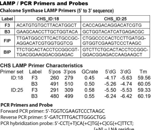
FIG 1Primers sets developed for sequence-specific LAMP and real-time PCR. The LAMP primer set ID:18 and ID:25 sequences are shown going from 5=to 3=with the F3, B3, FIP, and BIP nomenclature derived from Notomi et al. (14). The annealing positions corresponding to theA. thalianachalcone synthase (CHS) complete coding sequence (GenBank accession numberM20308.1), GC content (GCrate), 5=and 3=gene positions, free energy (5=dG/3=dG), and melting temperatures (Tm) are indicated. Also shown are the forward and reverse primer sequences (5=to 3=), along with the fluorescent hybridization probe sequence, used for real-time PCR.
FIG 2The 5=-to-3=sequence of the LAMP template. The annealing sites for the ID:18 F3 and B3 primers are underlined, while the annealing sites for the ID:25 F3 and B3 primers are shaded. The annealing site for the locked nucleic acid (LNA) hybridization probe for use in quantitative PCR (qPCR) is indi-cated in italics and gray. The adenosine residue complementary to amino-allyl dUTP nucleotide (Roche) incorporation (PciI site) is indicated in bold and underlined. PciI targets and cuts 5=...ACATGT...3= sequences to 5= -CATGT...3=products. As a result, the recombinant fragment digested is 1 bp shorter at the 5=end than the CHS plasmid sequence used to design the LAMP primers. LAMP recognizes and amplifies the recombinant construct and par-ent CHS plasmid equally well.
on August 17, 2020 by guest
http://cvi.asm.org/
serial dilutions of blocking buffer for 1 h at 25°C, washed the beads 3 times in 1 ml of casein-Tween-PBS by vortexing for 10 s, and then resuspended the beads with 50l of protein L/G LAMPole. After incubating for 1 h, we washed the beads 3 times with vortexing (as described above), resus-pended the beads in 10l 10 mM Tris-HCl (pH 8.0)– 0.05% Tween 20, and used 1l as the template for amplifying CHS templates by LAMP (25-l total volume) at 63°C. Here, we monitored the production of in-soluble Mg2⫹-pyrophosphate in real time using an LA-320C turbidimeter according to the manufacturer’s instructions. For instances in which we used real-time PCR to quantify the amount of LAMPole bound to beads (affinity measurements), we used a fluorescent hydrolysis probe contain-ing locked nucleic acid (LNA) nucleotides (5=-CCT[⫹T]CA[⫹C]TG [⫹C]CG[⫹C]TTCT, where [⫹N] is a LNA residue). For these measure-ments, we thermocycled bead-bound templates for 15 s at 95°C, 20 s at 60°C, and 60 s at 72°C. The quantities of LAMPole present in these sam-ples were calibrated by measurement of known numbers of CHS template with anr2value of 0.9988 over a 6-log range in concentration. We used the four-parameter logistic equation in the software package Prism v4.0 (GraphPad Software, Inc.) to fit a curve describing LAMPole activity to calculate the binding affinity constant according to previously published methods (5,21).
RESULTS
We designed primer sets that specifically annealed and amplified a
253-nucleotide (nt) element of the
A. thaliana
chalcone synthase
(CHS) gene (i) even in the presence of contaminating human or
pathogen DNA and (ii) conjugated to the protein L/G polypeptide
without inhibiting IgG-binding activity or interfering with the
ability to promote LAMP. We identified 2 primer sets out of 10
that promoted LAMP of the
A. thaliana
CHS template at 63°C by
assaying production of high-molecular-weight DNA ladders via
agarose gel electrophoresis (data not shown). The LAMP primer
set ID:18 and ID:25 sequences in
Fig. 1
are shown going from 5
=
to
3
=
, with F3, B3, FIP,
A. thaliana
CHS complete coding sequence
(GenBank accession number
M20308.1
), GC contents, 5
=
and 3
=
gene positions, free energy, and melting temperatures (
T
m)
indi-cated. Also shown are the forward and reverse primer sequences
(5
=
to 3
=
) that allow for more accurate DNA quantitation by
real-time PCR, along with the fluorescent hybridization probe
se-quence used to determine antibody-binding affinities as well as for
DNA quantitation.
While both ID:18 and ID:25 enabled detection of at least 100 fg
pET32(a)CHS as positive control (roughly 12,000 template
mol-ecules), ID:25 generated a more rapid LAMP reaction. We
con-firmed the primers to be 100% specific for
A. thaliana
CHS when
tested against
A. thaliana
chalcone isomerase (CHI) plasmid and
genomic DNAs from several protozoan parasites as negative
con-trols, including
Babesia microti
,
Plasmodium falciparum
, African
trypanosomes (
T. b. brucei
,
T. b. rhodesiense
, and
T. b. gambiense
)
(
Fig. 3
), and
Toxoplasma gondii
(not shown). DNA from
Rickettsia
prowazekii
, normal human blood (
Fig. 3
), and
Escherichia coli
and
blood from bacteremic patients (not shown) and also gave
nega-tive results. We considered precipitate occurring after 60 min to be
an artifact. Importantly, the amount of time needed to develop a
visual turbidity of
ⱖ
0.1 OD unit in these experiments was
in-versely proportional to the amount of pET32(a)CHS template,
demonstrating that the signal is quantitatively proportional to the
abundance of template (
Fig. 4
). Fortuitously, the ID:18 and ID:25
amplicons overlap in sequence, which allowed us to finalize our
LAMPole DNA design to include both overlapping amplicons and
an internal PCR amplicon containing a hybridization site for a
locked nucleic acid (LNA) (
24
) hydrolysis probe (see Materials
and Methods).
To construct the antibody-binding protein domain, we used a
recombinant protein originally created from the
immunoglobu-lin-binding domains of
Streptococcus
sp. protein G (which binds
mammalian immunoglobulin G [IgG] Fc chains) and
Peptostrep-tococcus
sp. protein L (which binds mammalian immunoglobulin
and
light chains) (
19
). We previously used this “L/G” domain
to construct a probe with PCR readout and quantified as few as
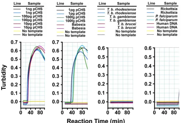
FIG 3Sensitivity and specificity of LAMP primers for pET32(a)CHS template. We serially diluted plasmid encodingA. thalianachalcone synthase (18) [pET32(a)CHS] or genomic DNA fromTrypanosoma bruceisubspecies,Babesia microti,Plasmodium falciparum,Toxoplasma gondii,E. coli,Rickettsia parkeri, bacteremic human patients, and normal human blood and assessed LAMP by monitoring turbidity in real-time. We used primer set ID:18 to direct LAMP in these reactions.
on August 17, 2020 by guest
http://cvi.asm.org/
21,559 and 1,016 molecules of an
E. coli
transcriptional initiation
factor 1 fusion protein (INFA-TAP) (
25
) and interleukin-6 (
26
) in
50
l, equivalent to detection limits of 716 and 34 aM, respectively
(
21
). Here, we directly adapted this IgG-binding domain to
syn-thesizing LAMPoles. We constructed our original Tadpoles
exclu-sively from recombinant protein and synthetic oligonucleotides
(60 to 85 bp) using “intein-mediated ligation” (
27
) to covalently
bond a single DNA element per protein domain. However, the
minimum size of templates that are amplifiable by LAMP averages
two hundred nucleotides (
3
). We ultimately want large quantities
of LAMPoles to be synthesized inexpensively so that they can be
used as field diagnostics. The cost per yield for using synthetic
DNA to manufacture LAMPoles would be prohibitive. This
con-clusion led us to develop an alternate synthetic route utilizing the
“Staudinger ligation” (
28
) to covalently bond recombinant DNA
to the L/G domain in limiting stoichiometry. Here, we conjugated
azide-modified DNA to phosphine-modified L/G protein to
cre-ate the IgG-binding LAMPole fusion. We verified that the L/G
domain remained active after conjugation to DNA by using a
hy-bridization LNA probe in qPCR to measure the
K
d(dissociation
constant) of the LAMPole for binding purified mouse polyclonal
IgG as previously described (
21
). We measured a
K
dof 250 nM for
mouse polyclonal IgG (Jackson Immunologicals) covalently
linked to M280 magnetic beads (Invitrogen). This affinity is less
than the
K
dof 3.4 nM measured for this protein linked to an 82-nt
DNA element (
21
).
As proof of concept that our LAMPole could be used to assay
antigen-specific antibodies, we measured levels of IgG that
recog-nize a specific
T. b. gambiense
antigen, variable surface
glycopro-tein (VSG), in the sera of vaccinated mice and a stage 2 HAT
patient. Various VSGs have traditionally been selected as antigenic
targets in serological assays for HAT (
13
,
22
). For all these assays,
we scavenged antiparasite IgG from sera using
T. b. gambiense
LiTat 1.3 VSG-coated magnetic beads and detected bound IgG by
forming a sandwich with our anti-IgG LAMPole (
Fig. 5
). Using
spike and recovery assays, we previously confirmed that no
sub-stances in human or mouse serum interfered with the use of
L/G-DNA fusions to measure IgG (
21
), and we found the same to be
true in these experiments. We accounted for cross-reactive
bind-ing of IgG to the beads and nonspecific bindbind-ing of the LAMPole by
subtracting the baseline signal obtained in replicates with neither
IgG nor antigen, and we confirmed equivalently low levels of
non-specific signal, even in negative-control serum. We formulated
assays with 0.576 mg/ml LAMPole (10-fold
K
d) to ensure that at
least 90% of available epitopes are occupied during measurement.
We initially formulated assay conditions using sera from naive
and VSG-vaccinated mice. We incubated 10
6VSG-coated
mag-netic beads with mouse sera and measured the amount of bound
antibody using the ID:25 primers (
Fig. 1
) to amplify the LAMPole
DNA element. When tested neat, only sera from VSG-vaccinated
mice (
n
⫽
4) yielded LAMP-dependent turbidity, in contrast to
naive controls (
n
⫽
2). We also serially diluted sera from one
immune mouse and one nonimmune mouse (in 1:10 steps) to
1:10
⫺5and found no loss of anti-VSG antibody signal at maximal
dilution (
Table 1
). However, after further dilution, anti-VSG
an-tibodies were detected in sera diluted to at least 1:10
⫺6. Based on
these findings, we incubated VSG-coated beads with the sera of a
patient with HAT and a healthy control (
Fig. 6
). We detected
anti-VSG antibodies in maximally diluted (1:10
⫺7) serum from a
patient with stage 2 HAT but not in serum from the healthy
con-trol. Thus, we observed a
⬎
6- to 7-log fold difference in signal
between sera from vaccinated versus control mice and from a
stage 2 HAT patient versus a control.
DISCUSSION
Our strategy to measure protein analytes (e.g., pathogen-specific
host antibodies) by LAMP is to use an adaptor probe that attaches
DNA oligonucleotides to the protein analyte to enable sensitive
and specific molecular detection of the protein-DNA LAMPole.
We found that we could measure protein analytes using LAMP as
a signal amplifier. Importantly, our goal was not to formulate a
better test for HAT,
per se
, but to demonstrate the LAMPole
con-cept as a technological advance applicable to improving the
mea-surement of a wide variety of protein analytes or pathogens with
high sensitivity and low cost. This is the first instance that we are
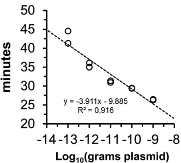
FIG 4Quantitative LAMP of CHS templates. Based on data inFig. 1, the amount of time necessary to accumulate turbidity above the threshold of de-tection (0.1 OD unit) is inversely proportional to the amount (100 fg to 1 ng) of startingA. thalianaCHS plasmid DNA template. The individual data points (n⫽2) are shown.
FIG 5LAMPole-based measurement of antigen-specific IgG. Measuring an-tigen-specific serology is a four-step process. (1) Capture. Antigen-coated beads (LiTat 1.3 VSG) are incubated with serum samples containing anti-VSG IgG (Ab). (2) Detect. After nonspecific IgG is washed away, bound IgG is detected by adding LAMPoles to create a molecular sandwich. (3) LAMP. The LAMPole sandwich is subjected to Bst polymerase-catalyzed LAMP to yield amplified high-molecular-weight DNA and insoluble pyrophosphate in com-plex with Mg2⫹. Here, the abundance of LAMPole retained on the bead surface
is proportional to the amount of specific anti-VSG IgG. (4) Visualize signal. Insoluble precipitate may be monitored in real time, or DNA may be directly visualized using intercalating dyes or gel electrophoresis.
on August 17, 2020 by guest
http://cvi.asm.org/
aware of in which a protein-DNA fusion has been used to combine
LAMP with immunoassays to measure protein analytes.
In its fully developed form, we expect that this technology may
be used in field situations without electricity, but for the sake of
this benchmark, we chose to detect turbidity using a turbidimeter
for increased precision. We envisioned the “field-deployed” assay
to be a two-step process in which a relevant biomarker of interest
(e.g., antigen) present in a patient’s sample (e.g., blood, serum,
saliva, urine, etc.) is captured onto magnetic beads coated with a
specific capture agent (e.g., capture antibody). The bound
bio-marker is then detected by addition of LAMPole to the beads, and
a visual signal is developed by supplementation with primers and
Bst polymerase. The inclusion of overlapping amplicons in the
DNA element of the LAMPole enables the use of two separate
LAMP primer sets to generate signal output and may be
con-firmed using an orthogonal amplification processes such as qPCR
for more precise quantification when needed. However, our
LAMPole fusion may also be embedded into existing and future
microfluidic platforms to provide signal amplification
in situ
.
While a colorimetric or turbidity output visible to the naked eye
could be used instead of microtiter plate readers in resource-poor
settings, this visual readout could also be useful in developed
B Vaccinated mouse CHS_ID:25 ⫹/⫹ ⫹/⫹ ⫹/⫹ ⫹/⫹ ⫹/⫹ ⫹/⫹ ⫹/⫹ ⫺/⫺
Control mouse ⫺/⫺ ⫺/⫺ ⫺/⫺ ⫺/⫺ ⫺/⫺ ND ND ND
a
The same serum samples were used in experiments A and B.
bDuplicate test samples were used.⫺/⫺, negative result;⫹/⫹, positive result; ND, not done.
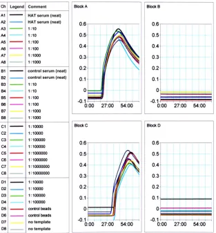
FIG 6Measurement of anti-VSG in a patient with HAT. Normal and HAT patient sera were incubated in duplicate with VSG-coated beads, followed by exposure to L/G LAMPole. We assayed the production of turbidity in real time. Samples incubated with HAT-positive serum were specifically amplified by CHS LAMP (blocks A and C), while normal serum and no-serum and no-template controls produced no signal (blocks B and D). We used primer set ID:25 to direct LAMP in these reactions.
on August 17, 2020 by guest
http://cvi.asm.org/
countries for POC diagnosis. When quantitative analysis is
impor-tant, real-time LAMP is also available.
The cost of LAMPole synthesis makes it possible to create a
wide array of probes for detecting all antigenic varieties expressed
by infectious agents that undergo antigenic variation, such as the
trypanosome parasites that cause HAT. Using our originally
pub-lished method for making Tadpoles that employed chemically
synthesized DNA elements (
5
), we estimate a reagent cost of about
$1.00 per assay. However, using the Staudinger ligation as
out-lined here, we estimate a reagent cost of about $0.0075 per assay.
Although this cost consideration is less important in developed
countries that can afford higher-priced methods, this issue is
sig-nificant in resource-limited countries where cost alone may
pre-vent test availability. Moreover, the modular nature of LAMPole
synthesis allows new probes to be easily reconfigured, which may
accommodate a multitude of antigenic variants corresponding to
geographic location.
Several multiplex LAMP methods are available (
29
,
30
), which
may be applied to amplifying mixtures of our LAMPole probes
and to simultaneously measure different target biomarkers in a
clinical sample. Using technologies that we describe here, the
cre-ation of antibody LAMPoles for antigen detection should now
also be possible. Together with intelligent LAMPole tail and
LAMP primer design, the creation of a single diagnostic
LAMP-based test for detection of host antipathogen antibodies (i.e.,
pro-tein L/G LAMPoles), pathogen antigens (antibody LAMPoles),
and pathogen DNA or RNA (LAMP or RT-LAMP) in a single
assay format should now be possible to increase the likelihood of
identifying the cause of the acute infection (increase sensitivity).
For example, an ideal LAMPole diagnostic test for acute dengue
might detect NS1 antigen, viral RNA, and IgM antibody; if it also
detected IgG, it could rapidly distinguish secondary from primary
dengue, which could have prognostic implications (higher risk of
severe dengue).
By merging LAMP and Tadpoles, we created a method that
may improve pathogen detection at the POC (
7
,
21
). We
previ-ously showed that the usefulness of traditional LAMP assays that
recognize multicopy gene targets for pathogen DNA detection can
be dramatically enhanced by sample pretreatment to lyse or
solu-bilize the pathogen(s) and facilitate release of its DNA prior to
assay, especially when the sample size is limited (
31
). Similar
mod-ifications could also be applied to the LAMPole-based assays
de-scribed here. Furthermore, we predict that LAMPoles will be a
suitable platform for the development of noninvasive saliva- and
potentially lachrymal (tear) fluid-based assays (
32
,
33
), with
sen-sitivities equivalent to those found with blood or CSF. Therefore,
the use of LAMPoles could provide cost-effective, ultrasensitive
antigen/antibody diagnostic assays with low technical
require-ments, which could improve acute diagnosis of neglected tropical
and other infections that plague the developing world and thereby
related clinical outcomes.
ACKNOWLEDGMENTS
We are grateful to J. Stephen Dumler, from Johns Hopkins University, for reading and editing the manuscript.
This research was supported in part by grants from the National Insti-tutes of Health (5R01AI082695 and 1R21AI079282) to D.J.G. and support from the French Ministry of Foreign Affairs in Gabon, Angola, and Cen-tral African Republic awarded to S.B.
The funders had no role in study design, data collection and analysis, decision to publish, or preparation of the manuscript.
D.J.G. conceived of the project. I.E.B., S.T.P., S.M., and D.J.G. dis-cussed and designed the experiments. D.J.G., O.V.N., K.Y., S.T.P., S.M., and I.E.B. performed the experiments. I.E.B., K.Y., O.V.N., S.T.P., S.M., S.B., M.E.R., and D.J.G. analyzed the data and made comments on the manuscript. I.E.B. and D.J.G. wrote the manuscript.
We declare no competing financial interests.
REFERENCES
1.Higuchi R, Dollinger G, Walsh PS, Griffith R. 1992. Simultaneous amplification and detection of specific DNA sequences. Biotechnology (NY)10:413– 417.http://dx.doi.org/10.1038/nbt0492-413.
2.Higuchi R, Fockler C, Dollinger G, Watson R. 1993. Kinetic PCR analysis: real-time monitoring of DNA amplification reactions. Biotech-nology (NY)11:1026 –1030.http://dx.doi.org/10.1038/nbt0993-1026. 3.Mori Y, Nagamine K, Tomita N, Notomi T.2001. Detection of
loop-mediated isothermal amplification reaction by turbidity derived from magnesium pyrophosphate formation. Biochem Biophys Res Commun
289:150 –154.http://dx.doi.org/10.1006/bbrc.2001.5921.
4.Goto M, Honda E, Ogura A, Nomoto A, Hanaki K.2009. Colormetric detection of loop-mediated isothermal amplification reaction by using htdroxy naphthol blue. Biotechniques46:167–172.http://dx.doi.org/10 .2144/000113072.
5.Burbulis I, Yamaguchi K, Gordon A, Carlson R, Brent R.2005. Using protein-DNA chimeras to detect and count small numbers of molecules. Nat Methods2:31–37.http://dx.doi.org/10.1038/nmeth729.
6.Evans J.2005. Tadpoles stick to protein analysis. Chem World18. 7.Nolan GP.2005. Tadpoles by the tail. Nat Methods2:11–12.http://dx.doi
.org/10.1038/nmeth0105-11.
8.Matovu E, Kazibwe AJ, Mugasa CM, Ndungu JM, Njiru ZK. 2012. Towards point-of-care diagnostic and staging tools for human African trypanosomiaisis. J Trop Med 2012:340538. http://dx.doi.org/10.1155 /2012/340538.
9.Bearinger JP, Dugan LC, Baker BR, Hall SB, Ebert K, Mioulet V, Madi M, King DP.2011. Development and initial results of a low cost, disposable, point-of-care testing device for pathogen detection. IEEE Trans Biomed Eng
58:805– 808.http://dx.doi.org/10.1109/TBME.2010.2089054.
10. Mori Y, Notomi T. 2009. Loop-mediated isothermal amplification (LAMP): a rapid, accurate, and cost-effective diagnostic method for infec-tious diseases. J Infect Chemother15:62– 69.http://dx.doi.org/10.1007 /s10156-009-0669-9.
11. Wastling SL, Picozzi K, Kakembo AS, Welburn SC.2010. LAMP for human African trypanosomiasis: a comparative study of detection for-mats. PLoS Negl Trop Dis4:e865.http://dx.doi.org/10.1371/journal.pntd .0000865.
12. Grab DJ, Lonsdale-Eccles J, Inoue N.2005. Lamp for tadpoles. Nat Methods 2:635. (Author reply, 2:635– 636.) http://dx.doi.org/10.1038 /nmeth0905-635a.
13. Lejon V, Jamonneau V, Solano P, Atchade P, Mumba D, Nkoy N, Bebronne N, Kibonja T, Balharbi F, Wierckx A, Boelaert M, Buscher P.
2006. Detection of trypanosome-specific antibodies in saliva, towards non-invasive serological diagnosis of sleeping sickness. Trop Med Int Health11:620 – 627.http://dx.doi.org/10.1111/j.1365-3156.2006.01620.x. 14. Notomi T, Okayama H, Masubuchi H, Yonekawa T, Watanabe K, Amino N, Hase T. 2000. Loop-mediated isothermal amplification of DNA. Nucleic Acids Res28:E63.http://dx.doi.org/10.1093/nar/28.12.e63. 15. Thekisoe OM, Kuboki N, Nambota A, Fujisaki K, Sugimoto C, Igarashi I, Yasuda J, Inoue N.2007. Species-specific loop-mediated isothermal amplification (LAMP) for diagnosis of trypanosomosis. Acta Trop102:
182–189.http://dx.doi.org/10.1016/j.actatropica.2007.05.004.
16. Thekisoe OM, Inoue N, Kuboki N, Tuntasuvan D, Bunnoy W, Borisut-suwan S, Igarashi I, Sugimoto C.2005. Evaluation of loop-mediated isothermal amplification (LAMP), PCR and parasitological tests for detec-tion ofTrypanosoma evansiin experimentally infected pigs. Vet Parasitol
130:327–330.http://dx.doi.org/10.1016/j.vetpar.2005.04.019.
17. Kuboki N, Inoue N, Sakurai T, Di Cello F, Grab DJ, Suzuki H, Sugimoto C, Igarashi I.2003. Loop-mediated isothermal amplification for detection of African trypanosomes. J Clin Microbiol41:5517–5524. http://dx.doi.org/10.1128/JCM.41.12.5517-5524.2003.
18. Pelletier MK, Burbulis IE, Winkel-Shirley B.1999. Disruption of specific flavonoid genes enhances the accumulation of flavonoid enzymes and
on August 17, 2020 by guest
http://cvi.asm.org/
totrophicum. J Biol Chem274:3923–3926.http://dx.doi.org/10.1074/jbc .274.7.3923.
21. Burbulis I, Yamaguchi K, Yu R, Resnekov O, Brent R.2007. Quantifying small numbers of antibodies with a ‘near-universal’ protein-DNA chi-mera. Nat Methods4:1011–1013.http://dx.doi.org/10.1038/nmeth1127. 22. Chappuis F, Loutan L, Simarro P, Lejon V, Büscher P.2005. Options for
field diagnosis of human african trypanosomiasis. Clin Microbiol Rev18:
133–146.http://dx.doi.org/10.1128/CMR.18.1.133-146.2005.
23. Stijlemans B, Conrath K, Cortez-Retamozo V, Van Xong H, Wyns L, Senter P, Revets H, De Baetselier P, Muyldermans S, Magez S.2004. Efficient targeting of conserved cryptic epitopes of infectious agents by single domain antibodies. African trypanosomes as paradigm. J Biol Chem
279:1256 –1261.http://dx.doi.org/10.1074/jbc.M307341200.
24. Braasch DA, Liu Y, Corey DR.2002. Antisense inhibition of gene ex-pression in cells by oligonucleotides incorporating locked nucleic acids: effect of mRNA target sequence and chimera design. Nucleic Acids Res
30:5160 –5167.http://dx.doi.org/10.1093/nar/gkf651.
25. Ghaemmaghami S, Huh WK, Bower K, Howson RW, Belle A, De-phoure N, O’Shea EK, Weissman JS.2003. Global analysis of protein expression in yeast. Nature 425:737–741. http://dx.doi.org/10.1038 /nature02046.
28. Staudinger H, Meyer J.1919. U¨ ber neue organische Phosphorverbind-ungen. III. Phosphinmethylenderivate und Phosphinimine. Helv Chim Acta2:635.http://dx.doi.org/10.1002/hlca.19190020164.
29. Crum NF, Aronson NE, Lederman ER, Rusnak JM, Cross JH.2005. History of U.S. military contributions to the study of parasitic diseases. Mil Med170:17–29.
30. Iseki H, Alhassan A, Ohta N, Thekisoe OM, Yokoyama N, Inoue N, Nambota A, Yasuda J, Igarashi I.2007. Development of a multiplex loop-mediated isothermal amplification (mLAMP) method for the simul-taneous detection of bovine Babesia parasites. J Microbiol Methods71:
281–287.http://dx.doi.org/10.1016/j.mimet.2007.09.019.
31. Grab DJ, Nikolskaia OV, Inoue N, Thekisoe MM, Morrison L, Gibson W, Dumler JS.2011. Using detergent to enhance detection sensitivity of African trypanosomes in human CSF and blood by Loop-mediated iso-thermal amplification (LAMP). PLoS Negl Trop Dis5:e1249.http://dx.doi .org/10.1371/journal.pntd.0001249.
32. Lolli F, Franciotta D.2010. Oligoclonal bands in tears. Mult Scler16:760. http://dx.doi.org/10.1177/1352458510367663.
33. Wartofsky L, Handelsman DJ.2010. Standardization of hormonal assays for the 21st century. J Clin Endocrinol Metab95:5141–5143.http://dx.doi .org/10.1210/jc.2010-2369.